
Solution-Mediated Polymorphic Transformation of L-Carnosine from Form II to Form I

1
College of Chemical Engineering, North China University of Science and Technology, Tangshan 063210, China
2
National Engineering Research Center of Industrial Crystallization Technology, School of Chemical Engineering and Technology, Tianjin University, Tianjin 300072, China
3
Graduate School, North China University of Science and Technology, Tangshan 063210, China
*
Authors to whom correspondence should be addressed.
Crystals 2022, 12(7), 1014; https://0-doi-org.brum.beds.ac.uk/10.3390/cryst12071014
Submission received: 28 June 2022
/
Revised: 18 July 2022
/
Accepted: 19 July 2022
/
Published: 21 July 2022
(This article belongs to the Special Issue Polymorphism in Crystals)
Abstract
:In this study, L-carnosine was chosen as the model compound to systematically study solution-mediated polymorphic transformation by online experiment and theoretical simulation. Form II, a new polymorph of L-carnosine, was developed using an antisolvent crystallization method. The properties of form I and form II L-carnosine were characterized by powder X-ray diffraction, polarizing microscope, thermal analysis, and Raman spectroscopy. In order to explore the relative stability, the solubility of L-carnosine form I and form II in a (water + DMAC) binary solvent mixture was determined by a dynamic method. During the solution-mediated polymorphic transformation process of L-carnosine in different solvents, Raman spectroscopy was employed to detect the solid-phase composition of suspension in situ, and the gravimetric method was used to measure the liquid concentration. In addition, the effect of the solvent on the transformation process was evaluated and analyzed. Finally, a mathematical model of dissolution–precipitation was established to simulate the kinetics of the polymorphic transformation process based on the experimental data. Taking the simulation results and the experimental data into consideration, the controlling step of solution-mediated polymorphic transformation was discussed.
1. Introduction
Polymorphism is defined as the ability of a compound to exist in multiple crystalline forms with different molecular arrangements or molecular conformations in a crystal lattice [1]. Polymorphism is a frequent phenomenon in the pharmaceutical industry. It is found that more than half of solid drugs have polymorphic forms. Due to differences in crystal structure, different polymorphs of the same solid drug generally present various physicochemical characteristics, such as powder property, melting point, enthalpy of fusion, dissolution behavior, and stability, which may lead to a different drug bioavailability, curative effect, and half-life of the drug [2]. As a consequence, the characterization and analysis of the physicochemical properties of different polymorphs, including the thermodynamic and kinetic properties of the polymorphic system, is essential to guide the development, manufacture, and application of solid drugs [3,4,5,6].
According to the Ostwald rule, solution-mediated polymorphic transformation would happen on a metastable form due to its higher Gibbs free energy, which finally transforms it into its stable polymorph [7]. Solution-mediated polymorphic transformation can be mainly divided into three steps: (i) dissolution of the metastable form, (ii) nucleation of the stable form, and (iii) growth of the stable form [8]. Additionally, the slowest step among (i)–(iii) was the so-called rate-controlling step. Based on examining the solution and solid-phase compositions for solution-mediated polymorphic transformation, O ’Mahony et al. summarized four kinds of principal scenarios, including “dissolution-controlled”, “growth-controlled”, “nucleation-dissolution-controlled”, and “nucleation-growth-controlled” polymorphic transformations [9].
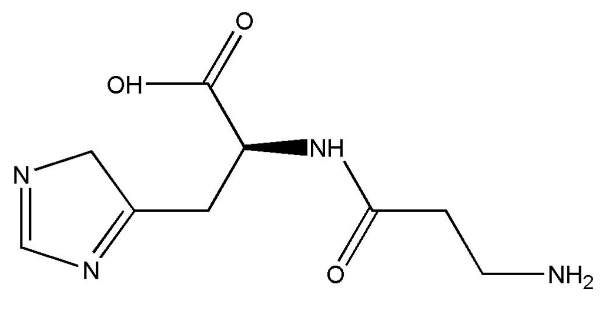
L-carnosine (C9H14N4O3, Figure 1, CAS Registry No. 305−84−0, molar mass: 226.235 g/mol), a bioactive peptide found in the brain and muscle tissues of mammal, was chosen as the model compound [10]. Owing to its strong antioxidant effects, L-carnosine has been widely used in treating ulcers, arthritis, atherosclerosis, cataracts, diabetes, hypertension, heart disease, and cancer. Through consulting a large volume of literature, it can be observed that studies about L-carnosine concentrate on its preparation, characterization, function, and application [11,12,13,14]. However, the polymorphism of L-carnosine has not been reported before. In this work, a new polymorph of L-carnosine was developed by the antisolvent crystallization method. It was named as form II, and the existing polymorph was named as form I. Different methods were employed to characterize and analyze these two forms of L-carnosine. Furthermore, the solvent-mediated polymorph transformation from form II to form I was investigated by online Raman, in which the influence of the solvent was further discussed. The rate-determining step in the transformation process was determined using the method of offline sampling, and the kinetics of crystal dissolution, nucleation, and growth were simulated and analyzed according to a dissolution-precipitation model.
2. Experimental Section
2.1. Materials
Form I of L-carnosine (≥0.990 mass fraction) was offered by Shanghai Yuanye Bio-Technology Co., Ltd., Shanghai, China. All the organic solvents used in the experiments, including methanol, ethanol, 2-propanol, acetone, dimethyl formamide (DMF), and dimethylacetamide (DMAC), were purchased from Tianjin Chemical Reagent No. 6 Factory, Tianjin China. The deionized water was supplied by Tianjin Yongqingyuan Co., Ltd., Tianjin, China. More details regarding the materials are listed in Table 1. All chemicals were used without further purification. Form II of L-carnosine was prepared in the laboratory by the antisolvent crystallization method, in which water acted as a solvent and DMF or DMAC served as an antisolvent.
2.2. Development of New Polymorph
The new polymorph of L-carnosine was developed by an antisolvent crystallization method through the following procedures. Firstly, 42 mL DMF or DMAC was gently poured into a 100 mL jacketed crystallizer, which was equipped with a mechanical stirrer. A thermostat (CF41, Julabo, Seelbach, Germany) was used to control the system at 303.15 K. Then, 2.45 g L-carnosine raw material was dissolved in water at room temperature to prepare a 0.175 g/mL L-carnosine solution. Finally, 14 mL L-carnosine solution was added into the jacketed crystallizer by a peristaltic pump (BT100-1F, Longer, Baoding, China) with a dropping rate of 333.3 μL/min. To fully mix the organic solvent and the L-carnosine solution, the mechanical stirrer was adjusted to 300 rpm during the entire experiment. Once the adding process finished, the suspension was filtered and dried in a vacuum oven at 298.15 K for further characterization.
2.3. Characterization Methods
2.3.1. Powder X-ray Diffraction
Powder X-ray diffraction, the most classic and commonly used method in qualitative and quantitative analysis for polymorphism, was used to measure the crystal form of L-carnosine. The data were collected by a D/max-2500 diffractometer (Rigaku, Tokyo, Japan) with Cu Ka radiation (0.15405 nm). Samples were determined at the diffraction angle (2θ) from 2° to 40° with a scanning rate of 8 °/min and a step size of 0.02°.
2.3.2. Polarizing Microscope
Polymorphism is one of the main factors that affect the morphology of solid drugs. A polarizing microscope (BX51, Olympus, Tokyo, Japan) was used to study the morphology of form I and form II of L-carnosine.
2.3.3. Thermal Analysis
Differential scanning calorimetry (1/500, Mettler-Toledo, Greifensee, Switzerland) and thermogravimetry (1/SF, Mettler Toledo, Greifensee, Switzerland) were carried out to obtain the melting temperature and decomposition temperature of form I and form II of L-carnosine. The measurements were conducted from 303.15 to 548.15 K at the rate of 2 K/min under the protection of a nitrogen atmosphere.
2.3.4. Raman Spectroscopy
A Raman spectrometer (RXN2, Mettler Toledo, Greifensee, Switzerland), equipped with an MR probe head and a PhAT probe head, was implemented to monitor the solid-phase composition of the suspension in situ during the solution-mediated polymorphic transformation process [15]. The data were collected in the wavenumber range from 150 to 1890 cm−1 at a laser wavelength of 514.5 nm.
2.4. Solubility Experiments
The solubility of L-carnosine form I and form II in binary solvent (water + DMAC) was determined using a dynamic method based on previous studies [16]. The molar ratio of water to DMAC was 9:1 in a binary solvent mixture. The solubility experiments were carried out in temperatures ranging from 278.25 K to 323.15 K under atmospheric pressure.
2.5. Solution-Mediated Polymorphic Transformation Experiments
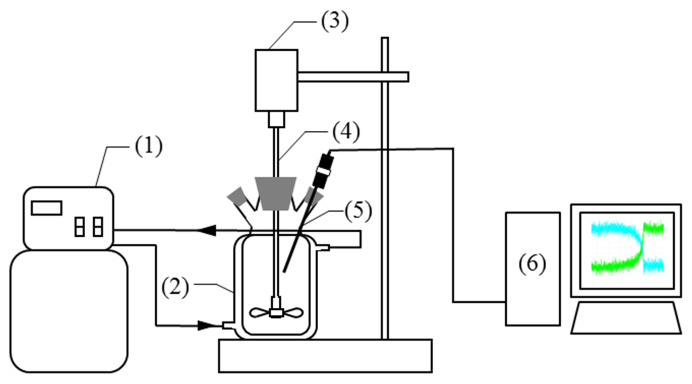
The polymorphic transformation experiments from form II to form I of L-carnosine were performed in six kinds of binary solvent mixtures, including water + methanol, water + ethanol, water + 2-propanol, water + acetone, water + DMF, and water + DMAC, which can be divided into two parts. Water + methanol, water + ethanol, water + 2-propanol, and water + acetone have been used in antisolvent crystallization to prepare form I, and water + DMAC and water + DMF were used to produce form II in this work. The volume ratio of water to organic solvent was 1:3 in a binary solvent mixture. First, 100 mL binary solvent mixture was gently poured into a 150 mL jacketed crystallizer, which was equipped with a mechanical stirrer. A thermostat (CF41, Julabo, Seelbach, Germany) was used to control the system at 303.15 K. Then, 3.00 g of form II of L-carnosine was added into the jacketed crystallizer to prepare the initial suspension of form II. The initial concentration of L-carnosine in the liquid phase is supersaturated for form I. To fully mix the suspension, the mechanical stirrer was adjusted to 300 rpm. During the polymorphic transformation process, a Raman MR probe was inserted into the slurry to monitor the solid-phase composition of the two polymorphs in situ. Meanwhile, the solid-phase composition was also measured and analyzed using powder X-ray diffraction by intermittent sampling [17]. In addition, the liquid concentration of L-carnosine was determined by the gravimetric method at a certain time interval [18]. All the transformation experiments were performed three times in this study. The experimental setup of solution-mediated polymorphic transformation between the two polymorphs of L-carnosine is shown in Figure 2.
2.6. Theoretical Model
In this study, a theoretical model of dissolution-precipitation was established to simulate the kinetics of the polymorphic transformation process from form II to form I of L-carnosine [19]. In the dissolution-precipitation model, the transformation process contains three steps: dissolution of the metastable form, nucleation of the stable form, and growth of the stable form. Based on these steps, the change in the amount of undissolved L-carnosine solid can be expressed via Equation (1).
where Ad and As represent the amounts of dissolved and undissolved solid L-carnosine. D, J, and G refer to the dissolution rate, nucleation rate, and growth rate, respectively.
Assuming the dissolution rate of the crystals in the suspension is size-independent, the dissolution rate can be defined as a proportional function of the amount of solid in the suspension, which is written as follows:
where subscript i stands for the i th polymorph of crystals. kdiss is the dissolution rate constant, which reflects the properties of the particles and solution [20].
The nucleation process, the first step of the crystallization process, is the spontaneous formation of clusters past the critical size [21]. At a microscopic level, the nucleation process can be simplified as the encounter of dissolved solute molecules. Therefore, the nucleation rate can be obtained as follows:
where knuc is the nucleation rate constant, which represents the possibility of generating aggregates. V and C are the volume and concentration of the solution. α refers to the nucleation molecularity index, denoting the average number of molecules needed to form nucleation.
During the growth process, discrete solute molecules in solution continuously aggregate onto the pre-existing crystals. Because of this, the growth rate is related to the concentration of the dissolved solute and the amount of undissolved solid in the suspension [22]. It can be shown as Equation (4).
where kgrowth is the growth rate constant, reflecting the reaction rate of dissolved solute molecules with pre-existing crystals.
Clearly, according to the above assumptions, the kinetics of undissolved L-carnosine solid can be calculated as in Equation (5).
where i = 1, 2 represents form I and form II of L-carnosine.
The relationship between solution concentration and undissolved solid amount can be described by Equation (6), when C = Ad/V is substituted into Equation (1).
When the system reaches equilibrium between the solid and liquid phases, the nucleation term can be ignored [19]. Thus, the dissolution rate equals the growth rate. Additionally, the solution concentration is the solubility of the solid solute, which can be described as following:
where Csol is the solubility of L-carnosine in the given binary solvent mixture.
Substituting Equation (7) into Equations (5) and (6), changes in the solution concentration and undissolved L-carnosine solid amount over time can be calculated by Equations (8) and (9), respectively.
where i = 1, 2, denotes form I and form II of L-carnosine.
Based on the experimental data and dissolution-precipitation model, the solution-mediated polymorphic transformation process of L-carnosine was simulated using MATLAB (2015 version). The model parameters, including kdiss, knuc, kgrowth, and α for form I and form II, were obtained by a nonlinear dynamic parameter fitting procedure based on the least squares method. In addition, the set ordinary differential equations were solved using the ode15s function with a variable integration step.
3. Results and Discussion
3.1. Characterizations
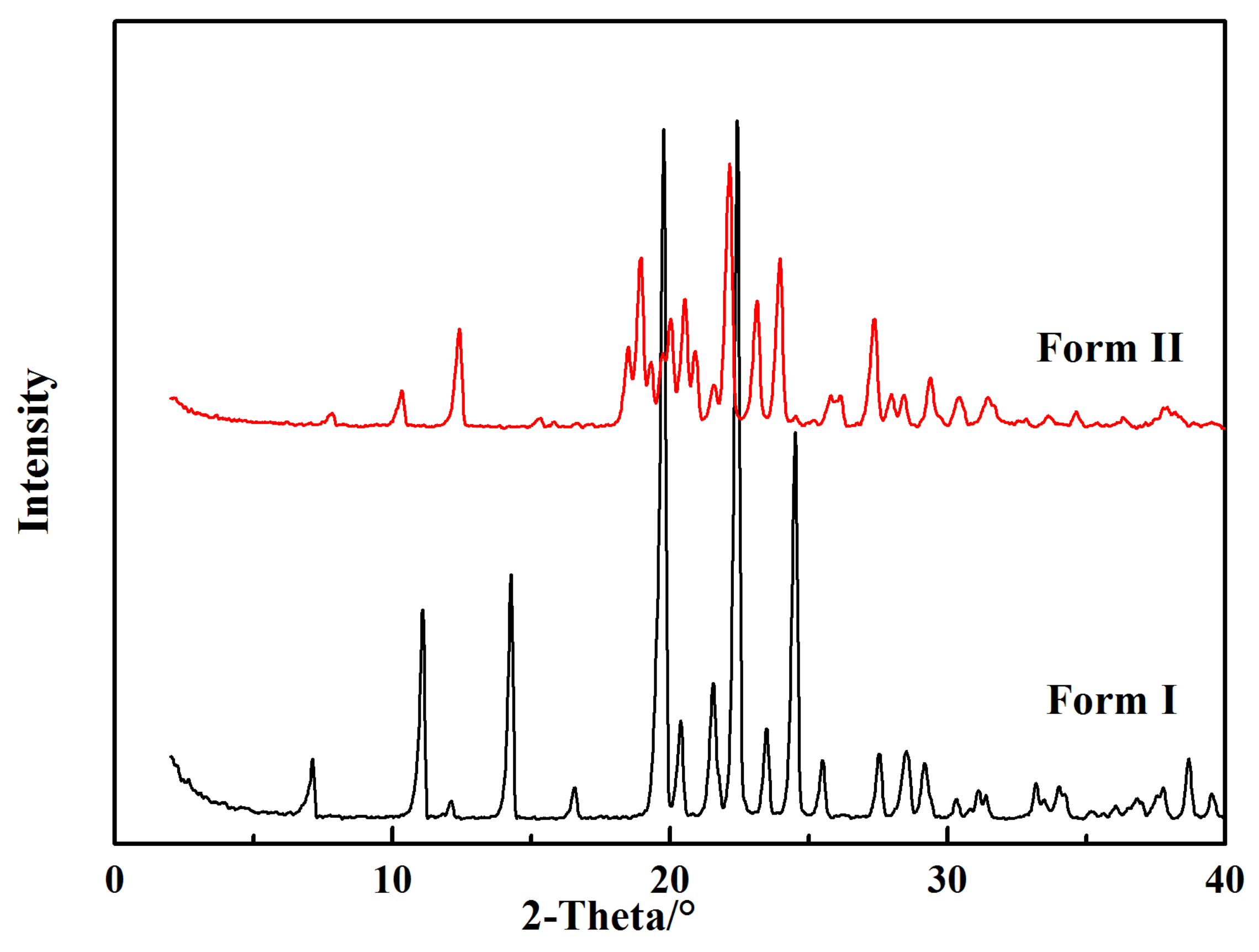
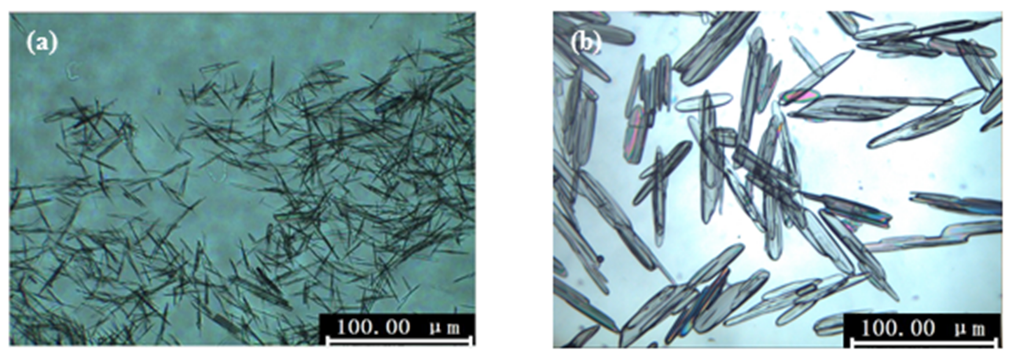
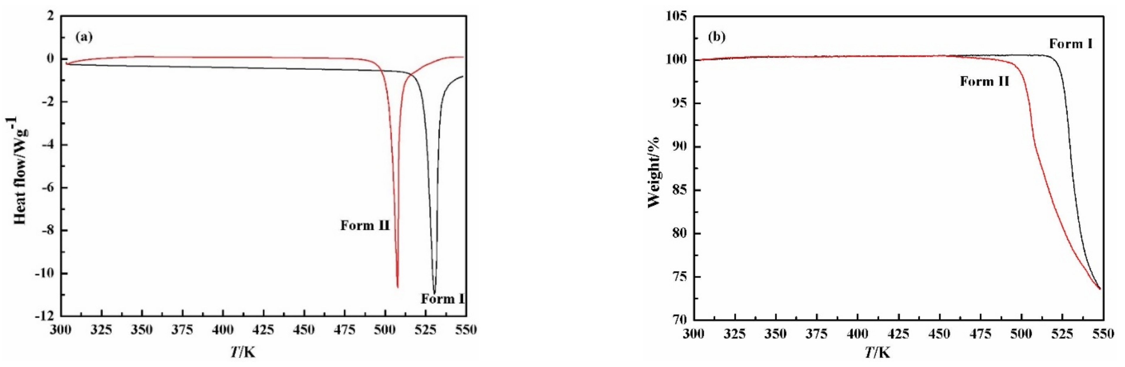
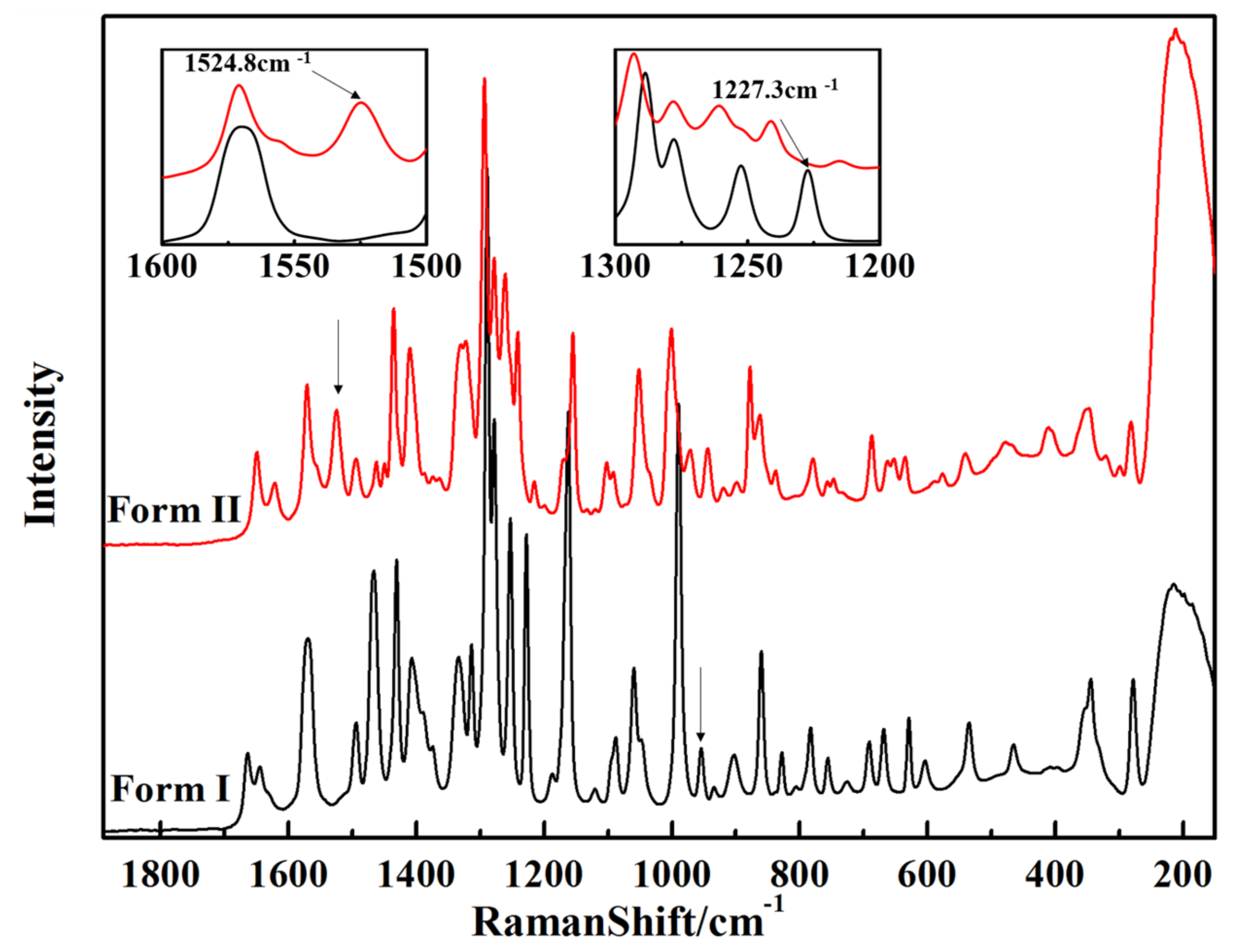
Polymorphs of L-carnosine, including form I and form II, were characterized by powder X-ray diffraction, polarizing microscope, thermal analysis, and Raman spectroscopy. The powder X-ray diffraction patterns are shown in Figure 3. It can be found that form I and form II exhibit distinct peaks, indicating that form II is a new polymorph of L-carnosine [12]. A polarizing microscope was utilized to research the crystal habit of the two polymorphs. As illustrated in Figure 4, form I exhibits needle-shaped crystals, whereas form II exhibits short rod-shaped crystals. Furthermore, the results of differential scanning calorimetry and thermogravimetry are displayed in Figure 5. It can be seen that there are obvious endothermic peaks at 517 K and 491 K for form I and form II of L-carnosine, respectively. Considering that samples of form I and form II started to decrease in weight at 517 K and 491 K, 517 K and 491 K were recognized as the decomposition temperatures of form I and form II, respectively. The results demonstrate that both polymorphs are decomposing before melting, and the chemical stability of form I is higher than that of form II. The Raman spectra of L-carnosine polymorphs are shown in Figure 6. Significantly distinguishable characteristic peaks of the two crystal forms are found in the wavelength range of 1600 cm−1–1500 cm−1 and 1300 cm−1–1200 cm−1, which indicates the difference in crystal structure. In this work, characteristic peaks at 1227.3 cm−1 and 1524.8 cm−1 were chosen to represent the changes in the solid content of form I and form II during the solution-mediated polymorphic transformation.
3.2. Solubility Data of L-carnosine Polymorphs
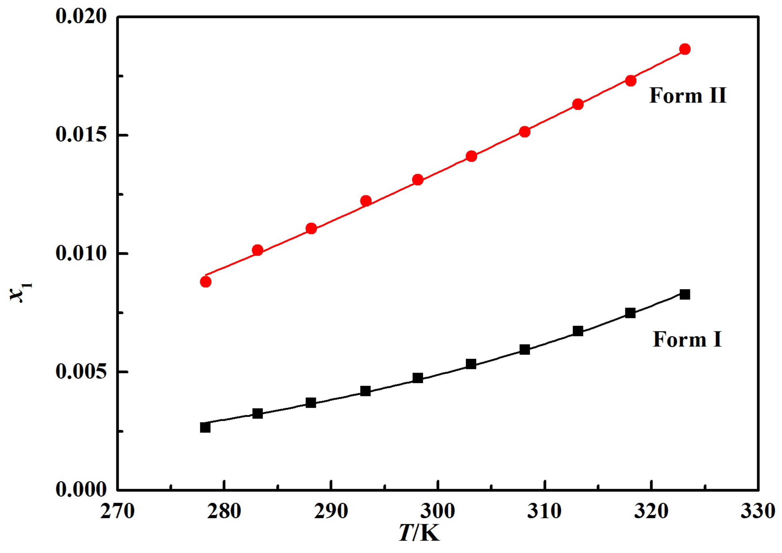
In this work, the solubility data of the L-carnosine polymorphs were measured to compare the stability of the two polymorphs. The mole fraction solubility of L-carnosine is graphically depicted in Figure 7. The results indicate that the solubility of the two polymorphs of L-carnosine is positively correlated with temperature. In addition, the solubility of form II is higher than that of form I throughout the whole temperature range studied. It confirms that form I is the stable form and form II is a metastable form, which is consistent with the results of thermal analysis [23].
3.3. Solution Mediated Transformation
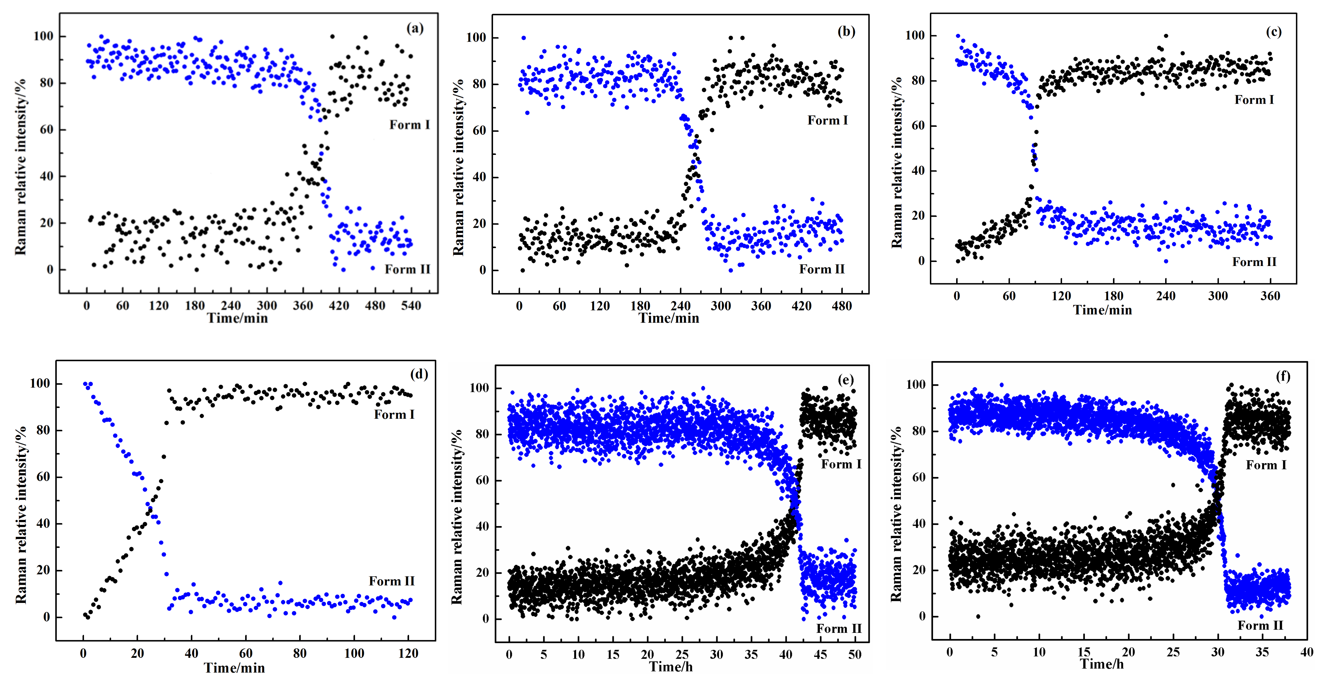
The solution-mediated polymorphic transformation of L-carnosine in different binary solvent mixtures of water + organic solvent, including methanol, ethanol, 2-propanol, acetone, DMF, and DMAC, was detected by online Raman spectroscopy. The results demonstrate that the transformation experiments of L-carnosine in a 100 mL binary solvent mixture were repeatable. The changes in Raman relative intensity during the polymorphic transformation process from form II to form I are shown in Figure 8. It can be seen that after the induction period of form I, the characteristic peak at 1227.3 cm−1 of form I increases with a corresponding decrease in the characteristic peak at 1524.8 cm−1 of form II. Finally, the characteristic peak of form II disappeared, and the characteristic peak intensity of form I remained basically stable, which means form II had completely transformed into form I at the end of this transformation process. In addition, the time needed for performing the crystal transformation of L-carnosine varies from tens of minutes to tens of hours in different binary solvent mixtures. The duration time of the transformation process for L-carnosine in the tested solvent systems was (water + acetone) < (water + 2-propanol) < (water + ethanol) < (water + methanol) < (water + DMAC) < (water + DMF), which represents the combined effects of solute conformation and solute−solvent interactions [24]. It indicates that the transformation rate from form II to form I of L-carnosine can be effectively regulated by changing the solvent.
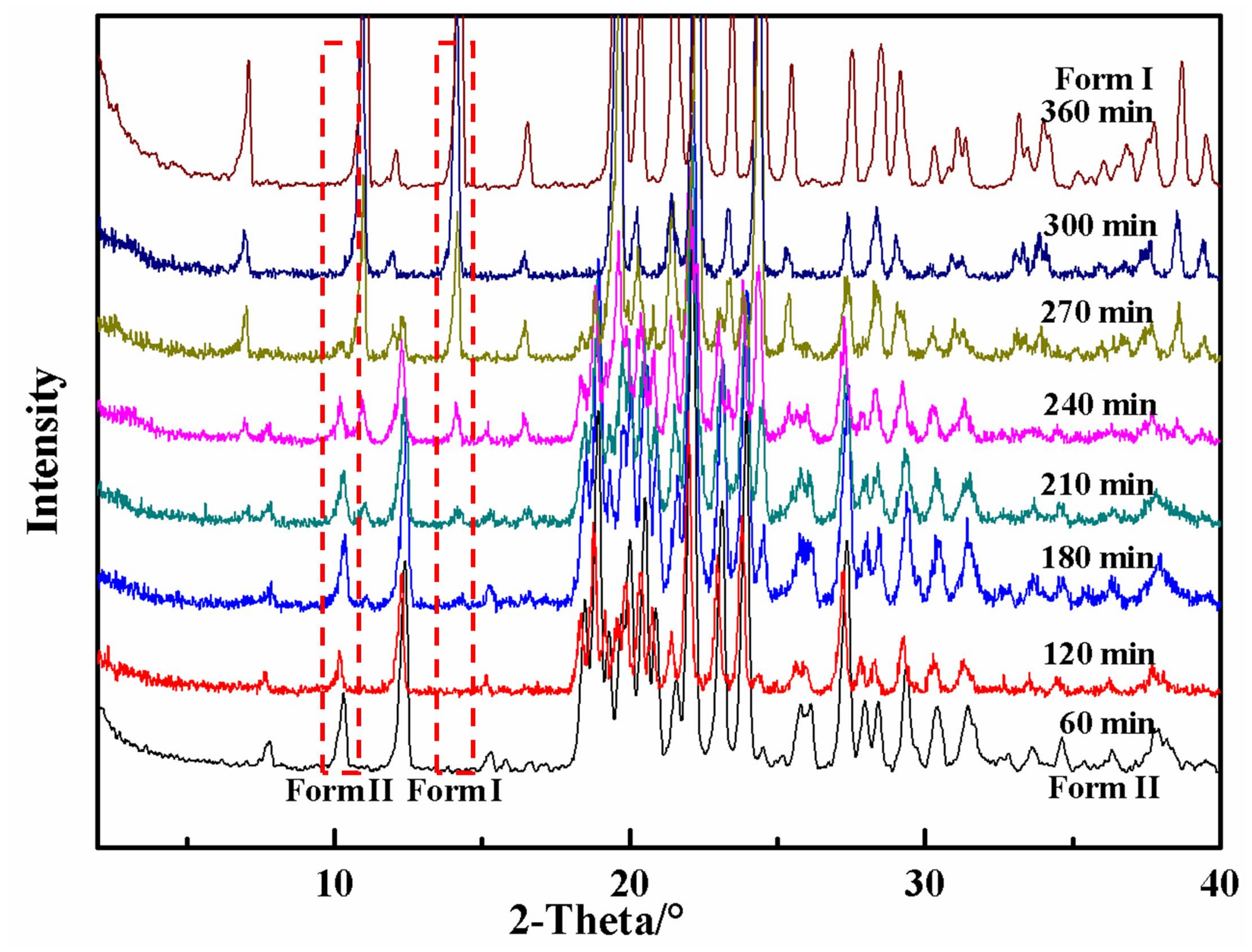
Powder X-ray diffraction was carried out at certain time intervals to verify the polymorphic transformation results from form II to form I of L-carnosine. The powder X-ray diffraction patterns of solid samples withdrawn from suspension in a water + ethanol binary solvent mixture are shown in Figure 9. It can be seen that the characteristic peak intensity of form I gradually increased, while the characteristic peak intensity of form II gradually decreased after 180 min. At this point, form I and form II of L-carnosine coexisted in the system. Finally, the characteristic peak of form II disappeared, while the characteristic peak intensity of form I reached its maximum. Accordingly, form I was the only polymorph detected in the solid phase after 300 min, which was consistent with the results of Raman spectroscopy.
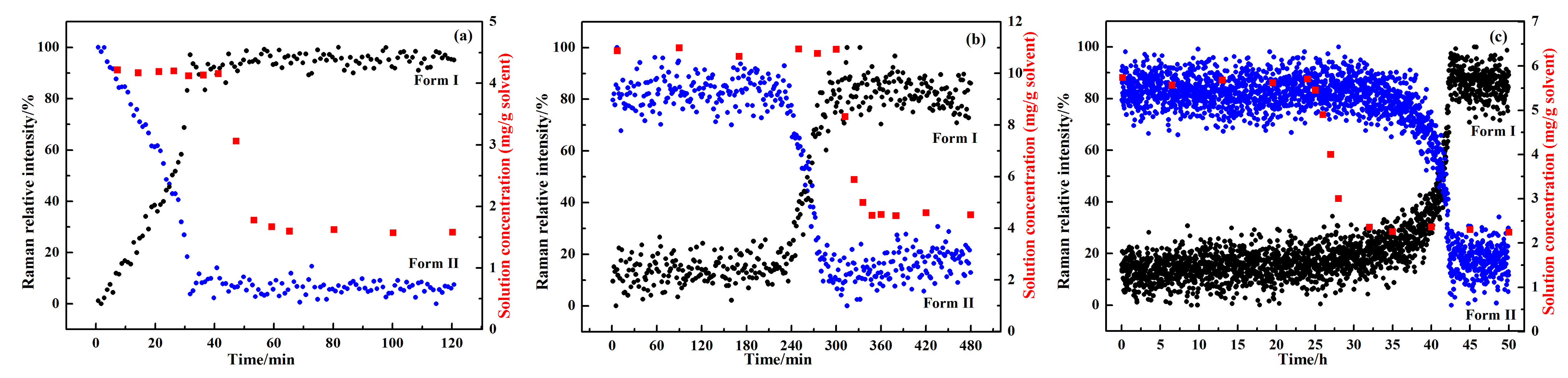
In combination with the changes of Raman relative intensity during the solution-mediated polymorphic transformation, the transformation process in the first kind of solvent systems can be divided into those with and without induction time. Therefore, three typical binary solvent mixtures, water + acetone, water + ethanol, and water + DMF, were chosen for further study, which are able to fully reflect the transformation mechanism. To explore the transformation mechanism, the solution concentration was measured at specific time intervals by the gravimetric method. The results in typical binary solvent mixtures, including water + acetone, water + ethanol, and water + DMF, are plotted in Figure 10. It can be seen that the driving force for transformation, namely the difference in solubility between the two polymorphs, decreased with the increase in solvent polarity. As a result, the transformation time for L-carnosine in the tested solvent systems increased with the increase in solvent polarity [25]. The transformation started immediately after form II of L-carnosine was added into a water + acetone binary solvent mixture. However, the induction time of the polymorphic transformation process in the water + ethanol and water + DMF binary solvent mixtures was 180 min and 30 h respectively, which indicates that the nucleation of form I is the controlling step of polymorphic transformation in water + ethanol and water + DMF binary solvent mixtures. In addition, the solution concentration in water + acetone and water + ethanol binary solvent mixtures held steady at the solubility of the metastable form II during the increase in form I, and started to decrease when form II had dissolved completely. It indicates that the dissolution rate of form II is higher than the growth rate of form I, and the growth of form I is the controlling step of polymorphic transformation in water + acetone and water + ethanol binary solvent mixtures. Nevertheless, the solution concentration in water + DMF decreased to the solubility of stable form I when the amount of form I started to increase, suggesting that the dissolution rate of form II is smaller than the growth rate of form I, and the dissolution of form I is the controlling step of polymorphic transformation in a water + DMF system. In conclusion, the polymorphic transformation of L-carnosine is “growth-controlled” in water + acetone solution, “nucleation-growth-controlled” in water + ethanol solution, and “nucleation-dissolution-controlled” in water + DMF solution [9].
3.4. Transformation Process Simulation
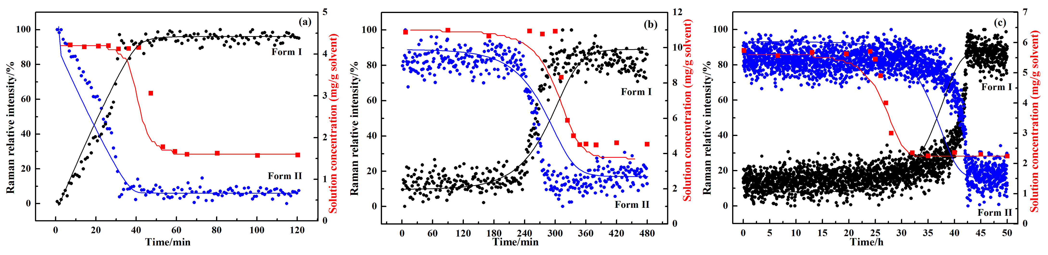
The dissolution-precipitation kinetics model was carried out to simulate and analyze the polymorphic transformation of L-carnosine based on the experimental data. The values of the model parameters are listed in Table 2. Figure 11 illustrate the calculated results of the liquid concentration and solid-phase composition of L-carnosine in three binary solvent systems. It was found that the simulated solution concentration curves can fit the experimental values well, and the simulated solid-phase compositions of the two polymorphs are also close to the measured online Raman data. The delicate discrepancies of the solid-phase composition in water + ethanol and water + DMF binary solvent mixtures are acceptable, considering the assumptions proposed to simplify the calculation process. The consistency between the model results and experimental data supposes that the model established in our work is reliable to simulate the solution-mediated polymorphic transformation process of L-carnosine.
4. Conclusions
In this paper, form II—a new polymorph of L-carnosine—was successfully developed by an antisolvent crystallization method. The properties of the two polymorphs were characterized by powder X-ray diffraction, polarizing microscope, thermal analysis, and Raman spectroscopy. The solubility of L-carnosine form I and form II in a (water + DMAC) binary solvent mixture was determined by a dynamic method, based on which the relative stability of the two forms was analyzed. The results suggest that form I is the thermodynamically stable form compared with form II. In addition, the solution-mediated polymorphic transformation of the two polymorphs in different binary solvent systems was detected by online and offline analytical techniques. It was found that the transformation process became longer with increasing solvent polarity. The polymorphic transformation of L-carnosine was “growth-controlled” in a water + acetone binary solvent, while “nucleation−growth” and “nucleation-dissolution” were the rate-controlling steps in water + ethanol and water + DMF binary solvents, respectively. A theoretical model of dissolution-precipitation was built to simulate the kinetics of the polymorphic transformation. It was verified that the simulated curves are basically consistent with the experimental results, which means that the model established in this work is reliable to simulate the solution-mediated polymorphic transformation process of L-carnosine.
Author Contributions
Investigation and writing—original draft preparation, Y.Z.; software, S.Z.; data curation, J.G.; visualization and project administration, C.L.; supervision and writing—review and editing, T.W. All authors have read and agreed to the published version of the manuscript.
Funding
This research was funded by the Fundamental Research Funds for the Universities of Hebei Province, grant number JQN2020030.
Institutional Review Board Statement
Not applicable.
Informed Consent Statement
Not applicable.
Data Availability Statement
Not applicable.
Acknowledgments
The authors are very grateful for the financial support of the Fundamental Research Funds for the Universities of Hebei Province (grant number JQN2020030).
Conflicts of Interest
The authors declare no conflict of interest.
References
- Brog, J.P.; Chanez, C.L.; Crochet, A.; Fromm, K.M. Polymorphism, what it is and how to identify it: A systematic review. Rsc Adv. 2013, 3, 16905–16931. [Google Scholar] [CrossRef] [Green Version]
- Zhou, Y.; Wang, J.; Xiao, Y.; Wang, T.; Huang, X. The Effects of Polymorphism on Physicochemical Properties and Pharmacodynamics of Solid Drugs. Curr. Pharm. Des. 2018, 24, 2375–2382. [Google Scholar] [CrossRef] [PubMed]
- Fandaruff, C.; Rauber, G.S.; Araya-Sibaja, A.M.; Pereira, R.N.; de Campos, C.E.M.; Rocha, H.V.A.; Monti, G.A.; Malaspina, T.; Silva, M.A.S.; Cuffini, S.L. Polymorphism of Anti-HIV Drug Efavirenz: Investigations on Thermodynamic and Dissolution Properties. Cryst. Growth Des. 2014, 14, 4968–4975. [Google Scholar] [CrossRef]
- Singhal, D.; Curatolo, W. Drug polymorphism and dosage form design: A practical perspective. Adv. Drug Deliv. Rev. 2004, 56, 335–347. [Google Scholar] [CrossRef] [PubMed]
- Yang, C.; Zhang, Z.; Zeng, Y.; Wang, J.; Wang, Y.; Ma, B. Structures and characterization of m-nisoldipine polymorphs. Cryst. Eng. Comm. 2012, 14, 2589–2594. [Google Scholar] [CrossRef]
- Kitamura, M. Polymorphism in the crystallization of L-glutamic acid. J. Cryst. Growth 1989, 96, 541–546. [Google Scholar] [CrossRef]
- Nývlt, J. The Ostwald Rule of Stages. Cryst. Res. Technol. 1995, 30, 443–449. [Google Scholar] [CrossRef]
- Guo, N.; Hou, B.; Wang, N.; Xiao, Y.; Huang, J.; Guo, Y.; Zong, S.; Hao, H. In Situ Monitoring and Modeling of the Solution-Mediated Polymorphic Transformation of Rifampicin: From Form II to Form I. J. Pharm. Sci. 2017, 107, 344–352. [Google Scholar] [CrossRef] [Green Version]
- O’Mahony, M.A.; Maher, A.; Croker, D.M.; Rasmuson, Å.C.; Hodnett, B.K. Examining Solution and Solid State Composition for the Solution-Mediated Polymorphic Transformation of Carbamazepine and Piracetam. Cryst. Growth Des. 2012, 12, 1925–1932. [Google Scholar] [CrossRef] [Green Version]
- Gulewitsch, W.; Amiradžibi, S. Ueber das Carnosin, eine neue organische Base des Fleischextractes. Eur. J. Inorg. Chem. 1900, 33, 1902–1903. [Google Scholar] [CrossRef] [Green Version]
- Sharif, S.; Schagen, D.; Toney, M.D.; Limbach, H.-H. Coupling of Functional Hydrogen Bonds in Pyridoxal-5‘-phosphate−Enzyme Model Systems Observed by Solid-State NMR Spectroscopy. J. Am. Chem. Soc. 2007, 129, 4440–4455. [Google Scholar] [CrossRef]
- Zhou, Y.; Wang, J.; Wang, T.; Wang, N.; Xiao, Y.; Zong, S.; Huang, X.; Hao, H. Self-Assembly of Monodispersed Carnosine Spherical Crystals in a Reverse Antisolvent Crystallization Process. Cryst. Growth Des. 2019, 19, 2695–2705. [Google Scholar] [CrossRef]
- Wu, J.W.; Liu, K.-N.; How, S.-C.; Chen, W.-A.; Lai, C.-M.; Liu, H.-S.; Hu, C.-J.; Wang, S.S.S. Carnosine’s Effect on Amyloid Fibril Formation and Induced Cytotoxicity of Lysozyme. PLoS ONE 2013, 8, e81982. [Google Scholar] [CrossRef] [Green Version]
- Banerjee, S.; Mukherjee, B.; Poddar, M.K.; Dunbar, G.L. Carnosine improves aging-induced cognitive impairment and brain regional neurodegeneration in relation to the neuropathological alterations in the secondary structure of amyloid beta (Aβ). J. Neurochem. 2021, 158, 710–723. [Google Scholar] [CrossRef]
- Jiang, C.; Wang, Y.; Yan, J.; Yang, J.; Xiao, L.; Hao, H. Formation Mechanism and Phase Transformation Behaviors of Pantoprazole Sodium Heterosolvate. Org. Process Res. Dev. 2015, 19, 1752–1759. [Google Scholar] [CrossRef]
- He, F.; Wang, Y.; Yin, Q.; Tao, L.; Lv, J.; Xu, Z.; Wang, J.; Hao, H. Effect of polymorphism on thermodynamic properties of cefamandole nafate. Fluid Phase Equilibria 2016, 422, 56–65. [Google Scholar] [CrossRef] [Green Version]
- Wu, S.; Du, S.; Chen, M.; Li, K.; Jia, L.; Zhang, D.; Macaringue, E.G.J.; Hou, B.; Gong, J. Crystal Structures and Phase Behavior of Sulfadiazine and a Method for the Preparation of Aggregates with Good Performance. Chem. Eng. Technol. 2017, 41, 532–540. [Google Scholar] [CrossRef]
- Zhou, Y.; Wang, J.; Wang, T.; Gao, J.; Huang, X.; Hao, H. Solubility and dissolution thermodynamic properties of L-carnosine in binary solvent mixtures. J. Chem. Thermodyn. 2020, 149, 106167. [Google Scholar] [CrossRef]
- Jakubiak, P.; Schuler, F.; Alvarez-Sánchez, R. Extension of the dissolution-precipitation model for kinetic elucidation of solvent-mediated polymorphic transformations. Eur. J. Pharm. Biopharm. 2016, 109, 43–48. [Google Scholar] [CrossRef]
- Zhu, M.; Wang, Y.; Li, F.; Bao, Y.; Huang, X.; Shi, H.; Hao, H. Theoretical Model and Experimental Investigations on Solution-Mediated Polymorphic Transformation of Theophylline: From Polymorph I to Polymorph II. Crystals 2019, 9, 260. [Google Scholar] [CrossRef] [Green Version]
- Xiao, Y.; Wang, J.; Huang, X.; Shi, H.; Zhou, Y.; Zong, S.; Hao, H.; Bao, Y.; Yin, Q. Determination Methods for Crystal Nucleation Kinetics in Solutions. Cryst. Growth Des. 2017, 18, 540–551. [Google Scholar] [CrossRef]
- Jakubiak, P.; Wagner, B.; Grimm, H.P.; Petrig-Schaffland, J.; Schuler, F.; Alvarez-Sánchez, R. Development of a Unified Dissolution and Precipitation Model and Its Use for the Prediction of Oral Drug Absorption. Mol. Pharm. 2016, 13, 586–598. [Google Scholar] [CrossRef]
- Grunenberg, A.; Henck, J.-O.; Siesler, H. Theoretical derivation and practical application of energy/temperature diagrams as an instrument in preformulation studies of polymorphic drug substances. Int. J. Pharm. 1996, 129, 147–158. [Google Scholar] [CrossRef]
- Kitamura, M.; Umeda, E.; Miki, K. Mechanism of Solvent Effect in Polymorphic Crystallization of BPT. Ind. Eng. Chem. Res. 2012, 51, 12814–12820. [Google Scholar] [CrossRef]
- Marcus, Y. The properties of organic liquids that are relevant to their use as solvating solvents. Chem. Soc. Rev. 1993, 22, 409–416. [Google Scholar] [CrossRef]
Figure 1.
The molecular structure of L-carnosine.

Figure 2.
The experimental setup of solution-mediated polymorphic transformation between the two polymorphs of L-carnosine: (1) thermostat; (2) crystallizer; (3) mechanical stirrer; (4) stirring paddle; (5) Raman probe; (6) computer.
Figure 2.
The experimental setup of solution-mediated polymorphic transformation between the two polymorphs of L-carnosine: (1) thermostat; (2) crystallizer; (3) mechanical stirrer; (4) stirring paddle; (5) Raman probe; (6) computer.

Figure 3.
Powder X-ray diffraction patterns of L-carnosine form I and form II.

Figure 4.
Polymorphic morphology of L-carnosine in a polarizing microscope: (a) form I; (b) form II.
Figure 4.
Polymorphic morphology of L-carnosine in a polarizing microscope: (a) form I; (b) form II.

Figure 5.
Thermal analysis curves of L-carnosine form I and form II: (a) differential scanning calorimetry; (b) thermogravimetry.
Figure 5.
Thermal analysis curves of L-carnosine form I and form II: (a) differential scanning calorimetry; (b) thermogravimetry.

Figure 6.
Raman spectra of L-carnosine form I and form II.

Figure 7.
Solubility of L-carnosine form I and form II in a binary solvent mixture of water and DMAC.
Figure 7.
Solubility of L-carnosine form I and form II in a binary solvent mixture of water and DMAC.

Figure 8.
Changes of Raman relative intensity during the solution-mediated polymorphic transformation of L-carnosine in different binary solvent mixtures: (a) water + methanol; (b) water + ethanol; (c) water + 2-propanol; (d) water + acetone; (e) water + DMF; (f) water + DMAC.
Figure 8.
Changes of Raman relative intensity during the solution-mediated polymorphic transformation of L-carnosine in different binary solvent mixtures: (a) water + methanol; (b) water + ethanol; (c) water + 2-propanol; (d) water + acetone; (e) water + DMF; (f) water + DMAC.

Figure 9.
Powder X-ray diffraction patterns at different times during the solution-mediated polymorphic transformation of L-carnosine in a water + ethanol binary solvent mixture.
Figure 9.
Powder X-ray diffraction patterns at different times during the solution-mediated polymorphic transformation of L-carnosine in a water + ethanol binary solvent mixture.

Figure 10.
Changes of solution concentration during the solution-mediated polymorphic transformation of L-carnosine in different solvent mixtures: (a) water + acetone; (b) water + ethanol; (c) water + DMF.
Figure 10.
Changes of solution concentration during the solution-mediated polymorphic transformation of L-carnosine in different solvent mixtures: (a) water + acetone; (b) water + ethanol; (c) water + DMF.

Figure 11.
Calculated solid composition profiles and solution concentration profile during the solution-mediated polymorphic transformation from form II to form I of L-carnosine in different solvent mixtures: (a) water + acetone; (b) water + ethanol; (c) water + DMF.
Figure 11.
Calculated solid composition profiles and solution concentration profile during the solution-mediated polymorphic transformation from form II to form I of L-carnosine in different solvent mixtures: (a) water + acetone; (b) water + ethanol; (c) water + DMF.


Table 1.
Sources and mass fraction purity of MAC chemicals used in this article.
| Chemical Name | Source | Mass Purity | Purification Method | Analysis Method |
|---|---|---|---|---|
| L-carnosine (Form I) | Shanghai Yuanye Bio-Technology Co., Ltd., China | ≥0.990 | None | HPLC a |
| Methanol | Tianjin Chemical Reagent No.6 Factory, China | >0.995 | None | GC b |
| Ethanol | Tianjin Chemical Reagent No.6 Factory, China | >0.995 | None | GC b |
| 2-Propanol | Tianjin Chemical Reagent No.6 Factory, China | >0.995 | None | GC b |
| Acetone | Tianjin Chemical Reagent No.6 Factory, China | >0.995 | None | GC b |
| DMF | Tianjin Chemical Reagent No.6 Factory, China | >0.995 | None | GC b |
| DMAC | Tianjin Chemical Reagent No.6 Factory, China | >0.995 | None | GC b |
| Deionized Water | Tianjin Yongqingyuan Co., Ltd., China | ≥18.25 MΩ·cm | None | CT c |
a High performance liquid chromatography, which was determined by Shanghai Yuanye Bio-Technology Co., Ltd., Shanghai, China. b Gas chromatography, which was determined by Tianjin Chemical Reagent No.6 Factory, Tianjin, China. c Conductivity test, which was carried out by Tianjin Yongqingyuan Co., Ltd., Tianjin, China.
Table 2.
Model parameters for the solution-mediated polymorphic transformation of L-carnosine in different solvent mixtures.
Table 2.
Model parameters for the solution-mediated polymorphic transformation of L-carnosine in different solvent mixtures.
| Solvent | Solid Form | kdiss/min−1 | Csol | knuc/min−1(g/mg)α−1 | kgrowth/min−1 | α |
|---|---|---|---|---|---|---|
| Water + Acetone | form I | 0.176 | 1.57 × 10⁻³ | 5.89 × 10⁻³ | 112 | 1.46 |
| form II | 0.157 | 4.21 × 10⁻³ | 5.53 × 10⁻² | 37.3 | 2.71 | |
| Water + Ethanol | form I | 0.758 | 4.49 × 10⁻³ | 4.81 × 10⁻⁵ | 169 | 1.74 |
| form II | 0.267 | 1.10 × 10⁻² | 2.22 × 10⁻² | 24.3 | 2.19 | |
| Water + DMF | form I | 0.318 | 2.24 × 10⁻³ | 4.65 × 10⁻⁶ | 142 | 1.87 |
| form II | 0.389 | 5.74 × 10⁻³ | 2.34 × 10⁻² | 67.8 | 2.79 |
Publisher’s Note: MDPI stays neutral with regard to jurisdictional claims in published maps and institutional affiliations. |
© 2022 by the authors. Licensee MDPI, Basel, Switzerland. This article is an open access article distributed under the terms and conditions of the Creative Commons Attribution (CC BY) license (https://creativecommons.org/licenses/by/4.0/).
Share and Cite
MDPI and ACS Style
Zhou, Y.; Zong, S.; Gao, J.; Liu, C.; Wang, T. Solution-Mediated Polymorphic Transformation of L-Carnosine from Form II to Form I. Crystals 2022, 12, 1014. https://0-doi-org.brum.beds.ac.uk/10.3390/cryst12071014
AMA Style
Zhou Y, Zong S, Gao J, Liu C, Wang T. Solution-Mediated Polymorphic Transformation of L-Carnosine from Form II to Form I. Crystals. 2022; 12(7):1014. https://0-doi-org.brum.beds.ac.uk/10.3390/cryst12071014
Chicago/Turabian StyleZhou, Yanan, Shuyi Zong, Jie Gao, Chunsong Liu, and Ting Wang. 2022. "Solution-Mediated Polymorphic Transformation of L-Carnosine from Form II to Form I" Crystals 12, no. 7: 1014. https://0-doi-org.brum.beds.ac.uk/10.3390/cryst12071014
Note that from the first issue of 2016, this journal uses article numbers instead of page numbers. See further details here.