Cutaneous Adverse Events Associated with Immune Checkpoint Inhibitors: A Review Article


1
Department of Dermatology, Kaohsiung Medical University Hospital, Kaohsiung Medical University, Kaohsiung 807378, Taiwan
2
Graduate Institute of Clinical Medicine, College of Medicine, Kaohsiung Medical University, Kaohsiung 807378, Taiwan
3
Department of Dermatology, School of Medicine, College of Medicine, Kaohsiung Medical University, Kaohsiung 807378, Taiwan
4
Neuroscience Research Center, Kaohsiung Medical University, Kaohsiung 807378, Taiwan
*
Author to whom correspondence should be addressed.
Curr. Oncol. 2022, 29(4), 2871-2886; https://0-doi-org.brum.beds.ac.uk/10.3390/curroncol29040234
Submission received: 28 March 2022
/
Revised: 15 April 2022
/
Accepted: 15 April 2022
/
Published: 18 April 2022
(This article belongs to the Special Issue Immune Checkpoint Inhibitors in Cancer Therapy: State of the Art and Future Perspectives)
Abstract
:Immune checkpoint inhibitors (ICIs) have emerged as novel options that are effective in treating various cancers. They are monoclonal antibodies that target cytotoxic T-lymphocyte antigen 4 (CTLA-4), programmed cell death 1 (PD-1), and programmed cell death-ligand 1 (PD-L1). However, activation of the immune systems through ICIs may concomitantly trigger a constellation of immunologic symptoms and signs, termed immune-related adverse events (irAEs), with the skin being the most commonly involved organ. The dermatologic toxicities are observed in nearly half of the patients treated with ICIs, mainly in the form of maculopapular rash and pruritus. In the majority of cases, these cutaneous irAEs are self-limiting and manageable, and continuation of the ICIs is possible. This review provides an overview of variable ICI-mediated dermatologic reactions and describes the clinical and histopathologic presentation. Early and accurate diagnosis, recognition of severe toxicities, and appropriate management are key goals to achieve the most favorable outcomes and quality of life in cancer patients.
1. Introduction
In recent decades, immune checkpoints inhibitors (ICIs) have been demonstrated to dramatically improve the overall survival for a broad spectrum of advanced malignancies [1,2,3]. These agents are monoclonal antibodies that target the immune checkpoint molecules, including cytotoxic T-lymphocyte antigen 4 (CTLA-4), programmed cell death 1 (PD-1), and programmed cell death-ligand 1 (PD-L1). To date, seven ICIs have been approved by the U.S. Food and Drug Administration (FDA). Since March 2011, when the first immune checkpoint inhibitor, ipilimumab (an anti-CTLA-4 agent), was approved for the treatment of advanced (either metastatic or unresectable) melanoma [4], additional therapies that target the PD-1/PD-L1 axis have been subsequently approved, showing promising therapeutic outcomes for various solid tumors and hematologic malignancies [5]. Nivolumab, pembrolizumab, and cemiplimab are anti-PD-1 agents, whereas atezolizumab, durvalumab, and avelumab are anti-PD-L1 agents. The indications of these FDA-approved ICIs are summarized in Table 1. Other novel therapies targeting the alternative inhibitory pathways are currently under investigation, including lymphocyte-activation gene 3 (LAG-3), T-cell immunoglobulin and ITIM domain (TIGIT), T-cell immunoglobulin and mucin-domain containing-3 (TIM-3), V-domain immunoglobulin suppressor of T-cell activation (VISTA), B7 homolog 3 protein (B7-H3), inducible T cell costimulatory (ICOS), and B and T lymphocyte attenuator (BTLA) [6]. While ICIs promote the reinvigoration of the anti-tumor T-cell response, the enhanced immunologic activation may result in a variety of autoimmune-like or inflammatory side effects, termed immune-related adverse events (irAEs), which can involve almost any organ system.
2. Biologic Mechanism of Immune Checkpoint Inhibition
Immune checkpoint molecules, primarily of CTLA-4 and PD-1, are negative regulators adopted by cancer cells to disguise themselves as regular components of the human body, dampen the immune responses, and escape from the assault of human immunity [7,8]. An overview of the action of immune checkpoint inhibition gives insight into the anti-tumor function of the immune checkpoint blockades and the pathogenesis of irAEs.
In the physiologic state, immunologic inhibitory pathways are achieved through a complex network of costimulatory and inhibitory signals in order to maintain the immune response within a desired physiological range [8]. Both CTLA-4 and PD-1 are predominantly expressed on the T-lymphocytes. At the priming phase of T-cell activation, CTLA-4 primarily attenuates T-cell activity through competition with the costimulatory molecule CD28 for binding to the stimulatory receptors CD80 and CD86 expressed on the antigen-presenting cells (APCs) [8]. Moreover, the CTLA-4 expression on the regulatory T-cells (Tregs) mediates an inhibitory immune effect [8]. The main function of PD-1 is to attenuate local T-cell responses through interaction with PD-L1 and PD-L2 on APCs, ultimately inhibiting T-cell-receptor signaling in the periphery [8]. Tumors themselves, as well as the tumor microenvironment (TME), can express multiple inhibitory pathways and associated molecules, leading to T-cell impairment and immune escape. When these pathways are blocked by the immune checkpoint therapies, T-cell responses are promoted, facilitating an effective anti-tumor function and the exuberant activation of self-reactive T-cells with the resultant autoimmunity, mainly considered as irAEs [9].
3. Immune-Related Adverse Events (irAEs)
The spectrum of organ systems affected by irAEs is very broad, with varying frequencies and severities being reported [10,11,12]. The degree of severity and the associated morbidity of irAEs are graded according to the Common Terminology Criteria for Adverse Events (CTCAE), and the frequency of irAEs usually depends on the agents administered, the exposure time, the dosage, as well as the patient’s medical condition [12,13].
Among the irAEs encountered in ICI-treated patients, a systematic review has documented that all-grades colitis, hypophysitis, and rash were more frequent with anti-CTLA-4 agents, whereas pneumonitis, hypothyroidism, arthralgia, and vitiligo were more common with anti-PD-1 blockades [11]. De Velasco G. et al. [10] reported that the incidence of any-grade irAEs was higher in patients receiving an anti-CTLA-4 agent (53.8%) compared with those receiving anti-PD-1 (26.5%) or anti-PD-L1 (17.1%), whereas Martins F. et al. [12] demonstrated that the severe irAEs of grade III or IV were more frequently seen in ICIs targeting CTLA-4 (10 to 30%) than those targeting PD-1 (~10%). In theory, anti-CTLA-4 blockades act on the early stage of T-cell priming mainly in lymphatic organs, enhancing the magnitude of T-cell proliferation or reducing Treg-mediated immunosuppression, while anti-PD-1/PD-L1 blockades act on the later phase of the immune response mainly in the periphery, reinvigorating the pre-existing T-cell activity [14,15,16]. The dissimilar mechanisms of different target molecules may explain why anti-CTLA-4 inhibitors are associated with more prominent irAEs than anti-PD-1/PD-L1 inhibitors. In addition to single therapy, dual therapies with anti-CTLA-4 and anti-PD-1/PD-L1 blockades aim to further augment the therapeutic response but also increase the risk of irAEs [15,17,18,19]. In a phase III trial, combination therapy with ipilimumab and nivolumab demonstrated higher efficacy in treating metastatic melanoma compared with either monotherapy; however, treatment-related complications of grade III or IV were found in 55.0% of those in the nivolumab-plus-ipilimumab group, which is higher than that in the ipilimumab group (27.3%) and the nivolumab group (16.3%) [19]. Another study reported that combination therapy was associated with the highest risk of severe irAEs (55%), followed by patients receiving anti-CTLA-4 (10 to 41.6%) and anti-PD-1 (12 to 20%) as monotherapy [18]. Martins F. et al. [12] reported that the incidence of fatal toxicities is estimated to be approximately 0.3% to 1.3%, with a tendency to occur earlier (median onset time ~14.5 days) in the course of treatment and evolve rapidly, especially in patients with combination therapy.
4. Cutaneous Immune-Related Adverse Events (cirAEs)
Dermatologic complications arise as the earliest and most frequently observed adverse events among all irAEs, affecting between 30 and 50% of patients on ICIs [2,12]. The symptoms may significantly impair patients’ quality of life, and even lead to a pause of immunotherapy treatment. Fortunately, the majority of cirAEs seem to be mild and manageable, but there are still a few serious events (grade III or IV) being observed [17]. Maculopapular rash, pruritus, lichenoid eruptions, and vitiligo are the most widely reported cutaneous adverse events, which are summarized in Table 2 [17,20,21]. Severe cutaneous adverse reactions (SCARs), consisting of Stevens–Johnson syndrome/toxic epidermal necrolysis (SJS/TEN), drug rash with eosinophilia and systemic symptoms (DRESS), and acute generalized exanthematous pustulosis (AGEP), are rare but potentially life-threatening [17]. Other less-frequent manifestations include ICI-induced dermatomyositis, Sweet syndrome, interstitial granulomatous dermatitis, pityriasis rubra pilaris-like erythroderma, and lupus-like cutaneous reaction [22,23,24,25,26].
Previous studies have documented that the occurrence of certain cirAEs may indicate a more favorable clinical outcome for the treatment of underlying malignancies [20,27,28]. Teulings HE. et al. [27] reported that the development of vitiligo in patients with melanoma treated with immunotherapy implied improved progression-free and overall survival. In a retrospective study, the presence of lichenoid and spongiotic dermatitis could be an indicator of favorable oncologic outcomes in a small cohort of patients receiving anti-PD-1/PD-L1 [28]. In contrast, Han Y. et. al. [15] stated that cirAEs are dose-independent and agent-specific immune reactions, which might not be a prognostic indicator for improved outcomes. Moreover, there are several currently known biomarkers for predicting cirAEs. Jia XH et al. [29] stated that patients with a positive rheumatoid factor (RF) greater than 15 IU/mL prior to ICI treatment were more likely to develop immune-related dermatologic toxicities. Another prospective observational study found that HLA-DRB1*11:01 was significantly associated with pruritus, suggestive of a genetic etiology in cirAEs [30].
Given that numerous cirAEs share similar clinical manifestations to spontaneous autoimmune or inflammatory dermatoses, it is difficult to distinguish the concurrent dermatological disease from the cirAEs, particularly when there is a long period of latency between the initiation of ICI treatment and the onset of cirAEs [31]. Moreover, other concomitant medications can also contribute to the cutaneous adverse effects via allergic mechanisms, which may be similar to the presentation of cirAEs [32]. In a retrospective study, 80% of the patients who developed lichenoid eruptions after anti-PD-1/PD-L1 therapy were concurrently taking medications that have been previously reported to cause a lichenoid reaction [21].

Table 2.
Summary of relatively common cutaneous adverse events associated with immune checkpoint inhibitors.
Table 2.
Summary of relatively common cutaneous adverse events associated with immune checkpoint inhibitors.
| Cutaneous irAEs | Clinical Features | Histopathological Findings | Mainly Associated ICIs | Suggested Managements |
|---|---|---|---|---|
| Maculopapular eruption | Pruritic erythematous macules and papules coalescing into thin plaques, mostly on the trunk and extremities | Superficial, perivascular lymphocytes and eosinophils infiltrate into the upper dermis, mild epidermal spongiosis [33] | Anti-CTLA-4 > anti-PD-1/PD-L1 | Symptomatic management with emollients, topical steroids, and oral antihistamines; consider systemic corticosteroids and withholding ICIs in severe cases [33] |
| Pruritus | May be concomitant with maculopapular rash or develop on normal- appearing skin | - | Anti-CTLA-4 > anti-PD-1/PD-L1 | Topical emollients or oral antihistamines; consider topical/systemic corticosteroids or topical calcineurin inhibitors in severe cases; other therapies include aprepitant, doxepin, gabapentin, pregabalin, and naloxone [3,34,35] |
| Lichenoid dermatitis | Erythematous-to- violaceous scaly plaques with a localized or generalized distribution, mostly on the trunk and extremities; mucosal involvement is rarely reported | Hyperkeratosis, hypergranulosis, a sawtooth rete ridge pattern, lichenoid and interface lymphocytic infiltrates, basal vacuolar changes, parakeratosis, epidermal spongiosis and necrosis, and eosinophils may present [3,36,37,38] | Anti-PD-1/PD-L1 | High-potency topical steroids; consider systemic corticosteroids and withholding ICIs in severe cases; other therapies include oral acitretin and phototherapy [39,40,41] |
| Psoriasiform dermatitis | Sharply bordered, scaly, and erythematous plaques, mostly at extensor sites | Hyperkeratosis, hypogranulosis, acanthosis with elongated rete ridges, perivascular lymphocytic infiltration [42,43] | Anti-PD-1/PD-L1 | Topical corticosteroids, topical vitamin D analogs, or topical retinoids; phototherapy (NB-UVB) [44]; other therapies include acitretin, apremilast, and methotrexate [45]; biologic agents and systemic steroids should be carefully used (TNF-α inhibitors are contraindicated) |
| Vitiligo-like depigmentation (VLD) | Multiple depigmented flecked lesions coalescing into patches on photoexposed areas; the Koebner phenomenon (-) | Dermal lymphocytic infiltrates and a lack of melanocytes [46] | Anti-PD-1/ PD-L1 > anti-CTLA-4 | No effective treatment |
| Bullous pemphigoid (BP) | Pruritic tense bullae overlying the urticarial plaques, mostly on the trunk and extremities | A subepidermal cleft with numerous eosinophils; DIF shows a linear deposition of C3 and IgG along the basement membrane zone | Anti-PD-1/PD-L1 | High-potency topical steroids or systemic corticosteroids depending on the extent of disease; other therapies include methotrexate, doxycycline, omalizumab, and rituximab [47,48,49,50,51] |
| SJS/TEN | Flaccid blister formation (Nikolsky’s sign +) and rapidly progressive and extensive epidermal necrosis and desquamation; mucosal involvement is common | Full-thickness epidermal necrolysis with extensive keratinocyte necrosis, subepidermal bullae, and dermal infiltrates with lymphocytes, eosinophils, and neutrophils | Anti-CTLA-4 > anti-PD-1/PD-L1 | Permanent cessation of ICIs, high-dose systemic corticosteroids and IVIG; intense supportive care (keeping a balance of electrolytes, fluid, and nutrition) and wound care; other therapies include TNF-α inhibitors, mycophenolate mofetil, cyclosporin, and plasmapheresis [52,53,54,55] |
Abbreviations: irAEs, immune-related adverse events; SJS/TEN, Stevens–Johnson syndrome/toxic epidermal necrolysis; DIF, direct immunofluorescence; ICIs, immune checkpoint inhibitors; anti-CTLA-4, anti-cytotoxic T-lymphocyte antigen 4; anti-PD-1, anti-programmed cell death 1; anti-PD-L1, anti-programmed cell death-ligand 1; NB-UVB, narrowband ultraviolet B; TNF-α, tumor necrosis factor-alpha.
4.1. Maculopapular Eruption (Eczema-like Dermatitis)
The development of maculopapular rash is observed in approximately 49% to 68% of patients receiving anti-CTLA-4 agents, compared with 20% of patients receiving anti-PD1/PDL-1 therapy, and the eruption usually occurs within the first 3 to 4 weeks after the initiation of ICI therapy [33,56,57]. The clinical presentation is relatively nonspecific and characterized by pruritic erythematous macules and papules coalescing into thin plaques, with the trunk and extremities mainly affected (Figure 1). The lesions usually spare the face, palms, and soles. In some cases, it appears as an exacerbation of a pre-existing skin condition, such as eczema or rosacea [33]. Histologically, superficial, perivascular lymphocytes and eosinophils infiltrate into the upper dermis, and mild epidermal spongiosis can be present [33].
It is important to be aware that on rare occasions, a maculopapular eruption may be the initial presentation of bullous pemphigoid (BP), SJS/TEN, or DRESS, which require a close follow-up [39]. The patient should be carefully assessed for the appearance of blister formation, mucosal involvement, skin pain, fever, lymphadenopathy, or erythroderma [39]. Laboratory investigation and skin biopsy should be considered if there is an evolution of lesions or the development of any concerning symptom or sign.
Treatments mainly include symptomatic management, and pruritus can be managed with emollients, topical steroids, and oral antihistamines. Since the symptoms are usually mild and self-limiting with resolution within 2 to 3 months, interruption or discontinuation of ICIs is not always necessary [33]. However, severe cases (grade III or above) may require systemic steroids and withholding ICI therapy.
4.2. Pruritus
Pruritus is among the most prevalent cutaneous adverse reactions to ICI therapy, with its all-grade incidence ranging from 13 to 20% with nivolumab and pembrolizumab, respectively [28,58,59]. A higher incidence was reported in patients treated with anti-CTLA-4 agents (25–36%), and the highest incidence was reported in patients treated with combination therapy (33–47%) [58]. Although it is typically concomitant with maculopapular rash, it can precede it or develop independently on normal-appearing skin. Symptoms are usually mild-to-moderate in severity (grade I or II), but high-grade pruritus occurs in less than 1% of patients and can severely impair the quality of life [2,59,60].
The treatment depends on the severity of the pruritus. Mild cases may respond to topical emollients or first-generation antihistamines, while topical or systemic glucocorticoids or topical calcineurin inhibitors should be considered in more severe cases [3,34]. The efficacy of aprepitant has been described in a Japanese patient with severe refractory pruritus during nivolumab treatment [61]. Additional medications, including doxepin, gabapentin, pregabalin, and naloxone, have been documented [3,34,35].
4.3. Lichenoid Dermatitis
Lichenoid drug eruptions are reported to be relatively common among cirAEs and occur more frequently in patients receiving anti-PD-1/PD-L1 blockades than in patients receiving anti-CTLA-4 blockades [36,37,39,60,62]. Lichenoid dermatitis affects nearly one-fifth of patients treated with anti-PD-1 agents, and the time to onset of lichenoid dermatologic toxicity ranges from 3 days to 13 months from the initiation of anti-PD-1 therapy [21,38]. The clinical presentation is characterized by erythematous-to-violaceous scaly plaques in a variety of distributions, with either discrete papules or plaques in a localized area or a more generalized distribution with a predilection for the trunk and extremities (Figure 2) [21,32]. While cutaneous lichenoid reactions have emerged as common side effects, involvement of the oral mucous membrane is rarely described [36,63,64,65]. In a case series, oral lichenoid eruptions were documented in 10 cases treated with pembrolizumab, nivolumab, or atezolizumab, whereas another report presented two cases developing ulcerative oral lichenoid reactions after nivolumab treatment [64,65]. Other clinical variants, including inverse presentation, bullous lichen planus pemphigoid, and erosive and hypertrophic variants, have been documented [38,66,67,68]. The pathologic features are similar to lichen planus, with the presence of hyperkeratosis, hypergranulosis, a saw-tooth rete ridge pattern, lichenoid and interface lymphocytic infiltrates, and basal vacuolar changes [36,37]. However, unlike typical lichen planus, parakeratosis, epidermal spongiosis and necrosis, and eosinophils may be present [3,36,37,38].
The treatment initially consists of high-potency topical steroids, and in most of the cases, interruption of ICI therapy is not necessary, while systemic corticosteroids and cessation of ICI therapy may be required in cases of high-grade toxicity. Alternative therapies for severe cases include oral acitretin and phototherapy, which were both reported to be effective [39,40,41]. It is also important to note that erosive oral or genital lichenoid reactions should be treated aggressively with systemic retinoids or oral prednisolone due to their scarring potential [39].
4.4. Psoriasiform Dermatitis
Psoriasiform dermatitis can be either de novo or a flare-up of pre-existing psoriasis in patients treated with anti-PD-1/PD-L1, and approximately 3% of patients in Japan treated with nivolumab developed psoriasis-like reactions [42,69,70,71,72]. In a study of 21 patients, the average duration between anti-PD1 initiation and psoriasis flare-up was about 50 days, which is a shorter duration than that of de novo psoriasiform eruptions (91 days) [69]. The typical presentation is plaque psoriasis, although guttate, pustular, inverse, and palmoplantar variants have been less frequently described [69,70,71]. The skin lesions are characterized by sharply bordered, erythematous scaly plaques, mostly at localized extensor sites. The histopathological features are similar to typical psoriasis vulgaris, with the presence of hyperkeratosis, hypogranulosis, acanthosis with elongated rete ridges, and a perivascular lymphocytic infiltration [42,43].
The immune mechanisms of ICI-mediated psoriasiform eruptions remain uncertain. In murine models, PD-1 blockade, either by a genetic deficiency or monoclonal antibody treatment, was found to enhance the production of interleukin (IL)-17A and IL-22 by activated γδ-low (GDL)-expressing T cells, promote neutrophil infiltration into the epidermis, and thereby induce psoriasiform skin inflammation [44,72,73,74].
Management should be carried out by applying a multidisciplinary approach. The initial treatment includes topical corticosteroids, topical vitamin D analogs, or topical retinoids. Phototherapy with narrowband ultraviolet B (NB-UVB) light may be helpful when used in conjunction [44]. Systemic options may be considered when topical treatment is ineffective. In a multicentric study of 115 European patients, acitretin, apremilast, and methotrexate were found to be efficacious and safe options for ICI-mediated psoriasis [45]. Biologic agents, particularly tumor necrosis factor (TNF)-α inhibitors, are contraindicated since they may promote the occurrence and progression of cancers [3,39,75,76]. IL-12/23 inhibitors, such as ustekinumab, act upstream of the immune signaling and may carry a higher risk of infection due to immunosuppression [39,77]. However, biologic agents targeting IL-23 or IL-17 may be considered in severe or recalcitrant cases given their selective inhibition of the T helper 17 (Th17) axis in psoriasis, minimal immunosuppressive effect, and rapid onset of action [39,78]. It is also important to note that, similar to spontaneous psoriasis, systemic steroids should be carefully prescribed, since they may carry the risk of a severe rebound of psoriasis upon steroid withdrawal [3,39,75].
4.5. Vitiligo-like Depigmentation (VLD)
Vitiligo-like depigmentation (VLD) appears most frequently in patients treated for melanoma, although other cancers have rarely been reported [46,79,80,81]. In a retrospective study in Italy, VLD was induced by anti-CTLA-4 inhibitors, anti-PD-1 inhibitors, and the combination therapy in 32%, 56%, and 12% of patients, respectively, with a median onset time of around 26 weeks [82]. Larsabal M. et al. [83] reported that ICI-induced vitiligo is distinct from idiopathic vitiligo in that it consists of multiple flecked lesions coalescing into patches on the photoexposed areas, and it is not associated with the Koebner phenomenon. The histologic features of VLD include an inflammatory infiltrate in the dermis with a predominance of T cells and a lack of melanocytes [3,46]. Immunotherapy-induced vitiligo potentially corresponds to a cross-reaction against melanocyte differentiation antigens (MART-1, gp100, and tyrosinase-related proteins 1 and 2) shared by healthy and malignant melanocytes, and cytotoxic T lymphocytes are thought to be the main effector cells that recognize these shared antigens, which were found to infiltrate both tumor and vitiligo tissues [46,84].
There is no definite treatment for ICI-induced vitiligo, and most of the cases with VLD do not resolve after discontinuation of ICIs [3]. Photoprotection with sunscreen and clothing should be encouraged to avoid sunburns, and camouflaging can be performed to limit the psychosocial impact [3,46]. Moreover, the occurrence of VLD in patients treated for melanoma may represent a positive prognostic factor, with a favorable response and prolonged overall survival [27,46,85].
4.6. Bullous Pemphigoid (BP)
Compared with other dermatoses, immunobullous disorders are relatively rarely reported in the literature, with most associated with anti-PD-1/PD-L1 blockades [47,68,86,87,88]. In a retrospective analysis including 853 patients receiving anti-PD-1/PD-L1, the incidence of bullous skin toxicity was approximately 1%, with bullous pemphigoid (BP) appearing to be the most common presentation, followed by bullous lichenoid dermatitis and linear IgA bullous dermatosis [47]. The clinical manifestation of BP is usually characterized by pruritic, tense bullae overlying the urticarial plaques mainly on the trunk and extremities (Figure 3); however, urticarial-like or eczematous rash may be the prodromal presentation or the “non-bullous” variants [89,90]. Involvement of the mucosal membrane is less frequent [48,63].
In addition to serologic investigations, the standard diagnostic work-up for bullous diseases comprises a dermatologic referral and biopsy specimens for initially establishing whether the site of splitting is intraepidermal or subepidermal [87]. Further assessments including direct immunofluorescence (DIF) and indirect immunofluorescence (IIF) are also necessary [87]. The histopathologic features are similar to those of classic BP, which include a subepidermal cleft with numerous eosinophils and linear deposition of complement component 3 (C3) and immunoglobulin G (IgG) along the basement membrane zone on DIF [87].
A number of theories have been developed to explain the immunologic mechanism of ICI-related BP. It is evident that BP is mediated by autoantibodies against BP180, the hemidesmosomal proteins that are expressed both on certain tumor cells (such as melanoma and non-small cell lung carcinoma) and the basement membrane of the skin [87,91]. In anti-PD-1/PD-L1-induced BP, it is possible that the reinvigoration of the T-cell response targets BP180 on cancer cells, as well as the basement membrane of the skin, thereby inducing BP [87].
As for the treatment strategy for ICI-induced BP, a mild presentation (grade I, < 10% body surface area (BSA)) may respond to high-potency topical steroids, whereas patients with more extensive (grade II and above, >10% BSA) eruptions or with mucosal involvement may require systemic corticosteroids as well as the interruption of ICI therapy, either temporary or permanent [47]. Other steroid-sparing agents include methotrexate, doxycycline with or without nicotinamide, and omalizumab, which are reported to be effective therapies [47,48,49,50]. Interestingly, in contrast to classic BP, which typically resolves upon discontinuation of the offending agent, ICI-induced BP may persist for several months after the cessation of the causative agent owing to prolonged immune activation [32,59,87]. The administration of rituximab, an anti-CD20 monoclonal antibody, may be considered in severe or recalcitrant cases, and successful use has been demonstrated in the literature [51,92,93,94].
4.7. Stevens–Johnson Syndrome (SJS)/Toxic Epidermal Necrolysis (TEN)
Severe cutaneous adverse reactions (SCARs), consisting of Stevens–Johnson syndrome/toxic epidermal necrolysis (SJS/TEN) and drug reaction with eosinophilia and systemic symptoms (DRESS), are rare dermatologic toxicities that can be potentially life-threatening and, hence, should be managed aggressively [52,95,96,97]. The occurrence of SCARs is related to both anti-CTLA-4 and anti-PD1/PD-L1 blockades, with the latency periods varying from 5 to 91 days [97,98]. In SJS/TEN, the constitutive symptoms, including fever, anorexia, and malaise, are followed by skin eruptions of flaccid blister formation with a positive Nikolsky’s sign and rapidly progressive and extensive epidermal necrosis and desquamation. Mucosal involvement of the oral tract, gastrointestinal tract, respiratory tract, and genitalia may occur [99,100]. It is important to note that nonspecific morbilliform eruptions may precede the severe drug reactions; therefore, the careful monitoring of patients with morbilliform rash is necessary to assess a possible evolution [39,95]. Biopsy specimens typically reveal full-thickness epidermal necrolysis with extensive keratinocyte necrosis, subepidermal bullae, and varying degrees of inflammation containing lymphocytes, eosinophils, and neutrophils in the superficial dermis.
In these severe cases, permanent ICI cessation is necessary. The mainstay of management requires intense supportive care ensuring the homeostasis of fluid and electrolytes, as well as minimizing the infectious risks with wound care and topical or systemic antibiotics treatment. High-dose systemic corticosteroids (methylprednisolone at 1 to 2 mg/kg/day) and intravenous immunoglobulin (IVIG) should be administered [52,53]. Additional medications, such as TNF-α inhibitors (infliximab or etanercept), mycophenolate mofetil, or cyclosporin, may be considered [52,53,54]. Plasmapheresis can be used in some cases [39,55].
4.8. Other Less-Common cirAEs
Other less-common cutaneous immune-related adverse events reported as case reports or case series are summarized in Table 3.
5. Conclusions
Owing to the increasingly widespread use of ICI therapy in cancer treatment, a better understanding of irAEs is warranted. Both physicians and patients should be well-educated about these adverse events. Maculopapular eruption, pruritus, and SJS/TEN are more commonly seen with anti-CTLA-4 blockades, whereas lichenoid dermatitis, psoriasiform dermatitis, vitiligo-like depigmentation, and bullous pemphigoid more frequently occur in patients treated with anti-PD-1/PD-L1 blockades. While the majority of cutaneous adverse reactions are usually mild, severe cirAEs such as STS/TEN and DRESS are life-threating and require the cessation of ICIs. Early and accurate diagnosis, recognition of severe adverse effects, as well as appropriate management are key goals in treating patients receiving ICIs. It is important that a multidisciplinary team involving oncologists and dermatologists be engaged in the assessment of cirAEs, providing patients with better care and an important opportunity to continue to benefit from the anti-tumor treatment.
Author Contributions
C.-H.C. and S.Y. drafted the manuscript. H.-S.Y. revised the manuscript. All authors have read and agreed to the published version of the manuscript.
Funding
This study was supported by grants from the Taiwan Ministry of Science and Technology (MOST-108-2314-B-037-081, MOST-108-2628-B-037-004, MOST-109-2628-B-037-013, and MOST-110-2628-B-037-007) to SY and grants from Kaohsiung Medical University Hospital (KMUH108-8T03, KMUH109-9R67, and KMUH110-0R61) to SY. This study was supported partially by Kaohsiung Medical University Research Center Grants (KMU-TC109B03 and KMU-TC110B03).
Institutional Review Board Statement
Not applicable.
Informed Consent Statement
Written informed consent was obtained from the patients to publish this paper.
Data Availability Statement
Not applicable.
Conflicts of Interest
The authors declare no conflict of interest.
References
- Abdel-Wahab, N.; Shah, M.; Lopez-Olivo, M.A.; Suarez-Almazor, M.E. Use of Immune Checkpoint Inhibitors in the Treatment of Patients With Cancer and Preexisting Autoimmune Disease: A Systematic Review. Ann. Intern. Med. 2018, 168, 121–130. [Google Scholar] [CrossRef] [PubMed]
- Geisler, A.N.; Phillips, G.S.; Barrios, D.M.; Wu, J.; Leung, D.Y.M.; Moy, A.P.; Kern, J.A.; Lacouture, M.E. Immune checkpoint inhibitor-related dermatologic adverse events. J. Am. Acad. Dermatol. 2020, 83, 1255–1268. [Google Scholar] [CrossRef] [PubMed]
- Quach, H.T.; Johnson, D.B.; LeBoeuf, N.R.; Zwerner, J.P. Dewan AK.Cutaneous adverse events caused by immune checkpoint inhibitors. J. Am. Acad. Dermatol. 2021, 85, 956–966. [Google Scholar] [CrossRef] [PubMed]
- Graziani, G.; Tentori, L.; Navarra, P. Ipilimumab: A novel immunostimulatory monoclonal antibody for the treatment of cancer. Pharmacol. Res. 2012, 65, 9–22. [Google Scholar] [CrossRef] [PubMed]
- Alsaab, H.O.; Sau, S.; Alzhrani, R.; Tatiparti, K.; Bhise, K.; Kashaw, S.K.; Iyer, A.K. PD-1 and PD-L1 Checkpoint Signaling Inhibition for Cancer Immunotherapy: Mechanism, Combinations, and Clinical Outcome. Front. Pharmacol. 2017, 8, 561. [Google Scholar] [CrossRef] [PubMed]
- Lee, J.B.; Ha, S.J.; Kim, H.R. Clinical Insights Into Novel Immune Checkpoint Inhibitors. Front. Pharmacol. 2021, 12, 681320. [Google Scholar] [CrossRef]
- Li, B.; Chan, H.L.; Chen, P. Immune Checkpoint Inhibitors: Basics and Challenges. Curr. Med. Chem. 2019, 26, 3009–3025. [Google Scholar] [CrossRef]
- Wei, S.C.; Duffy, C.R.; Allison, J.P. Fundamental Mechanisms of Immune Checkpoint Blockade Therapy. Cancer Discov. 2018, 8, 1069–1086. [Google Scholar] [CrossRef] [Green Version]
- Dai, S.; Jia, R.; Zhang, X.; Fang, Q.; Huang, L. The PD-1/PD-Ls pathway and autoimmune diseases. Cell Immunol. 2014, 290, 72–79. [Google Scholar] [CrossRef]
- De Velasco, G.; Je, Y.; Bossé, D.; Awad, M.M.; Ott, P.A.; Moreira, R.B.; Schutz, F.; Bellmunt, J.; Sonpavde, G.P.; Hodi, F.S.; et al. Comprehensive Meta-analysis of Key Immune-Related Adverse Events from CTLA-4 and PD-1/PD-L1 Inhibitors in Cancer Patients. Cancer Immunol. Res. 2017, 5, 312–318. [Google Scholar] [CrossRef] [Green Version]
- Khoja, L.; Day, D.; Chen, T.W.-W.; Siu, L.L.; Hansen, A.R. Tumour- and class-specific patterns of immune-related adverse events of immune checkpoint inhibitors: A systematic review. Ann. Oncol. 2017, 28, 2377–2385. [Google Scholar] [CrossRef] [PubMed]
- Martins, F.; Sofiya, L.; Sykiotis, G.P.; Lamine, F.; Maillard, M.; Fraga, M.; Shabafrouz, K.; Ribi, C.; Cairoli, A.; Guex-Crosier, Y.; et al. Adverse effects of immune-checkpoint inhibitors: Epidemiology, management and surveillance. Nat. Rev. Clin. Oncol. 2019, 16, 563–580. [Google Scholar] [CrossRef] [PubMed]
- Common Terminology Criteria for Adverse Events (CTCAE) v5.0. Available online: https://ctep.cancer.gov/protocoldevelopment/electronic_applications/docs/CTCAE_v5_Quick_Reference_8.5x11.pdf (accessed on 19 February 2022).
- Buchbinder, E.I.; Desai, A. CTLA-4 and PD-1 Pathways: Similarities, Differences, and Implications of Their Inhibition. Am. J. Clin. Oncol. 2016, 39, 98. [Google Scholar] [CrossRef] [PubMed] [Green Version]
- Han, Y.; Wang, J.; Xu, B. Cutaneous adverse events associated with immune checkpoint blockade: A systematic review and meta-analysis. Crit. Rev. Oncol. Hematol. 2021, 163, 103376. [Google Scholar] [CrossRef] [PubMed]
- Chang, C.-Y.; Park, H.; Malone, D.C.; Wang, C.-Y.; Wilson, D.L.; Yeh, Y.-M.; Boemmel-Wegmann, S.V.; Lo-Ciganic, W.-H. Immune Checkpoint Inhibitors and Immune-Related Adverse Events in Patients With Advanced Melanoma: A Systematic Review and Network Meta-analysis. JAMA Netw. Open 2020, 3, e201611. [Google Scholar] [CrossRef]
- Collins, L.K.; Chapman, M.S.; Carter, J.B.; Samie, F.H. Cutaneous adverse effects of the immune checkpoint inhibitors. Curr. Probl. Cancer 2017, 41, 125–128. [Google Scholar] [CrossRef]
- Haanen, J.B.A.G.; Carbonnel, F.; Robert, C.; Kerr, K.M.; Peters, S.; Larkin, J.; Jordan, K.; ESMO Guidelines Committee. Management of toxicities from immunotherapy: ESMO Clinical Practice Guidelines for diagnosis, treatment and follow-up †. Ann. Oncol. 2017, 28, iv119–iv142. [Google Scholar] [CrossRef]
- Larkin, J.; Chiarion-Sileni, V.; Gonzalez, R.; Grob, J.J.; Cowey, C.L.; Lao, C.D.; Schadendorf, D.; Dummer, R.; Smylie, M.; Rutkowski, P.; et al. Combined Nivolumab and Ipilimumab or Monotherapy in Untreated Melanoma. N. Engl. J. Med. 2015, 373, 23–34. [Google Scholar] [CrossRef] [Green Version]
- Belum, V.R.; Benhuri, B.; Postow, M.A.; Hellmann, M.D.; Lesokhin, A.M.; Segal, N.H.; Motzer, R.J.; Wu, S.; Busam, K.J.; Wolchok, J.D.; et al. Characterisation and management of dermatologic adverse events to agents targeting the PD-1 receptor. Eur. J. Cancer 2016, 60, 12–25. [Google Scholar] [CrossRef] [Green Version]
- Shi, V.J.; Rodic, N.; Gettinger, S.; Leventhal, J.S.; Neckman, J.P.; Girardi, M.; Bosenberg, M.; Choi, J.N. Clinical and Histologic Features of Lichenoid Mucocutaneous Eruptions Due to Anti-Programmed Cell Death 1 and Anti-Programmed Cell Death Ligand 1 Immunotherapy. JAMA Dermatol. 2016, 152, 1128–1136. [Google Scholar] [CrossRef] [Green Version]
- Coustal, C.; Du Thanh, A.; Roubille, F.; Assenat, E.; Maria, A.T.J. Rare cutaneous toxicity of immune checkpoint inhibitors: A case of durvalumab-induced dermatomyositis. Eur. J. Cancer 2021, 155, 25–27. [Google Scholar] [CrossRef] [PubMed]
- Trinidad, C.; Nelson, K.C.; Oliva, I.C.G.; Torres-Cabala, C.A.; Nagarajan, P.; Tetzlaff, M.T.; Ivan, D.; Hwu, W.-J.; Prieto, V.G.; Curry, J.L.; et al. Dermatologic toxicity from immune checkpoint blockade therapy with an interstitial granulomatous pattern. J. Cutan. Pathol. 2018, 45, 504–507. [Google Scholar] [CrossRef] [PubMed]
- Coleman, E.; Panse, G.; Haldas, J.; Gettinger, S.N.; Leventhal, J.S. Pityriasis rubra pilaris-like erythroderma in the setting of pembrolizumab therapy responsive to acitretin. JAAD Case Rep. 2018, 4, 669–671. [Google Scholar] [CrossRef] [PubMed] [Green Version]
- Takeda, K.; Nakano, K.; Udagawa, S.; Fukuda, N.; Nishizawa, A.; Ono, M.; Urasaki, T.; Tomomatsu, J.; Mochizuki, T.; Shiga, T.; et al. Chilblain lupus-like cutaneous reaction associated with systemic lupus erythematosus induced by immune checkpoint inhibitor. Rheumatology 2021, 61, e13–e14. [Google Scholar] [CrossRef] [PubMed]
- Pintova, S.; Sidhu, H.; Friedlander, P.A.; Holcombe, R.F. Sweet’s syndrome in a patient with metastatic melanoma after ipilimumab therapy. Melanoma Res. 2013, 23, 498–501. [Google Scholar] [CrossRef]
- Teulings, H.E.; Limpens, J.; Jansen, S.N.; Zwinderman, A.H.; Reitsma, J.B.; Spuls, P.I.; Luiten, R.M. Vitiligo-like depigmentation in patients with stage III-IV melanoma receiving immunotherapy and its association with survival: A systematic review and meta-analysis. J. Clin. Oncol. 2015, 33, 773–781. [Google Scholar] [CrossRef] [PubMed]
- Min Lee, C.K.; Li, S.; Tran, D.C.; Zhu, G.A.; Kim, J.; Kwong, B.Y.; Chang, A.L.S. Characterization of dermatitis after PD-1/PD-L1 inhibitor therapy and association with multiple oncologic outcomes: A retrospective case-control study. J. Am. Acad. Dermatol. 2018, 79, 1047–1052. [Google Scholar] [CrossRef]
- Jia, X.-H.; Geng, L.-Y.; Jiang, P.-P.; Xu, H.; Nan, K.-J.; Yao, Y.; Jiang, L.-L.; Sun, H.; Qin, T.-J.; Guo, H. The biomarkers related to immune related adverse events caused by immune checkpoint inhibitors. J. Exp. Clin. Cancer Res. 2020, 39, 284. [Google Scholar] [CrossRef]
- Hasan Ali, O.; Berner, F.; Bomze, D.; Fässler, M.; Diem, S.; Cozzio, A.; Jörger, M.; Früh, M.; Driessen, C.; Lenz, T.L.; et al. Human leukocyte antigen variation is associated with adverse events of checkpoint inhibitors. Eur. J. Cancer 2019, 107, 8–14. [Google Scholar] [CrossRef]
- Gault, A.; Anderson, A.E.; Plummer, R.; Stewart, C.; Pratt, A.G.; Rajan, N. Cutaneous immune-related adverse events in patients with melanoma treated with checkpoint inhibitors. Br. J. Dermatol. 2021, 185, 263–271. [Google Scholar] [CrossRef]
- Okiyama, N.; Tanaka, R. Immune-related adverse events in various organs caused by immune checkpoint inhibitors. Allergol. Int. 2022, 71, 169–178. [Google Scholar] [CrossRef] [PubMed]
- Minkis, K.; Garden, B.C.; Wu, S.; Pulitzer, M.P.; Lacouture, M.E. The risk of rash associated with ipilimumab in patients with cancer: A systematic review of the literature and meta-analysis. J. Am. Acad. Dermatol. 2013, 69, e121–e128. [Google Scholar] [CrossRef] [PubMed]
- Wu, J.; Lacouture, M.E. Pruritus Associated with Targeted Anticancer Therapies and Their Management. Dermatol. Clin. 2018, 36, 315–324. [Google Scholar] [CrossRef] [PubMed]
- Kwatra, S.G.; Ständer, S.; Kang, H. PD-1 Blockade-Induced Pruritus Treated with a Mu-Opioid Receptor Antagonist. N. Engl. J. Med. 2018, 379, 1578–1579. [Google Scholar] [CrossRef] [PubMed]
- Tetzlaff, M.T.; Nagarajan, P.; Chon, S.; Huen, A.; Diab, A.; Omar, P.; Aung, P.P.; Torres-Cabala, C.A.; Mays, S.R.; Prieto, V.G.; et al. Lichenoid Dermatologic Toxicity From Immune Checkpoint Blockade Therapy: A Detailed Examination of the Clinicopathologic Features. Am. J. Dermatopathol. 2017, 39, 121–129. [Google Scholar] [CrossRef] [PubMed]
- Schaberg, K.B.; Novoa, R.A.; Wakelee, H.A.; Kim, J.; Cheung, C.; Srinivas, S.; Kwong, B.Y. Immunohistochemical analysis of lichenoid reactions in patients treated with anti-PD-L1 and anti-PD-1 therapy. J. Cutan. Pathol. 2016, 43, 339–346. [Google Scholar] [CrossRef]
- Hwang, S.J.; Carlos, G.; Wakade, D.; Byth, K.; Kong, B.Y.; Chou, S.; Carlino, M.S.; Kefford, R.; Fernandez-Penas, P. Cutaneous adverse events (AEs) of anti-programmed cell death (PD)-1 therapy in patients with metastatic melanoma: A single-institution cohort. J. Am. Acad. Dermatol. 2016, 74, 455–461.e1. [Google Scholar] [CrossRef] [Green Version]
- Muntyanu, A.; Netchiporouk, E.; Gerstein, W.; Gniadecki, R.; Litvinov, I.V. Cutaneous Immune-Related Adverse Events (irAEs) to Immune Checkpoint Inhibitors: A Dermatology Perspective on Management. J. Cutan. Med. Surg. 2021, 25, 59–76. [Google Scholar] [CrossRef]
- Donaldson, M.; Owen, J.L.; Chae, Y.K.; Choi, J.N. Management of Persistent Pruritus and Lichenoid Reaction Secondary to Nivolumab With Narrowband Ultraviolet B Phototherapy. Front. Oncol. 2018, 8, 405. [Google Scholar] [CrossRef]
- Lindner, A.K.; Schachtner, G.; Tulchiner, G.; Staudacher, N.; Steinkohl, F.; Nguyen, V.A.; Horninger, W.; Pichler, R. Immune-related lichenoid mucocutaneous erosions during anti-PD-1 immunotherapy in metastatic renal cell carcinoma—A case report. Urol. Case Rep. 2019, 23, 1–2. [Google Scholar] [CrossRef]
- Ohtsuka, M.; Miura, T.; Mori, T.; Ishikawa, M.; Yamamoto, T. Occurrence of Psoriasiform Eruption During Nivolumab Therapy for Primary Oral Mucosal Melanoma. JAMA Dermatol. 2015, 151, 797–799. [Google Scholar] [CrossRef] [PubMed] [Green Version]
- Suzuki, M.; Matsumoto, S.; Takeda, Y.; Sugiyama, H. Systemic Psoriasiform Dermatitis Appeared after the Administration of Pembrolizumab. Intern. Med. 2020, 59, 871–872. [Google Scholar] [CrossRef] [PubMed] [Green Version]
- Ma, V.T.; Katzman, C.S.; Palmbos, P.L.; Patel, R.M.; Gudjonsson, J.E.; Alva, A.S. NB-UVB phototherapy in the treatment of anti-PD-1 inhibitor induced psoriasis: A case report. Curr. Probl. Cancer Case Rep. 2020, 1, 100004. [Google Scholar] [CrossRef]
- Nikolaou, V.; Sibaud, V.; Fattore, D.; Sollena, P.; Ortiz-Brugués, A.; Giacchero, D.; Romano, M.C.; Riganti, J.; Lallas, K.; Peris, K.; et al. Immune checkpoint-mediated psoriasis: A multicenter European study of 115 patients from the European Network for Cutaneous Adverse Event to Oncologic Drugs (ENCADO) group. J. Am. Acad. Dermatol. 2021, 84, 1310–1320. [Google Scholar] [CrossRef] [PubMed]
- Hua, C.; Boussemart, L.; Mateus, C.; Routier, E.; Boutros, C.; Cazenave, H.; Viollet, R.; Thomas, M.; Roy, S.; Benannoune, N. Association of Vitiligo With Tumor Response in Patients With Metastatic Melanoma Treated with Pembrolizumab. JAMA Dermatol. 2016, 152, 45–51. [Google Scholar] [CrossRef] [Green Version]
- Siegel, J.; Totonchy, M.; Damsky, W.; Berk-Krauss, J.; Castiglione, F., Jr.; Sznol, M.; Petrylak, D.P.; Fischbach, N.; Goldberg, S.B.; Decker, R.H.; et al. Bullous disorders associated with anti-PD-1 and anti-PD-L1 therapy: A retrospective analysis evaluating the clinical and histopathologic features, frequency, and impact on cancer therapy. J. Am. Acad. Dermatol. 2018, 79, 1081–1088. [Google Scholar] [CrossRef]
- Haug, V.; Behle, V.; Benoit, S.; Kneitz, H.; Schilling, B.; Goebeler, M.; Gesierich, A. Pembrolizumab-associated mucous membrane pemphigoid in a patient with Merkel cell carcinoma. Br. J. Dermatol. 2018, 179, 993–994. [Google Scholar] [CrossRef]
- Lopez, A.T.; Geskin, L. A Case of Nivolumab-Induced Bullous Pemphigoid: Review of Dermatologic Toxicity Associated with Programmed Cell Death Protein-1/Programmed Death Ligand-1 Inhibitors and Recommendations for Diagnosis and Management. Oncologist 2018, 23, 1119–1126. [Google Scholar] [CrossRef] [Green Version]
- Barrios, D.M.; Phillips, G.S.; Geisler, A.N.; Trelles, S.R.; Markova, A.; Noor, S.J.; Quigley, E.A.; Haliasos, H.C.; Moy, A.P.; Schram, A.M.; et al. IgE blockade with omalizumab reduces pruritus related to immune checkpoint inhibitors and anti-HER2 therapies. Ann. Oncol. 2021, 32, 736–745. [Google Scholar] [CrossRef]
- Sowerby, L.; Dewan, A.K.; Granter, S.; Gandhi, L.; LeBoeuf, N.R. Rituximab Treatment of Nivolumab-Induced Bullous Pemphigoid. JAMA Dermatol. 2017, 153, 603–605. [Google Scholar] [CrossRef]
- Keerty, D.; Koverzhenko, V.; Belinc, D.; LaPorta, K.; Haynes, E. Immune-Mediated Toxic Epidermal Necrolysis. Cureus 2020, 12, e9587. [Google Scholar] [CrossRef] [PubMed]
- Si, X.; He, C.; Zhang, L.; Liu, X.; Li, Y.; Wang, H.; Guo, X.; Zhou, J.; Duan, L.; Wang, M.; et al. Management of immune checkpoint inhibitor-related dermatologic adverse events. Thorac. Cancer 2020, 11, 488–492. [Google Scholar] [CrossRef] [PubMed] [Green Version]
- Vivar, K.L.; Deschaine, M.; Messina, J.; Divine, J.M.; Rabionet, A.; Patel, N.; Harrington, M.A.; Seminario-Vidal, L. Epidermal programmed cell death-ligand 1 expression in TEN associated with nivolumab therapy. J. Cutan. Pathol. 2017, 44, 381–384. [Google Scholar] [CrossRef] [PubMed]
- Kim, M.C.; Khan, H.N. Nivolumab-Induced Toxic Epidermal Necrolysis: Rare but Fatal Complication of Immune Checkpoint Inhibitor Therapy. Cureus 2021, 13, e15017. [Google Scholar] [CrossRef]
- Bottlaender, L.; Amini-Adle, M.; Maucort-Boulch, D.; Robinson, P.; Thomas, L.; Dalle, S. Cutaneous adverse events: A predictor of tumour response under anti-PD-1 therapy for metastatic melanoma, a cohort analysis of 189 patients. J. Eur. Acad. Dermatol. Venereol. 2020, 34, 2096–2105. [Google Scholar] [CrossRef]
- Curry, J.L.; Tetzlaff, M.T.; Nagarajan, P.; Drucker, C.; Diab, A.; Hymes, S.R.; Duvic, M.; Hwu, W.-J.; Wargo, J.A.; Cabala, C.A.T.C.; et al. Diverse types of dermatologic toxicities from immune checkpoint blockade therapy. J. Cutan. Pathol. 2017, 44, 158–176. [Google Scholar] [CrossRef]
- Sibaud, V. Dermatologic Reactions to Immune Checkpoint Inhibitors: Skin Toxicities and Immunotherapy. Am. J. Clin. Dermatol. 2018, 19, 345–361. [Google Scholar] [CrossRef]
- Apalla, Z.; Papageorgiou, C.; Lallas, A.; Delli, F.; Fotiadou, C.; Kemanetzi, C.; Lazaridou, E. Cutaneous Adverse Events of Immune Checkpoint Inhibitors: A Literature Review. Dermatol. Pract. Concept. 2021, 11, e2021155. [Google Scholar] [CrossRef]
- Sibaud, V.; Meyer, N.; Lamant, L.; Vigarios, E.; Mazieres, J.; Delord, J.P. Dermatologic complications of anti-PD-1/PD-L1 immune checkpoint antibodies. Curr. Opin. Oncol. 2016, 28, 254–263. [Google Scholar] [CrossRef]
- Ito, J.; Fujimoto, D.; Nakamura, A.; Nagano, T.; Uehara, K.; Imai, Y.; Tomii, K. Aprepitant for refractory nivolumab-induced pruritus. Lung Cancer 2017, 109, 58–61. [Google Scholar] [CrossRef]
- Joseph, R.W.; Cappel, M.; Goedjen, B.; Gordon, M.; Kirsch, B.; Gilstrap, C.; Bagaria, S.; Jambusaria-Pahlajani, A. Lichenoid dermatitis in three patients with metastatic melanoma treated with anti-PD-1 therapy. Cancer Immunol. Res. 2015, 3, 18–22. [Google Scholar] [CrossRef] [PubMed] [Green Version]
- Fässler, M.; Rammlmair, A.; Feldmeyer, L.; Suter, V.G.A.; Gloor, A.D.; Horn, M.; Deml, K.; Beltraminelli, H.; Borradori, L. Mucous membrane pemphigoid and lichenoid reactions after immune checkpoint inhibitors: Common pathomechanisms. J. Eur. Acad. Dermatol. Venereol. 2020, 34, e112–e115. [Google Scholar] [CrossRef] [PubMed]
- Obara, K.; Masuzawa, M.; Amoh, Y. Oral lichenoid reaction showing multiple ulcers associated with anti-programmed death cell receptor-1 treatment: A report of two cases and published work review. J. Dermatol. 2018, 45, 587–591. [Google Scholar] [CrossRef] [PubMed]
- Sibaud, V.; Eid, C.; Belum, V.R.; Combemale, P.; Barres, B.; Lamant, L.; Mourey, L.; Gomez-Roca, C.; Estilo, C.L.; Motzer, R.; et al. Oral lichenoid reactions associated with anti-PD-1/PD-L1 therapies: Clinicopathological findings. J. Eur. Acad. Dermatol. Venereol. JEADV 2017, 31, e464–e469. [Google Scholar] [CrossRef]
- Guggina, L.M.; Yanes, D.A.; Choi, J.N. Inverse lichenoid drug eruption associated with nivolumab. JAAD Case Rep. 2016, 3, 7–9. [Google Scholar] [CrossRef] [Green Version]
- Strickley, J.D.; Vence, L.M.; Burton, S.K.; Callen, J.P. Nivolumab-induced lichen planus pemphigoides. Cutis 2019, 103, 224–226. [Google Scholar]
- Chang, H.-C.; Chang, Y.-S.; Lee, H.-L.; Lin, M.-H. Bullous lichen planus-like reactions in a patient with renal cancer after receiving anti-programmed cell death-1 therapy. Dermatol. Sin. 2020, 38, 55–58. [Google Scholar]
- Bonigen, J.; Raynaud-Donzel, C.; Hureaux, J.; Kramkimel, N.; Blom, A.; Jeudy, G.; Breton, A.-L.; Hubiche, T.; Bedane, C.; Legoupil, D.; et al. Anti-PD1-induced psoriasis: A study of 21 patients. J. Eur. Acad. Dermatol. Venereol. 2017, 31, e254–e257. [Google Scholar] [CrossRef]
- De Bock, M.; Hulstaert, E.; Kruse, V.; Brochez, L. Psoriasis Vulgaris Exacerbation during Treatment with a PD-1 Checkpoint Inhibitor: Case Report and Literature Review. Case Rep. Dermatol. 2018, 10, 190–197. [Google Scholar] [CrossRef]
- Seervai, R.N.H.; Heberton, M.; Cho, W.C.; Gill, P.; Murphy, M.B.; Aung, P.P.; Nagarajan, P.; Torres-Cabala, C.A.; Patel, A.B.; Ruiz-Bañobre, J.; et al. Severe de novo pustular psoriasiform immune-related adverse event associated with nivolumab treatment for metastatic esophageal adenocarcinoma. J. Cutan. Pathol. 2021; online ahead of print. [Google Scholar] [CrossRef]
- Imai, Y.; Ayithan, N.; Wu, X.; Yuan, Y.; Wang, L.; Hwang, S.T. Cutting Edge: PD-1 Regulates Imiquimod-Induced Psoriasiform Dermatitis through Inhibition of IL-17A Expression by Innate γδ-Low T Cells. J. Immunol. 2015, 195, 421–425. [Google Scholar] [CrossRef] [Green Version]
- Yu, S.; Wu, X.; Shi, Z.; Huynh, M.; Jena, P.K.; Sheng, L.; Zhou, Y.; Han, D.; Wan, Y.-J.Y.; Hwang, S.T. Diet-induced obesity exacerbates imiquimod-mediated psoriasiform dermatitis in anti-PD-1 antibody-treated mice: Implications for patients being treated with checkpoint inhibitors for cancer. J. Dermatol. Sci. 2020, 97, 194–200. [Google Scholar] [CrossRef] [PubMed]
- Pantelyushin, S.; Haak, S.; Ingold, B.; Kulig, P.; Heppner, F.L.; Navarini, A.A.; Becher, B. Rorγt+ innate lymphocytes and γδ T cells initiate psoriasiform plaque formation in mice. J. Clin. Investig. 2012, 122, 2252–2256. [Google Scholar] [CrossRef] [PubMed] [Green Version]
- Tattersall, I.W.; Leventhal, J.S. Cutaneous Toxicities of Immune Checkpoint Inhibitors: The Role of the Dermatologist. Yale J. Biol. Med. 2020, 93, 123–132. [Google Scholar] [PubMed]
- Montfort, A.; Colacios, C.; Levade, T.; Andrieu-Abadie, N.; Meyer, N.; Ségui, B. The TNF Paradox in Cancer Progression and Immunotherapy. Front. Immunol. 2019, 10, 1818. [Google Scholar] [CrossRef] [Green Version]
- Savage, L.J.; Wittmann, M.; McGonagle, D.; Helliwell, P.S. Ustekinumab in the Treatment of Psoriasis and Psoriatic Arthritis. Rheumatol. Ther. 2015, 2, 1–16. [Google Scholar] [CrossRef] [Green Version]
- Hawkes, J.E.; Yan, B.Y.; Chan, T.C.; Krueger, J.G. Discovery of the IL-23/IL-17 Signaling Pathway and the Treatment of Psoriasis. J. Immunol. 2018, 201, 1605–1613. [Google Scholar] [CrossRef]
- Yun, S.J.; Oh, I.-J.; Park, C.K.; Kim, Y.-C.; Kim, H.B.; Kim, H.-K.; Hong, A.R.; Kim, I.-Y.; Ahn, S.-J.; Na, K.-J.; et al. Vitiligo-like depigmentation after pembrolizumab treatment in patients with non-small cell lung cancer: A case report. Transl. Lung Cancer Res. 2020, 9, 1585–1590. [Google Scholar] [CrossRef]
- Yin, E.S.; Totonchy, M.B.; Leventhal, J.S. Nivolumab-associated vitiligo-like depigmentation in a patient with acute myeloid leukemia: A novel finding. JAAD Case Rep. 2017, 3, 90–92. [Google Scholar] [CrossRef] [Green Version]
- Liu, R.C.; Consuegra, G.; Chou, S.; Peñas, P.F. Vitiligo-like depigmentation in oncology patients treated with immunotherapies for nonmelanoma metastatic cancers. Clin. Exp. Dermatol. 2019, 44, 643–646. [Google Scholar] [CrossRef]
- Guida, M.; Strippoli, S.; Maule, M.; Quaglino, P.; Ramondetta, A.; Sileni, V.C.; Cappellini, G.A.; Queirolo, P.; Ridolfi, L.; Del Vecchio, M.; et al. Immune checkpoint inhibitor associated vitiligo and its impact on survival in patients with metastatic melanoma: An Italian Melanoma Intergroup study. ESMO Open 2021, 6, 100064. [Google Scholar] [CrossRef]
- Larsabal, M.; Marti, A.; Jacquemin, C.; Rambert, J.; Thiolat, D.; Dousset, L.; Taieb, A.; Dutriaux, C.; Prey, S.; Boniface, K.; et al. Vitiligo-like lesions occurring in patients receiving anti-programmed cell death-1 therapies are clinically and biologically distinct from vitiligo. J. Am. Acad. Dermatol. 2017, 76, 863–870. [Google Scholar] [CrossRef] [PubMed]
- Lommerts, J.E.; Bekkenk, M.W.; Luiten, R.M. Vitiligo induced by immune checkpoint inhibitors in melanoma patients: An expert opinion. Expert Opin. Drug Saf. 2021, 20, 883–888. [Google Scholar] [CrossRef] [PubMed]
- De Golian, E.; Kwong, B.Y.; Swetter, S.M.; Pugliese, S.B. Cutaneous Complications of Targeted Melanoma Therapy. Curr. Treat. Options Oncol. 2016, 17, 57. [Google Scholar] [CrossRef] [PubMed]
- Wang, X.; Suppa, M.; Bruderer, P.; Sirtaine, N.; Aspeslagh, S.; Kerger, J. A Late Dermatologic Presentation of Bullous Pemphigoid Induced by Anti-PD-1 Therapy and Associated with Unexplained Neurological Disorder. Case Rep. Oncol. 2021, 14, 861–867. [Google Scholar] [CrossRef] [PubMed]
- Naidoo, J.; Schindler, K.; Querfeld, C.; Busam, K.; Cunningham, J.; Page, D.B.; Postow, M.A.; Weinstein, A.; Lucas, A.S.; Ciccolini, K.T.; et al. Autoimmune Bullous Skin Disorders with Immune Checkpoint Inhibitors Targeting PD-1 and PD-L1. Cancer Immunol. Res. 2016, 4, 383–389. [Google Scholar] [CrossRef] [Green Version]
- Carlos, G.; Anforth, R.; Chou, S.; Clements, A.; Fernandez-Peñas, P. A case of bullous pemphigoid in a patient with metastatic melanoma treated with pembrolizumab. Melanoma Res. 2015, 25, 265–268. [Google Scholar] [CrossRef]
- Ba, A.T.L.; Khanna, T.; Antonov, N.; Audrey-Bayan, C.; Geskin, L. A review of bullous pemphigoid associated with PD-1 and PD-L1 inhibitors. Int. J. Dermatol. 2018, 57, 664–669. [Google Scholar]
- Chou, P.-S.; Chou, T.-C.; Chang, C.-H.; Yu, S.; Lee, C.-H. Chronic eczematous dermatitis in patients with neurodegenerative diseases may be an early marker of bullous pemphigoid. Med. Hypotheses 2017, 103, 86–89. [Google Scholar] [CrossRef]
- Hammers, C.M.; Stanley, J.R. Mechanisms of Disease: Pemphigus and Bullous Pemphigoid. Annu. Rev. Pathol. 2016, 11, 175–197. [Google Scholar] [CrossRef] [Green Version]
- Sharma, P.; Barnes, M.; Nabeel, S.; LiPera, W. Pembrolizumab-Induced Bullous Pemphigoid Treated With Rituximab. JCO Oncol. Pract. 2020, 16, 764–766. [Google Scholar] [CrossRef]
- Wesolow, J.T.; Jalali, S.; Clark, L.D. A Rare Case of Bullous Pemphigoid Secondary to Checkpoint Inhibitor Immunotherapy: A Tense Situation. Cureus 2021, 13, e16169. [Google Scholar] [CrossRef] [PubMed]
- Ridpath, A.V.; Rzepka, P.V.; Shearer, S.M.; Scrape, S.R.; Olencki, T.E.; Kaffenberger, B.H. Novel use of combination therapeutic plasma exchange and rituximab in the treatment of nivolumab-induced bullous pemphigoid. Int. J. Dermatol. 2018, 57, 1372–1374. [Google Scholar] [CrossRef]
- Bs, N.J.M.; Ravi, V.; Cheng, K.; Bach, D.Q.; Worswick, S. Stevens-Johnson syndrome and toxic epidermal necrolysis-like reactions to checkpoint inhibitors: A systematic review. Int. J. Dermatol. 2020, 59, e183–e188. [Google Scholar]
- Chirasuthat, P.; Chayavichitsilp, P. Atezolizumab-Induced Stevens-Johnson Syndrome in a Patient with Non-Small Cell Lung Carcinoma. Case Rep. Dermatol. 2018, 10, 198–202. [Google Scholar] [CrossRef] [PubMed]
- Raschi, E.; Antonazzo, I.C.; La Placa, M.; Ardizzoni, A.; Poluzzi, E.; De Ponti, F. Serious Cutaneous Toxicities with Immune Checkpoint Inhibitors in the U.S. Food and Drug Administration Adverse Event Reporting System. Oncologist 2019, 24, e1228–e1231. [Google Scholar] [CrossRef] [Green Version]
- Chen, C.-B.; Wu, M.-Y.; Ng, C.Y.; Lu, C.-W.; Wu, J.; Kao, P.-H.; Yang, C.-K.; Peng, M.-T.; Huang, C.-Y.; Chang, W.-C.; et al. Severe cutaneous adverse reactions induced by targeted anticancer therapies and immunotherapies. Cancer Manag. Res. 2018, 10, 1259–1273. [Google Scholar] [CrossRef] [Green Version]
- Salati, M.; Pifferi, M.; Baldessari, C.; Bertolini, F.; Tomasello, C.; Cascinu, S.; Barbieri, F. Stevens-Johnson syndrome during nivolumab treatment of NSCLC. Ann. Oncol. 2018, 29, 283–284. [Google Scholar] [CrossRef]
- Brahmer, J.R.; Lacchetti, C.; Schneider, B.J.; Atkins, M.B.; Brassil, K.J.; Caterino, J.M.; Chau, I.; Ernstoff, M.S.; Gardner, J.M.; Ginex, P.; et al. Management of Immune-Related Adverse Events in Patients Treated with Immune Checkpoint Inhibitor Therapy: American Society of Clinical Oncology Clinical Practice Guideline. J. Clin. Oncol. 2018, 36, 1714–1768. [Google Scholar] [CrossRef]
- Zarbo, A.; Belum, V.; Sibaud, V.; Oudard, S.; Postow, M.; Hsieh, J.; Motzer, R.; Busam, K.; Lacouture, M. Immune-related alopecia (areata and universalis) in cancer patients receiving immune checkpoint inhibitors. Br. J. Dermatol. 2017, 176, 1649–1652. [Google Scholar] [CrossRef]
- Assi, H.; Wilson, K.S. Immune toxicities and long remission duration after ipilimumab therapy for metastatic melanoma: Two illustrative cases. Curr. Oncol. 2013, 20, e165–e169. [Google Scholar] [CrossRef] [Green Version]
- Antoury, L.; Maloney, N.J.; Bach, D.Q.; Goh, C.; Cheng, K. Alopecia areata as an immune-related adverse event of immune checkpoint inhibitors: A review. Dermatol. Ther. 2020, 33, e14171. [Google Scholar] [CrossRef] [PubMed]
- Dasanu, C.A.; Lippman, S.M.; Plaxe, S.C. Persistently curly hair phenotype with the use of nivolumab for squamous cell lung cancer. J. Oncol. Pharm. Pract. 2017, 23, 638–640. [Google Scholar] [CrossRef] [PubMed]
- Gulati, N.; Carvajal, R.D.; Postow, M.A.; Wolchok, J.D.; Krueger, J.G. Definite regression of cutaneous melanoma metastases upon addition of topical contact sensitizer diphencyprone to immune checkpoint inhibitor treatment. Exp. Dermatol. 2016, 25, 553–554. [Google Scholar] [CrossRef] [PubMed] [Green Version]
- Fujimura, T.; Furudate, S.; Kakizaki, A.; Kambayashi, Y.; Haga, T.; Hashimoto, A.; Aiba, S. Contact immunotherapy enhances the therapeutic effects of nivolumab in treating in-transit melanoma: Two cases reports. J. Dermatol. 2016, 43, 686–689. [Google Scholar] [CrossRef] [PubMed]
- Gkiozos, I.; Kopitopoulou, A.; Kalkanis, A.; Vamvakaris, I.N.; Judson, M.A.; Syrigos, K.N. Sarcoidosis-Like Reactions Induced by Checkpoint Inhibitors. J. Thorac. Oncol. 2018, 13, 1076–1082. [Google Scholar] [CrossRef] [Green Version]
- Tetzlaff, M.T.; Nelson, K.; Diab, A.; Staerkel, G.A.; Nagarajan, P.; Torres-Cabala, C.A.; Chasen, B.A.; Wargo, J.A.; Prieto, V.G.; Amaria, R.N.; et al. Granulomatous/sarcoid-like lesions associated with checkpoint inhibitors: A marker of therapy response in a subset of melanoma patients. J. ImmunoTherapy Cancer 2018, 6, 14. [Google Scholar] [CrossRef]
- Laroche, A.; Chinchilla, E.A.; Bourgeault, E.; Doré, M.-A. Erythema Nodosum as the Initial Presentation of Nivolumab-Induced Sarcoidosis-Like Reaction. J. Cutan. Med. Surg. 2018, 22, 627–629. [Google Scholar] [CrossRef]
- Pach, J.; Moody, K.; Ring, N.; Panse, G.; Zhang, M.; Deverapalli, S.; Leventhal, J. Erythema nodosum-like panniculitis associated with immune checkpoint inhibitor therapy: Two cases reporting a rare cutaneous adverse event. JAAD Case Rep. 2021, 13, 118–120. [Google Scholar] [CrossRef]
- Tetzlaff, M.T.; Jazaeri, A.A.; Torres-Cabala, C.A.; Korivi, B.R.; Landon, G.A.; Nagarajan, P.; Choksi, A.; Chen, L.; Uemura, M.; Aung, P.; et al. Erythema nodosum-like panniculitis mimicking disease recurrence: A novel toxicity from immune checkpoint blockade therapy-Report of 2 patients. J. Cutan. Pathol. 2017, 44, 1080–1086. [Google Scholar] [CrossRef]
- Seban, R.-D.; Vermersch, C.; Champion, L.; Bonsang, B.; Roger, A.; Ghidaglia, J. Immune-Related Erythema Nodosum Mimicking in Transit Melanoma Metastasis on [18F]-FDG PET/CT. Diagnostics 2021, 11, 747. [Google Scholar] [CrossRef]
- Ravi, V.; Maloney, N.J.; Worswick, S. Neutrophilic dermatoses as adverse effects of checkpoint inhibitors: A review. Dermatol. Ther. 2019, 32, e13074. [Google Scholar] [CrossRef] [PubMed]
- Gormley, R.; Wanat, K.; Elenitsas, R.; Giles, J.; McGettigan, S.; Schuchter, L.; Takeshita, J. Ipilimumab-associated Sweet syndrome in a melanoma patient. J. Am. Acad. Dermatol. 2014, 71, e211–e213. [Google Scholar] [CrossRef] [PubMed] [Green Version]
- Yaşar, H.A.; Akkus, E.; Heper, A.O.; Akay, B.N.; Urun, Y.; Utkan, G. Sweet’s syndrome under ipilimumab therapy and a brief comparison of the cases in literature. J. Oncol. Pharm. Pract. 2020, 26, 1762–1764. [Google Scholar] [CrossRef] [PubMed]
- Welborn, M.E.; Kubicki, S.L.; Patel, A.B. Pyoderma Gangrenosum Following Initiation of Immune Checkpoint Inhibitor Therapy. J. Immunother. Precis. Oncol. 2020, 1, 82–84. [Google Scholar] [CrossRef]
- Tsibris, H.; Lian, C.; Ho, A. Pembrolizumab-associated pyoderma gangrenosum in a patient with metastatic squamous cell carcinoma. Dermatol. Online J. 2021, 27, 27. [Google Scholar] [CrossRef]
- Rudolph, B.M.; Staib, F.; Von Stebut, E.; Hainz, M.; Grabbe, S.; Loquai, C. Neutrophilic disease of the skin and intestines after ipilimumab treatment for malignant melanoma-simultaneous occurrence of pyoderma gangrenosum and colitis. Eur. J. Dermatol. 2014, 24, 268–269. [Google Scholar] [CrossRef]
- Thomas, R.; Patel, H.; Scott, J. Dermatomyositis Flare With Immune Checkpoint Inhibitor Therapy for Melanoma. Cureus 2021, 13, e14387. [Google Scholar] [CrossRef]
- Shibata, C.; Kato, J.; Toda, N.; Imai, M.; Fukumura, Y.; Arai, J.; Kurokawa, K.; Kondo, M.; Takagi, K.; Kojima, K.; et al. Paraneoplastic dermatomyositis appearing after nivolumab therapy for gastric cancer: A case report. J. Med. Case Rep. 2019, 13, 168. [Google Scholar] [CrossRef]
- Estenaga, A.; Rodriguez-Garijo, N.; Tomás-Velázquez, A.; Antoñanzas-Pérez, J.; Alvarez-Gigli, M.L.; García-Tobar, L.; Espaa-Alonso, A.; Salido-Vallejo, R. Immunotherapy-intensified paraneoplastic dermatomyositis. Indian J. Dermatol. Venereol. Leprology. 2021, 88, 93–96. [Google Scholar] [CrossRef]
- Koelzer, V.H.; Buser, T.; Willi, N.; Rothschild, S.I.; Wicki, A.; Schiller, P.; Cathomas, G.; Zippelius, A.; Mertz, K.D. Grover’s-like drug eruption in a patient with metastatic melanoma under ipilimumab therapy. J. Immunother. Cancer 2016, 4, 47. [Google Scholar] [CrossRef] [Green Version]
- Uemura, M.; Fa’ak, F.; Haymaker, C.; McQuail, N.; Sirmans, E.; Hudgens, C.W.; Barbara, L.; Bernatchez, C.; Curry, J.L.; Hwu, P.; et al. A case report of Grover’s disease from immunotherapy-a skin toxicity induced by inhibition of CTLA-4 but not PD-1. J. Immun. Ther. Cancer 2016, 4, 55. [Google Scholar] [CrossRef] [PubMed] [Green Version]
- Lu, J.; Thuraisingam, T.; Chergui, M.; Nguyen, K. Nivolumab-associated DRESS syndrome: A case report. JAAD Case Rep. 2019, 5, 216–218. [Google Scholar] [CrossRef] [PubMed] [Green Version]
- Ai, L.; Gao, J.; Zhao, S.; Li, Q.; Cui, Y.-H.; Liu, Q.; Wu, D.; Wang, Y.; Jin, X.; Ji, Y.; et al. Nivolumab-associated DRESS in a genetic susceptible individual. J. Immunother. Cancer 2021, 9, e002879. [Google Scholar] [CrossRef] [PubMed]
- Matsubara, T.; Uchi, H.; Haratake, N.; Takamori, S.; Toyozawa, R.; Miura, N.; Yamaguchi, M.; Seto, T.; Takenoyama, M. Acute Generalized Exanthematous Pustulosis Caused by the Combination of Pembrolizumab Plus Chemotherapy in a Patient With Squamous-Cell Carcinoma. Clin. Lung Cancer 2020, 21, e54–e56. [Google Scholar] [CrossRef]
- Page, B.; Borradori, L.; Beltraminelli, H.; Yawalkar, N.; Hunger, R. Acute generalized exanthematous pustulosis associated with ipilimumab and nivolumab. J. Eur. Acad. Dermatol. Venereol. 2017, 32, e256–e257. [Google Scholar] [CrossRef]
- Chang, H.-C.; Ko, P.-H.; Chang, Y.-S.; Chou, P.-C. Acute generalized exanthematous pustulosis induced by atezolizumab in a patient with lung adenocarcinoma. Lung Cancer 2021, 159, 175–176. [Google Scholar] [CrossRef]
- Hwang, S.J.; Carlos, G.; Wakade, D.; Sharma, R.; Fernandez-Penas, P. Ipilimumab-induced acute generalized exanthematous pustulosis in a patient with metastatic melanoma. Melanoma Res. 2016, 26, 417–420. [Google Scholar] [CrossRef]
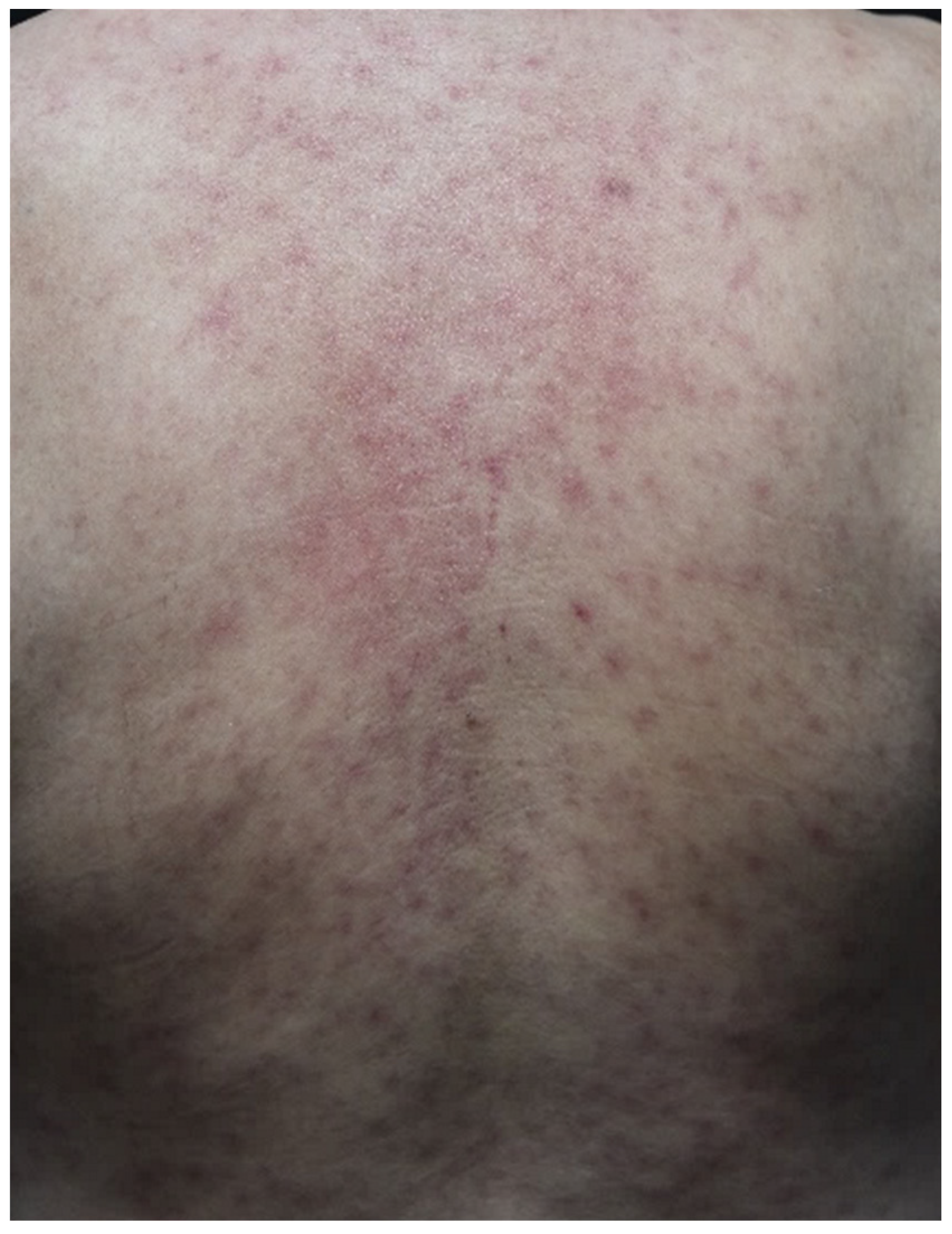
Figure 1.
Maculopapular eruption. Diffuse, asymptomatic, erythematous maculopapular rash on the trunk and four extremities in a patient with hepatocellular carcinoma who started atezolizumab treatment 15 days prior.
Figure 1.
Maculopapular eruption. Diffuse, asymptomatic, erythematous maculopapular rash on the trunk and four extremities in a patient with hepatocellular carcinoma who started atezolizumab treatment 15 days prior.

Figure 2.
Lichenoid dermatitis. Scattered pruritic, violaceous-to-erythematous, flat-topped scaly papules and plaques on the scalp, face, bilateral dorsal hands, and anterior chest, with a predilection for the sun-exposed area, in a patient with lung cancer receiving atezolizumab.
Figure 2.
Lichenoid dermatitis. Scattered pruritic, violaceous-to-erythematous, flat-topped scaly papules and plaques on the scalp, face, bilateral dorsal hands, and anterior chest, with a predilection for the sun-exposed area, in a patient with lung cancer receiving atezolizumab.

Figure 3.
Bullous pemphigoid. Multiple pruritic tense bullae with erosions on the background of urticarial patches on the trunk and four extremities in a patient with metastatic lung cancer treated with nivolumab.
Figure 3.
Bullous pemphigoid. Multiple pruritic tense bullae with erosions on the background of urticarial patches on the trunk and four extremities in a patient with metastatic lung cancer treated with nivolumab.

Table 1.
Summary of immune checkpoint inhibitors approved by the Food and Drug Administration.
| ICIs | Target | Indications |
|---|---|---|
| Ipilimumab | CTLA-4 | CRC, HCC, melanoma, mesothelioma, NSCLC, RCC |
| Nivolumab | PD-1 | CRC, esophageal SCC, HCC, HL, HNSCC, melanoma, mesothelioma, NSCLC, RCC, urothelial carcinoma |
| Pembrolizumab | PD-1 | breast cancer, cervical cancer, CRC, CSCC, endometrial carcinoma, esophageal carcinoma, gastric carcinoma, HCC, HL, HNSCC, melanoma, mesothelioma, MCC, NSCLC, large B-cell lymphoma, RCC, SCLC, urothelial carcinoma |
| Cemiplimab | PD-1 | BCC, CSCC, NSCLC |
| Atezolizumab | PD-L1 | breast cancer, HCC, melanoma, NSCLC, SCLC, urothelial carcinoma |
| Durvalumab | PD-L1 | NSCLC, SCLC, urothelial carcinoma |
| Avelumab | PD-L1 | MCC, RCC, urothelial carcinoma |
Abbreviations: ICIs, immune checkpoints inhibitors; CTLA-4, cytotoxic T-lymphocyte antigen 4; CRC, colorectal cancer; PD-1, programmed cell death 1; HCC, hepatocellular carcinoma; NSCLC, non-small cell lung cancer; RCC, renal cell carcinoma; SCC, squamous cell carcinoma; HL, Hodgkin’s lymphoma; HNSCC, head and neck squamous cell carcinoma; CSCC, cutaneous squamous cell carcinoma; MCC, Merkel cell carcinoma; SCLC, small cell lung cancer; BCC, basal cell carcinoma; PD-L1, programmed cell death receptor-1 ligand.
Table 3.
Other less-common dermatologic toxicities associated with immune checkpoint inhibitors.
| Less-Common cirAEs | Description | Suggested Managements |
|---|---|---|
| Alopecia areata/ universalis [101,102,103] |
|
|
| Sarcoidosis/ sarcoidosis-like reactions [107,108,109] |
| Dependent on the extent of Involvement.
|
| Erythema nodosum (EN) [109,110,111,112] |
|
|
| Sweet syndrome [26,113,114,115] |
|
|
| Pyoderma gangrenosum (PG) [113,116,117,118] |
|
|
| Dermatomyositis (DM) [22,119,120,121] |
|
|
| Grover’s disease (GD) [122,123] |
|
|
| Drug reaction with eosinophilia and systemic symptoms (DRESS) [124,125] |
|
|
| Acute generalized exanthematous pustulosis (AGEP) [126,127,128,129] |
|
|
Abbreviations: cirAEs, cutaneous immune-related adverse events; ICIs, immune checkpoint inhibitors; DPCP, diphenylcyclopropenone; SADBE, squaric acid dibutylester; NSAID, non-steroidal anti-inflammatory drug; anti-TIF1-γ antibody, anti-transcription intermediary factor 1-gamma antibody; CK, creatine kinase; CRP, c-reactive protein; ESR, erythrocyte sedimentation rate; IVIG, intravenous immunoglobulin; SCARs, severe cutaneous adverse reactions.
Publisher’s Note: MDPI stays neutral with regard to jurisdictional claims in published maps and institutional affiliations. |
© 2022 by the authors. Licensee MDPI, Basel, Switzerland. This article is an open access article distributed under the terms and conditions of the Creative Commons Attribution (CC BY) license (https://creativecommons.org/licenses/by/4.0/).
Share and Cite
MDPI and ACS Style
Chen, C.-H.; Yu, H.-S.; Yu, S. Cutaneous Adverse Events Associated with Immune Checkpoint Inhibitors: A Review Article. Curr. Oncol. 2022, 29, 2871-2886. https://0-doi-org.brum.beds.ac.uk/10.3390/curroncol29040234
AMA Style
Chen C-H, Yu H-S, Yu S. Cutaneous Adverse Events Associated with Immune Checkpoint Inhibitors: A Review Article. Current Oncology. 2022; 29(4):2871-2886. https://0-doi-org.brum.beds.ac.uk/10.3390/curroncol29040234
Chicago/Turabian StyleChen, Chieh-Hsun, Hsin-Su Yu, and Sebastian Yu. 2022. "Cutaneous Adverse Events Associated with Immune Checkpoint Inhibitors: A Review Article" Current Oncology 29, no. 4: 2871-2886. https://0-doi-org.brum.beds.ac.uk/10.3390/curroncol29040234