Brief Report

New Euthemistid Damsel–Dragonfly from the Middle Jurassic of Northern China (Odonata, Isophlebioidea, Euthemistidae)

1
College of Life Sciences, Capital Normal University, Xisanhuanbeilu 105, Haidian District, Beijing 100048, China
2
Guangdong Provincial Key Lab of Geological Processes and Mineral Resources, School of Earth Sciences and Engineering, Sun Yat-Sen University, Guangzhou 510275, China
3
Ningcheng National Geopark Administration, Chifeng 024200, China
4
Key Laboratory of Conservation and Application in Biodiversity of South China, School of Life Sciences, Guangzhou University, Guangzhou Higher Education Mega Center, #230 Waihuanxi Road, Guangzhou 510006, China
*
Authors to whom correspondence should be addressed.
Submission received: 22 February 2024 / Revised: 15 March 2024 / Accepted: 15 March 2024 / Published: 22 March 2024
(This article belongs to the Special Issue Diversity of Fossil and Recent Insect Faunae)

Abstract

:
A new genus and new species of the euthemistid, Kidaneuthemis ningchengensis gen. et sp. nov., is described from the Middle Jurassic of Inner Mongolia, China. It can be assigned to the Euthemistidae by the several long intercalary veins between RP1 and IR1, IR1 and RP2, as well as between RP2 and IR2 and between IR2 and RP3/4. Kidaneuthemis ningchengensis gen. et sp. nov. distinguishes from the other two genera of this family by the presence of not less than eight intercalary veins between MP and wing margin; about three rows of cells in the distal part between MA and MP; the base of IR2 is two cells distal to that of RP3/4. In addition, a revision of the family Euthemistidae has been proposed.

1. Introduction

The Isophlebioptera Bechly, 1996, is an ancient and large clade that prospered during the Triassic, Jurassic, and Early Cretaceous in Europe and Central Asia and was subdivided into four subgroups: Euthemistidae, Parazygoptera, Selenothemistidae, and Isophlebiida [1,2]. Up to now, only three species have involved two genera: Euthemis cellulata Pritykina, 1968 [3], Euthemis multivenosa Pritykina, 1968 [3], and Sinoeuthemis daohugouensis. Li, Nel, and Ren (2013) [2] have been reported in the family Euthemistidae. Of these, only one species, Sinoeuthemis daohugouensis, was described from the Jiulongshan Formation, Middle Jurassic of Daohugou, Inner Mongolia, China, where a large number of fossil animals (insects, dinosaurs, mammals, etc.) and plants (gymnosperm, ferns, etc.) have been found in this Lagerstätte [4,5,6].
Herein, we describe a new genus and species, Kidaneuthemis ningchengensis gen. et sp. nov., from the Middle Jurassic Jiulongshan Formation. This well-preserved fossil shows some important characteristics of the Euthemistidae, such as the several long intercalary veins between RP1 and IR1, IR1 and RP2, as well as between RP2 and IR2, and between IR2 and RP3/4; an extremely narrow post-discoidal area; numerous secondary antenodal cross-veins between the anterior wing margin and ScP distal of Ax2; and discoidal cells opened in the forewing and closed in the hind wing. As the second genus of the Euthemistidae found in China during the Mesozoic, K. ningchengensis gen. et sp. nov. has well preserved head, thorax, legs, wings, and abdomen, especially the female ovipositor, which can contribute to the further understanding of the morphology and taxonomy of Euthemistidae. In addition, a revision of the family Euthemistidae has been proposed.

2. Materials and Methods

2.1. Materials

This study is based on one specimen from the Middle Jurassic Jiulongshan Formation in Daohugou Village, Wuhua Township, Ningcheng County, Inner Mongolia, China. The specimens (holotype No. NCG012-1, No. NCG012-2) described herein is deposited in the Ningcheng Geological Park, Chifeng, Inner Mongolia. Based on the exploration of the stratigraphy, the age of the Jiulongshan Formation is approximately 164–165 Ma [5,6,7,8]. The specimens were observed and photographed using a stereoscopic Nikon SMZ25 microscope with an attached Nikon DS-Ri2 digital camera system. Line drawings were prepared with Affinity Designer 1.10 and Adobe Photoshop 2024.

2.2. Institutional Abbreviations

The nomenclature of the dragonfly wing venation used in this paper is based on the interpretations of Riek [9] and Riek and Kukalová-Peck [10], as modified by Nel et al. [11] and Bechly [12]. The wing abbreviations are as follows: AA, anterior anal; Arc, arculus; Ax0, Ax1, Ax2, primary antenodal crossveins; CuAa, distal branch of anterior cubitus; CuAb, proximal branch of anterior cubitus; IR1, IR2, intercalary radial veins; MA, distal branch of median anterior; MAb, posterior branch of median anterior; MP, posterior median; N, nodus; ‘O’, oblique vein; Pt, pterostigma; RA, anterior radius; RP, posterior radius; C, costa; ScP, posterior subcosta; Sn, subnodal cross-vein; Cr, nodal cross-vein; D.C., discoidal cell.

3. Results

3.1. Systematic Paleontology

Order Odonata Fabricius, 1793
Clade Isophlebioptera Bechly, 1996
Superfamily Isophlebioidea Handlirsch, 1906
Family Euthemistidae Pritykina, 1968
Type genus: Euthemis Pritykina, 1968; other genera included Sinoeuthemis Li, Nel, and Ren, 2013 and Kidaneuthemis gen. nov.
Emended familial diagnosis. Forewing: IR1 long, basally zigzagged, then smooth and slightly curved distally; postdiscoidal area extremely narrow, with one row of cells; areas between RP2 and IR2, IR2 and RP3/4, RP3/4 and MA, as well as between MA and MP before N separated by only one row of cells apart; area between RP3/4 and MA widen distally, while area between MA and MP slightly narrowed distally.
Other characteristics as in the diagnosis of Pritykina [3] and Li et al. [2]: several long intercalary veins between RP1 and IR1, IR1 and RP2, as well as between RP2 and IR2, and between IR2 and RP3/4 (intercalaries parallel to main longitudinal veins without apparent origin on them but originating in cross-venation); extremely narrow postdiscoidal area; not petiolate; numerous secondary antenodal crossveins between anterior wing margin and ScP distal of Ax2; discoidal cells opened in forewing and closed in hind wing; hind wing subdiscoidal area transverse, posteriorly closed, short and broad, with vein CuAb making a strong angle with AA; hind wing ‘gaff’ not very long; RP2 aligned with subnodus; crossveins in hind wing postdiscoidal space are not very long and not oblique.
Genus Kidaneuthemis gen. nov. Liu, Ren et Yang (契丹优美蜓属)
urn:lsid:zoobank.org:act:A4D5A72C-22F1-4BF8-A335-210514B379AA
Type species. Kidaneuthemis ningchengensis gen. et sp. nov. Liu, Ren et Yang
Etymology. The generic name is derived from the Latin “Kidani” (from which the locality of Ningcheng was once the capital of the Liao Dynasty, founded by the Khitan people) and Euthemis (type genus of this family).
Diagnosis. Fore- and hind wings falcate, slender, and longer than 100 mm. Forewing characteristics include (1) Pt short; (2) IR1 long, basally zigzagged, then smooth curved distally; (3) numerous intercalary veins between IRl and RP1, IR1 and RP2, RP2 and IR2, IR2 and RP3/4, as well as RP3/4 and MA, and these areas widened distally; (4) area between MA and MP narrows slightly distally, spaced three rows of cells; (5) discoidal space basally opened; (6) 10 near-parallel intercalary cross-veins in area between the MP and the wing margin; (7) CuAa short, divided into three branches. Hind wing: (1) Pt elongate; (2) IR1 long, basally slightly zigzagged, smooth, and curved distally; (3) presence of four cross-veins in area between the base of RP2 and the base of RP3/4; (4) discoidal cell basally closed; (5) lots of polygonal groups of cells separated by veinlets and intercalary veins between RPl and IR1, IR1 and RP2, RP2 and IR2, IR2 and RP3/4, as well as RP3/4 and MA; (6) cubital area broad, CuAa with three posterior branches; (7) not less than nine longer intercalary cross-veins between the MP and the wing margin. Body: ovipositor weakly curved and elongated, expanding beyond the end of the abdomen.
Kidaneuthemis ningchengensis gen. et sp. nov. Liu, Ren et Yang (宁城契丹优美蜓)
urn:lsid:zoobank.org:act:D5433533-0742-43E2-89DC-C7520E5D23BD
Etymology. The specific epithet is from the locality “Ningcheng county”, where the holotype yielded.
Diagnosis. As for the genus.
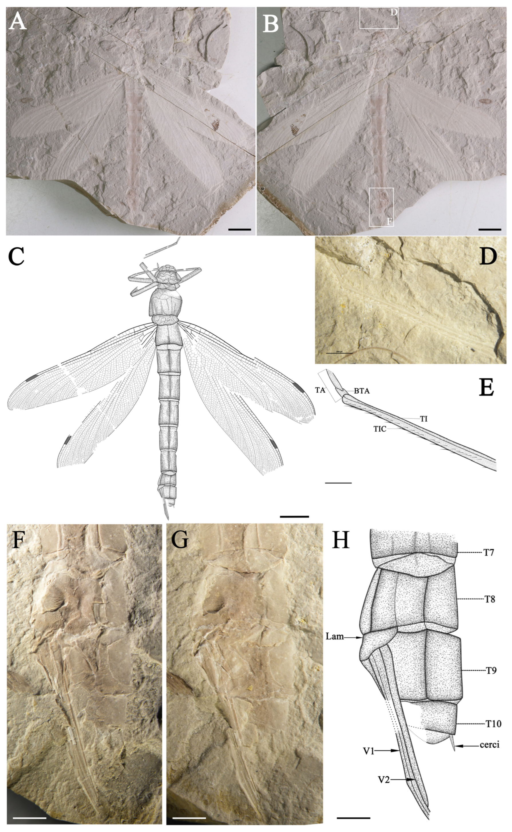
Holotype. No. NCG012-1, No. NCG012-2 (part and counterpart) Female. Imprint of a relatively complete specimen. Fore- and hind wings preserved almost intact with slight overlapped (Figure 1A–C and Figure 2).
Type locality and horizon. Jiulongshan Formation, Middle Jurassic (late Callovian); Daohugou Village, Wuhua Township, Ningcheng County, Inner Mongolia, China.

Description

Holotype. Female (hind wing anal margin rounded, preserved with ovipositor). Imprint of a relatively complete specimen. The insect carapace retains the head, thorax, two forelegs, one midleg and one hindleg, the abdomen, the female ovipositor, and the cerci attached to the most terminal abdominal segment. Fore- and hind wings preserved almost intact, with wing bases slightly overlapped.
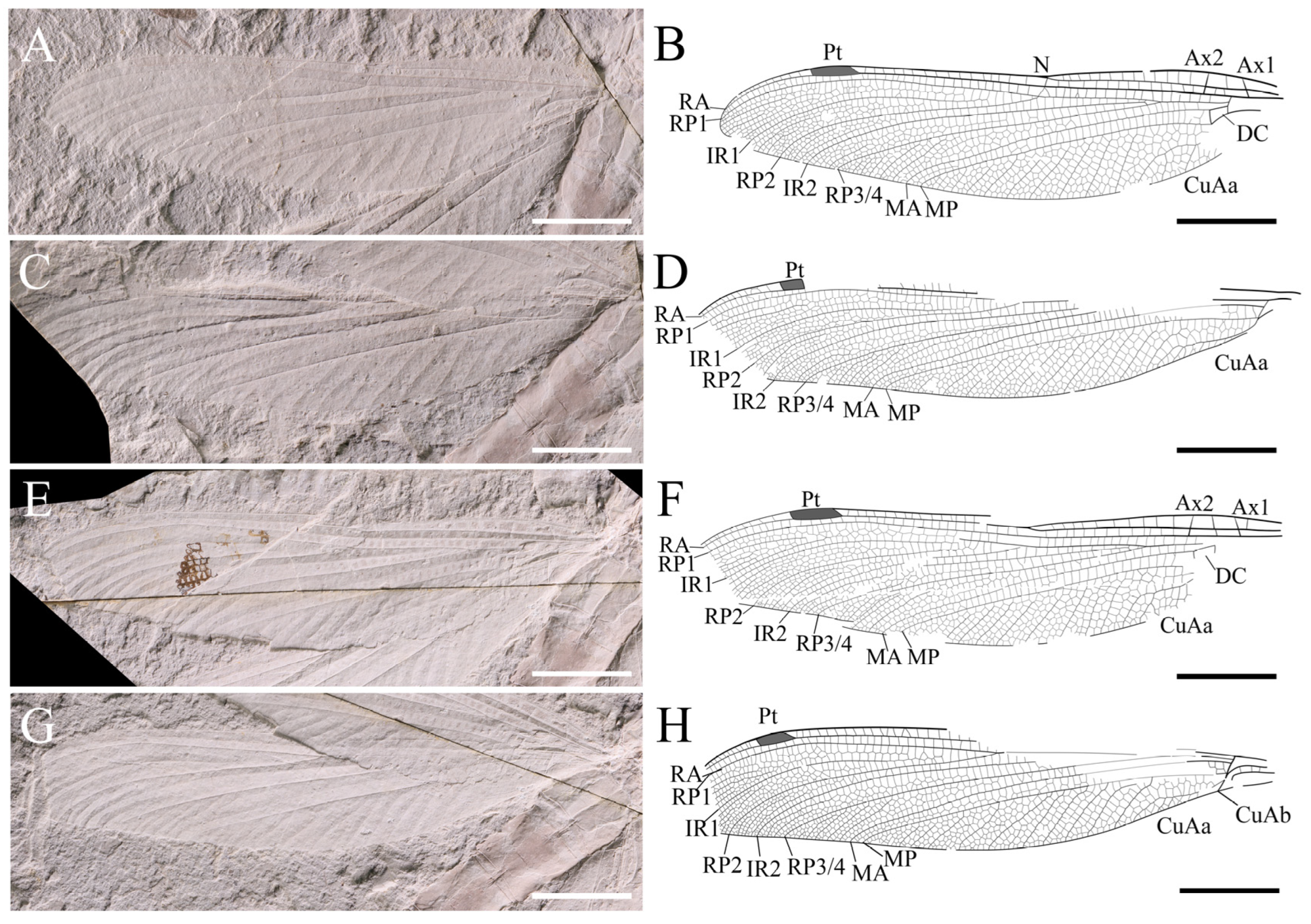
Forewing (left and right wings combined). Wing without petiolates. 117.2 mm long and 24.3 mm wide; ratio of width to length: 0.21; nodus invisible; from pterostigma to apex 14.9 mm; pterostigma 7.0 mm long, 1.8 mm wide. Discoidal space basally opened. Antenodal cross-veins out of sight, while not less than 20 posterior cross-veins before pterostigma. Pt shorter, covering four cells. IR1 long, originating from RP1, zigzagged at base, smooth distally, more or less parallel to RP1; RP2 originating at subnodus; RP2 and IR2 gently curved and parallel in basal part, with only one row of intervening cells, about seven or eight rows of cells in distal part. Area between RP2 and IR2 somewhat widened distally near posterior wing margin; area between RP3/4 and MA separated by only one row of cells at base and then broadened distally, with 14 rows of cells near posterior wing margin; zigzagged intercalary veins present in area between IRl and RP1, IR1 and RP2, RP2 and IR2, IR2 and RP3/4, as well as RP3/4 and MA; post-discoidal area with one row of cells in basal, becoming two rows of cells distally; MA fluctuates slightly at the origin of IR1, causing area between MA and MP to contract near wing margins. MAb 2.2 mm long and partially aligned with basal part of ‘gaff’. MP straight area between MP and CuA with 2–3 rows of cells; and area between MP and wing margin wide, with the widest space not less than seven rows of cells and about 10 intercalary veins originating from MP. CuA divided into CuAa and CuAb. CuAb points towards the posterior margin of the wing and short, CuAa runs almost parallel to the wing margin for some distance, forming a short and narrow cubito-anal area; CuAa branches towards the posterior end, with more than three posterior branches and two rows of cells at its broadest part. Area between MP and posterior wing margin broad, with about eight rows of cells at the widest part, and 10 almost parallel intercalary veins in this area.
Hind wing (left and right wings combined). Wing without short petiole; preserved length 114.2 mm, width 26.2 mm; ratio of width to length: 0.23. Distance from base to nodus 46.5 mm, distance from wing base to arculus 10.3 mm, from arculus to nodus 37.3 mm, from nodus to apex 67.8 mm, from nodus to pterostigma 39.0 mm, from pterostigma to apex 21.2 mm, from nodus to arculus 36.2 mm. Pterostigma 9.2 mm long, 1.7 mm wide. Primary antenodal cross-veins strong; Ax0 not preserved; Ax1 2.9 mm basal of arculus, disposed obliquely to ScP and R + MA; and Ax2 5.3 mm distal of arculus, with inverted obliquity. R + MA nearly straight; RP and MA parallel at base, separated at a slightly obtuse angle from RA in arculus; 14/12 secondary antenodal cross-veins present between C and ScP (left/right hind wing, respectively); thirteen secondary antenodal cross-veins present between ScP and RA distal of Ax2; and 19 antesubnodal cross-veins present between RA and RP, extending from Arc to Sn. Arculus, distinctly angled. Discoidal cells basally closed and free of cross-veins. MP nearly straight, slightly curved upwards two cells away from Ax2, and spaced two–three rows of cells away from CuAa. CuAa short and zigzagged, retaining only three branches and basally somewhat parallel to the posterior wing margin, with three or four rows of cells between. Area between the first and second branches of the left hind wing, CuAa wider than that of the right hind wing. The stem of the second branch of left hind wing, CuAa more curved, while part of right hind wing straight. Nodal well preserved; Sn fluctuates in the middle of the vein and aligns with the slightly tilted Cr. 16 postnodal cross-veins present between C and RA, not aligned with 21 cross-veins between RA and RP1 before Pterostigma. Area between RA and RP1 with one row of cells, two rows of cells exist after Pt. Pterostigma more elongated than the forewing and with a weak Pt-brace. IR1 long, smooth, and slightly zigzagged, originating at RP1 at a distance of six cells from Sn and more or less parallel to RP1. Area between RP1 and IR1, RP2 and IR2, and IR2 and RP3/4 strongly expanded in distal part, and more intercalary veins are present in these areas. RP2 aligned with subnodus; RP2 and IR2 smoothly curved, with only one row of intervening cells in basal part and about eight rows of cells in distal part. IR2 with an apparent origin in RP3/4; base of IR2 with two cells and 3.1 mm distal of base of RP3/4 in right hind wing. Area between RP3/4 and MA, separated by one row of cells basally and distally, strongly widened and presented with five subparallel intercalary veins of different lengths. MA slightly undulated in the middle of N and Pt. Post -discoidal area narrow distally, with one row of cells at the base between MA and MP and three rows of cells at the distal. Area between MP and wing margin board with approximately nine rows of cells spaced apart at the widest point, and about nine or 10 near-parallel intercalary veins in this area.
Body (part and counterpart combined). Total body length (from head to end of abdomen, including anal appendages) 172.0 mm. Head 17.1 mm long and 22.3 mm wide, with compound eyes large, preserved clypeus, post clypeus, and frons. Thorax 24.2 mm long and 20.2 mm wide, relatively well preserved, showing ventral view. Legs elongated, retaining four in part. Forelegs relatively well preserved, femur about 16.3 mm long, tibia 17.4 mm long, preserving two rows of tibial comb; tarsus retain only two-segmented, 1.8 mm long, in which basitarsus 0.84 mm long in counterpart; meso-leg partly preserved, femur about 16.9 mm long, tibia 16.2 mm long; meta-leg party preserved as well, femur more complete, about 18.4 mm, while tibia partially preserved (Figure 1D,E). The abdomen long and cylindrical, with 122.9 mm long, 18.0 mm at the widest, and 10.4 mm at the narrowest. The first, eighth, ninth, and tenth abdominal segments are shorter than the others. The second abdominal segment is 13.2 mm long, the third 15.5 mm long, and the third to seventh abdominal segments approximately the same length, decreasing in width. The eighth abdominal segment 8.2 mm long, the ninth 9.2 mm long, and the tenth 3.9 mm long. The presence of gonapophyses in the counterpart (valvulae of the cone-shaped ovipositor) between the eighth and ninth abdominal segments 22.7 mm long. The gonapophyses strongly sclerotized, only weakly curved, and extremely elongated, with the ovipositor extending far beyond the end of the abdomen. In addition, on the ventral margin of the last abdominal segment, one tiny structure visible, maybe the cercus (Figure 1F–H).

4. Discussion

The Isophlebioptera Bechly [12] is divided into four subgroups: Euthemistidae, Parazygoptera, Selenothemistidae and Isophlebiida [1,2]. As a result of the following characters: RP3/4 is slightly curved, more or less parallel to RP2 for some distance, and the space between RP3/4 and MA is significantly enlarged at the distal near posterior wing margin; the area between MA and MP is very narrow, with only one row of cells in between; and the distal space between MP and CuAa is strongly expanded [11]. Kidaneuthemis gen. nov. can be easily classified into the Isophlebioptera.
According to the diagnosis of Euthemistidae [2,3], there are several long intercalary veins parallel to the main longitudinal veins, with no apparent origin on them, but in cross-venation, between IR1 and RP1 and between IR1 and RP2, as well as between RP3/4 and IR2 and between IR2 and RP2. Additionally, there is an extremely narrow post-discoidal area without petiolate; numerous secondary antenodal cross-veins between the anterior wing margin and ScP distal of Ax2; discoidal cells opened in the forewing and closed in the hind wing; RP2 aligned with the subnodus; and cross-veins in the hind wing post-discoidal space that are not very long and not oblique. Kidaneuthemis gen. nov. is undoubtedly a member of this family.
The Euthemistidae comprises two Mesozoic genera: Euthemis Pritykina [3] and Sinoeuthemis Li et al. [2]. Kidaneuthemis gen. nov. is distinguished from the two genera by the presence of numerous (not less than nine) near-parallel intercalary veins in the area between the MP and the wing margin. Furthermore, compared to Euthemis, there is one row of cells at the base between MA and MP and three rows of cells at the distal in Kidaneuthemis gen. nov., instead of only one row. The difference with Sinoeuthemis is that Kidaneuthemis gen. nov. has two cells based on IR2 and RP3/4 instead of Sinoeuthemis with five cells.
This specimen has a relatively well-preserved, sclerotized, and slightly curved ovipositor tube attached to the eighth abdominal segment. Its structure looks very similar to that of extant dragonfly ovipositors [13], such as being overall sclerotized and curved and having the same oblique position in the cranial direction.
The ovipositor of female dragonflies falls into two main categories: ovipositor open-ended vulvar lamina and tubes with a saw-like or conical shape. The former is present in several anisopteran families, while the latter is present in all Zygoptera, Epiophlebiidae, and basal Anisoptera, especially in Petaluridae, Austropetaliidae, and Aeshnidae [14]. Depending on the morphology of the ovipositor, the spawning pattern varies greatly. The vulvar lamina is used to lay eggs by dipping the tip of the abdomen into the water during flight. The other type of ovipositor is used to insert a substrate, such as plant tissue. Kidaneuthemis ningchengensis gen. et sp. nov. possesses a long and strongly dependent saw-like ovipositor. Usually, this type of ovipositor is used to insert their eggs into the mud at the stream’s edge, called endophytic oviposition, which allows the eggs to be effectively protected, increasing the survival rate of the offspring [15]. So, K. ningchengensis gen. et sp. nov. maybe lived in a warm, humid climate and was close to streams or lakes.

5. Conclusions

A new euthemistid dragonfly, Kidaneuthemis ningchengensis gen. et sp. nov., is described from the Middle Jurassic Jiulongshan Formation of northern China. This is the second genus of the Euthemistidae discovered in China, which is rarely preserved with a complete female ovipositor in the fossil. This discovery enriches the diversity of the Euthemistidae and presents more complete morphological information about this family for further study.

Author Contributions

Conceptualization, Q.Y. and D.R.; methodology, Q.Y., D.R., Y.L. and C.S.; validation, Q.Y., D.R. and C.S.; investigation, Q.Y., D.R., C.S., J.S. and Y.L.; resources, Q.Y., D.R. and J.S.; data curation, Q.Y., D.R. and Y.L.; writing—original draft preparation, Y.L.; writing—review and editing, Q.Y., D.R., C.S., J.S. and Y.L.; supervision, Q.Y. and D.R.; project administration, Q.Y., D.R. and C.S.; funding acquisition, Q.Y., D.R. and C.S. All authors have read and agreed to the published version of the manuscript.

Funding

This research was funded by the National Natural Science Foundation of China, grant numbers 32070425, 42072012, and 32020103006, and the Natural Science Foundation of Guangdong Province, grant number 2021A1515012508.

Institutional Review Board Statement

Not applicable.

Data Availability Statement

This published work and the nomenclatural acts it contains have been registered in ZooBank, the online registration system for the ICZN (International Code of Zoological Nomenclature). The LSID (Life Science Identifier) for this publication is urn:lsid:zoobank.org:pub:0F0C5401-6006-4AF9-8B4E-A69BFBB0FD14.

Acknowledgments

We thank the Ningcheng National Geopark for providing the type specimen for us to study. The first author would like to express her sincere gratitude to Nan Yang and Ziqiang Xu (Capital Normal University) for their technical support.

Conflicts of Interest

The authors declare no conflicts of interest.

References

  1. Fleck, G.; Nel, A. The first isophlebioid dragonfly (Odonata: Isophlebioptera: Campterophlebiidae) from the Mesozoic of China. Palaeontology 2002, 45, 1123–1136. [Google Scholar] [CrossRef]
  2. Li, Y.J.; Nel, A.; Shih, C.K. The first euthemistid damsel-dragonfly from the Middle Jurassic of China (Odonata, Epiproctophora, Sophlebioptera). ZooKeys 2013, 261, 41–50. [Google Scholar] [CrossRef] [PubMed]
  3. Pritykina, L.N. Strekozy Karatau (Odonata) [Dragonflies from Karatau (Odonata)]. In Yurskie Nasekomye Karatau [Fossil insects from Karatau]; Panfilov, D.V., Ed.; Academy of Sciences of the S.S.S.R.; Nauka: Moscow, Russia, 1968; pp. 26–55. (In Russian) [Google Scholar]
  4. Huang, D.Y. The Daohugou Biota; Shanghai Science and Technology Publisher: Shanghai, China, 2016; p. 332. [Google Scholar]
  5. Ren, D.; Shih, C.K.; Gao, T.P.; Wang, Y.J.; Yao, Y.Z. Rhythms of Insect Evolution-Evidence from the Jurassic and Cretaceous in Northern China; Wiley Blackwell: Hoboken, NJ, USA, 2019; p. 710. [Google Scholar]
  6. Ren, D.; Gao, K.Q.; Guo, Z.G.; Ji, S.A.; Tan, J.J.; Song, Z. Statigraphic division of the Jurassic in the Daohugou area, Ningcheng, Inner Mongolia. Geol. Bull. China 2002, 21, 584–591. (In Chinese) [Google Scholar]
  7. Gao, T.P.; Shih, C.K.; Ren, D. Behaviors and interactions of insects in Mid-Mesozoic ecosystems of Northeastern China. Annu. Rev. Entomol. 2021, 66, 337–354. [Google Scholar] [CrossRef] [PubMed]
  8. Wang, B.; Li, J.F.; Fang, Y.; Zhang, H.C. Preliminary elemental analysis of fossil insects from the Middle Jurassic of Daohugou, Inner Mongolia and its taphonomic implications. Chin. Sci. Bull. 2009, 54, 783–787. [Google Scholar] [CrossRef]
  9. Riek, E.F. A new collection of insects from the Upper Triassic of South Africa. Ann. Natal Mus. 1976, 22, 791–820. [Google Scholar]
  10. Riek, E.F.; Kukalová-Peck, J. A new interpretation of dragonfly wing venation based upon Early Carboniferous fossils from Argentina (Insecta: Odonatoidea) and basic character states in pterygote wings. Can. J. Zool. 1984, 62, 1150–1166. [Google Scholar] [CrossRef]
  11. Nel, A.; Martínez-Delclὸs, X.; Paicheler, J.C.; Henrotay, M. Les ‘Anisozygoptera’ Fossiles. Phylogénie et Classification (Odonata); Société Française d’Odonatologie: Bois-d’Arcy, France, 1993; p. 311. [Google Scholar]
  12. Bechly, G. Morphologische Untersuchungen am Flügelgeäder der rezenten Libellen und deren Stammgruppenvertreter (Insecta; Pterygota; Odonata), unter besonderer Berücksichtigung der Phylogenetischen Systematik und des Grundplanes der Odonata. Petalura 1996, 2, 1–402. [Google Scholar]
  13. Schneider, T.; Vierstraete, A.; Müller, O.; van Pelt, G.J.; Caspers, M.; Ikemeyer, D.; Snegovaya, N.; Dumont, H.J. Taxonomic revision of eastern part of Western Palaearctic Cordulegaster using molecular phylogeny and morphology, with the description of two new species (Odonata: Anisoptera: Cordulegastridae). Diversity 2021, 13, 667. [Google Scholar] [CrossRef]
  14. Bechly, G. New results concerning the morphology of the most ancient dragonflies (Insecta: Odonatoptera) from the Namurian of Hagen-Vorhalle (Germany). J. Zool. Syst. Evol. Res. 2001, 39, 209–226. [Google Scholar] [CrossRef]
  15. Tillyard, R.J. The Biology of Dragonflies (Odonata or Paraneuroptera); Cambridge University Press: London, UK, 1917; p. 396. [Google Scholar]
Figure 1. Holotype of Kidaneuthemis ningchengensis gen. et sp. nov., (A,B), photographs of the holotype, No. NCG012-1, No. NCG012-2 (part and counterpart); (C), line drawing of the holotype; (D,E), region of the foreleg (as located on (B)). TA, tarsus; BTA, basitarsus; TI, tibia; TIC, tibial comb; (FH), photographs of the ovipositor (lateral view) (as located on (F)). Lam, basal plate of ovipositor (lamina valvarum); V1, first valvulae/ventral valvifers; V2, second valvulae/inner valvulae. Scale bars = 20 mm (AC) and 2 mm (DH).
Diversity 16 00191 g001
Figure 2. Holotype of Kidaneuthemis ningchengensis gen. et sp. nov., (A,B), photograph and line drawing of left hind wing; (C,D), photograph and line drawing of left forewing; (E,F), photograph and line drawing of right hind wing; (G,H), photograph and line drawing of right forewing. Scale bars = 20 mm.
Diversity 16 00191 g002
Disclaimer/Publisher’s Note: The statements, opinions and data contained in all publications are solely those of the individual author(s) and contributor(s) and not of MDPI and/or the editor(s). MDPI and/or the editor(s) disclaim responsibility for any injury to people or property resulting from any ideas, methods, instructions or products referred to in the content.

