Article

Diversity of Flea Species (Siphonaptera) and Their Vector-Borne Pathogens from Bats (Chiroptera) in Lithuania

by
Povilas Sakalauskas
*,
Indrė Lipatova
,
Loreta Griciuvienė
,
Irma Ražanskė
,
Justina Snegiriovaitė
and
Algimantas Paulauskas
Department of Biology, Faculty of Natural Sciences, Vytautas Magnus University, K. Donelaičio Str. 58, LT-44248 Kaunas, Lithuania
*
Author to whom correspondence should be addressed.
Submission received: 29 February 2024 / Revised: 18 March 2024 / Accepted: 20 March 2024 / Published: 22 March 2024

Abstract

:
Bat fleas (Insecta: Siphonaptera: Ischnopsyllidae) are highly host-specific ectoparasites distributed worldwide (except Antarctica). The identification of flea species and the detection of flea-borne pathogens plays a significant role not only in veterinary and public health, but also in providing insights into the ecology, behaviour, and geographical distribution of fleas and flea-associated pathogens. The present study aimed to conduct a morphological and molecular identification of fleas collected from Lithuanian bats, as well as to screen for the presence of vector-borne pathogens. A total of 58 flea specimens were collected from bats in Lithuania. Based on morphological analysis, seven flea species were identified (Ischnopsyllus variabilis, I. octactenus, I. simplex, I. elongatus, I. hexactenus, Nycteridopsylla pentactena, and N. eusarca) and screened for the presence of Anaplasma spp., Borrelia spp., Babesia spp., Bartonella spp., Rickettsia spp., and Mycoplasma spp. Molecular analysis of fleas’ COI and 18S rRNA genes showed that the flea species collected from bats are closely genetically related. Bartonella DNA was detected in bat fleas (41.4%). No DNA of Anaplasma, Borrelia, Babesia, Rickettsia, and Mycoplasma was amplified in the tested samples. The Wolbachia sp. was accidentally found in I. elongatus. The phylogenetic analysis of four Bartonella genes (16S-23S rRNA ITS, gltA, rpoB, and groEL) revealed specificity to bats or bats’ ectoparasites. This is the first report about bat flea species and the pathogens in bat fleas in Lithuania. New host records for bat fleas were also reported. This study extends the knowledge about bat fleas and their role as vectors of different pathogens.

1. Introduction

Fleas (Siphonaptera) are globally distributed blood-sucking ectoparasites of mammals and birds that can play an important role in transmitting various pathogens. Currently, more than 2500 species in 16 families of fleas are known [1]. Fleas of the Ischnopsyllidae family, consisting of 20 genera and 125 species, are mainly parasites of bats [2]. The fleas of this family are highly host-specific but occasionally can be found on other bat species [3].
Bats (Chiroptera) are known as the second largest group of mammals, following rodents [4,5]. They form dense and large colonies and are characterized by a long lifespan [6]. These traits create appropriate conditions for bats to be reservoir hosts for different pathogens [7]. During the COVID-19 pandemic period, bats triggered a great interest [8]. Many research studies have focused on bat-associated viruses. Meanwhile, information about the bacterial pathogens in bats and their ectoparasites is limited [7,9].
In Lithuania, 15 bat species belonging to the Vespertilionidae family were recorded [10,11]. Only a few studies have focused on the distribution of Lithuanian bats [10,12]. Meanwhile, the fauna of bat ectoparasites has not been investigated and the species composition of fleas on Lithuanian bats is not known. Currently, no information about the pathogens circulating in Lithuanian bats and their ectoparasites is available. To date, only a small number of studies have been published about the diversity of bat flea species [13,14,15,16] and the pathogens they might carry in Europe [17,18]. This highlights a need for studies to obtain knowledge about bat fleas and their role as vectors of different pathogens. Accordingly, the aims of this study were (i) to perform a morphological and molecular identification of the fleas collected from Lithuanian bats and (ii) to screen the presence of vector-borne pathogens (Anaplasma spp., Babesia spp., Bartonella spp., Borrelia spp., Rickettsia spp., and Mycoplasma spp.) in bat fleas.

2. Materials and Methods

2.1. Flea Collection and Species Identification

Fleas were collected from both live and deceased bats by brushing the fur with tweezers, between 2019 and 2021. Live bats were either observed during ringing activities at the Ventės Ragas Ornithological Station or incidentally found in buildings. The carcasses of collected bats were stored at −40 °C and inspected for fleas before dissections. All aspects of this study were performed according to guidelines established for the ethical treatment of animals and complied with current Lithuanian regulations.
The fleas collected from each bat were placed in separate 1.5 mL tubes with 70% ethanol and were kept at +4 °C until investigation. Fleas were morphologically identified at the species level with taxonomic keys [19]. DNA was extracted from each specimen individually, using 2.5% ammonium hydroxide, while retaining flea exoskeletons [20]. The flea exoskeletons were mounted on permanent slides in Canada balsam [21]. The subsequent identification of the species was carried out by analysing the 18S ribosomal RNA (18S rRNA) and cytochrome c oxidase subunit I (COI) genes.
Conventional PCR was performed to amplify a ~1000 bp fragment of flea 18S rRNA using one primer set 18ai 5′-CCT GAG AAA CGG CTA CCA CAT C-3’ and 18bi 5′-GAG TCT CGT TCG TTA TCG GA–3’ [22]. Each 20 µL reaction contained 10 µL of 2x DreamTaq Green PCR Master Mix (Thermo Fisher Scientific, Vilnius, Lithuania), 5 µL of nuclease-free water, 1 µL (10 mM) of each primer, and 3 µL of DNA. The PCR reaction conditions were as follows: initial denaturation at 94° for 2 min; followed by 35 cycles of denaturation at 94° for 1 min, annealing at 58° for 1 min, and extension at 72° for 1 min; with a final extension at 72° for 5 min, after the completion of all cycles. To amplify a 709 bp fragment of the COI gene, conventional PCR with primers LCO1490 5′-GGT CAA CAA ATC ATA AAG ATA TTG G-3’ and HCO2198 5′-TAA ACT TCA GGG TGA CCA AAA AAT CA-3’ was conducted [23]. The PCR reaction was performed in a 20 µL mixture containing 10 µL of 2x DreamTaq Green PCR Master Mix (Thermo Fisher Scientific, Vilnius, Lithuania), 2.6 µL of nuclease-free water, 1.7 µL (10 mM) of each primer, and 4 µL of DNA. The PCR reaction conditions were as follows: initial denaturation at 94° for 1 min; followed by 5 cycles at 94° for 1 min, at 45° for 1.5 min, and at 72° for 1.5 min; followed by 35 cycles at 94° for 1 min, at 50° for 1.5 min, and at 72° for 1 min; with a final extension at 72° for 5 min after the completion of all cycles. All purified PCR products were prepared for DNA sequencing.

2.2. Pathogen DNA Amplification

Screening for the presence of six pathogens, Anaplasma spp., Borrelia spp., Babesia spp., Bartonella spp., Rickettsia spp., and Mycoplasma spp., was conducted by using different PCR assays with specific primers, as previously described (Supplementary Table S1) [24,25,26,27,28,29,30,31,32,33,34,35,36,37,38,39,40,41].
Multiplex-tandem (MT) real-time PCR for vector-borne pathogens was used to screen the samples. MT real-time PCR for Anaplasma, Borrelia, and Babesia species targeting msp2, 23S rRNA, and 18S rRNA genes, respectively, was run, as previously described [24,36]. To further screen MT real-time PCR for Bartonella and Rickettsia species targeting ssrA and gltA genes, respectively, was used, as previously described [31,36]. Results were considered positive when repeats yielded cycle threshold (Ct) values <38.00. All flea samples were further identified via conventional and nested PCRs targeting the specific genes of different pathogens. All PCR reactions were performed as previously described (Supplementary Tables S1 and S2). Positive (DNA of target pathogen, confirmed by sequencing) and negative (sterile, double-distilled water) controls were included in each PCR run. PCR products were visualized under UV light after electrophoresis on 1.5% agarose gel with ethidium bromide.

2.3. Sequencing

PCR products of all positive samples were extracted from agarose gel and purified using the GeneJET™ Gel Extraction Kit (Thermo Fisher Scientific, Vilnius, Lithuania) and sent to a sequencing service (Macrogen, Amsterdam, The Netherlands). The obtained DNA sequences were edited using the MegaX software package v10.2.6 [42] and aligned with each other, as well as with the previously published sequences in GenBank using the ClustalW multiple alignment option and BLASTn. The most appropriate model of nucleotide substitution for each alignment data set was determined according to the Bayesian information criterion (BIC). The phylogenetic tree was conducted using the Maximum Likelihood method and Tamura 3-parameter model. Outgroup taxa were obtained from GenBank. Bootstrap support was calculated by means of 1000 replicates.
Sequences for representative samples obtained in this study were submitted to the GenBank database under the accession numbers PP057939–PP057943 and PP407542–PP407543 for the flea 18S rRNA gene, PP425999–PP426007 for the flea COI gene, PP430158–PP430160 for the Wolbachia, PP296528–PP296548 for the Bartonella 16S-23S rRNA ITS region, PP333876–PP333895 for the Bartonella groEL gene, PP333854–PP333875 for the Bartonella rpoB gene, and PP333838–PP333853 for the Bartonella gltA gene. Detailed information about the uploaded Bartonella sequences to GenBank, with accession numbers, is provided in Supplementary Table S3.

3. Results

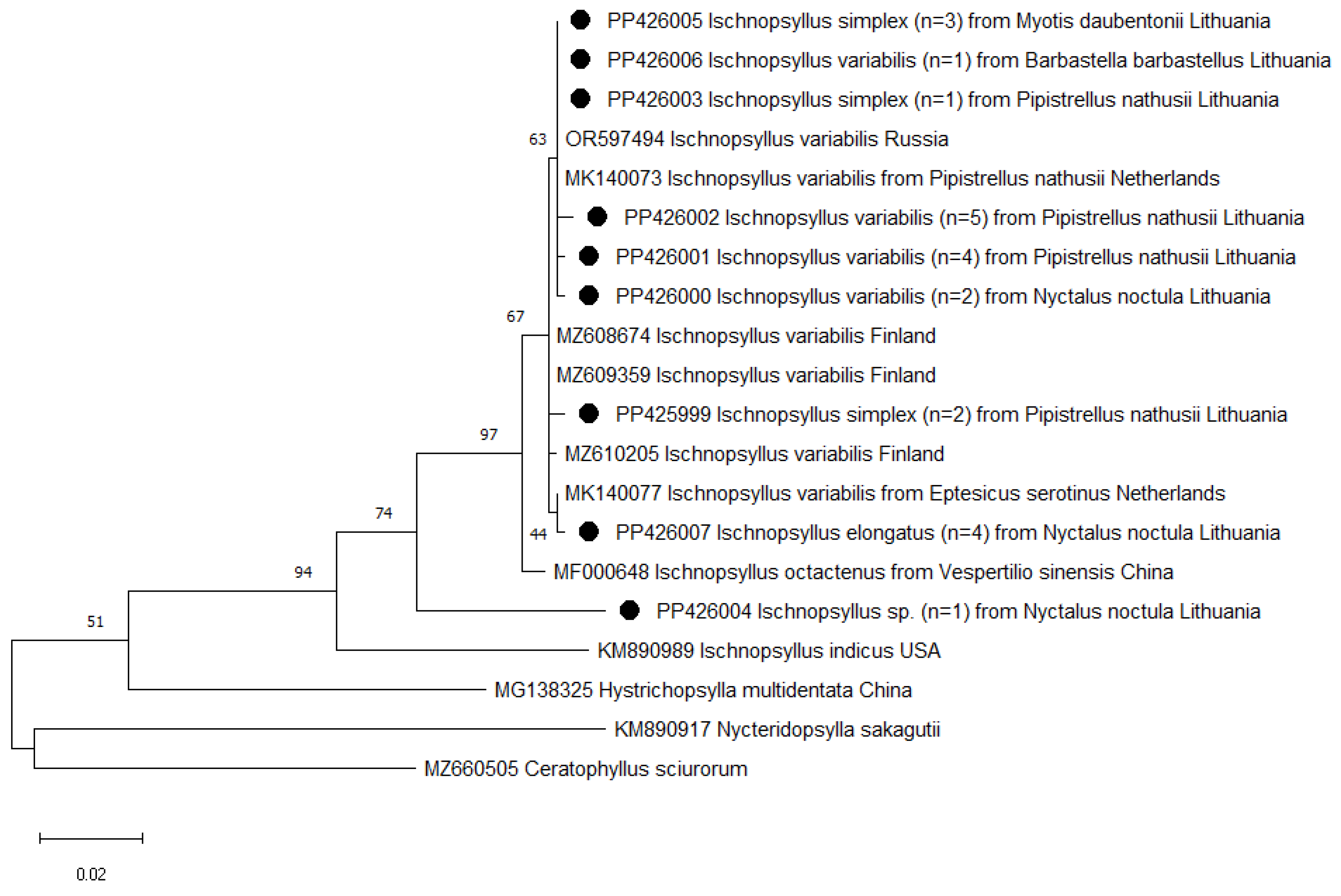
3.1. Identification of Flea Species

A total of 58 fleas were collected from 41 bats, representing 6 species, Pipistrellus nathusii (Keyserling and Blasius, 1839) (n = 32), Pipistrellus pygmaeus (Leach, 1825) (n = 2), Nyctalus noctula (Schreber, 1774) (n = 4), Eptesicus nilssonii (Keyserling and Blasius, 1839) (n = 1), Myotis daubentonii (Kuhl, 1817) (n = 1), and Barbastella barbastellus (Schreber, 1774) (n = 1). Seven species of fleas were identified morphologically, Ischnopsyllus variabilis (Wagner, 1898) (n = 24), Ischnopsyllus octactenus (Kolenati, 1856) (n = 16), Ischnopsyllus simplex (Rothschild, 1906) (n = 6), Ischnopsyllus elongatus (Curtis, 1832) (n = 4), Nycteridopsylla pentactena (Kolenati, 1856) (n = 5), Ischnopsyllus hexactenus (Kolenati, 1856) (n = 1), and Nycteridopsylla eusarca (Dampf, 1908) (n = 1) (Supplementary Figures S1–S7). One flea specimen collected from N. noctula was unidentified to species level (Table 1).
Analysis of the 18S rRNA data revealed low variability, showing only one nucleotide difference between the Ischnopsyllus and Nycteridopsylla genus (G and C, respectively). At the genus level, all obtained sequences (n = 23) were 100% identical to each other and to sequences deposited in GenBank (Figure 1). Meanwhile, the partial sequences of the COI gene showed divergences on an individual level among flea specimens. A total of six variable positions were found between I. variabilis and four between I. simplex specimens (Supplementary Table S4). However, different bat flea species did not form separate clusters (Figure 2). All sequences from Lithuanian bat fleas had a maximum similarity to fleas from GenBank, varying between 93.8 and 100%. Moreover, the presence of Wolbachia endosymbionts was detected in three I. elongatus specimens using COI gene fragments. The NCBI BLAST analysis revealed that these sequences were 96.9–99.7% similar to the Wolbachia endosymbiont found in other insect species.

3.2. Detection of Pathogens

Results of real-time PCR revealed the presence of Anaplasma (3.4%, 2/58), Borrelia (5.2%, 3/58), Babesia (8.6%, 5/58), and Bartonella (41.4%, 24/58) DNA in bat fleas (Table 2). No DNA of Rickettsia was detected in the tested samples. DNA from fleas was further tested using a conventional and nested PCR for additional Anaplasma spp., Borrelia spp., Babesia spp., Bartonella spp., Rickettsia spp., and Mycoplasma spp. identification. Only Bartonella DNA was successfully detected in bat fleas (39.7%, 23/58) using nested PCR (Table 2). All Bartonella-positive samples were subjected to sequence analysis.

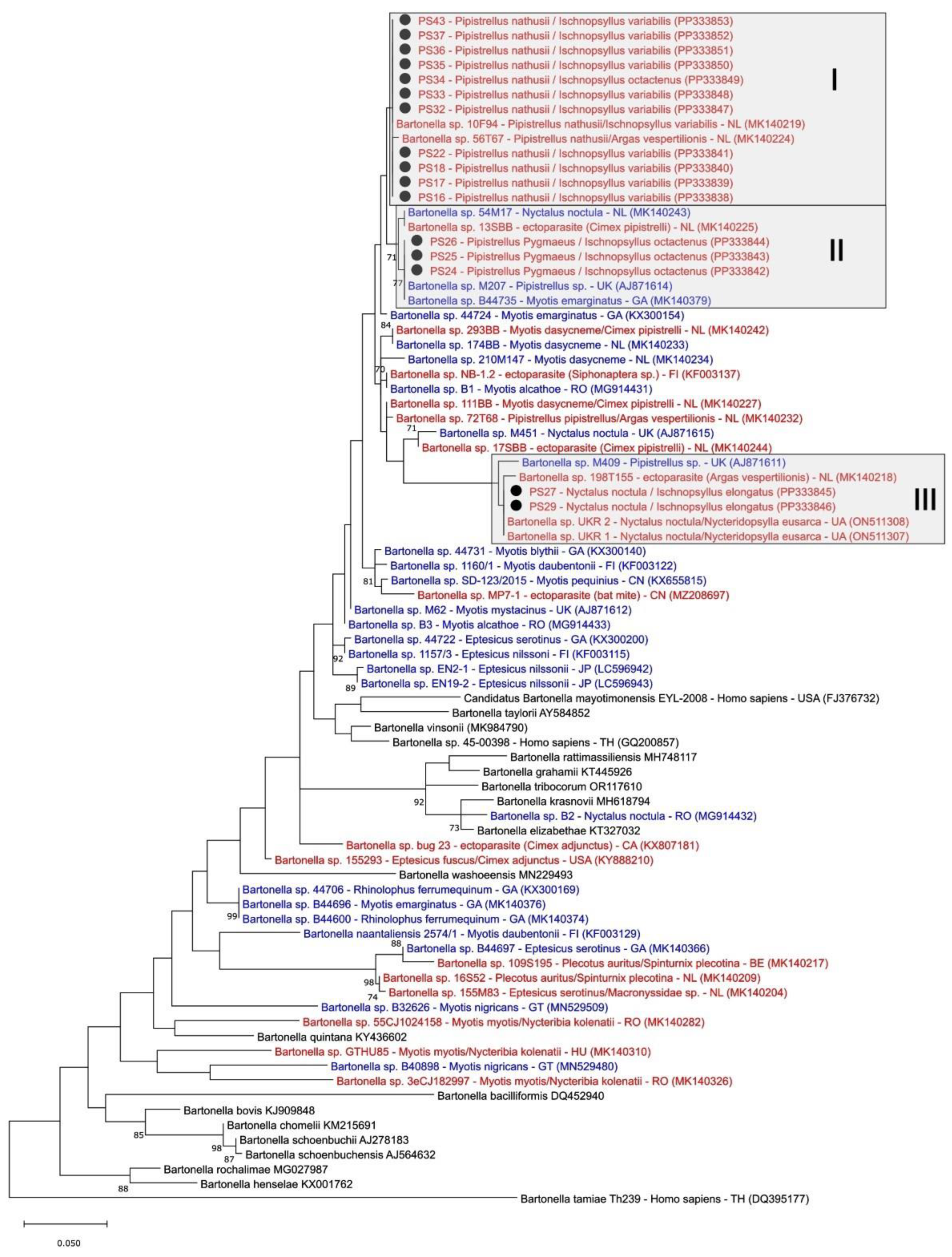
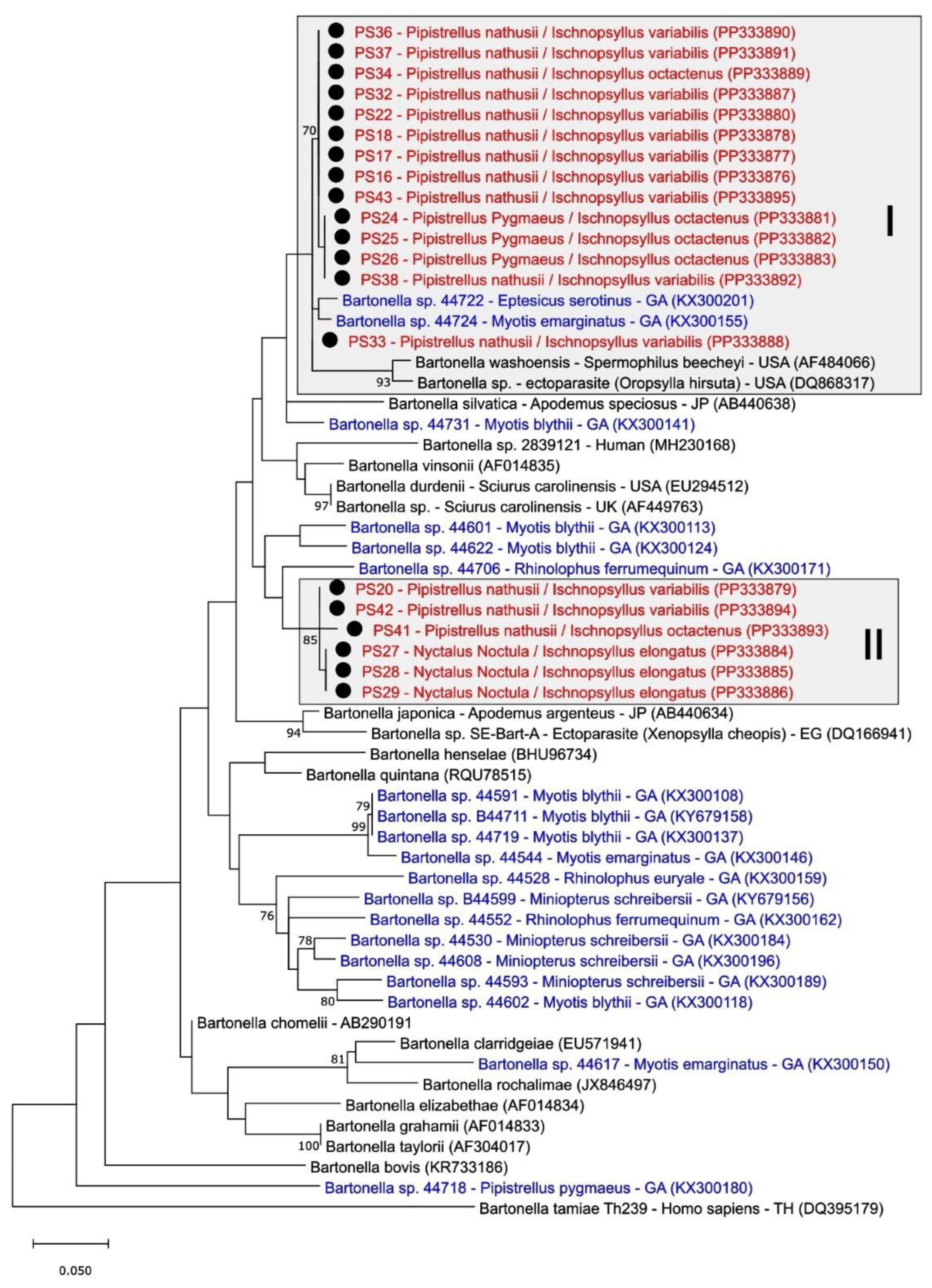
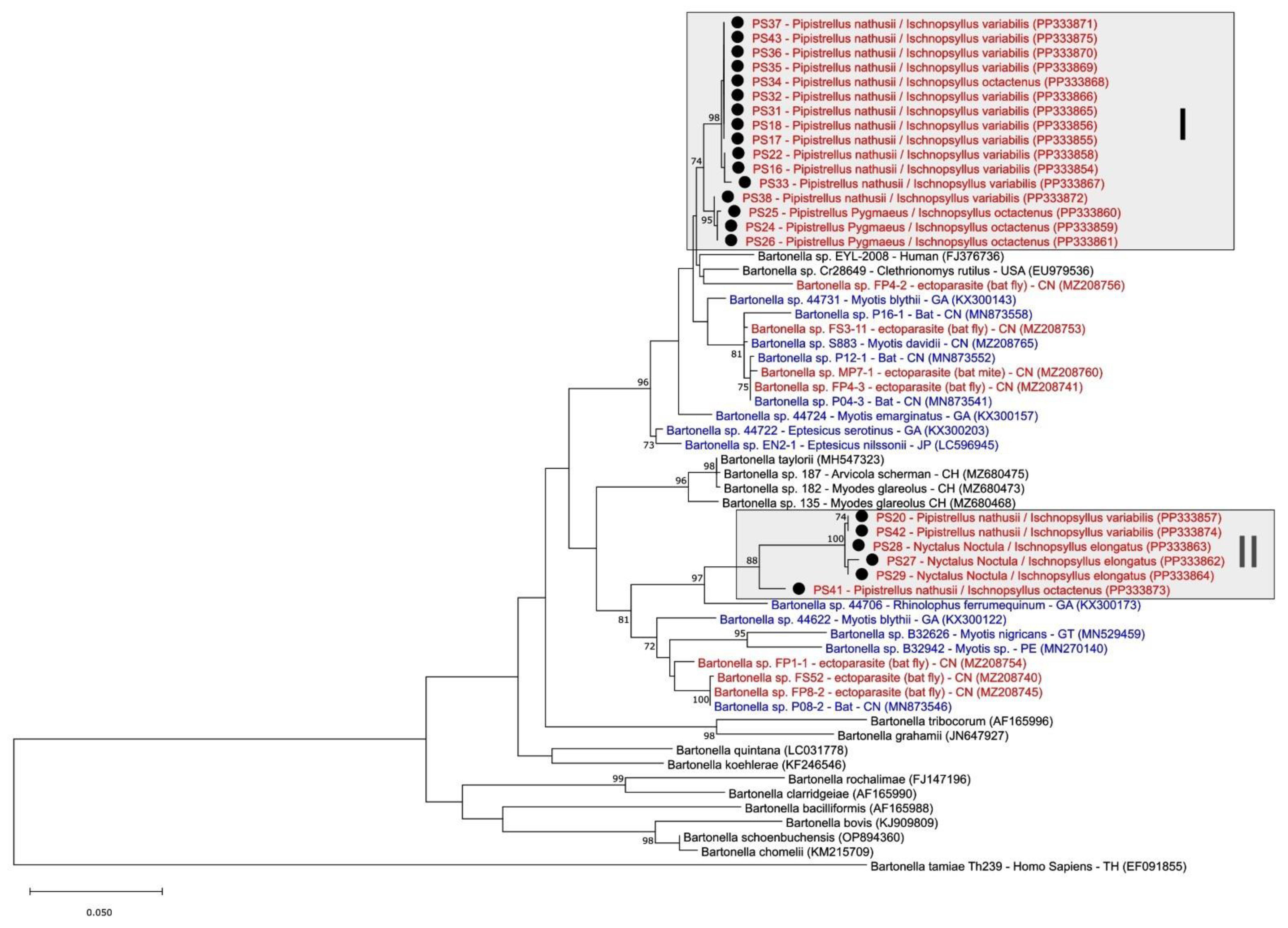
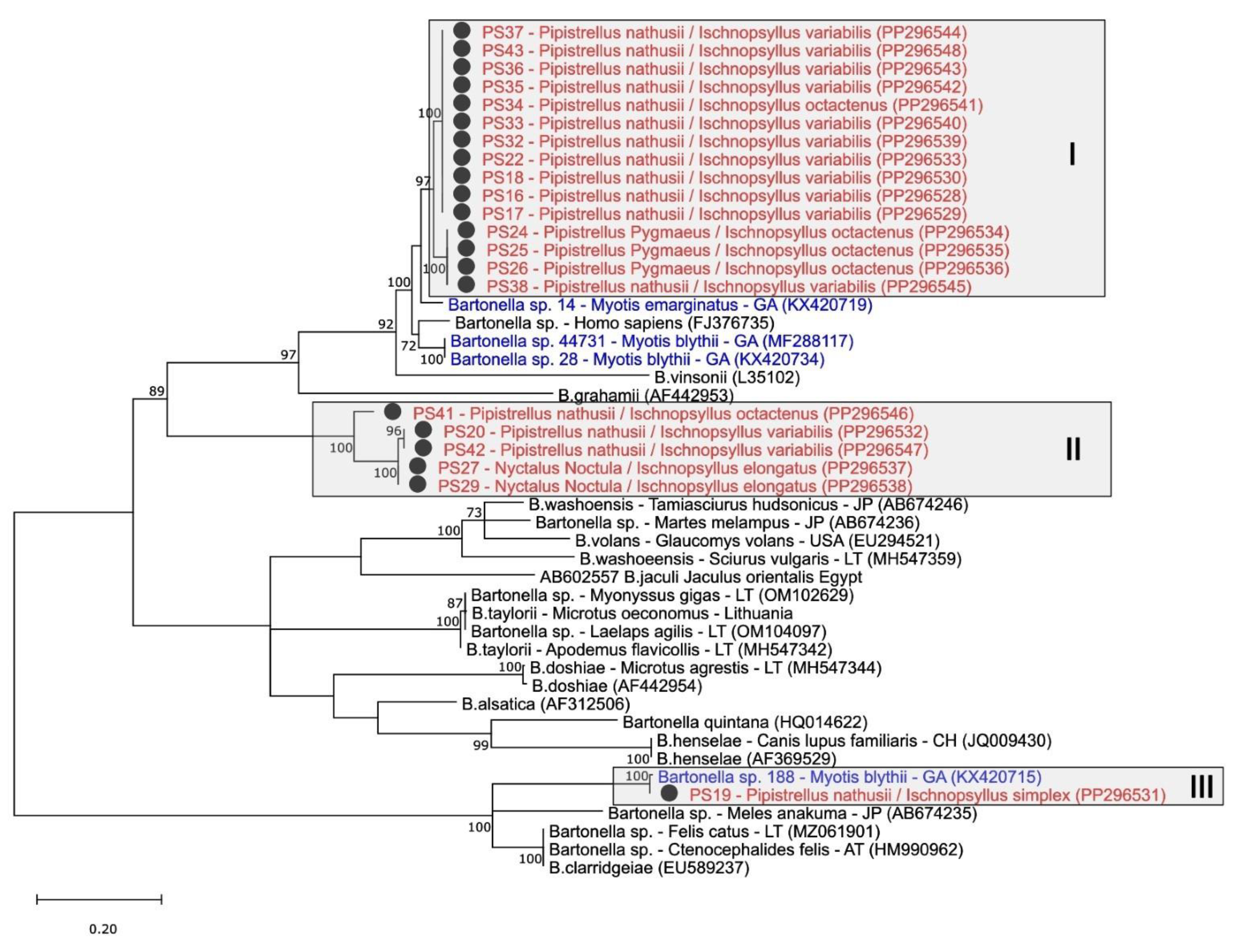
3.3. Phylogenetic Analysis of Bartonella spp. in Fleas

The phylogenetic analysis of Bartonella spp. detected from bat fleas of three species (I. variabilis, I. octactenus, and I. elongatus) collected from three different bat species (P. nathusii, P. pygmaeus, and N. noctula) was conducted through the examination of partial gltA, groEL, rpoB, and 16S-23S rRNA ITS sequences.
A total of 16 partial gltA gene (334 bp) sequences from fleas were obtained in this study. The derived sequences were clustered in three separate groups and had 23 variable sites between each other. The BLAST analysis showed a 97–100% similarity with Bartonella sp. sequences from the GenBank database. The sequences belonging to the first cluster were 100% identical to the Bartonella sp. sequences from the I. variabilis species flea collected from P. nathusii from the Netherlands. The second cluster sequences were 100% identical to sequences derived from two bat species (Myotis emarginatus (Geoffroy Saint-Hilaire, 1806) from Georgia and Pipistrellus sp. from United Kingdon). The sequences belonging to the third cluster had a 97% similarity with the sequences derived from the N. eusarca species of fleas from Ukraine (Figure 3).
A total of 20 partial groEL gene (290 bp) sequences were obtained in this study. Derived sequences were clustered in 2 separate clades and had 21 variable sites between each other. The BLAST analysis of sequences from the first cluster showed 97–99% similarity with Bartonella sp. sequences from M. emarginatus and Eptesicus serotinus (Schreber, 1774) bat species from Georgia. The rest of the sequences obtained in this study formed the second cluster, which is unique and exhibited only a 94–95% similarity with sequences from Rhinolophus ferrumequinum (Schreber, 1774) and Myotis blythii (Tomes, 1857) from Georgia (Figure 4).
A total of 22 partial rpoB gene (819–831 bp) sequences were obtained in this study. Derived sequences were clustered in 2 separate clades and had 100 variable sites between each other. The BLAST analysis of sequences from the first cluster showed a 97–98% similarity with sequences from the Clethrionomys rutilus (Pallas, 1779) vole from the USA and the Myotis davidii (Peters, 1869) bat from China. The sequence from the second cluster had a 93–96% similarity with sequences from the bat fly from China and the h. ferrumequinum bat from Georgia and formed a unique cluster (Figure 5).
A total of 21 partial (717–836 bp) 16S-23S rRNA ITS region sequences were obtained in this study. Derived sequences were clustered in 3 separate clades and had 290 variable sites between each other. The first clade sequences clustered near the Bartonella sp. sequence from M. emarginatus from Georgia and had an 89% similarity. The second clade compounded five unique sequences, obtained in our study, and exhibited the highest similarity (90%) to the Bartonella sequences from the Cimex adjunctus (Barber, 1939) bat bug from the USA. The third clade contains only one sequence derived from our study which has a 99.8% similarity to the Bartonella sp. from M. blythii from Georgia (Figure 6).

4. Discussion

The present study is the first report on fleas infesting bats in Lithuania. This finding presents seven flea species (I. variabilis, I. octactenus, I. simplex, I. elongatus, I. hexactenus, N. pentactena, and N. eusarca), extending the knowledge about the Lithuanian flea fauna. Also, it presents new data about the distribution of fleas belonging to the Ischnopsyllidae family in Europe.
Ischnopsyllus octactenus and I. variabilis were the main flea species collected from bats in this study. They are distributed in Europe, North Africa, and Central Asia [21]. In this study, I. octactenus was collected from both P. nathusii and P. pygmaeus, while I. variabilis was collected from P. nathusii, N. noctula and B. barbastellus. As far as we know, this is the first report of the I. variabilis flea species being collected from B. barbastellus. Pipistrellus spp. Bats are the principal host of these fleas [3]. However, there are records of findings on other bat species [3,15,43].
Six specimens of the flea I. simplex were collected from the M. daubentonii and P. nathusii bat species. To the best of our knowledge, this is the first report of the I. simplex flea species parasitizing P. nathusii. Previously, it was recorded on bats of Myotis spp., Rhinolophus spp., B. barbastellus, Plecotus auritus, Eptesicus nilssonii, Vespertilio murinus, and P. pipistrellus from European countries [3,15,43].
Ischnopsyllus elongatus was found on the N. noctula bat species in this study. Nyctalus noctula is the principal host of this flea species. It is distributed in Europe and Asia and was also previously recorded on other bat species [3,15,21,43].
Bats of Plecotus auritus and Myotis sp. are the principal hosts for fleas of the I. hexactenus species [3,21]. In this study, one specimen of I. hexactenus was found on B. barbastellus. This is not surprising as it has also been detected in other European countries [13,15,21]. Moreover, this flea species was reported from bats of the Pipistrellus, Plecotus, and Eptesicus genera [3,13,15].
According to Table 1, two winter flea species were found, as follows: N. eusarca and N. pentactena. One specimen of N. eusarca has been observed from N. noctula, which is the true host of these rare fleas [3,15,18,21]. Meanwhile, five specimens of N. pentactena were found on E. nilssonii. These flea species are distributed in European countries. Previously, they were recorded from bats of B. barbastellus, Plecotus spp., Myotis spp., Pipistrellus spp., Eptesicus serotinus, Miniopterus schreibersii, and Rhinolophus ferrumequinum [3,15,18,21].
The identification of flea species based on morphological characteristics can sometimes lead to misidentifications. Furthermore, recent studies have shown the presence of cryptic species between Siphonaptera [44,45]. For this reason, molecular data are needed to confirm the species of fleas. According to Figure 1 and Figure 2, two of the most frequently used genes (COI and 18S rRNA) did not reveal intraspecific variation. The phylogenetic analysis of the 18S rRNA gene allowed us to separate fleas only to the genus level. This analysis was complicated by the fact that currently, only three 18S rRNA gene sequences of Ischnopsyllus spp. and five of Nycteridopsylla spp. are deposited in the GenBank. Moreover, previous studies conducted with other flea species also demonstrated that the phylogenetic analysis of 18S rRNA shows differences only above the species level [46]. Despite a larger number of COI gene sequences in the GenBank (n = 18), a detailed phylogenetic analysis was not feasible due to the fact that most of the sequences are from the I. variabilis flea species. Unlike the 18S rRNA gene, this gene revealed that all analysed bat flea species belonged to a single cluster. According to other authors, bat fleas belong to a monophyletic family [2,47]. Possibly, flea species of the Ischnopsyllidae family may be closely genetically related to each other; therefore, further research with additional genetic markers is needed in the future.
Wolbachia endosymbionts were accidentally detected in I. elongatus flea species. Previous studies indicated that the LCO1490 and HCO2198 primers targeting the COI gene may lead to the amplification of Wolbachia DNA rather than the host’s [48,49]. Also, bat fleas as hosts for Wolbachia spp. have been detected in recent studies [18,50,51]. However, the detection of Wolbachia endosymbionts in bat fleas is not unexpected, considering their widespread occurrence in many insect species.
In this study, we screened the fleas collected from bats for pathogens, including Anaplasma, Borrelia, Babesia, Bartonella, Rickettsia, and Mycoplasma, which have been previously detected in bats or their ectoparasites [5,7,9,17,18,52]. Only the detection of Borrelia spp. (3/58) and Anaplasma sp. (2/58) were exclusively achieved through real-time PCR (Table 2). However, efforts to amplify those samples for sequence analysis were unsuccessful, leaving the samples without further identification. Furthermore, real-time PCR screening showed a 41.4% (24/58) overall infection rate with Bartonella spp. in the tested fleas. Bats, in particular, are recognized as reliable reservoirs for novel Bartonella species, contributing significantly to the overall diversity observed in these bacteria. While most studies have focused on detecting Bartonella in the blood and associated ectoparasites of bats, such as ticks, mites, cimicid bugs, and bat flies [5,9,17,18,52,53], only a few have reported on the prevalence of Bartonella in bat-associated fleas [5,18,54]. Five genetic loci (ssrA, gltA, rpoB, groEL, and 16S-23S rRNA ITS) have been used for Bartonella characterization in this study. More detailed studies are needed to confirm fleas as competent vectors of Bartonella in bats. To the best of our knowledge, this is the first comprehensive study on the prevalence of Bartonella in fleas associated with bats in Europe. The Bartonella sp. has previously been detected only in N. eusarca fleas (7 positive out of 100 tested) in Ukraine [18], Sternopsylla texanus (Jordan and Rothschild, 1921) fleas (1 positive out of 11 tested) in the USA [54], Thaumapsylla breviceps (Rothschild, 1907) fleas (4 positive out of 61 tested) in South Africa [5], Ischnopsyllus needhami (Hsu Yinch’i, 1935) fleas (79 positive out of 174 tested) in Japan [55], and unidentified bat fleas (2 positive pools out of 3 tested) in China [56]. The BLAST analysis and topology of the phylogenetic trees showed that the Bartonella sequences derived in this study were similar to other bat-associated Bartonella sequences from the GenBank database and shared the same clusters at all four tested loci (gltA, rpoB, groEL, and 16S-23S rRNA ITS). These findings confirm many other authors’ conclusions about the high host/vector specificity of bat-associated Bartonella [18,39,55,57,58,59].

5. Conclusions

To conclude, this study showed that the flea species diversity on Lithuanian bats is high. Herein, we report the first detection of the I. variabilis flea species on the B. barbastellus bat species and the I. simplex flea species on the P. nathusii bat species. The phylogenetic analysis of flea species indicated the necessity for additional research. In the future, Ischnopsyllidae family fleas should be further explored using additional or other genetic markers that would help confirm the taxonomy of flea species. Additionally, this is the first report of Bartonella infection in bat fleas in the Baltic region. More research is needed to specify Bartonella strains for different bats and their flea species.

Supplementary Materials

The following supporting information can be downloaded at: https://0-www-mdpi-com.brum.beds.ac.uk/article/10.3390/d16040192/s1, Table S1: PCR target genes and primers that were used in this study; Table S2: Detailed PCR protocols for Bartonella spp. detection; Table S3: GenBank accession numbers for Bartonella sp. from Lithuanian bat fleas; Table S4: Variable nucleotide positions of COI gene fragments in studied bat flea species; Figure S1: Ischnopsyllus variabilis general view of male (a) and female (b); Figure S2: Ischnopsyllus octactenus general view of male (a) and female (b); Figure S3: Ischnopsyllus simplex general view of male (a) and female (b); Figure S4: Ischnopsyllus elongatus general view of male (a) and female (b); Figure S5: Nycteridopsylla pentactena general view of male (a) and female (b); Figure S6: Ischnopsyllus hexactenus general view of male; Figure S7: Nycteridopsylla eusarca general view of male.

Author Contributions

Conceptualization, P.S. and A.P.; methodology, P.S., I.L., L.G., I.R., J.S. and A.P.; software, P.S., I.L., L.G. and I.R.; investigation, P.S., I.L., L.G., I.R. and J.S.; resources, P.S.; writing—original draft preparation, P.S., I.L., L.G., I.R., J.S. and A.P.; writing—review and editing, P.S., I.L., L.G., I.R., J.S. and A.P.; visualization, P.S., I.L., L.G., I.R., J.S. and A.P.; supervision, A.P.; funding acquisition, I.R. and A.P. All authors have read and agreed to the published version of the manuscript.

Funding

This research was partially funded by the Science Fund of Vytautas Magnus University, grant number P-N-23-01.

Institutional Review Board Statement

Not applicable.

Data Availability Statement

Data are contained within the manuscript and Supplementary Materials.

Conflicts of Interest

The authors declare no conflicts of interest.

References

  1. Bitam, I.; Dittmar, K.; Parola, P.; Whiting, M.F.; Raoult, D. Fleas and flea-borne diseases. Int. J. Infect. Dis. 2010, 14, e667–e676. [Google Scholar] [CrossRef]
  2. Whiting, M.F.; Whiting, A.S.; Hastriter, M.W.; Dittmar, K. A molecular phylogeny of fleas (Insecta: Siphonaptera): Origins and host associations. Cladistics 2008, 24, 677–707. [Google Scholar] [CrossRef]
  3. Whitaker, A.P. Fleas (Siphonaptera) (Handbooks for the Identification of British Insects), 2nd ed.; Royal Entomological Society: London, UK, 2007; pp. 44–57. [Google Scholar]
  4. Bejec, G.A.; Bucol, L.A.; Ancog, A.B.; Pagente, A.C.; Panerio, J.J.M.; Bejec, A.L.N.; Belanizo, J.D.; Tuastomban, D.J.S.; Jose, R.P. Diversity of bat ectoparasites from the caves of selected key biodiversity areas in Central Visayas, Philippines. Biodiversitas 2023, 24, 1693–1703. [Google Scholar] [CrossRef]
  5. Szentiványi, T.; Heintz, A.C.; Markotter, W.; Wassef, J.; Christe, P.; Glaizot, O. Vector-borne protozoan and bacterial pathogen occurrence and diversity in ectoparasites of the Egyptian Rousette bat. Med. Vet. Entomol. 2023, 37, 189–194. [Google Scholar] [CrossRef]
  6. Munshi-South, J.; Wilkinson, G.S. Bats and birds: Exceptional longevity despite high metabolic rates. Ageing. Res. Rev. 2010, 9, 12–19. [Google Scholar] [CrossRef]
  7. Szentivanyi, T.; McKee, C.; Jones, G.; Foster, J.T. Trends in bacterial pathogens of bats: Global distribution and knowledge gaps. Transbound. Emerg. Dis. 2023, 2023, 9285855. [Google Scholar] [CrossRef]
  8. Ahn, M.; Wang, L.F. Translation from bats to humans beyond infectious diseases. J. Exp. Med. 2021, 218, e20211223. [Google Scholar] [CrossRef]
  9. Han, H.J.; Li, Z.M.; Li, X.; Liu, J.X.; Peng, Q.M.; Wang, R.; Gu, X.L.; Jiang, Y.; Zhou, C.M.; Li, D.; et al. Bats and their ectoparasites (Nycteribiidae and Spinturnicidae) carry diverse novel Bartonella genotypes, China. Transbound. Emerg. Dis. 2022, 69, e845–e858. [Google Scholar] [CrossRef]
  10. Pauza, D.H.; Pauziene, A.N. Bats of Lithuania: Distribution, status and protection. Mammal. Rev. 1998, 28, 53–68. [Google Scholar] [CrossRef]
  11. Lithuanian Ornithological Society. New Bat Species Recorded in Lithuania—Greater Mouse-Eared Bat (Myotis myotis). Available online: https://www.birdlife.lt/new-bat-species-recorded-in-lithuania-greater-mous (accessed on 5 December 2023).
  12. Baranauskas, K. Bats (Chiroptera) found in bat boxes in Southeastern Lithuania. Ekologija 2007, 53, 34–37. [Google Scholar]
  13. Jaunbauere, G.; Salmane, I.; Spungis, V. Occurrence of bat ectoparasites in Latvia. Latv. Entomol. 2008, 45, 38–42. [Google Scholar]
  14. Burazerovic, J.; Orlova, M.; Obradovic, M.; Cirovic, D.; Tomanovic, S. Patterns of abundance and host specificity of bat ectoparasites in the Central Balkans. J. Med. Entomol. 2018, 55, 20–28. [Google Scholar] [CrossRef]
  15. Léger, C. Bat parasites (Acari, Anoplura, Cestoda, Diptera, Hemiptera, Nematoda, Siphonaptera, Trematoda) in France (1762–2018): A literature review and contribution to a checklist. Parasite 2020, 27, 61. [Google Scholar] [CrossRef]
  16. Ivanova-Aleksandrova, N.; Dundarova, H.; Neov, B.; Emilova, R.; Georgieva, I.; Antova, R.; Kirov, K.; Pikula, J.; Zukalová, K.; Zukal, J. Ectoparasites of cave-dwelling bat species in Bulgaria. Proc. Zool. Soc. 2022, 75, 463–468. [Google Scholar] [CrossRef]
  17. Veikkolainen, V.; Vesterinen, E.J.; Lilley, T.M.; Pulliainen, A.T. Bats as reservoir hosts of human bacterial pathogen, Bartonella mayotimonensis. Emerg. Infect. Dis. 2014, 20, 960–967. [Google Scholar] [CrossRef]
  18. Vlaschenko, A.; Răileanu, C.; Tauchmann, O.; Muzyka, D.; Bohodist, V.; Filatov, S.; Rodenko, O.; Tovstukha, I.; Silaghi, C. First data on bacteria associated with bat ectoparasites collected in Kharkiv oblast, Northeastern Ukraine. Parasit. Vectors. 2022, 15, 443. [Google Scholar] [CrossRef]
  19. Rosicky, B. Fauna ČSR Blechy—Aphaniptera; ČSAV: Praha, Czech Republic, 1957. (In Czech) [Google Scholar]
  20. Rijpkema, S.; Golubic, D.; Molkenboer, M.; Verbeek-De Kruif, N.; Schellekens, J. Identification of four genomic groups of Borrelia burgdorferi sensu lato in Ixodes ricinus ticks collected in a Lyme borreliosis endemic region of Northern Croatia. Exp. Appl. Acarol. 1996, 20, 23–30. [Google Scholar] [CrossRef]
  21. Brinck-Lindroth, G.; Smit, F.G.A.M. The Fleas (Siphonaptera) of Fennoscandia and Denmark; Brill, Leiden: Boston, MA, USA, 2007. [Google Scholar]
  22. Whiting, M.F. Mecoptera is paraphyletic: Multiple genes and phylogeny of Mecoptera and Siphonaptera. Zool. Scr. 2002, 31, 93–104. [Google Scholar] [CrossRef]
  23. Folmer, O.; Black, M.; Hoeh, W.; Lutz, R.; Vrijenhoek, R. DNA primers for amplification of mitochondrial cytochrome c oxidase subunit I from diverse metazoan invertebrates. Mol. Mar. Biol. Biotechnol. 1994, 3, 294–299. [Google Scholar]
  24. Ražanskė, I.; Rosef, O.; Radzijevskaja, J.; Bratchikov, M.; Griciuvienė, L.; Paulauskas, A. Prevalence and co-infection with tick-borne Anaplasma phagocytophilum and Babesia spp. in red deer (Cervus elaphus) and roe deer (Capreolus capreolus) in Southern Norway. Int. J. Parasitol. 2019, 8, 127–134. [Google Scholar]
  25. de la Fuente, J.; Massung, R.F.; Wong, S.J.; Chu, F.K.; Lutz, H.; Meli, M.; von Loewenich, F.D.; Grzeszczuk, A.; Torina, A.; Caracappa, S.; et al. Sequence analysis of the msp4 gene of Anaplasma phagocytophilum strains. J. Clin. Microbiol. 2005, 43, 1309–1317. [Google Scholar] [CrossRef]
  26. Bown, K.J.; Lambin, X.; Ogden, N.H.; Petrovec, M.; Shaw, S.E.; Woldehiwet, Z.; Birtles, R.J. High-resolution genetic fingerprinting of European strains of Anaplasma phagocytophilum by use of multilocus variable-number tandem-repeat analysis. J. Clin. Microbiol. 2007, 45, 1771–1776. [Google Scholar] [CrossRef]
  27. Massung, R.F.; Slater, K.; Owens, J.H.; Nicholson, W.L.; Mather, T.N.; Solberg, V.B.; Olson, J.G. Nested PCR assay for detection of granulocytic ehrlichiae. J. Clin. Microbiol. 1998, 36, 1090–1095. [Google Scholar] [CrossRef]
  28. Sumner, J.W.; Nicholson, W.L.; Massung, R.F. PCR amplification and comparison of nucleotide sequences from the groESL heat shock operon of Ehrlichia species. J. Clin. Microbiol. 1997, 35, 2087–2092. [Google Scholar] [CrossRef]
  29. Liz, J.S.; Sumner, J.W.; Pfister, K.; Brossard, M. PCR detection and serological evidence of granulocytic ehrlichial infection in roe deer (Capreolus capreolus) and chamois (Rupicapra rupicapra). J Clin Microbiol. 2002, 40, 892–897. [Google Scholar] [CrossRef]
  30. Rar, V.A.; Epikhina, T.I.; Livanova, N.N.; Panov, V.V. Genetic diversity of Babesia in Ixodes persulcatus and small mammals from North Ural and West Siberia, Russia. Parasitol. 2011, 138, 175–182. [Google Scholar] [CrossRef]
  31. Mardosaitė-Busaitienė, D.; Radzijevskaja, J.; Balčiauskas, L.; Bratchikov, M.; Jurgelevičius, V.; Paulauskas, A. Prevalence and diversity of Bartonella species in small rodents from coastal and continental areas. Sci. Rep. 2019, 9, 9285855. [Google Scholar] [CrossRef]
  32. Kaewmongkol, G.; Kaewmongkol, S.; Burmej, H.; Bennett, M.D.; Fleming, P.A.; Adams, P.J.; Wayne, A.F.; Ryan, U.; Irwin, P.J.; Fenwick, S.G. Diversity of Bartonella species detected in arthropod vectors from animals in Australia. Comp. Immunol. Microbiol. Infect. Dis. 2011, 34, 411–417. [Google Scholar] [CrossRef]
  33. Norman, A.F.; Regnery, R.; Jameson, P.; Greene, C.; Krause, D.C. Differentiation of Bartonella-like isolates at the species level by PCR-restriction fragment length polymorphism in the citrate synthase gene. J Clin Microbiol. 1995, 33, 1797–1803. [Google Scholar] [CrossRef]
  34. Kim, C.M.; Kim, J.Y.; Yi, Y.H.; Lee, M.J.; Cho, M.R.; Shah, D.H.; Klein, T.A.; Kim, H.C.; Song, J.W.; Chong, S.T.; et al. Detection of Bartonella species from ticks, mites and small mammals in Korea. J. Vet. Sci. 2005, 6, 327–334. [Google Scholar] [CrossRef]
  35. Kaewmongkol, G. Detection and Characterization of Bartonella Species in Western Australia. Ph.D. Thesis, Murdoch University, Perth, Australia, 2012. [Google Scholar]
  36. Sakalauskas, P.; Lipatova, I.; Radzijevskaja, J.; Paulauskas, A. Pathogen screeningin the red fox (Vulpes vulpes) from lithuania. Biologija 2020, 65, 283–295. [Google Scholar] [CrossRef]
  37. Demaerschalck, I.; Ben Messaoud, A.; De Kesel, M.; Hoyois, B.; Lobet, Y.; Hoet, P.; Bigaignon, G.; Bollen, A.; Godfroid, E. Simultaneous presence of different Borrelia burgdorferi genospecies in biological fluids of Lyme disease patients. J. Clin. Microbiol. 1995, 33, 602–608. [Google Scholar] [CrossRef]
  38. Bunikis, J.; Garpmo, U.; Tsao, J.; Berglund, J.; Fish, D.; Barbour, A.G. Sequence typing reveals extensive strain diversity of the Lyme borreliosis agents Borrelia burgdorferi in North America and Borrelia afzelii in Europe. Microbiol. 2004, 150, 1741–1755. [Google Scholar] [CrossRef]
  39. Wodecka, B.; Rymaszewska, A.; Sawczuk, M.; Skotarczak, B. Detectability of tick borne agents DNA in the blood of dogs, undergoing treatment for borreliosis. Ann. Agric. Environ. Med. 2009, 16, 914. [Google Scholar]
  40. Miťková, K.; Berthová, L.; Kalúz, S.; Kazimírová, M.; Burdová, L.; Kocianová, E. First detections of Rickettsia helvetica and R. monacensis in ectoparasitic mites (Laelapidae and Trombiculidae) infesting rodents in south-western Slovakia. Parasitol. Res. 2015, 114, 2465–2472. [Google Scholar] [CrossRef]
  41. Varanat, M.; Maggi, R.G.; Linder, K.E.; Breitschwerdt, E.B. Molecular prevalence of Bartonella, Babesia, and hemotropic Mycoplasma sp. in dogs with splenic disease. J. Vet. Intern. Med. 2011, 25, 1284–1291. [Google Scholar] [CrossRef]
  42. Kumar, S.; Stecher, G.; Li, M.; Knyaz, C.; Tamura, K. MEGA X: Molecular Evolutionary Genetics Analysis across Computing Platforms. Mol. Biol. Evol. 2018, 35, 1547–1549. [Google Scholar] [CrossRef]
  43. Krištofík, J.; Danko, Š. Arthropod ectoparasites (Acarina, Heteroptera, Diptera, Siphonaptera) of bats in Slovakia. Vespertilio 2012, 16, 167–189. [Google Scholar]
  44. Azrizal-Wahid, N.; Sofian-Azirun, M.; Low, V.L. New insights into the haplotype diversity of the cosmopolitan cat flea Ctenocephalides felis (Siphonaptera: Pulicidae). Vet. Parasitol. 2020, 281, 109102. [Google Scholar] [CrossRef]
  45. Zurita, A.; García-Sánchez, Á.M.; Cutillas, C. Comparative molecular and morphological study of Stenoponia tripectinata tripectinata (Siphonaptera: Stenoponiidae) from the Canary Islands and Corsica. Bull. Entomol. Res. 2022, 112, 681–690. [Google Scholar] [CrossRef]
  46. Hornok, S.; Beck, R.; Farkas, R.; Grima, A.; Otranto, D.; Kontschán, J.; Takács, N.; Horváth, G.; Szőke, K.; Szekeres, S.; et al. High mitochondrial sequence divergence in synanthropic flea species (Insecta: Siphonaptera) from Europe and the Mediterranean. Parasit. Vectors. 2018, 11, 221. [Google Scholar] [CrossRef]
  47. Bossard, R.L.; Lareschi, M.; Urdapilleta, M.; Cutillas, C.; Zurita, A. Flea (Insecta: Siphonaptera) family diversity. Diversity 2023, 15, 1096. [Google Scholar] [CrossRef]
  48. Gerth, M.; Saeed, A.; White, J.A.; Bleidorn, C. Extensive screen for bacterial endosymbionts reveals taxon-specific distribution patterns among bees (Hymenoptera, Anthophila). FEMS Microbiol. Microb. Ecol. 2015, 91, fiv047. [Google Scholar] [CrossRef]
  49. Bleidorn, C.; Henze, K. A new primer pair for barcoding of bees (Hymenoptera: Anthophila) without amplifying the orthologous coxA gene of Wolbachia bacteria. BMC Res. Notes. 2021, 14, 427. [Google Scholar] [CrossRef]
  50. Dittmar, K.; Whiting, M.F. New Wolbachia endosymbionts from Nearctic and Neotropical fleas (Siphonaptera). J. Parasitol. 2004, 90, 953–957. [Google Scholar] [CrossRef]
  51. Li, K.; Stanojević, M.; Stamenković, G.; Ilić, B.; Paunović, M.; Lu, M.; Pešić, B.; Đurić Maslovara, I.; Siljic, M.; Cirkovic, V.; et al. Insight into diversity of bacteria belonging to the order Rickettsiales in 9 arthropods species collected in Serbia. Sci. Rep. 2019, 9, 18680. [Google Scholar] [CrossRef] [PubMed]
  52. Hornok, S.; Kovács, R.; Meli, M.L.; Gönczi, E.; Hofmann-Lehmann, R.; Kontschán, J.; Gyuranecz, M.; Dán, A.; Molnár, V. First detection of bartonellae in a broad range of bat ectoparasites. Vet. Microbiol. 2012, 159, 541–543. [Google Scholar] [CrossRef] [PubMed]
  53. Urushadze, L.; Bai, Y.; Osikowicz, L.; McKee, C.; Sidamonidze, K.; Putkaradze, D.; Imnadze, P.; Kandaurov, A.; Kuzmin, I.; Kosoy, M. Prevalence, diversity, and host associations of Bartonella strains in bats from Georgia (Caucasus). PLoS Negl. Trop. Dis. 2017, 11, e0005428. [Google Scholar] [CrossRef]
  54. Reeves, W.K.; Rogers, T.E.; Durden, L.A.; Dasch, G.A. Association of Bartonella with the fleas (Siphonaptera) of rodents and bats using molecular techniques. J. Vector Ecol. 2007, 32, 118–122. [Google Scholar] [CrossRef]
  55. Nabeshima, K.; Sato, S.; Brinkerhoff, R.J.; Amano, M.; Kabeya, H.; Itou, T.; Maruyama, S. Prevalence and Genetic Diversity of Bartonella Spp. in Northern Bats (Eptesicus nilssonii) and Their Blood-Sucking Ectoparasites in Hokkaido, Japan. Microb. Ecol. 2023, 85, 298–306. [Google Scholar] [CrossRef] [PubMed]
  56. Kuang, G.; Zhang, J.; Yang, W.; Pan, H.; Han, X.; Yang, L.; Wang, J.; Yang, T.; Song, Z.; Feng, Y.; et al. Molecular Detection and Phylogenetic Analyses of Diverse Bartonella Species in Bat Ectoparasites Collected from Yunnan Province, China. Pathogens. 2022, 11, 1283. [Google Scholar] [CrossRef]
  57. Han, H.J.; Wen, H.L.; Zhao, L.; Liu, J.W.; Luo, L.M.; Zhou, C.M.; Qin, X.R.; Zhu, Y.L.; Zheng, X.X.; Yu, X.J. Novel Bartonella Species in Insectivorous Bats, Northern China. PLoS ONE 2017, 12, e0167915. [Google Scholar] [CrossRef] [PubMed]
  58. McKee, C.D.; Kosoy, M.Y.; Bai, Y.; Osikowicz, L.M.; Franka, R.; Gilbert, A.T.; Boonmar, S.; Rupprecht, C.E.; Peruski, L.F. Diversity and phylogenetic relationships among Bartonella strains from Thai bats. PLoS ONE 2017, 12, e0181696. [Google Scholar] [CrossRef] [PubMed]
  59. Ikeda, P.; Marinho Torres, J.; Perles, L.; Lourenço, E.C.; Herrera, H.M.; de Oliveira, C.E.; Zacarias Machado, R.; André, M.R. Intra- and Inter-Host Assessment of Bartonella Diversity with Focus on Non-Hematophagous Bats and Associated Ectoparasites from Brazil. Microorganisms 2020, 8, 1822. [Google Scholar] [CrossRef] [PubMed]
Figure 1. Phylogenetic tree of the Ischnopsyllidae flea species, based on the 18S rRNA gene sequences constructed using the Maximum Likelihood method and Tamura 3-parameter model. Numbers on the tree indicate bootstrap support (values <50% not shown). Samples sequenced in the present study are marked ●. The number of samples represented by the sequence is given in parentheses (n = x).
Diversity 16 00192 g001
Figure 2. Phylogenetic tree of the Ischnopsyllidae flea species based on the COI gene sequences constructed using the Maximum Likelihood method and Tamura 3-parameter model. Numbers on the tree indicate bootstrap support (values < 50% not shown). Samples sequenced in the present study are marked ●. The number of samples represented by the sequence is given in parentheses (n = x).
Diversity 16 00192 g002
Figure 3. Phylogenetic tree (Maximum Likelihood, Tamura 3-parameter model, 1000 bootstrap replicates) based on 285 bp alignment of Bartonella sp. gltA gene sequences. Samples sequenced in the present study are marked ●. The blue colour indicates sequences derived from bat hosts, while the red colour sequences indicates those from ectoparasites associated with bats. The Roman numerals (I, II, and III) indicate clusters.
Diversity 16 00192 g003
Figure 4. Phylogenetic tree (Maximum Likelihood, Tamura 3-parameter model, 1000 bootstrap replicates) based on 290 bp alignment of Bartonella sp. groEL gene sequences. Samples sequenced in the present study are marked ●. The blue colour indicates sequences derived from bat hosts, while the red colour indicates sequences from ectoparasites associated with bats. The Roman numerals (I, II) indicate clusters.
Diversity 16 00192 g004
Figure 5. Phylogenetic tree (Maximum Likelihood, Tamura 3-parameter model, 1000 bootstrap replicates) based on 757 bp alignment of Bartonella sp. rpoB gene sequences. Samples sequenced in the present study are marked ●. The blue colour indicates sequences derived from bat hosts, while the red colour indicates sequences from ectoparasites associated with bats. The Roman numerals (I, II) indicate clusters.
Diversity 16 00192 g005
Figure 6. Phylogenetic tree (Maximum Likelihood, Tamura 3-parameter model, 1000 bootstrap replicates) based on 574 bp alignment of Bartonella sp. 16S-23S rRNA ITS sequences. Samples sequenced in the present study are marked ●. The blue colour indicates sequences derived from bat hosts, while the red colour indicates sequences from ectoparasites associated with bats. The Roman numerals (I, II, and III) indicate clusters.
Diversity 16 00192 g006
Table 1. Sex and number of flea species collected from bats in Lithuania.
Bat SpeciesFlea Number, Sex, and Species
P. nathusii3 ♂, 10 ♀ I. octactenus
3 ♂, 17 ♀ I. variabilis
2 ♂, 1 ♀ I. simplex
P. pygmaeus0 ♂, 3 ♀ I. octactenus
N. noctula1 ♂, 0 ♀ N. eusarca
1 ♂, 3 ♀ I. elongatus
1 ♂, 2 ♀ I. variabilis
0 ♂, 1 ♀ Ischnopsyllus sp.
E. nilssonii2 ♂, 3 ♀ N. pentactena
M. daubentonii2 ♂, 1 ♀ I. simplex
B. barbastellus1 ♂, 0 ♀ I. hexactenus
0 ♂, 1 ♀ I. variabilis
Total number (female/male ratio)16 ♂, 42 ♀ (2.625, χ2 = 58.34)
Table 2. Summary of vector-borne pathogen detection results from fleas collected from bats in Lithuania.
PathogenGeneI.v. *
n/N
I.o. *
n/N
I.s. *
n/N
I.e. *
n/N
I.h. *
n/N
N.p. *
n/N
N.e. *
n/N
I.sp. *
n/N
Total
n/N
AnaplasmaRT-PCR msp20/241/160/60/40/11/50/10/12/58
msp40/240/160/60/40/10/50/10/10/58
16S rRNA0/240/160/60/40/10/50/10/10/58
groESL0/240/160/60/40/10/50/10/10/58
BorreliaRT-PCR 23S rRNA1/240/160/60/40/11/50/10/12/58
ospA0/240/160/60/40/10/50/10/10/58
16S-23S rrs-rrlA ITS0/240/160/60/40/10/50/10/10/58
flA0/240/160/60/40/10/50/10/10/58
BabesiaRT-PCR 18S rRNA3/240/161/60/40/10/50/10/14/58
18S rRNA0/240/160/60/40/10/50/10/10/58
BartonellaRT-PCR ssrA14/246/161/63/40/10/50/10/124/58
16S-23S rRNA ITS14/246/161/62/40/10/50/10/123/58
gltA13/245/160/62/40/10/50/10/120/58
rpoB14/246/160/63/40/10/50/10/123/58
groEL14/246/160/63/40/10/50/10/123/58
RickettsiaRT-PCR gltA0/240/160/60/40/10/50/10/10/58
gltA0/240/160/60/40/10/50/10/10/58
Mycoplasma16S rRNA0/240/160/60/40/10/50/10/10/58
* I.v.—I. variabilis; I.o.—I. octactenus; I.s.—I. simplex; I.e.—I. elongatus; I.h.—I. hexactenus; N.p.—N. pentactena; N.e.—N. eusarca; I.sp.—Ischnopsyllus sp.; N—number of tested fleas, n—number of PCR-positive fleas.
Disclaimer/Publisher’s Note: The statements, opinions and data contained in all publications are solely those of the individual author(s) and contributor(s) and not of MDPI and/or the editor(s). MDPI and/or the editor(s) disclaim responsibility for any injury to people or property resulting from any ideas, methods, instructions or products referred to in the content.

