
Data on the Quantification of Aspartate, GABA and Glutamine Levels in the Spinal Cord of Larval Sea Lampreys after a Complete Spinal Cord Injury

 ,
, 
Abstract
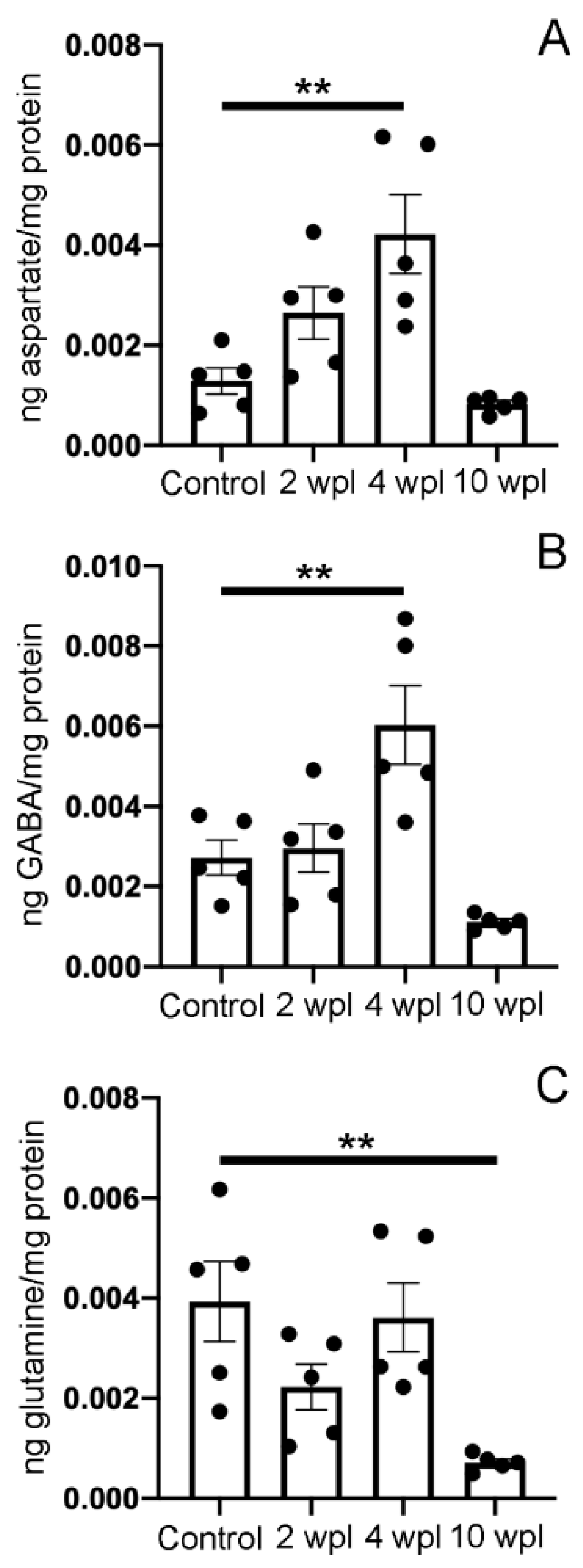
:1. Summary
2. Data Description
3. Methods
3.1. Animals and Complete SCI
3.2. High-Performance Liquid Chromatography
3.3. Statistical Analyses
Supplementary Materials
Author Contributions
Funding
Institutional Review Board Statement
Informed Consent Statement
Data Availability Statement
Acknowledgments
Conflicts of Interest
References
- Katz, H.R.; Fouke, K.E.; Losurdo, N.A.; Morgan, J.R. Recovery of Burrowing Behavior After Spinal Cord Injury in the Larval Sea Lamprey. Biol. Bull. 2020, 239, 174–182. [Google Scholar] [CrossRef] [PubMed]
- Cohen, A.H.; Abdelnabi, M.; Guan, L.; Ottinger, M.A.; Chakrabarti, L. Changes in distribution of serotonin induced by spinal injury in larval lampreys: Evidence from immunohistochemistry and HPLC. J. Neurotrauma 2005, 22, 172–188. [Google Scholar] [CrossRef] [PubMed]
- Cornide-Petronio, M.E.; Fernández-López, B.; Barreiro-Iglesias, A.; Rodicio, M.C. Traumatic injury induces changes in the expression of the serotonin 1A receptor in the spinal cord of lampreys. Neuropharmacology 2014, 77, 369–378. [Google Scholar] [CrossRef] [PubMed]
- Fernández-López, B.; Valle-Maroto, S.M.; Barreiro-Iglesias, A.; Rodicio, M.C. Neuronal release and successful astrocyte uptake of aminoacidergic neurotransmitters after spinal cord injury in lampreys. Glia 2014, 62, 1254–1269. [Google Scholar] [CrossRef] [PubMed]
- Fernández-López, B.; Romaus-Sanjurjo, D.; Cornide-Petronio, M.E.; Gómez-Fernández, S.; Barreiro-Iglesias, A.; Rodicio, M.C. Full anatomical recovery of the dopaminergic system after a complete spinal cord injury in lampreys. Neural Plast. 2015, 2015, 350750. [Google Scholar] [CrossRef]
- Fernández-López, B.; Barreiro-Iglesias, A.; Rodicio, M.C. Anatomical recovery of the spinal glutamatergic system following a complete spinal cord injury in lampreys. Sci. Rep. 2016, 6, 37786. [Google Scholar] [CrossRef]
- Romaus-Sanjurjo, D.; Valle-Maroto, S.M.; Barreiro-Iglesias, A.; Fernández-López, B.; Rodicio, M.C. Anatomical recovery of the GABAergic system after a complete spinal cord injury in lampreys. Neuropharmacology 2018, 131, 389–402. [Google Scholar] [CrossRef]
- Parker, D.; McClelland, T.J. Neuromodulator interactions and spinal cord injury in lamprey. Neural Regen. Res. 2018, 13, 643–644. [Google Scholar] [CrossRef]
- Parker, D. The Lesioned Spinal Cord Is a “New” Spinal Cord: Evidence from Functional Changes after Spinal Injury in Lamprey. Front. Neural Circuits 2017, 11, 84. [Google Scholar] [CrossRef] [Green Version]
- Becker, M.; Parker, D. Time course of functional changes in locomotor and sensory systems after spinal cord lesions in lamprey. J. Neurophysiol. 2019, 121, 2323–2335. [Google Scholar] [CrossRef]
- Valle-Maroto, S.M.; Barreiro-Iglesias, A.; Fernández-López, B.; Rodicio, M.C. Data on the recovery of glycinergic neurons after spinal cord injury in lampreys. Data Brief 2020, 28, 105092. [Google Scholar] [CrossRef]
- Sobrido-Cameán, D.; Fernández-López, B.; Pereiro, N.; Lafuente, A.; Rodicio, M.C.; Barreiro-Iglesias, A. Taurine Promotes Axonal Regeneration after a Complete Spinal Cord Injury in Lampreys. J. Neurotrauma 2020, 37, 899–903. [Google Scholar] [CrossRef]
- Sobrido-Cameán, D.; Robledo, D.; Sánchez, L.; Rodicio, M.C.; Barreiro-Iglesias, A. Serotonin inhibits axonal regeneration of identifiable descending neurons after a complete spinal cord injury in lampreys. Dis. Models Mech. 2019, 12, dmm037085. [Google Scholar] [CrossRef] [Green Version]
- Sobrido-Cameán, D.; Rodicio, M.C.; Barreiro-Iglesias, A. Data on the effect of a muscimol treatment in caspase activation in descending neurons of lampreys after a complete spinal cord injury. Data Brief 2018, 21, 2037–2041. [Google Scholar] [CrossRef] [PubMed]
- Romaus-Sanjurjo, D.; Ledo-García, R.; Fernández-López, B.; Hanslik, K.; Morgan, J.R.; Barreiro-Iglesias, A.; Rodicio, M.C. GABA promotes survival and axonal regeneration in identifiable descending neurons after spinal cord injury in larval lampreys. Cell Death Dis. 2018, 9, 663. [Google Scholar] [CrossRef]
- Romaus-Sanjurjo, D.; Rodicio, M.C.; Barreiro-Iglesias, A. Gamma-aminobutyric acid (GABA) promotes recovery from spinal cord injury in lampreys: Role of GABA receptors and perspective on the translation to mammals. Neural Regen. Res. 2019, 14, 1695–1696. [Google Scholar]
- Sobrido-Cameán, D.; Robledo, D.; Romaus-Sanjurjo, D.; Pérez-Cedrón, V.; Sánchez, L.; Rodicio, M.C.; Barreiro-Iglesias, A. Inhibition of Gamma-Secretase Promotes Axon Regeneration after a Complete Spinal Cord Injury. Front. Cell Dev. Biol. 2020, 8, 173. [Google Scholar] [CrossRef]
- de Sousa, N.; Santos, D.; Monteiro, S.; Silva, N.; Barreiro-Iglesias, A.; Salgado, A.J. Role of Baclofen in Modulating Spasticity and Neuroprotection in Spinal Cord Injury. J. Neurotrauma 2021, in press. [Google Scholar] [CrossRef]
- Fleck, M.W.; Henze, D.A.; Barrionuevo, G.; Palmer, A.M. Aspartate and glutamate mediate excitatory synaptic transmission in area CA1 of the hippocampus. J. Neurosci. 1993, 13, 3944–3955. [Google Scholar] [CrossRef] [PubMed]
- Villar-Cerviño, V.; Barreiro-Iglesias, A.; Anadón, R.; Rodicio, M.C. Aspartate immunoreactivity in the telencephalon of the adult sea lamprey: Comparison with GABA immunoreactivity. Brain Res. Bull. 2008, 75, 246–250. [Google Scholar] [CrossRef]
- Villar-Cerviño, V.; Fernández-López, B.; Rodicio, M.C.; Anadón, R. Aspartate-containing neurons of the brainstem and rostral spinal cord of the sea lamprey Petromyzon marinus: Distribution and comparison with γ-aminobutyric acid. J Comp. Neurol. 2014, 522, 1209–1231. [Google Scholar] [CrossRef] [PubMed]
- Barreiro-Iglesias, A.; Zhang, G.; Selzer, M.E.; Shifman, M.I. Complete spinal cord injury and brain dissection protocol for subsequent wholemount in situ hybridization in larval sea lamprey. J. Vis. Exp. 2014, 92, e51494. [Google Scholar] [CrossRef] [PubMed] [Green Version]
- Zhang, G.; Jin, L.Q.; Sul, J.Y.; Haydon, P.G.; Selzer, M.E. Live imaging of regenerating lamprey spinal axons. Neurorehabil. Neural Repair 2005, 19, 46–57. [Google Scholar] [CrossRef] [PubMed]
- Caride, A.; Fernández Pérez, B.; Cabaleiro, T.; Lafuente, A. Daily pattern of pituitary glutamine, glutamate, and aspartate content disrupted by cadmium exposure. Amino Acids 2010, 38, 1165–1172. [Google Scholar] [CrossRef]

Publisher’s Note: MDPI stays neutral with regard to jurisdictional claims in published maps and institutional affiliations. |
© 2021 by the authors. Licensee MDPI, Basel, Switzerland. This article is an open access article distributed under the terms and conditions of the Creative Commons Attribution (CC BY) license (https://creativecommons.org/licenses/by/4.0/).
Share and Cite
Fernández-López, B.; Pereiro, N.; Lafuente, A.; Rodicio, M.C.; Barreiro-Iglesias, A. Data on the Quantification of Aspartate, GABA and Glutamine Levels in the Spinal Cord of Larval Sea Lampreys after a Complete Spinal Cord Injury. Data 2021, 6, 54. https://0-doi-org.brum.beds.ac.uk/10.3390/data6060054
Fernández-López B, Pereiro N, Lafuente A, Rodicio MC, Barreiro-Iglesias A. Data on the Quantification of Aspartate, GABA and Glutamine Levels in the Spinal Cord of Larval Sea Lampreys after a Complete Spinal Cord Injury. Data. 2021; 6(6):54. https://0-doi-org.brum.beds.ac.uk/10.3390/data6060054
Chicago/Turabian StyleFernández-López, Blanca, Natividad Pereiro, Anunciación Lafuente, María Celina Rodicio, and Antón Barreiro-Iglesias. 2021. "Data on the Quantification of Aspartate, GABA and Glutamine Levels in the Spinal Cord of Larval Sea Lampreys after a Complete Spinal Cord Injury" Data 6, no. 6: 54. https://0-doi-org.brum.beds.ac.uk/10.3390/data6060054