Impact of Chromosome 9 Numerical Imbalances in Oral Squamous Cell Carcinoma: A Pilot Grid-Based Centromere Analysis

 , , , ,
Abstract
:1. Introduction
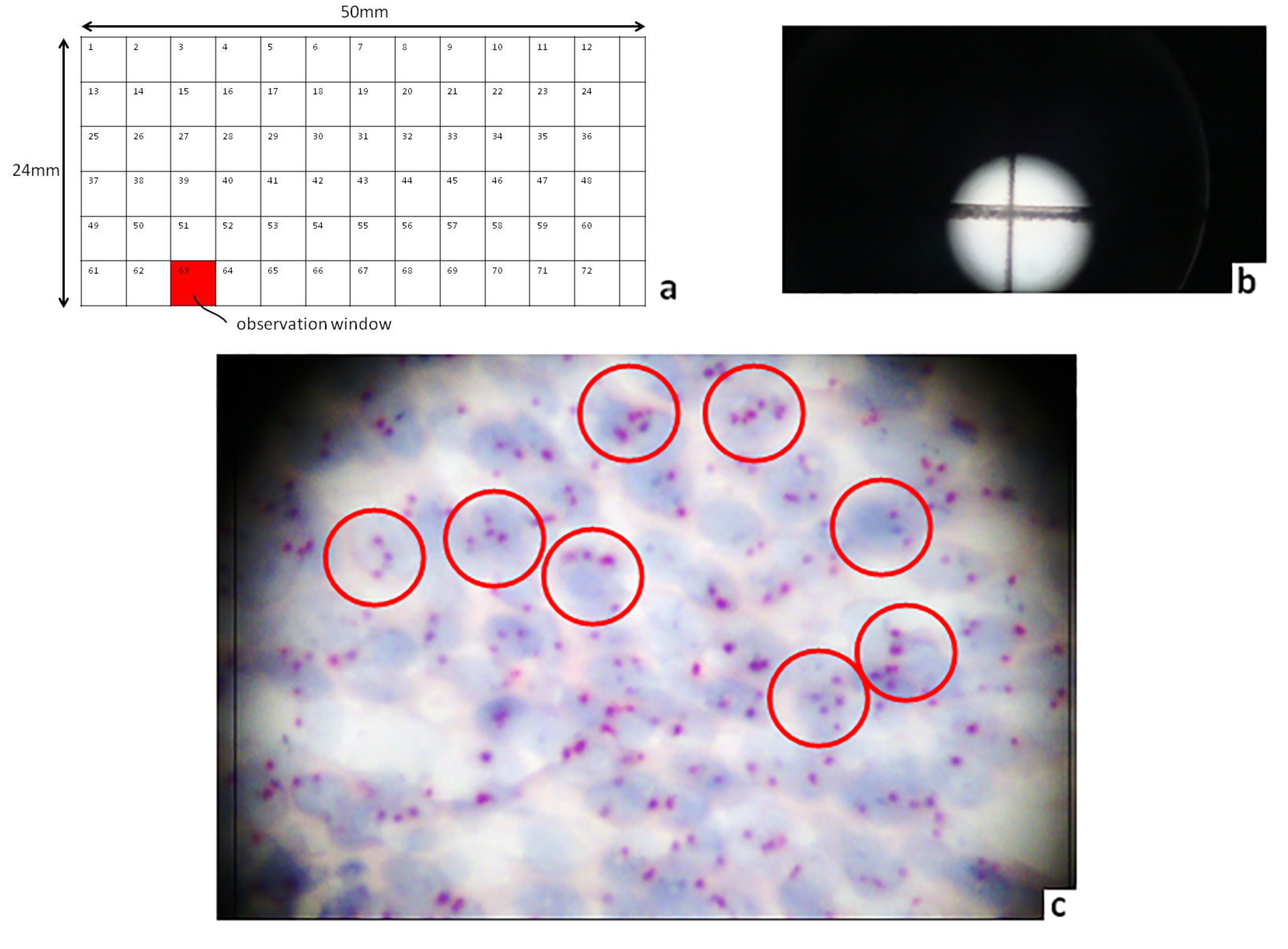
2. Results
3. Discussion
4. Materials and Methods
4.1. Study Group
4.2. Chromogenic In Situ Hybridization (CISH) Assay
4.3. Slide Screening Process
4.4. Statistical Analysis
5. Conclusions
6. Patents
Author Contributions
Funding
Acknowledgments
Conflicts of Interest
Abbreviations
| AJCC7 | American Joint Committee Cancer seventh edition |
| OSCC | Oral squamous cell carcinoma |
| CI | Chromosome instability |
| CISH | Chromogenic in situ hybridization |
| CEN | Centromere enumeration |
| LOH | Loss of heterozygosity |
| WHO | World Health Organization |
| GCS | Grid coverslip |
| FLM | Femtosecond laser micromachining |
| DAB | 3,3-diaminobenzidine |
References
- Albertson, D.G. Chromosome aberrations in solid tumours. Nat. Genet. 2003, 34, 369–376. [Google Scholar] [CrossRef] [PubMed]
- Ali, J.; Sabiha, B.; Jan, U.H.; Haider, S.A.; Khan, A.A.; Ali, S.S. Genetic etiology of oral cancer. Oral Oncol. 2017, 70, 23–28. [Google Scholar] [CrossRef] [PubMed]
- Grégoire, V.; Lefevre, J.-L.; Licitra, L.; Felip, E. EHNS-ESMO-ESTRO GWG. Squamous cell carcinoma of the head and neck: EHNS–ESMO–ESTRO clinical practice guidelines for diagnosis, treatment and follow-up. Ann. Oncol. 2010, 21, 184–186. [Google Scholar] [CrossRef]
- Kang, H.; Kiess, A.; Chung, C.H. Emerging biomarkers in head and neck cancer in the era of genomics. Nat. Rev. Clin. Oncol. 2010, 12, 11–26. [Google Scholar] [CrossRef] [PubMed]
- Reder, H.; Wagner, S.; Gamerdinger, U.; Sandmann, S.; Wuerdemann, N.; Braeuninger, A.; Dugas, M.; Gattenioehner, S.; Klussmann, J.P.; Wittenkindt, C. Genetic alterations in human papillomavirus-associated oropharyngeal squamous cell carcinoma of patients with treatment failure. Oral Oncol. 2019, 93, 59–65. [Google Scholar] [CrossRef] [PubMed]
- Stratton, M.R.; Campbell, P.J.; Futreal1, A.P. The cancer genome. Nature 2009, 458, 719–724. [Google Scholar] [CrossRef] [PubMed] [Green Version]
- Vera-Roman, J.M.; Rubio-Martinez, L.A. Comparative assays for the HER-2/neu oncogene status in breast cancer. Arch. Pathol. Lab. Med. 2004, 128, 627–633. [Google Scholar]
- Sholl, L.M.; Iafrate, A.J.; Chou, Y.P.; Wu, M.T.; Goan, Y.-G.; Su, L.; Huang, Y.-T.; Christiani, D.C.; Chirieac, L.R. Validation of chromogenic in situ hybridization for detection of EGFR copy number amplification in nonsmall cell lung carcinoma. Mod. Pathol. 2007, 20, 1028–1035. [Google Scholar] [CrossRef] [Green Version]
- Tsiambas, E.; Karameris, A.; Lygeros, M.; Athanasiou, A.E.; Salemis, N.S.; Gourgiotis, S.; Ragkos, V.; Metaxas, G.E.; Vilaras, G.; Patsouris, E. Gene numerical imbalances in cytological specimens based on fluorescence/chromogenic in situ hybridization analysis. J. BUON 2012, 17, 593–599. [Google Scholar]
- Yarom, N.; Shani, T.; Amariglio, N.; Taicher, S.; Kaplan, I.; Vered, M.; Rechavi, G.; Trakhtenbrot, L.; Hirshberg, A. Chromosomal numerical aberrations in oral lichen planus. J. Dent. Res. 2009, 88, 427–432. [Google Scholar] [CrossRef]
- Pierssens, D.D.C.G.; Borgemeester, M.C.; van der Heijden, S.J.H.; Peutz-Kootstra, C.J.; Ruland, A.M.; Haesevoets, A.M.; Kessler, P.A.W.H.; Kremer, B.; Speel, E.J.M. Chromosome instability in tumor resection margins of primary OSCC is a predictor of local recurrence. Oral Oncol. 2007, 66, 14–21. [Google Scholar] [CrossRef] [PubMed]
- Hardisson, D.; Alvarez-Marcos, C.; Salas-Bustamante, A.; Alonso-Guervós, M.; Sastre, N.; Sampedro, A. Numerical aberrations of chromosomes 8, 9, 11, and 17 in squamous cell carcinoma of the pharynx and larynx: A fluorescence in situ hybridization and DNA flow cytometric analysis of 50 cases. Oral Oncol. 2004, 40, 409–417. [Google Scholar] [CrossRef] [PubMed]
- Sato, H.; Uzawa, N.; Takahashi, N.-I.; Myo, K.; Ohyama, Y.; Amagasa, T. Prognostic utility of chromosomal instability detected by fluorescence in situ hybridization in fine-needle aspirates fromoralsquamous cell carcinomas. BMC Cancer 2010, 10, 182–186. [Google Scholar] [CrossRef] [PubMed] [Green Version]
- Lin, S.C.; Chen, Y.J.; Kao, S.Y.; Hsu, M.S.; Lin, C.H.; Yang, S.C.; Liu, T.Y.; Chang, K.W. Chromosomal changes in betel-associated oral squamous cell carcinomas and their relationship to clinical parameters. Oral Oncol. 2002, 38, 266–273. [Google Scholar] [CrossRef]
- Bernardes, V.F.; Diniz, M.G.; Silva, J.C.; Moraes, D.C.; De Marco, L.; Gomes, C.C.; Gomez, R.S. Lack of association between denture trauma and loss of heterozygosity confronts the proposed pathologic role of chronic mucosal trauma in oral carcinogenesis. J. Oral Pathol. Med. 2019, 48, 421–423. [Google Scholar] [CrossRef]
- Wang, X.; Chen, S.; Chen, X.; Zhang, C.; Liang, X. Tumor-related markers in histologically normal margins correlate with locally recurrentoral squamous cellcarcinoma: A retrospective study. J. Oral Pathol. Med. 2016, 45, 83–88. [Google Scholar] [CrossRef] [PubMed]
- Towle, R.; Tsui, I.F.L.; Zhu, Y.; MacLellan, S.; Poh, C.F.; Garnis, C. Recurring DNA copy number gain at chromosome 9p13 plays a role in the activation of multiple candidate oncogenes in progressing oral premalignant lesions. Cancer Med. 2014, 3, 1170–1184. [Google Scholar] [CrossRef] [PubMed]
- Murali, A.; Sailasree, R.; Sebastian, P.; Kumar, R.R.; Varghese, V.T.; Kannan, S. Loss of heterozygosity of D9S162: Molecular predictor for treatment response in oral carcinoma. Oral Oncol. 2011, 47, 571–576. [Google Scholar] [CrossRef]
- Ohta, S.; Uemura, H.; Matsui, Y.; Ishiguro, H.; Fujinami, K.; Kondo, K.; Miyamoto, H.; Yazawa, T.; Danenberg, K.; Danenberg, P.V.; et al. Alterations of p16 and p14ARF genes and their 9p21 locus in oral squamous cell carcinoma. Oral Surg. Oral Med. Oral Pathol. Oral Radiol. Endod. 2009, 107, 81–91. [Google Scholar] [CrossRef]
- Gebhart, E.; Liehr, T.; Wolff, E.; Wiltfang, J.; Koscielny, S.; Ries, J. Loss of 9p21 is embedded in a complex but consistent pattern of genomic imbalances in oral squamous cell carcinomas. Cytogenet. Genome Res. 2003, 101, 106–112. [Google Scholar] [CrossRef]
- Schwarz, S.; Bier, J.; Driemel, O.; Reichert, T.E.; Hauke, S.; Arndt Hartmann, A.; Brockhoff, A.G. Losses of 3p14 and 9p21 as shown by fluorescence in situ hybridization are early events in tumorigenesis of oral squamous cell carcinoma and already occur in simple keratosis. Cytometry A 2008, 73, 305–311. [Google Scholar] [CrossRef] [PubMed]
- Lingen, M.W.; Chang, K.W.; McMurray, S.J.; Solt, D.B.; Kies, M.S.; Mittal, B.B.; Haines, G.K.; Pelzer, H.J. Overexpression of p53 in squamous cell carcinoma of the tongue in young patients with no known risk factors is not associated with mutations in exons 5–9. Head Neck 2000, 22, 328–335. [Google Scholar] [CrossRef]
- Mascitti, M.; Tempesta, A.; Togniet, L.; Capodiferro, S.; Troiano, G.; Rubini, C.; Maiorano, E.; Santarelli, A.; Favia, G.; Limongelli, L. Histological Features and Survival in Young Patients with HPV Negative Oral Squamous Cell Carcinoma. Oral Dis. 2020. [Google Scholar] [CrossRef] [PubMed]
- Pickering, C.R.; Zhang, J.; Neskey, D.M.; Zhao, M.; Jasser, S.A.; Wang, J.; Ward, A.; Tsai, C.J.; Alves, M.V.O.; Zhou, J.H.; et al. Squamous cell carcinoma of the oral tongue in young non-smokers is genomically similar to tumors in older smokers. Clin. Cancer Res. 2014, 20, 3842–3848. [Google Scholar] [CrossRef] [Green Version]
- Tsiambas, E.; Riziotis, C.; Mastronikolis, N.S.; Peschos, D.; Mortakis, A.; Kyroysis, G.; Mastronikolis, S.N.; Batistatou, A.; Lazaris, A.C.; Patsouris, E.; et al. Comparative p16IKN4A Expression in Laryngeal Carcinoma and Cervical Cancer Precursors: A Real-time Grid-based Immunocytochemistry Analysis. Anticancer Res. 2018, 38, 5805–5810. [Google Scholar] [CrossRef]
- Barnes, L.; Eveson, J.W.; Reichart, P.; Sidransky, D. Pathology and Genetics: Head and Neck Tumours; WHO IARC Press: Lyon, France, 2005; pp. 118–130. [Google Scholar]
- Tsiambas, E.; Riziotis, C. Implementation of a real-time reference and calibration grid platform for improved screening–mapping in Pap test slides. Pathol. Int. 2017, 67, 24–31. [Google Scholar] [CrossRef] [Green Version]


| Clinicopathological Parameters | Chromosome 9 (CEN) | p Value | ||
|---|---|---|---|---|
| OSCC (n = 50) | N/D | P | ||
| 42/50 (84%) | 8/50 (16%) | |||
| n (%) | n | n | ||
| Gender | 0.572 | |||
| Male (mean age: 54.9) | 44 (88%) | 36/50 (72%) | 8/50 (16%) | |
| Female (mean age: 49.1) | 6 (12%) | 6/50 (12%) | 0/50 (0%) | |
| HPV history | 0.923 | |||
| Positive | 18 (36%) | 15/50 (30%) | 3/50 (6%) | |
| Negative | 32 (64%) | 27/50 (54%) | 5/50 (10%) | |
| Grade | 0.036 | |||
| 1 | 18 (18%) | 18/50 (36%) | 0/50 (0%) | |
| 2 | 21 (58%) | 19/50 (38%) | 2/50 (4%) | |
| 3 | 11 (24%) | 3/50 (6%) | 6/50 (12%) | |
| Stage | 0.841 | |||
| I | 9 (18%) | 9/50 (18%) | 0/50 (0%) | |
| II | 26 (52%) | 22/50 (44%) | 4/50 (8%) | |
| III | 15 (30%) | 11/50 (22%) | 4/50 (8%) | |
| Smoking status | 0.661 | |||
| Current | 38 (76%) | 31/50 (62%) | 7/50 (14%) | |
| Former | 12 (24%) | 11/50 (22%) | 1/50 (2%) | |
© 2020 by the authors. Licensee MDPI, Basel, Switzerland. This article is an open access article distributed under the terms and conditions of the Creative Commons Attribution (CC BY) license (http://creativecommons.org/licenses/by/4.0/).
Share and Cite
Kyrodimos, E.; Chrysovergis, A.; Mastronikolis, N.; Tsiambas, E.; Riziotis, C.; Roukas, D.; Fotiades, P.; Stavraka, C.; Ragos, V.; Paschopoulos, M.; et al. Impact of Chromosome 9 Numerical Imbalances in Oral Squamous Cell Carcinoma: A Pilot Grid-Based Centromere Analysis. Diagnostics 2020, 10, 501. https://0-doi-org.brum.beds.ac.uk/10.3390/diagnostics10070501
Kyrodimos E, Chrysovergis A, Mastronikolis N, Tsiambas E, Riziotis C, Roukas D, Fotiades P, Stavraka C, Ragos V, Paschopoulos M, et al. Impact of Chromosome 9 Numerical Imbalances in Oral Squamous Cell Carcinoma: A Pilot Grid-Based Centromere Analysis. Diagnostics. 2020; 10(7):501. https://0-doi-org.brum.beds.ac.uk/10.3390/diagnostics10070501
Chicago/Turabian StyleKyrodimos, Efthymios, Aristeidis Chrysovergis, Nicholas Mastronikolis, Evangelos Tsiambas, Christos Riziotis, Dimitrios Roukas, Panagiotis Fotiades, Chara Stavraka, Vasileios Ragos, Minas Paschopoulos, and et al. 2020. "Impact of Chromosome 9 Numerical Imbalances in Oral Squamous Cell Carcinoma: A Pilot Grid-Based Centromere Analysis" Diagnostics 10, no. 7: 501. https://0-doi-org.brum.beds.ac.uk/10.3390/diagnostics10070501