Effect of Baseline Characteristics and Tumor Burden on Vaspin Expression and Progressive Disease in Operable Colorectal Cancer


Abstract
:1. Introduction
2. Materials and Methods
2.1. Data Sets
2.2. Immunohistochemistry of Vaspin Expression
2.3. Statistical Analysis
3. Results
4. Discussion
5. Conclusions
Supplementary Materials
Author Contributions
Funding
Conflicts of Interest
References
- McDougall, J.A.; Blair, C.K.; Wiggins, C.L.; Goodwin, M.B.; Chiu, V.K.; Rajput, A.; Kinney, A.Y. Socioeconomic disparities in health-related quality of life among colorectal cancer survivors. J. Cancer Surviv. 2019, 13, 459–467. [Google Scholar] [CrossRef] [PubMed]
- Thong, M.S.Y.; Koch-Gallenkamp, L.; Jansen, L.; Bertram, H.; Eberle, A.; Holleczek, B.; Waldeyer-Sauerland, M.; Waldmann, A.; Zeissig, S.R.; Brenner, H.; et al. Age-specific health-related quality of life in long-term and very long-term colorectal cancer survivors versus population controls - a population-based study. Acta Oncol. 2019, 58, 801–810. [Google Scholar] [CrossRef]
- Riechelmann, R.P.; Srimuninnimit, V.; Bordonaro, R.; Kavan, P.; Di Bartolomeo, M.; Maiello, E.; Cicin, I.; Garcia-Alfonso, P.; Chau, I.; Fedyanin, M.Y.; et al. Aflibercept plus folfiri for second-line treatment of metastatic colorectal cancer: Observations from the global aflibercept safety and health-related quality-of-life program (asqop). Clin. Colorectal Cancer 2019, 18, 183–191. [Google Scholar] [CrossRef] [PubMed] [Green Version]
- Lu, X.; Cai, Y.; Xia, L.; Ju, H.; Zhao, X. Treatment modalities and relative survival in patients with brain metastasis from colorectal cancer. Biosci. Trends 2019, 13, 182–188. [Google Scholar] [CrossRef] [PubMed] [Green Version]
- Liu, Y.; Yang, M.; Li, B.; Xu, K.; Gao, X.; Li, J.; Wei, H.; Huang, Q.; Xu, W.; Xiao, J. Development of a novel model for predicting survival of patients with spine metastasis from colorectal cancer. Eur. Spine J. 2019, 28, 1491–1501. [Google Scholar] [CrossRef] [PubMed]
- Levy, M.; Visokai, V.; Lipska, L.; Topolcan, O. Tumor markers in staging and prognosis of colorectal carcinoma. Neoplasma 2008, 55, 138. [Google Scholar] [PubMed]
- Sasaki, K.; Morioka, D.; Conci, S.; Margonis, G.A.; Sawada, Y.; Ruzzenente, A.; Kumamoto, T.; Iacono, C.; Andreatos, N.; Guglielmi, A. The tumor burden score: A new “metro-ticket” prognostic tool for colorectal liver metastases based on tumor size and number of tumors. Annals Surg. 2018, 267, 132–141. [Google Scholar] [CrossRef] [PubMed]
- Poornakala, S.; Prema, N.S. A study of morphological prognostic factors in colorectal cancer and survival analysis. Indian J. Pathol. Microbiol. 2019, 62, 36–42. [Google Scholar] [CrossRef] [PubMed]
- Sun, G.; Li, Y.; Peng, Y.; Lu, D.; Zhang, F.; Cui, X.; Zhang, Q.; Li, Z. Impact of the preoperative prognostic nutritional index on postoperative and survival outcomes in colorectal cancer patients who underwent primary tumor resection: A systematic review and meta-analysis. Int. J. Colorectal Dis. 2019, 34, 681–689. [Google Scholar] [CrossRef] [PubMed]
- Kam, A.E.; Pendurti, G.; Shah, U.H.; Ghalib, M.H.; Chaudhary, I.; Chuy, J.; Rajdev, L.; Kaubisch, A.; Aparo, S.; Mantzaris, I.; et al. Survival outcome and prognostic model of patients with colorectal cancer on phase 1 trials. Investig. New Drugs 2019, 37, 490–497. [Google Scholar] [CrossRef]
- Oshi, M.; Margonis, G.A.; Sawada, Y.; Andreatos, N.; He, J.; Kumamoto, T.; Morioka, D.; Wolfgang, C.L.; Tanaka, K.; Weiss, M.J.; et al. Higher tumor burden neutralizes negative margin status in hepatectomy for colorectal cancer liver metastasis. Ann. Surg. Oncol. 2019, 26, 593–603. [Google Scholar] [CrossRef]
- Erdogan, S.; Sezer, S.; Baser, E.; Gun-Eryilmaz, O.; Gungor, T.; Uysal, S.; Yilmaz, F.M. Evaluating vaspin and adiponectin in postmenopausal women with endometrial cancer. Endocr. Relat. Cancer 2013, 20, 669–675. [Google Scholar] [CrossRef] [PubMed] [Green Version]
- Jabbari, S.; Hedayati, M.; Yaghmaei, P.; Parivar, K. Medullary thyroid carcinoma--circulating status of vaspin and retinol binding protein-4 in iranian patients. Asian Pac. J. Cancer Prev. 2015, 16, 6507–6512. [Google Scholar] [CrossRef] [PubMed] [Green Version]
- Dwipayana, I.M.P.; Semadi, I.M.S.; Gotera, W.; Saraswati, M.R.; Suastika, K. Vaspin in developing obesity (vande-ob); the correlation of waist circumference and visceral fat percentage with vaspin levels in patients with type ii diabetes mellitus. Open Access Maced. J. Med. Sci. 2019, 7, 50–52. [Google Scholar] [CrossRef] [PubMed]
- Lee, J.A.; Park, H.S.; Song, Y.S.; Jang, Y.J.; Kim, J.H.; Lee, Y.J.; Heo, Y.S. Relationship between vaspin gene expression and abdominal fat distribution of korean women. Endocr. J. 2011, 58, 639–646. [Google Scholar] [CrossRef] [Green Version]
- Salek-Maghsoudi, A.; Hassani, S.; Momtaz, S.; Shadboorestan, A.; Ganjali, M.R.; Ghahremani, M.H.; Hosseini, R.; Norouzi, P.; Abdollahi, M. Biochemical and molecular evidence on the role of vaspin in early detection of the insulin resistance in a rat model of high-fat diet and use of diazinon. Toxicology 2019, 411, 1–14. [Google Scholar] [CrossRef]
- Fazeli, M.S.; Dashti, H.; Akbarzadeh, S.; Assadi, M.; Aminian, A.; Keramati, M.R.; Nabipour, I. Circulating levels of novel adipocytokines in patients with colorectal cancer. Cytokine 2013, 62, 81–85. [Google Scholar] [CrossRef] [Green Version]
- Deng, B.C.; Yun, Y.H.; Liang, Y.Z.; Cao, D.S.; Xu, Q.S.; Yi, L.Z.; Huang, X. A new strategy to prevent over-fitting in partial least squares models based on model population analysis. Anal. Chim. Acta 2015, 880, 32–41. [Google Scholar] [CrossRef]
- Gromski, P.S.; Muhamadali, H.; Ellis, D.I.; Xu, Y.; Correa, E.; Turner, M.L.; Goodacre, R. A tutorial review: Metabolomics and partial least squares-discriminant analysis--a marriage of convenience or a shotgun wedding. Anal. Chim. Acta 2015, 879, 10–23. [Google Scholar] [CrossRef]
- Sanchez, G. Pls path modeling with r. Berkeley Trowchez Ed. 2013, 383, 2013. Available online: https://www.gastonsanchez.com/PLS_Path_Modeling_with_R.pdf (accessed on 8 October 2020).
- Li, Y.; Xing, C.; Wei, M.; Wu, H.; Hu, X.; Li, S.; Sun, G.; Zhang, G.; Wu, B.; Zhang, F.; et al. Combining red blood cell distribution width (rdw-cv) and cea predict poor prognosis for survival outcomes in colorectal cancer. J. Cancer 2019, 10, 1162–1170. [Google Scholar] [CrossRef] [PubMed] [Green Version]
- Chen, L.; Jiang, B.; Di, J.; Zhang, C.; Wang, Z.; Zhang, N.; Xing, J.; Cui, M.; Yang, H.; Yao, Z.; et al. Predictive value of preoperative detection of cea and ca199 for prognosis in patients with stage ii-iii colorectal cancer. Zhonghua Wei Chang Wai Ke Za Zhi 2015, 18, 914–919. [Google Scholar] [PubMed]
- Cymbaluk-Ploska, A.; Chudecka-Glaz, A.; Jagodzinska, A.; Pius-Sadowska, E.; Sompolska-Rzechula, A.; Machalinski, B.; Menkiszak, J. Evaluation of biologically active substances promoting the development of or protecting against endometrial cancer. Onco. Targets Ther. 2018, 11, 1363–1372. [Google Scholar] [CrossRef] [PubMed] [Green Version]
- Xu, X.; Wen, J.; Lu, Y.; Ji, H.; Zhuang, J.; Su, Y.; Liu, B.; Li, H.; Xu, Y. Impact of age on plasma vaspin concentration in a group of normal chinese people. J. Endocrinol. Investig. 2017, 40, 143–151. [Google Scholar] [CrossRef] [PubMed] [Green Version]
- Cho, J.K.; Han, T.K.; Kang, H.S. Combined effects of body mass index and cardio/respiratory fitness on serum vaspin concentrations in korean young men. Eur. J. Appl. Physiol. 2010, 108, 347–353. [Google Scholar] [CrossRef] [PubMed]
- Li, J.; Li, Q.; Zhu, Y.C.; Wang, Y.K.; Gao, C.P.; Li, X.Y.; Ji, T.; Bai, S.J. Association of vaspin rs2236242 gene variants with type 2 diabetes and obesity in a chinese population: A prospective, single-center study. J. Cell Physiol. 2019. [Google Scholar] [CrossRef]
- Mierzynski, R.; Poniedzialek-Czajkowska, E.; Dluski, D.; Patro-Malysza, J.; Kimber-Trojnar, Z.; Majsterek, M.; Leszczynska-Gorzelak, B. Nesfatin-1 and vaspin as potential novel biomarkers for the prediction and early diagnosis of gestational diabetes mellitus. Int. J. Mol. Sci. 2019, 20, 159. [Google Scholar] [CrossRef] [Green Version]
- Yang, W.; Li, Y.; Tian, T.; Wang, L. Serum vaspin concentration in elderly type 2 diabetes mellitus patients with differing body mass index: A cross-sectional study. Biomed. Res. Int. 2017, 2017, 4875026. [Google Scholar] [CrossRef]
- Rodrigues, V.S.; do Valle Junior, R.F.; Sanches Fernandes, L.F.; Pacheco, F.A.L. The assessment of water erosion using partial least squares-path modeling: A study in a legally protected area with environmental land use conflicts. Sci. Total Environ. 2019, 691, 1225–1241. [Google Scholar] [CrossRef]
- Bates, T.C.; Maes, H.; Neale, M.C. Umx: Twin and path-based structural equation modeling in r. Twin Res. Hum. Genet. 2019, 22, 27–41. [Google Scholar] [CrossRef] [Green Version]
- Li, X.; Gill, R.; Cooper, N.G.; Yoo, J.K.; Datta, S. Modeling microrna-mrna interactions using pls regression in human colon cancer. BMC Med. Genomics 2011, 4, 44. [Google Scholar] [CrossRef] [PubMed] [Green Version]
- Evermann, J.; Tate, M. Comparing Out-of-Sample Predictive Ability of Pls, Covariance, and Regression Models. 2014. Available online: https://eprints.qut.edu.au/114787/ (accessed on 8 October 2020).
- Hosseinzadeh, P.; Javanbakht, M.; Alemrajabi, M.; Gholami, A.; Amirkalali, B.; Sohrabi, M.; Zamani, F. The association of dietary intake of calcium and vitamin d to colorectal cancer risk among iranian population. Asian Pac. J. Cancer Prev. 2019, 20, 2825–2830. [Google Scholar] [CrossRef] [PubMed]
- Yang, K.; Li, X.; Forman, M.R.; Monahan, P.O.; Graham, B.H.; Joshi, A.; Song, M.; Hang, D.; Ogino, S.; Giovannucci, E.L.; et al. Pre-diagnostic leukocyte mitochondrial DNA copy number and colorectal cancer risk. Carcinogenesis 2019, 40, 1462–1468. [Google Scholar] [CrossRef] [PubMed]


| Variables | Characteristics | Disease-Free (n = 55) | Progressive Disease (n = 34) | p |
|---|---|---|---|---|
| Age | Age ≥ 65 years | 65.8 ± 13.2 | 63 ± 12.7 | 0.324 |
| Sex | Male: Female | 34: 21 | 25: 9 | 0.256 |
| BMI | Body mass index ≥ 24 | 22.8 ± 3.6 | 23.7 ± 4.1 | 0.256 |
| DM | Diabetes mellitus | 15 (27.3%) | 13 (38.2%) | 0.279 |
| Grade | Grade III | 6 (10.9%) | 2 (5.9%) | 0.345 |
| Stage | Stage III–IV | 18 (32.7%) | 26 (76.5%) | <0.001 |
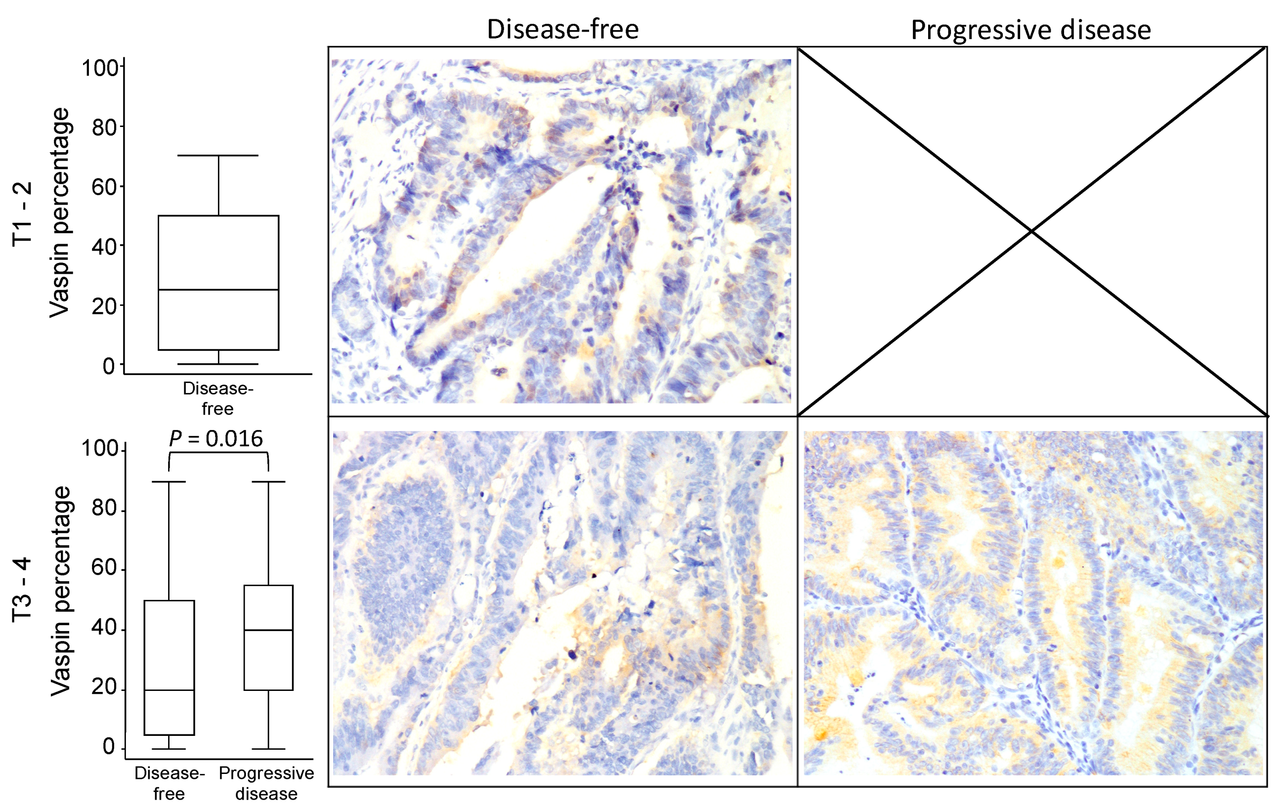
| Tumor | Tumor size (T3–4) | 37 (67.3%) | 34 (100.0%) | <0.001 |
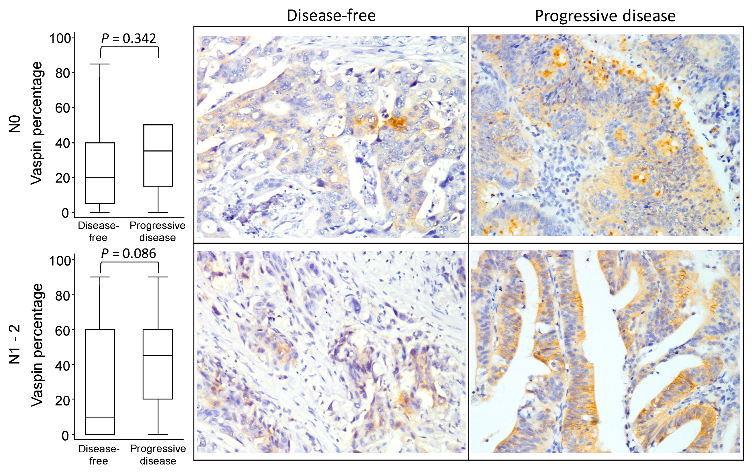
| Node | Lymph node invasion (N1–2) | 18 (32.7%) | 23 (67.6%) | 0.001 |
| PNI | Perineurial invasion | 28 (50.9%) | 27 (79.4%) | 0.007 |
| VCI | Vascular invasion | 20 (36.4%) | 23 (67.6%) | 0.004 |
| CEA | CEA level (ng/mL) | 3.5 (0.7–110) | 11.3 (1.7–1228) | 0.003 |
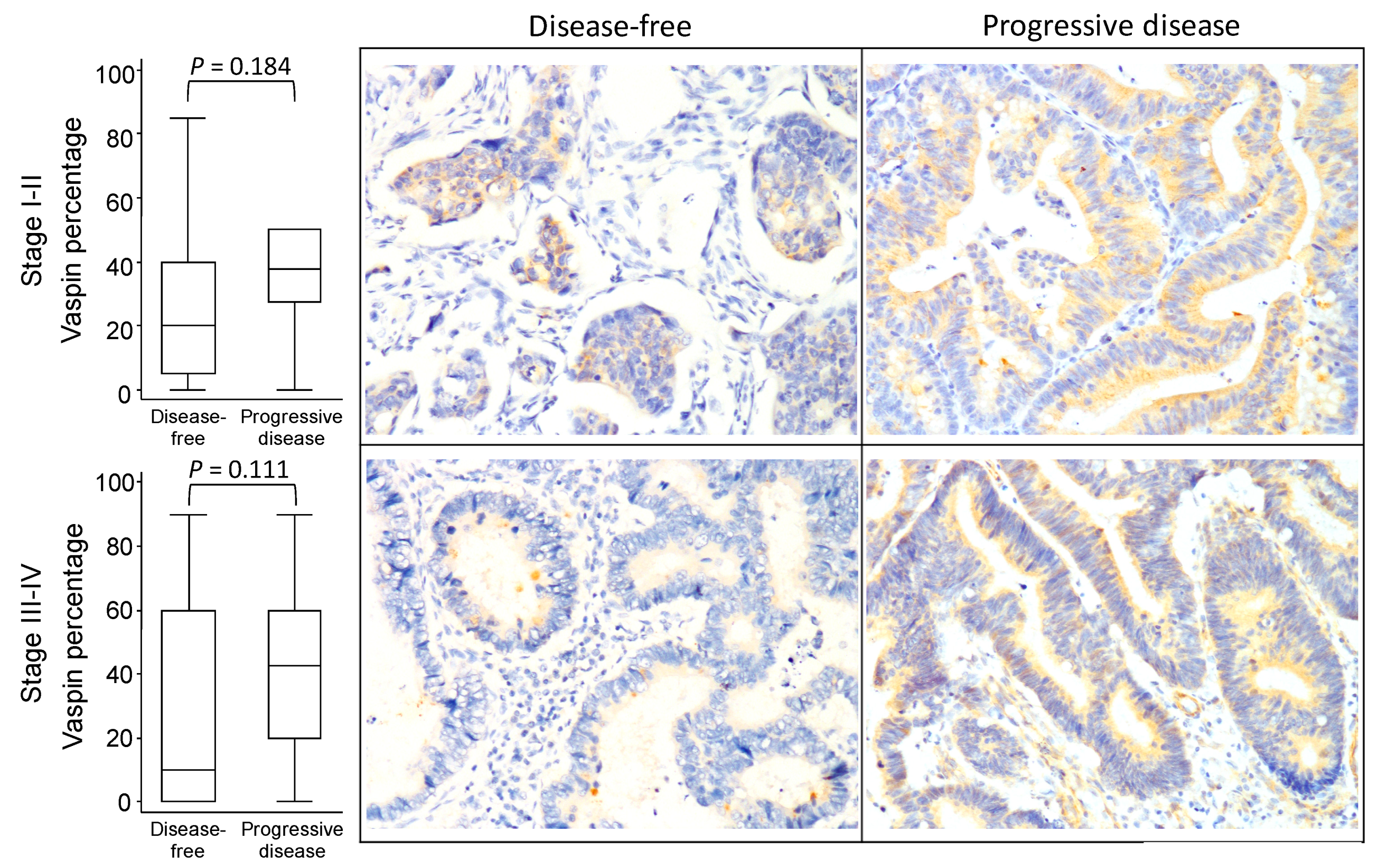
| Vaspin | Vaspin percentage | 27.4 ± 26.0 | 40.1 ± 23.1 | 0.021 |
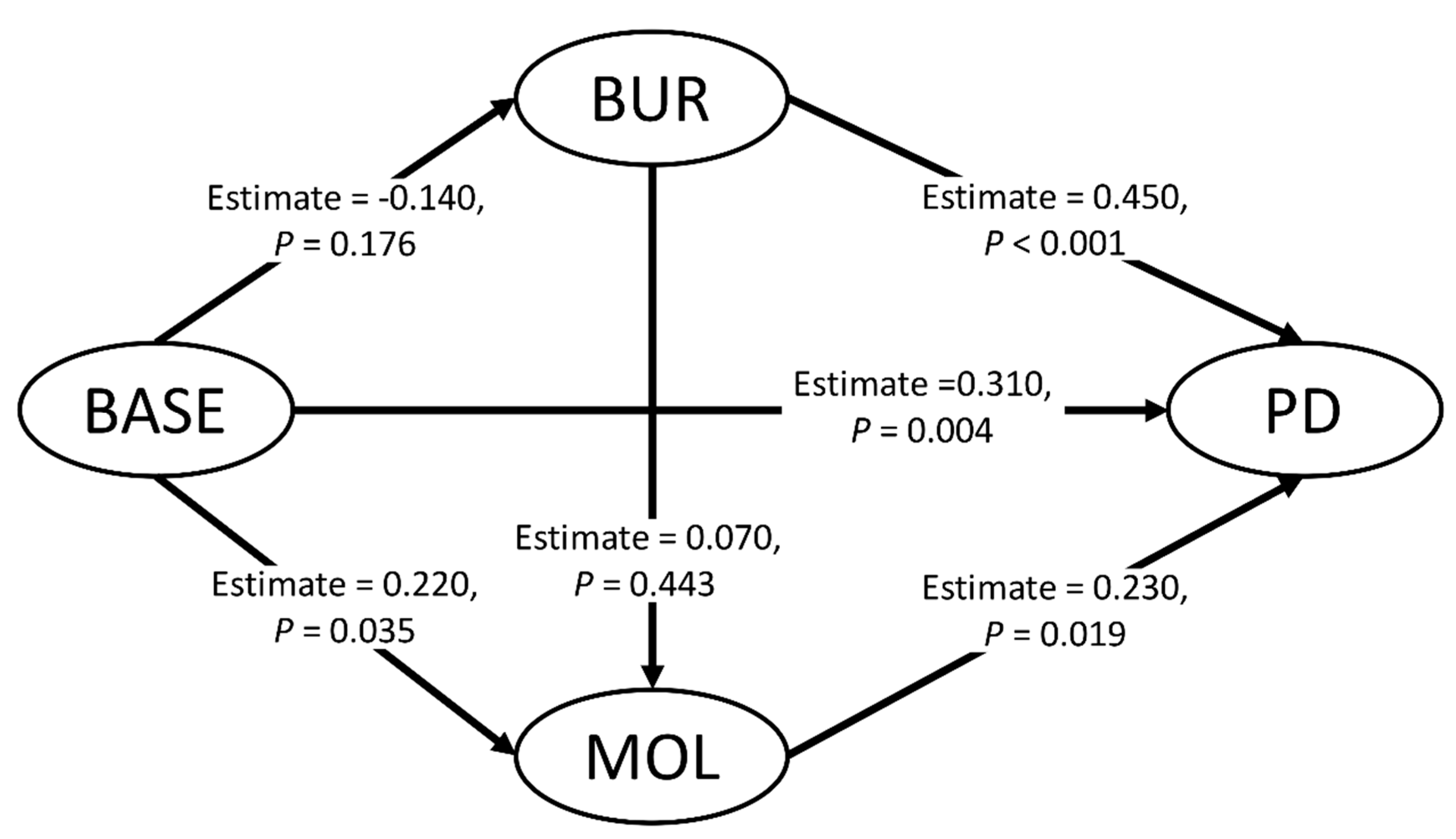
| Variables | Latent Variables | BASE | BUR | MOL | META | Communality | Redundancy | |
|---|---|---|---|---|---|---|---|---|
| 1 | Age | BASE | 0.750 | −0.196 | 0.176 | -0.106 | 0.563 | 0.000 |
| 2 | Sex | BASE | 0.079 | 0.241 | 0.186 | 0.120 | 0.006 | 0.000 |
| 3 | BMI | BASE | 0.171 | −0.068 | −0.088 | 0.122 | 0.029 | 0.000 |
| 4 | DM | BASE | 0.643 | −0.041 | 0.059 | 0.115 | 0.414 | 0.000 |
| 5 | Grade | BUR | −0.088 | 0.115 | 0.064 | −0.085 | 0.013 | 0.000 |
| 6 | Stage | BUR | −0.026 | 0.789 | 0.248 | 0.425 | 0.622 | 0.013 |
| 7 | Tumor | BUR | −0.139 | 0.648 | 0.140 | 0.396 | 0.420 | 0.009 |
| 8 | Node | BUR | −0.042 | 0.771 | 0.282 | 0.340 | 0.594 | 0.012 |
| 9 | PNI | BUR | −0.039 | 0.633 | 0.240 | 0.285 | 0.401 | 0.008 |
| 10 | VCI | BUR | −0.262 | 0.630 | 0.032 | 0.304 | 0.396 | 0.008 |
| 11 | CEA | MOL | 0.053 | 0.234 | 0.742 | 0.312 | 0.550 | 0.067 |
| 12 | Vaspin | MOL | 0.207 | 0.183 | 0.774 | 0.244 | 0.600 | 0.073 |
| 13 | Progressive disease | META | 0.047 | 0.503 | 0.365 | 1.000 | - | - |
© 2020 by the authors. Licensee MDPI, Basel, Switzerland. This article is an open access article distributed under the terms and conditions of the Creative Commons Attribution (CC BY) license (http://creativecommons.org/licenses/by/4.0/).
Share and Cite
Kan, J.-Y.; Lee, Y.-C.; Lin, Y.-D.; Ho, W.-Y.; Moi, S.-H. Effect of Baseline Characteristics and Tumor Burden on Vaspin Expression and Progressive Disease in Operable Colorectal Cancer. Diagnostics 2020, 10, 801. https://0-doi-org.brum.beds.ac.uk/10.3390/diagnostics10100801
Kan J-Y, Lee Y-C, Lin Y-D, Ho W-Y, Moi S-H. Effect of Baseline Characteristics and Tumor Burden on Vaspin Expression and Progressive Disease in Operable Colorectal Cancer. Diagnostics. 2020; 10(10):801. https://0-doi-org.brum.beds.ac.uk/10.3390/diagnostics10100801
Chicago/Turabian StyleKan, Jung-Yu, Yi-Chen Lee, Yu-Da Lin, Wan-Yi Ho, and Sin-Hua Moi. 2020. "Effect of Baseline Characteristics and Tumor Burden on Vaspin Expression and Progressive Disease in Operable Colorectal Cancer" Diagnostics 10, no. 10: 801. https://0-doi-org.brum.beds.ac.uk/10.3390/diagnostics10100801