
Early Detection of Diabetic Peripheral Neuropathy: A Focus on Small Nerve Fibres

 ,
,  , , and
Abstract
:1. Introduction
1.1. Economic and Functional Consequences of Small Fibre Degeneration
1.2. Pathogenesis
1.3. Evidence in Favour of Early Intervention for DPN
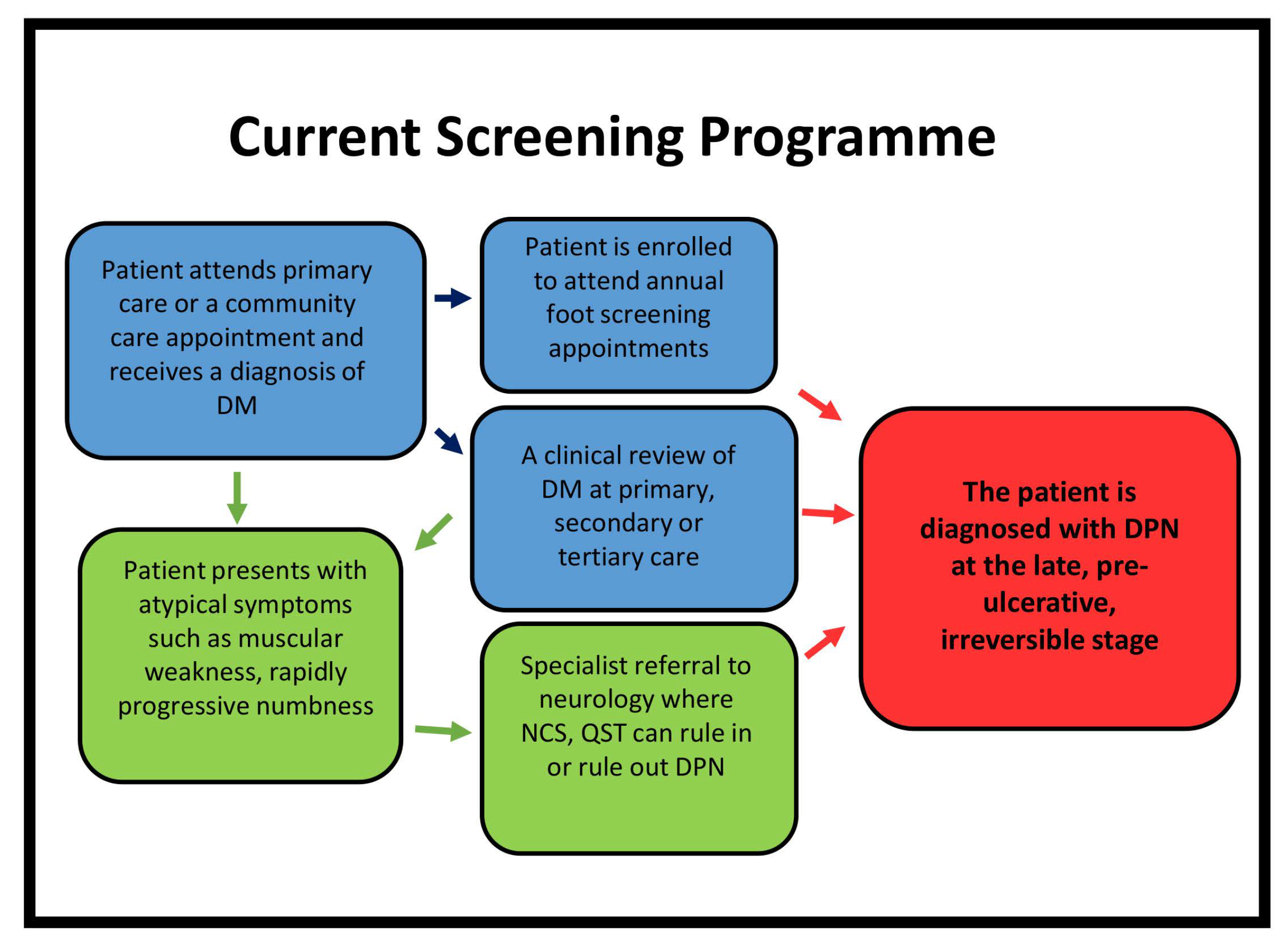
1.4. Current Clinical Assessment of Neuropathy
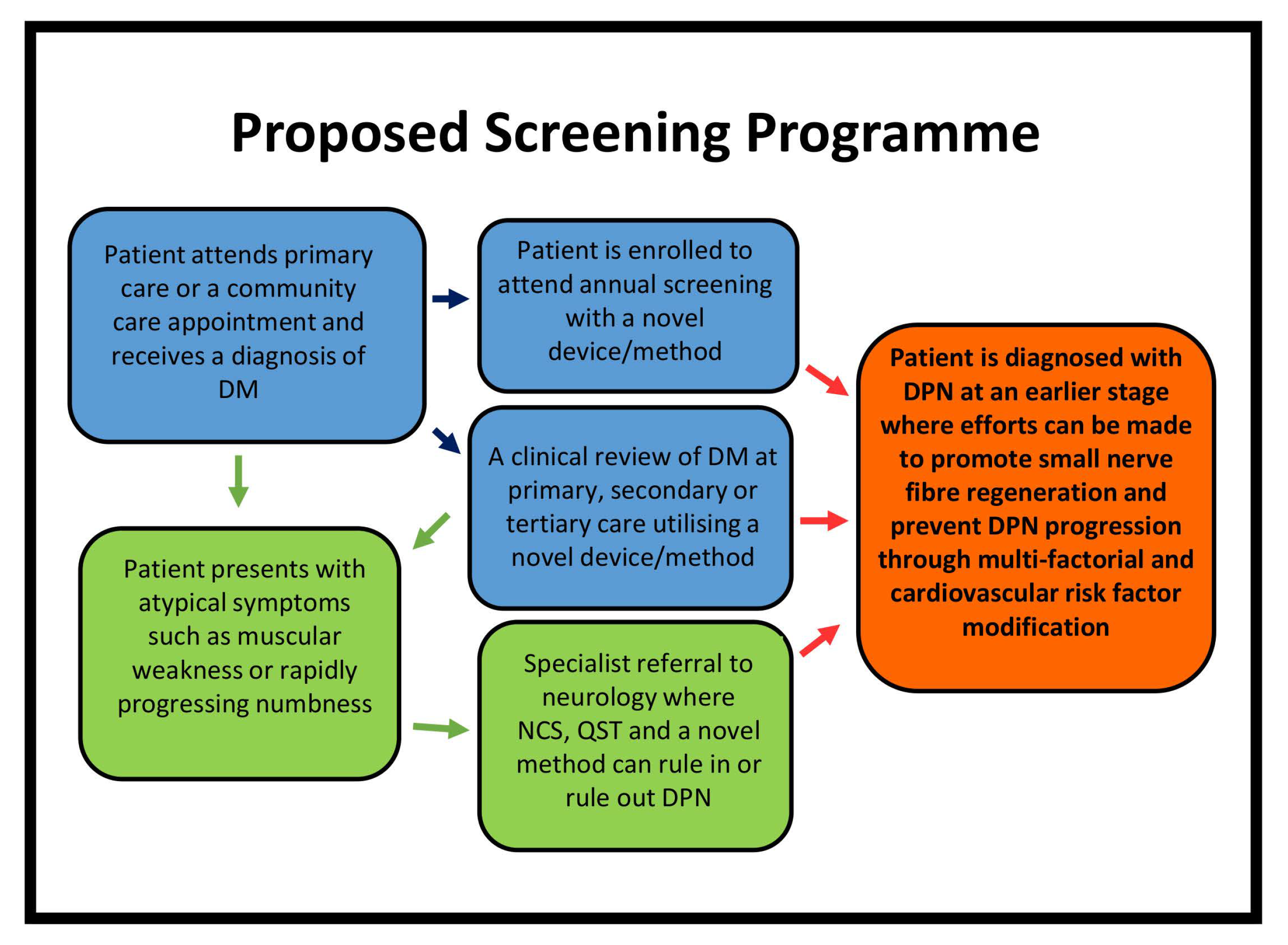
1.5. Methods
2. Screening and Diagnostic Tools
2.1. Composite Scoring Systems
2.2. Thermal and Vibration Perception Thresholds
2.3. German Network on Neuropathic Pain
2.4. Evoked Potentials
2.5. Microneurography
2.6. Current Perception Threshold
2.7. NC-Stat DPN Check
2.8. Skin Biopsy
2.9. Sudoscan
2.10. Neuropad
2.11. Laser Doppler Flare
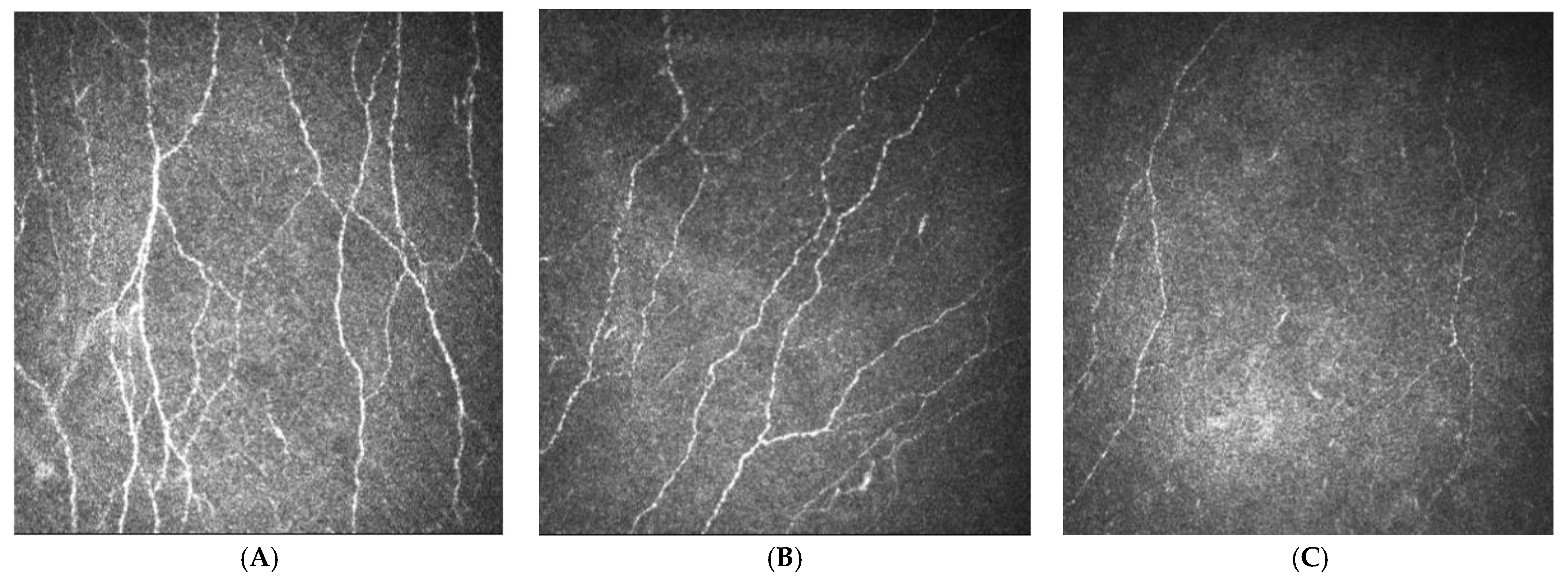
2.12. Corneal Confocal Microscopy
2.13. Limitations of This Review
3. Discussion
Author Contributions
Funding
Acknowledgments
Conflicts of Interest
Appendix A

| Citation | Participants | Comparator Test (s) | Cut Disease off Values Used | Threshold Value Used on Index Test | Test | AUC | Sensitivity % | Specificity % | PPV | NPV | +LR | −LR |
|---|---|---|---|---|---|---|---|---|---|---|---|---|
| McArthur et al., 1998 [190] | Control = 98 Sensory Neuropathy = 20 | No Comparator Test | NA | >the 5th percentile of the normative data | IENFD Distal Leg | - | 45 | 97 | 92 | 90 | - | - |
| Chien et al., 2001 [179] | Control = 55 Idiopathic = 28 DM = 5 Vasculitis = 2 | NCS | Reduced amplitudes of sensory action potential in the median, ulnar and sural nerves | >the 5th percentile of the normative data | IENFD Distal Forearm | - | 70 | 94 | 84 | 90 | - | - |
| HDT | - | |||||||||||
| CDT | - | IENFD Distal Leg | - | 80 | 95 | 84 | 93 | - | - | |||
| Neurological Examination | - | |||||||||||
| Koskinen et al., 2005 [189] | DPN = 12 Idiopathic/CIPN /Amyloidosis = 10 CIDP = 7 Control = 15 | Neurological Examination | - | - | NPEA Distal Leg | - | 90 | 95 | 95 | 91 | - | - |
| VPT | - | |||||||||||
| CDT | - | |||||||||||
| NCS | - | |||||||||||
| Vlcková-Moravcová et al., 2008 [185] | SFN = 58 MFN = 41 Control = 37 | NDS | (none = 0–2, mild = 3–5, moderate 6–8, severe 9–10) | ≤8.80/mm | IENFD All neuropathy participants | 0.89 | 79.7 | 82.1 | - | - | - | - |
| NSS | ≥1 | ≤7.77/mm | IENFD SFN Group | 0.88 | 68.6 | 95 | - | - | - | - | ||
| MNSI | ≥2 | ≤8.80/mm | IENFD MFN Group | 0.87 | 72.2 | 79.6 | - | - | - | - | ||
| CDT | CDT—see below * | |||||||||||
| HDT | WDT—see below * | |||||||||||
| Touch | - | |||||||||||
| Proprioception | - | |||||||||||
| Vibration | - | |||||||||||
| NCS | Abnormal conduction study from at least two nerves | |||||||||||
| Devigili et al., 2008 [133] | LFN = 22 SFN = 67 MFN = 21 Total = 110 | Thermal Thresholds | >95th percentile values of participants compared to age and sex matched controls considered abnormal | 7.63/mm | IENFD Total Distal Leg | 0.90 | 82.8 | 90 | 89.4 | 87.5 | - | - |
| NCS | SNAP <20 mv and <50 m/s (fifth to second finger) SNAP <6 and >42 m/s (sural) CMAP—A difference of at least 50% in SNAP and CMAP amplitude to define significant asymmetry (peroneal, tibial, ulnar and median) | 12.8/mm | IENFD Total Proximal Thigh | 0.82 | 95.5 | 57.5 | - | - | - | - | ||
| Laser Doppler Flowmetry | - | >95th percentile values of participants compared to age and sex matched controls considered abnormal | CDT and HDT | 0.57 | 56.7 | 36.5 | 48.7 | 44.2 | - | - | ||
| LEPs | Lower intensity compared to 18 age and sex matched controls | |||||||||||
| Nebuchennykh et al., 2009 [102] | SFN = 45 MFN = 165 Total = 210 | NISLL | - | 10.3 fibres/mm | Distal IENFD—SFN | 0.75 | 78 | 64 | - | - | - | - |
| NCS | >2 SD compared to reference normal values in ≥2 nerves | 9.8 fibres/mm | Distal IENFD MFN and SFN | 0.77 | 73 | 69 | - | - | - | - | ||
| QST | For reference values see [182] | |||||||||||
| Alam et al., 2017 [101] | Control = 27 T1D without DPN = 30 T1D with DPN = 31 | NSP | - | <4.5 fibres/mm | T1D with DPN | 0.73 | 61 | 80 | - | - | - | - |
| McGill-SF-2 | ||||||||||||
| CDT | ≤24.7 °C | |||||||||||
| HDT | ≥38.0 °C | |||||||||||
| VPT | ≥13 V |
| Citation | Participants | Comparator Test (s) | Cut off Value | Threshold Value Used on Index Test | Test and Target Condition | AUC | Sensitivity (%) | Specificity (%) | PPV | NPV | +LR | −LR |
|---|---|---|---|---|---|---|---|---|---|---|---|---|
| Chatzikosma et al., 2016 [172] | T2D = 114 Controls = 46 | NDS | ≥3 | Abnormality of ≥1 in ≥1 leg | Sural SNAP/SNCV—DPN | - | 90.4 | 86.1 | 79.1 | 93.9 | 6.51 | 0.11 |
| Perkins et al., 2006 [174] | T1D = 8 T2D = 64 | Reference NCS | ≥1 sign or symptom + abnormality of at least two parameters in at least two nerves [83] | Abnormality in either SNCV or SNAP | Sural SNAP/SNCV—DPN | - | 92 | 82 | 92 | 82 | - | - |
| Kural et al., 2019 [277] | T2D (n = 168) | Reference NCS | DPN ruled in if the sum z-score >2 [278] | <6 µV Both Legs | Sural SNAP—DPN | - | 96 | 71 | 54 | 98 | - | - |
| >4 and ≤6 µV Both Legs | Sural SNAP—DPN | - | 95 | 84 | 68 | 98 | - | - | ||||
| Perkins et al., 2008 [173] | DM = 72 | Reference NCS; PMNAP, PMNCV, tibial SNCV, tibial SNAP, median SNAP, median SNCV | ≥1 sign or symptom + abnormality of at least two parameters in at least two nerves [83] | Abnormality in either SNCV or SNAP | Sural SNAP—DPN | - | 88 | 82 | - | - | - | - |
| Scarr et al., 2018 [175] | T1D = 68 Controls = 71 | Reference NCS; SNAP SNCV | ≤7.2 µV for participants ≤ 65 years old, ≤5.5 µV for participants ≥ 65 years old ≤40 m/s | Abnormality in either SNCV (≤44 m/s) or SNAP (<6 µV) or both measures | SNAP only SNCV only SNAP/SNCV SNAP and SNCV | - - - 0.88 | 80 81 86 66 | 80 82 79 97 | - - - - | - - - - | - * - * - * - * | - - - - |
| Citation | Participants | Comparator Test (s) | Cut Disease off Values Used | Threshold Value Used on Index Test | Test and Target Condition | AUC | Sensitivity (%) | Specificity (%) | PPV | NPV | +LR | −LR |
|---|---|---|---|---|---|---|---|---|---|---|---|---|
| Krieger et al., 2018 [279] | T2D = 47 DPN =27 Non-DPN = 20 Controls = 16 | NDS | Mild (3–4) Moderate (5–6) Severe (7–10) | ESC of ≤80.00 μS Foot | Sudoscan—DPN | 0.70 | 70 | 53 | - | - | - | - |
| NDS | ESC of ≤75.00 μS Hand | Sudoscan—DPN | 0.71 | 85 | 50 | - | - | - | - | |||
| Goel et al., 2017 [210] | Total (T2D) = 523 DPN = 110 Non-DPN = 413 | NDS | DPN = ≥6 Non-DPN = <6 | Mean ESC <60 μS | Sudoscan—DPN | 0.88 | 85 | 85 | - | - | - | - |
| VPT—DPN | 0.84 | 72 | 90 | - | - | - | - | |||||
| VPT | >15 considered abnormal | NDS—DPN | 0.6 | 52 | 60 | - | - | - | - | |||
| Smith et al., 2014 [211] | Total = 97 Suspected DSP = 55 Control = 42 | UENS | - | ≤ 70 μS | Sudoscan—DPN | 0.76 | 77 | 67 | 59 | 83 | - | - |
| IENFD | - | |||||||||||
| QSART | - | - | IENFD—DPN | 0.75 | 63 | 63 | 73 | 52 | - | - | ||
| NCS | - | |||||||||||
| Selvarajah et al., 2015 [208] | Total = 70 T1D = 45 Control = 25 | NTSS NISLL TDT VPT NCS CARTS | >1 Symptom/sign and abnormal NCS | ≤ 77.0 μS | Sudoscan DPN | 0.85 | 87.5 | 76.2 | - | - | - | - |
| Autonomic Function Score | >30.0% Cardiac Autonomic Neuropathy Risk Score | Sudoscan CAN | 0.75 | 65 | 80 | - | - | - | - | |||
| Yajnik et al., 2012 [280] | T2D = 265 | MNSI VPT Ewing’s CART | >2 >21 V HRV E/I Ratio <1.21 HRV 30/15 Ratio <1.03 HRV during Valsalva manoeuvre <1.20 Orthostatic BP response >20 mmHg | <52.0 μS | Sudoscan DPN or CAN | 0.71 | 73 | 62 | - | - | - | - |
| Casellini et al., 2013 [207] | T1D = 83 Control = 210 | Modified NISLL HDT CDT QAFT | - ≥12.4 °C ≥10.5 °C - | >60 µS = Normal 60–40 µS = Moderate <40 µS = Severe | Sudoscan Hand—DPN | 0.86 | 78.3 | 85.7 | 61 | 93.2 | 5.48 | 0.25 |
| Sudoscan Feet—DPN | 0.88 | 78.3 | 92.3 | 74.6 | 93.72 | 10.28 | 0.23 | |||||
| Modified NISLL | 0.84 | 76.6 | 85.7 | 95.8 | 46.1 | 5.37 | 0.27 | |||||
| Carbajal-Ramírez et al., 2019 [281] | Total T2D = 221 T2D <5 years = 170 T2D ≥5 years = 51 | MNSI VPT 10-g Monofilament | - - - | <60 μS = abnormal hands AND/OR <70 μS = abnormal feet | Sudoscan T2D <5 years Sudoscan T2D ≥5 years | 0.66 0.72 | 91 97 | - - | 88 87 | - - | - - | - - |
| Binns-Hall et al., 2018 [282] | Total = 236 (T2D = 97.8%) No DPN = 163 Mild DPN = 34 Moderate DPN = 19 Severe DPN = 20 | TCNS 10-g Monofilament NCS (DPN Check) | TCNS- >5 Inability to feel ≥2 sites taken to indicate DPN If either SNAP was >4 µV and/or SNCV was <40 m/s | ≤58.5 µS (>60 µS = Normal <60 µS = DPN) ≤4.3 ≤46.3 | Sudoscan— DPN | 0.75 | 77.4 | 68.3 | - | - | - | - |
| NC-Stat DPN Check SNAP | 0.84 | 84.3 | 72.3 | - | - | - | - | |||||
| NC-Stat DPN Check SNCV | 0.81 | 68.3 | 80 | - | - | - | - | |||||
| 10-g Monofilament DPN | 0.61 | 30 | 92.7 | - | - | - | - | |||||
| Sudoscan + NC-Stat DPN Check | 0.73 | 93.2 | 52.8 | - | - | - | - | |||||
| Eranki et al., 2013 * [283] | T2D = 309 | VPT | - VPT ≥ 15 | Sudoscan VPT | 0.68 0.65 | 82 82 | 61 55 | - - | - - | - - | - - |
| Citation | Participants | Comparator Test (s) | Cut Disease off Values Used | Threshold Value Used on Index Test | Test and Target Condition | AUC | Sensitivity (%) | Specificity (%) | PPV | NPV | +LR | −LR |
|---|---|---|---|---|---|---|---|---|---|---|---|---|
| Papanas et al., 2011 [218] | T2D = 251 | NDS | Mild ≥ 3 | <365 s | Neuropad DPN | 0.91 | 95 | 75 | 99 | 21 | 3.79 | 0.07 |
| NDS | Moderate ≥ 6 | ≤1005 s | Neuropad DPN | 0.96 | 91 | 96 | 96 | 92 | 24.4 | 0.09 | ||
| NDS | Severe ≥ 9 | ≥1190 s | Neuropad DPN | 0.97 | 91 | 95 | 84 | 98 | 19 | 0.09 | ||
| Papanas et al., 2005 [213] | T2D = 104 | DNI | Moderate = 2.5–4.5 | Normal—A complete colour change (blue to pink) in both feet after 10 min. | Neuropad DPN | - | 94.4 | 69.7 | - | - | - | - |
| Severe = 5–8 | Abnormal—Partial or incomplete colour change in one foot after 10 min. | |||||||||||
| Liatis et al., 2007 [284] | Total = 117 T1D = 9 T2D = 108 | NSS | ≥3 | Normal—A complete colour change (blue to pink) in both feet after 10 min. Abnormal—Partial or incomplete colour change in one foot after 10 min. | Neuropad DPN | - | 86 | 67.2 | 66.2 | 86.5 | - | - |
| Ewing’s Score | ≥2 | Neuropad CAN | - | 59.1 | 46.5 | 40.6 | 64.7 | - | - | |||
| Ewing’s Score | ≥6 | Neuropad CAN | - | 80.9 | 50 | 26.0 | 92.1 | - | - | |||
| Papanas et al., 2007 [217] | T2D = 120 | MNSI | 0—none 1—mild 2—moderate 3—severe neuropathy | ≤530 s | Neuropad DPN | - | 97 | 100 | - | - | - | - |
| ≤1000 s | Neuropad DPN | - | 100 | 97 | - | - | - | - | ||||
| <1440 s | Neuropad DPN | - | 93 | 100 | - | - | - | - | ||||
| >1440 s | Neuropad DPN | - | 100 | 99 | - | - | - | - | ||||
| Papanas et al., 2008 [285] | T2D = 154 | NDS | Abnormal >25 V Intermediate 15–25 V Normal <15 V | Neuropad—DPN | - | 97.8 | 67.2 | - | - | - | - | |
| VPT—DPN | - | 78.9 | 85.9 | - | - | - | - | |||||
| Quattrini et al., 2008 [220] | Total n = 57 T1D = 20 T2D = 37 | NDS | ≥3 | Partial or incomplete colour change in one foot after 10 min | Neuropad— DPN | - | 85 | 45 | 69 | 71 | - | - |
| Tentolouris et al., 2008 [216] | Total = 156 T1D = 7 T2D = 149 | NSS | Normal (pink colour bi-laterally) or abnormal (blue colour or any other combinations of colours bilaterally) | Neuropad— DPN | - | 87 | 66 | 94 | 79 | - | - | |
| Bilen et al., 2007 [286] | T2D = 105 | Corrected QT Interval | >0.440 s | Blue indicated sudomotor dysfunction and pink indicated normal sudomotor function | Neuropad— CAN | - | 87.5 | 43.1 | 48.6 | 84.8 | - | - |
| Freitas et al., 2009 [287] | T2D = 40 DPN+ = 22 DPN− = 18 | NDS | ≥6 | Absence of a colour change from blue to pink, or incomplete change (blue and pink mixed) were considered to correspond to an altered test | Neuropad— DPN | - | 100 | 44 | 69 | 100 | - | - |
| Ishibashi et al., 2014 [288] | Control = 28 T2D = 78 DPN Stage 1 = 23 DPN Stage 2 = 28 DPN Stage 3 = 20 DPN Stage 4 = 7 | 1. Subjective symptom in bilateral lower limbs or feet 2. Loss of or decreased ankle jerk 3. Decreased VPT assessed | ≥2 | >815 s | Neuropad— DPN | - | 83.1 | 84.0 | - | - | - | - |
| Manes et al., 2014 [219] | Total T2D = 1010 Sudomotor Dysfunction = 441 No Sudomotor Dysfunction = 569 | NDS | >6 | Partial colour change = 1 No colour change = 2 Blue to pink = 0 Neuropad score ≥ 1 Neuropad score = 2 | Neuropad Sudomotor Dysfunction | - | 85.6 | 71.2 | 51.2 | 93.3 | - | - |
| Neuropad DPN | - | 94.9 | 70.2 | 46.3 | 98.1 | - | - | |||||
| Neuropad Sudomotor Dysfunction | - | 52 | 96 | 82 | 85 | - | - | |||||
| Neuropad DPN | - | 64 | 96 | 82 | 91 | - | - | |||||
| Kamenov et al., 2010 [289] | Total = 264 T1D = 61 T2D = 203 | NDS NDS | >3 >6 | Neuropad Neuropad | - - | 76.3 79.3 | 56.1 42.9 | 86.3 62.8 | 39.5 63.0 | - - | - - | |
| Ponirakis et al., 2014 [290] | T2D = 50 | DNS | >2 | Scanned images of Neuropads applied (n = 50) were estimated by three masked observers on two separate occasions on a scale of 0-100% colour change | Neuropad * | 66 | 70 | 50 | 63 | 57 | 1.4 | 0.6 |
| VPT | >14 volts | Neuropad * | 73 | 83 | 53 | 45 | 39 | 1.77 | 0.32 | |||
| SNAP | <3 µV | Neuropad * | 70 | 70 | 64 | 26 | 92 | 1.94 | 0.47 | |||
| SNCV | <43 m/s | Neuropad * | 63 | 64 | 54 | 45 | 72 | 1.39 | 0.67 | |||
| PMNAP | <2 mV | Neuropad * | 69 | 82 | 50 | 31 | 91 | 1.64 | 0.36 | |||
| PMNCV | <42 m/s | Neuropad * | 70 | 81 | 54 | 59 | 78 | 1.76 | 0.35 | |||
| WPT | >43 °C | Neuropad * | 60 | 68 | 49 | 26 | 44 | 1.33 | 0.65 | |||
| CNFL | <24 no/mm2 | Neuropad * | 67 | 74 | 60 | 54 | 78 | 1.85 | 0.43 | |||
| CNFD | <14 mm/mm2 | Neuropad * | 85 | 83 | 80 | 49 | 95 | 4.12 | 0.21 | |||
| NSS | (Subjective Test) | Neuropad * | 68 | 78 | 60 | 34 | 91 | 1.95 | 0.37 | |||
| Ziegler et al., 2011 [222] | T1D = 52 | NDS | ≥2 | Normal—A complete colour change (blue to pink) in both feet after <10 min. Abnormal—Partial or incomplete colour change in one foot after 10, 15, or 20 min | Neuropad—DPN 10 min | - | 87.5 | 47.7 | 23.3 | 95.4 | - | - |
| Neuropad— DPN 15 min | - | 50 | 70.4 | 23.5 | 88.6 | - | - | |||||
| Neuropad— DPN 20 min | - | 12.5 | 84.1 | 12.5 | 84.1 | - | - | |||||
| Neuropad *— 10 min | - | 80 | 44.7 | 13.3 | 95.4 | - | - | |||||
| Neuropad *— 15 min | - | 80 | 72.3 | 23.5 | 97.1 | - | - | |||||
| Neuropad *— 20 min | - | 20 | 85.1 | 12.5 | 90.9 | - | - | |||||
| Ziegler et al., 2011 [222] | T2D = 99 | NDS | ≥2 | Normal—A complete colour change (blue to pink) in both feet after <10 min. Abnormal—Partial or incomplete colour change in one foot after 10, 15, or 20 min | Neuropad— DPN 10 min | - | 65.1 | 48.2 | 49.1 | 64.3 | - | - |
| Neuropad— DPN 15 min | - | 48.8 | 76.8 | 61.8 | 66.1 | - | - | |||||
| Neuropad— DPN 20 min | - | 34.9 | 89.3 | 71.4 | 64.1 | - | - | |||||
| Neuropad *— 10 min | - | 67.7 | 47.1 | 36.8 | 76.2 | - | - | |||||
| Neuropad *— 15 min | - | 45.2 | 70.6 | 41.2 | 73.8 | - | - | |||||
| Neuropad *— 20 min | - | 32.3 | 83.8 | 47.6 | 73.1 | - | - |
| Citation | Participants | Comparator Test (s) | Cut-off Value * | Index Test Threshold | Test and Target Condition | AUC | Sensitivity | Specificity | PPV | NPV | +LR | −LR |
|---|---|---|---|---|---|---|---|---|---|---|---|---|
| Ahmed et al., 2012 [291] | T1D = 89 Control = 64 | TCNS LDIFlare | - - | ≥11.50 mm/mm2 | CNFL—DPN | - | - | 93 | 83 | - | 8.5 | - |
| ≥14.00 mm/mm2 | CNFL—DPN | 0.88 | 85 | 84 | 76 | 90 | 5.3 | 0.18 | ||||
| ≥15.80 mm/mm2 | CNFL—DPN | - | 91 | - | - | 91 | - | 0.16 | ||||
| - | CNFD | 0.84 | - | - | - | - | - | - | ||||
| - | CNBD | 0.73 | - | - | - | - | - | - | ||||
| Perkins et al., 2018 [271] | Total = 998 T1D = 516 T2D = 482 | Reference standard based on recommendations [78] | Abnormal nerve conduction and signs or symptoms of neuropathy [6] | 12.5 mm/mm2 | Automated CNFL T1D—DPN | 0.77 | 73 | 69 | 50 | 86 | 2.32 | 0.39 |
| 12.3 mm/mm2 | Automated CNFL T2D—DPN | 0.68 | 69 | 63 | 66 | 66 | 1.86 | 0.49 | ||||
| 12.3 mm/mm2 | Automated CNFL T1D and T2D—DPN | 0.77 | 67 | 66 | 59 | 74 | 1.97 | 0.50 | ||||
| Total <8.6 mm/mm2 | Automated CNFL T1D and T2D—DPN | - | 88 | 88 | - | - | - | - | ||||
| Alam et al., 2017 [101] | T1D with neuropathy = 31 Control Participants = 27 | CDT HDT VPT NCS Autonomic Function Tests IENFD | ≤24.7 °C ≥38.0 °C ≥13 V | 25 no/mm2 36.5 no/mm2 16.8 mm/mm2 <4.5 no/mm | CNFD—DPN CNBD—DPN CNFL—DPN IENFD | 0.81 0.67 0.74 0.73 | 77 58 61 61 | 79 79 86 80 | - - - - | - - - - | - - - - | - - - - |
| Chen et al., 2015 [292] | T1D = 63 Control = 26 | DNS NDS CDT HDT VPT NCT (PMNamp) | ≥1 ≥3 NS NS NS PMNCV of <42 m/s | 2 SD below the mean of the control group | Manual CNFD—DPN CNFL—DPN CNBD—DPN Automated CNFD—DPN CNFL—DPN CNBD—DPN IENFD | 0.82 0.70 0.59 0.80 0.77 0.80 0.66 | 82 59 17 60 59 29 53 | 71 74 96 83 80 98 76 | - - - - - - - | - - - - - - - | - - - - - - - | - - - - - - - |
| Edwards et al., 2014 [293] | NDS 10-g Monofilament NCS | Abnormal nerve conduction and signs or symptoms of neuropathy | - | CNFL | 0.64 | 32 | 87 | - | - | - | - | |
| - | Tortuosity-standardised CNFL | 0.67 | 38 | 88 | - | - | - | - | ||||
| Ishibashi et al., 2012 [251] | T1D = 38 Control = 38 | Signs and symptoms VPT Reflex Testing | ≥2 of the following: bilateral lower limb symptoms, impaired reflexes and abnormal VPT | 11 mm/mm2 | CNFL | - | 87 | 77 | - | - | - | - |
| 28.4 mm2 | CNFD | - | 87 | 76 | - | - | - | - | ||||
| 25.2/0.1 mm | CNFT Grade | - | 92 | 94 | - | - | - | - | ||||
| 2.47 | Beading Frequency | - | 81 | 79 | - | - | - | - | ||||
| Ponirakis et al., 2015 [294] | DPN− = 64 DPN+ = 46 | NDS PMNCV Signs and Symptoms | >2 >42 μV ≥1 | <14 mm/mm2 <18 no/mm2 <24 no/mm2 <4 no./mm >42 °C >2 >14 <3 μV <43 μV <2 μV <42 μV | CNFL CNFD CNBD IENFD HDT NDS VPT Sural SNAP Sural SNCV PMNAP PMNCV | 0.80 0.82 0.79 0.63 0.69 0.67 0.75 0.86 0.62 0.63 0.60 | 89 88 83 65 75 71 80 85 66 67 62 | 75 78 72 54 60 58 71 83 61 54 58 | - - - - - - - - - - - | - - - - - - - - - - - | - - - - - - - - - - - | - - - - - - - - - - - |
| Tavakoli et al., 2015 [295] | DAN+ve = 19 DAN−ve = 15 Control = 18 | CASS | >2 | <4.78 mm/mm2 <23.26 no/mm2 <19.53 no/mm2 | CNFL—DAN CNFD—DAN CNBD—DAN | 0.90 0.91 0.88 | 86 86 100 | 78 78 56 | - - - | - - - | - - - | - - - |
| Pritchard et al., 2015 [296] | Control = 80 T1D = 107 of which; DPN+ = 25 DPN− = 82 | NDS * | ≥3 ≥3 | ≤17.9 ≤18.6 | CNFL Centre CNFL Whorl | 0.76 0.77 | 90 80 | 50 60 | - - | - - | 2.0 2.2 | 0.2 0.3 |
| Wang et al., 2020 [297] | Total = 220 Control = 48 T2D = 172 | Signs and Symptoms PMNCV SSNCV SSNAMP PCMAP | ≥42 m/s ≥42 m/s ≥6 µV ≥2 mV | <15.3 n/mm2 <39 n/mm2 <25.68 n/mm2 | CNFL CNBD CNFD | 0.70 0.67 0.66 | 80 85 78 | 59 47 52 | - - - | - - - | 1.98 1.61 1.65 | 0.33 0.31 0.41 |
| Petropoulos et al., 2014 ** [261] | T1/2D = 186 DPN+ = 100 DPN− = 86 Controls = 55 | PMNAP Sural SNAP PMNCV HDT | <1.4 µV <5.5 µV <42.0 m/s >41 °C | 15.8 mm/mm2 18.7 no/mm2 41.7 no/mm2 14.6 mm/mm2 14.9 no/mm2 14.9 no/mm2 19.4 mm/mm2 23.1 no/mm2 47.1 no/mm2 16.1 mm/mm2 18.9 no/mm2 23.4 no/mm2 19.7 mm/mm2 25.4 no/mm2 49.0 no/mm2 16.0 mm/mm2 19.7 no/mm2 24.9 no/mm2 19.2mm/mm2 24.0 no/mm2 47.2 no/mm2 15.9 mm/mm2 17.9 no/mm2 22.9 no/mm2 | CNFL Manual CNFD Manual CNBD Manual CNFL Auto CNFD Auto CNBD Auto CNFL Manual CNFD Manual CNBD Manual CNFL Auto CNFD Auto CNBD Auto CNFL Manual CNFD Manual CNBD Manual CNFL Auto CNFD Auto CNBD Auto CNFL Manual CNFD Manual CNBD Manual CNFL Auto CNFD Auto CNBD Auto | 0.82 0.84 0.75 0.84 0.84 0.79 0.70 0.74 0.65 0.77 0.72 0.70 0.73 0.74 0.68 0.79 0.74 0.67 0.67 0.69 0.65 0.68 0.67 0.64 | 77 79 73 77 76 74 68 72 61 72 18.9 23.4 74 78 69 74 80 68 63 63 65 61 63 60 | 76 78 68 74 72 73 67 67 56 66 56 54 63 70 61 71 61 52 61 62 55 61 60 58 | - - - - - - - - - - - - - - - - - - - - - - - - | - - - - - - - - - - - - - - - - - - - - - - - - | 3.2 4.6 2.3 3.3 3.4 2.9 2.1 1.9 1.4 2.1 1.9 1.4 2.0 2.6 1.8 2.6 2.2 1.4 1.6 1.7 1.4 1.5 1.5 1.4 | 0.3 0.3 0.4 0.2 0.3 0.3 0.5 0.4 0.7 0.4 0.4 0.7 0.4 0.3 0.5 0.4 0.3 0.6 0.6 0.6 0.7 0.7 0.6 0.7 |
| Ferdousi et al., 2020 [265] | Total = 490 Control = 72 T1D =149 T2D = 269 | NDS VPT CDT HDT Sural SNCV | >2 | 21.6 mm/mm2 23.95 no/mm2 58.54 no/mm2 | CNFL CNFD CNBD | 0.68 0.71 0.70 | 64 58 69 | 67 83 65 | - - - | - - - | - - - | - - - |
| Ferdousi et al., 2021 [298] | Control = 72 T1D = 149 T2D = 269 | NSP NDS Pinprick VPT CPT HPT DB-HRV NCS Sural SNAP Sural SNCV PMNCV | >2 <40.0 m/s | 24 mm/mm2 29.4 no/mm2 64.58 no/mm2 | CNFL CNFD CNBD | 0.73 0.81 0.74 | 66.7 73.5 66.7 | 66.4 74.4 66.7 | - - - | - - - | - - - | - - - |
References
- Cho, N.H.; Shaw, J.E.; Karuranga, S.; Huang, Y.; da Rocha Fernandes, J.D.; Ohlrogge, A.W.; Malanda, B. IDF Diabetes Atlas: Global estimates of diabetes prevalence for 2017 and projections for 2045. Diabetes Res. Clin. Pract. 2018, 138, 271–281. [Google Scholar] [CrossRef] [PubMed]
- Bhutani, J.; Bhutani, S. Worldwide burden of diabetes. Indian J. Endocrinol. Metab. 2014, 18, 868–870. [Google Scholar] [CrossRef] [PubMed]
- Global Burden of Disease Cancer Collaboration; Fitzmaurice, C.; Allen, C.; Barber, R.M.; Barregard, L.; Bhutta, Z.A.; Brenner, H.; Dicker, D.J.; Chimed-Orchir, O.; Dandona, R.; et al. Global, Regional, and National Cancer Incidence, Mortality, Years of Life Lost, Years Lived with Disability, and Disability-Adjusted Life-years for 32 Cancer Groups, 1990 to 2015: A Systematic Analysis for the Global Burden of Disease Study. JAMA Oncol. 2017, 3, 524–548. [Google Scholar] [CrossRef] [PubMed]
- Singh, R.; Kishore, L.; Kaur, N. Diabetic peripheral neuropathy: Current perspective and future directions. Pharmacol. Res. 2014, 80, 21–35. [Google Scholar] [CrossRef]
- Callaghan, B.C.; Cheng, H.T.; Stables, C.L.; Smith, A.L.; Feldman, E.L. Diabetic neuropathy: Clinical manifestations and current treatments. Lancet Neurol. 2012, 11, 521–534. [Google Scholar] [CrossRef] [Green Version]
- Tesfaye, S.; Boulton, A.J.M.; Dyck, P.J.; Freeman, R.; Horowitz, M.; Kempler, P.; Lauria, G.; Malik, R.A.; Spallone, V.; Vinik, A.; et al. Diabetic neuropathies: Update on definitions, diagnostic criteria, estimation of severity, and treatments. Diabetes Care 2010, 33, 2285–2293. [Google Scholar] [CrossRef] [Green Version]
- Pirart, J. Diabetes mellitus and its degenerative complications: A prospective study of 4400 patients observed between 1947 and 1973 (3rd and last part) (author’s transl.). Diabetes Metab. 1977, 3, 245–256. [Google Scholar]
- Gordois, A.; Scuffham, P.; Shearer, A.; Oglesby, A.; Tobian, J.A. The health care costs of diabetic peripheral neuropathy in the US. Diabetes Care 2003, 26, 1790–1795. [Google Scholar] [CrossRef] [Green Version]
- Feldman, E.L.; Callaghan, B.C.; Pop-Busui, R.; Zochodne, D.W.; Wright, D.E.; Bennett, D.L.; Bril, V.; Russell, J.W.; Viswanathan, V. Diabetic neuropathy. Nat. Rev. Dis. Primers 2019, 5, 41. [Google Scholar] [CrossRef]
- Malik, R.; Veves, A.; Tesfaye, S.; Smith, G.; Cameron, N.; Zochodne, D.; Lauria, G. Small Fiber Neuropathy: Role in the diagnosis of Diabetic Sensorimotor Polyneuropathy. Diabetes Metab. Res. Rev. 2011, 27, 678–684. [Google Scholar] [CrossRef]
- Breiner, A.; Lovblom, L.E.; Perkins, B.A.; Bril, V. Does the Prevailing Hypothesis That Small-Fiber Dysfunction Precedes Large-Fiber Dysfunction Apply to Type 1 Diabetic Patients? Diabetes Care 2014, 37, 1418. [Google Scholar] [CrossRef] [PubMed] [Green Version]
- Ziegler, D.; Papanas, N.; Zhivov, A.; Allgeier, S.; Winter, K.; Ziegler, I.; Bruggemann, J.; Strom, A.; Peschel, S.; Kohler, B.; et al. Early detection of nerve fiber loss by corneal confocal microscopy and skin biopsy in recently diagnosed type 2 diabetes. Diabetes 2014, 63, 2454–2463. [Google Scholar] [CrossRef] [PubMed] [Green Version]
- Dobretsov, M.; Romanovsky, D.; Stimers, J.R. Early diabetic neuropathy: Triggers and mechanisms. World J. Gastroenterol. 2007, 13, 175–191. [Google Scholar] [CrossRef] [PubMed]
- Azmi, S.; Jeziorska, M.; Ferdousi, M.; Petropoulos, I.N.; Ponirakis, G.; Marshall, A.; Alam, U.; Asghar, O.; Atkinson, A.; Jones, W.; et al. Early nerve fibre regeneration in individuals with type 1 diabetes after simultaneous pancreas and kidney transplantation. Diabetologia 2019, 62, 1478–1487. [Google Scholar] [CrossRef] [PubMed] [Green Version]
- Smith, A.G.; Russell, J.; Feldman, E.L.; Goldstein, J.; Peltier, A.; Smith, S.; Hamwi, J.; Pollari, D.; Bixby, B.; Howard, J.; et al. Lifestyle intervention for pre-diabetic neuropathy. Diabetes Care 2006, 29, 1294–1299. [Google Scholar] [CrossRef] [Green Version]
- Tavakoli, M.; Mitu-Pretorian, M.; Petropoulos, I.N.; Fadavi, H.; Asghar, O.; Alam, U.; Ponirakis, G.; Jeziorska, M.; Marshall, A.; Efron, N.; et al. Corneal confocal microscopy detects early nerve regeneration in diabetic neuropathy after simultaneous pancreas and kidney transplantation. Diabetes 2013, 62, 254–260. [Google Scholar] [CrossRef] [Green Version]
- Abbott, C.A.; Malik, R.A.; van Ross, E.R.; Kulkarni, J.; Boulton, A.J. Prevalence and characteristics of painful diabetic neuropathy in a large community-based diabetic population in the U.K. Diabetes Care 2011, 34, 2220–2224. [Google Scholar] [CrossRef] [Green Version]
- Veves, A.; Backonja, M.; Malik, R.A. Painful diabetic neuropathy: Epidemiology, natural history, early diagnosis, and treatment options. Pain Med. 2008, 9, 660–674. [Google Scholar] [CrossRef]
- Pruitt, J., 3rd; Moracho-Vilrriales, C.; Threatt, T.; Wagner, S.; Wu, J.; Romero-Sandoval, E.A. Identification, prevalence, and treatment of painful diabetic neuropathy in patients from a rural area in South Carolina. J. Pain Res. 2017, 10, 833–843. [Google Scholar] [CrossRef] [Green Version]
- Attal, N.; Lanteri-Minet, M.; Laurent, B.; Fermanian, J.; Bouhassira, D. The specific disease burden of neuropathic pain: Results of a French nationwide survey. Pain 2011, 152, 2836–2843. [Google Scholar] [CrossRef]
- Finnerup, N.B.; Attal, N.; Haroutounian, S.; McNicol, E.; Baron, R.; Dworkin, R.H.; Gilron, I.; Haanpää, M.; Hansson, P.; Jensen, T.S.; et al. Pharmacotherapy for neuropathic pain in adults: A systematic review and meta-analysis. Lancet Neurol. 2015, 14, 162–173. [Google Scholar] [CrossRef] [Green Version]
- Quattrini, C.; Tesfaye, S. Understanding the impact of painful diabetic neuropathy. Diabetes Metab. Res. Rev. 2003, 19 (Suppl. S1), S2–S8. [Google Scholar] [CrossRef] [PubMed]
- Gore, M.; Brandenburg, N.A.; Dukes, E.; Hoffman, D.L.; Tai, K.-S.; Stacey, B. Pain severity in diabetic peripheral neuropathy is associated with patient functioning, symptom levels of anxiety and depression, and sleep. J. Pain Symptom Manag. 2005, 30, 374–385. [Google Scholar] [CrossRef] [PubMed]
- Stewart, W.F.; Ricci, J.A.; Chee, E.; Hirsch, A.G.; Brandenburg, N.A. Lost productive time and costs due to diabetes and diabetic neuropathic pain in the US workforce. J. Occup. Environ. Med. 2007, 49, 672–679. [Google Scholar] [CrossRef]
- Scholz, J.; Finnerup, N.B.; Attal, N.; Aziz, Q.; Baron, R.; Bennett, M.I.; Benoliel, R.; Cohen, M.; Cruccu, G.; Davis, K.D.; et al. The IASP classification of chronic pain for ICD-11: Chronic neuropathic pain. Pain 2019, 160, 53–59. [Google Scholar] [CrossRef] [Green Version]
- Torrance, N.; Elliott, A.M.; Lee, A.J.; Smith, B.H. Severe chronic pain is associated with increased 10 year mortality. A cohort record linkage study. Eur. J. Pain 2010, 14, 380–386. [Google Scholar] [CrossRef]
- Ziegler, D.; Strom, A.; Lobmann, R.; Reiners, K.; Rett, K.; Schnell, O. High prevalence of diagnosed and undiagnosed polyneuropathy in subjects with and without diabetes participating in a nationwide educational initiative (PROTECT study). J. Diabetes Complicat. 2015, 29, 998–1002. [Google Scholar] [CrossRef]
- Ponirakis, G.; Elhadd, T.; Chinnaiyan, S.; Dabbous, Z.; Siddiqui, M.; Al-Muhannadi, H.; Petropoulos, I.; Khan, A.; Ashawesh, K.A.E.; Dukhan, K.M.O.; et al. Prevalence and risk factors for painful diabetic neuropathy in secondary healthcare in Qatar. J. Diabetes Investig. 2019, 10, 1558–1564. [Google Scholar] [CrossRef] [Green Version]
- Ponirakis, G.; Elhadd, T.; Chinnaiyan, S.; Dabbous, Z.; Siddiqui, M.; Al-Muhannadi, H.; Petropoulos, I.N.; Khan, A.; Ashawesh, K.A.E.; Dukhan, K.M.O.; et al. Prevalence and management of diabetic neuropathy in secondary care in Qatar. Diabetes Metab. Res. Rev. 2020, 36, e3286. [Google Scholar] [CrossRef]
- Vinik, A.I. Management of neuropathy and foot problems in diabetic patients. Clin. Cornerstone 2003, 5, 38–55. [Google Scholar] [CrossRef]
- Vinik, A.I.; Park, T.S.; Stansberry, K.B.; Pittenger, G.L. Diabetic neuropathies. Diabetologia 2000, 43, 957–973. [Google Scholar] [CrossRef] [PubMed]
- Margolis, D.J.; Jeffcoate, W. Epidemiology of foot ulceration and amputation: Can global variation be explained? Med. Clin. 2013, 97, 791–805. [Google Scholar] [CrossRef]
- Larsson, J.; Agardh, C.-D.; Apelqvist, J.; Stenström, A. Long-term prognosis after healed amputation in patients with diabetes. Clin. Orthop. Relat. Res. 1998, 350, 149–158. [Google Scholar] [CrossRef]
- Tentolouris, N.; Al-Sabbagh, S.; Walker, M.G.; Boulton, A.J.; Jude, E.B. Mortality in diabetic and nondiabetic patients after amputations performed from 1990 to 1995: A 5-year follow-up study. Diabetes Care 2004, 27, 1598–1604. [Google Scholar] [CrossRef] [Green Version]
- Icks, A.; Scheer, M.; Morbach, S.; Genz, J.; Haastert, B.; Giani, G.; Glaeske, G.; Hoffmann, F. Time-dependent impact of diabetes on mortality in patients after major lower extremity amputation: Survival in a population-based 5-year cohort in Germany. Diabetes Care 2011, 34, 1350. [Google Scholar] [CrossRef] [Green Version]
- Bakker, K.; Apelqvist, J.; Lipsky, B.A.; Van Netten, J.J. The 2015 IWGDF guidance documents on prevention and management of foot problems in diabetes: Development of an evidence-based global consensus. Diabetes Metab. Res. Rev. 2016, 32 (Suppl. S1), 2–6. [Google Scholar] [CrossRef] [Green Version]
- Tchero, H.; Kangambega, P.; Lin, L.; Mukisi-Mukaza, M.; Brunet-Houdard, S.; Briatte, C.; Retali, G.R.; Rusch, E. Cost of diabetic foot in France, Spain, Italy, Germany and United Kingdom: A systematic review. Ann. Endocrinol. 2018, 79, 67–74. [Google Scholar] [CrossRef]
- NHS. Annual Diabetes Foot Care Report. 2014. Available online: https://www.england.nhs.uk/south/wp-content/uploads/sites/6/2017/07/se-clinical-network-foot-care-annual-report-october-2016.pdf (accessed on 5 May 2020).
- Feldman, E.L.; Nave, K.A.; Jensen, T.S.; Bennett, D.L.H. New Horizons in Diabetic Neuropathy: Mechanisms, Bioenergetics, and Pain. Neuron 2017, 93, 1296–1313. [Google Scholar] [CrossRef] [Green Version]
- Cameron, N.E.; Eaton, S.E.M.; Cotter, M.A.; Tesfaye, S. Vascular factors and metabolic interactions in the pathogenesis of diabetic neuropathy. Diabetologia 2001, 44, 1973–1988. [Google Scholar] [CrossRef] [Green Version]
- Greene, D.A.; Stevens, M.J.; Obrosova, I.; Feldman, E.L. Glucose-induced oxidative stress and programmed cell death in diabetic neuropathy. Eur. J. Pharmacol. 1999, 375, 217–223. [Google Scholar] [CrossRef]
- Zhou, J.; Zhou, S. Inflammation: Therapeutic Targets for Diabetic Neuropathy. Mol. Neurobiol. 2014, 49, 536–546. [Google Scholar] [CrossRef] [PubMed]
- Obrosova, I.G.; Li, F.; Abatan, O.I.; Forsell, M.A.; Komjáti, K.; Pacher, P.; Szabó, C.; Stevens, M.J. Role of poly(ADP-ribose) polymerase activation in diabetic neuropathy. Diabetes 2004, 53, 711–720. [Google Scholar] [CrossRef] [PubMed] [Green Version]
- Obrosova, I.G.; Drel, V.R.; Oltman, C.L.; Mashtalir, N.; Tibrewala, J.; Groves, J.T.; Yorek, M.A. Role of nitrosative stress in early neuropathy and vascular dysfunction in streptozotocin-diabetic rats. Am. J. Physiol. Endocrinol. Metab. 2007, 293, E1645–E1655. [Google Scholar] [CrossRef] [PubMed] [Green Version]
- Vareniuk, I.; Pacher, P.; Pavlov, I.A.; Drel, V.R.; Obrosova, I.G. Peripheral neuropathy in mice with neuronal nitric oxide synthase gene deficiency. Int. J. Mol. Med. 2009, 23, 571–580. [Google Scholar] [CrossRef] [Green Version]
- Kobayashi, M.; Zochodne, D.W. Diabetic neuropathy and the sensory neuron: New aspects of pathogenesis and their treatment implications. J. Diabetes Investig. 2018, 9, 1239–1254. [Google Scholar] [CrossRef]
- Hosseini, A.; Abdollahi, M. Diabetic neuropathy and oxidative stress: Therapeutic perspectives. Oxidative Med. Cell. Longev. 2013, 2013, 168039. [Google Scholar] [CrossRef]
- Lukic, I.K.; Humpert, P.M.; Nawroth, P.P.; Bierhaus, A. The RAGE pathway: Activation and perpetuation in the pathogenesis of diabetic neuropathy. Ann. N. Y. Acad. Sci. 2008, 1126, 76–80. [Google Scholar] [CrossRef]
- Casellini, C.M.; Barlow, P.M.; Rice, A.L.; Casey, M.; Simmons, K.; Pittenger, G.; Bastyr, E.J.; Wolka, A.M.; Vinik, A.I. A 6-month, randomized, double-masked, placebo-controlled study evaluating the effects of the protein kinase C-beta inhibitor ruboxistaurin on skin microvascular blood flow and other measures of diabetic peripheral neuropathy. Diabetes Care 2007, 30, 896–902. [Google Scholar] [CrossRef] [Green Version]
- Jack, M.; Wright, D. Role of advanced glycation endproducts and glyoxalase I in diabetic peripheral sensory neuropathy. Transl. Res. 2012, 159, 355–365. [Google Scholar] [CrossRef] [Green Version]
- Geraldes, P.; King, G.L. Activation of protein kinase C isoforms and its impact on diabetic complications. Circ. Res. 2010, 106, 1319–1331. [Google Scholar] [CrossRef] [Green Version]
- So, K. Roles of TRPA1 in Painful Dysesthesia. Yakugaku Zasshi 2020, 140, 1–6. [Google Scholar] [CrossRef] [PubMed]
- Pek, S.L.T.; Lim, S.C.; Ang, K.; Kwan, P.Y.; Tang, W.E.; Sum, C.F.; Tavintharan, S. Endothelin-1 predicts incident diabetic peripheral neuropathy in Type 2 Diabetes: A cohort study. Eur. J. Endocrinol. 2020, 182, 429–438. [Google Scholar] [CrossRef] [PubMed]
- Tesfaye, S.; Chaturvedi, N.; Eaton, S.E.M.; Ward, J.D.; Manes, C.; Ionescu-Tirgoviste, C.; Witte, D.R.; Fuller, J.H. Vascular Risk Factors and Diabetic Neuropathy. N. Engl. J. Med. 2005, 352, 341–350. [Google Scholar] [CrossRef] [PubMed] [Green Version]
- The DCCT Research Group. The Effect of Intensive Treatment of Diabetes on the Development and Progression of Long-Term Complications in Insulin-Dependent Diabetes Mellitus. N. Engl. J. Med. 1993, 329, 977–986. [Google Scholar] [CrossRef]
- The DCCT Research Group. Effect of intensive diabetes treatment on nerve conduction in the Diabetes Control and Complications Trial. Ann. Neurol. 1995, 38, 869–880. [Google Scholar] [CrossRef]
- Epidemiology of Diabetes Interventions and Complications (EDIC). Epidemiology of Diabetes Interventions and Complications (EDIC). Design, implementation, and preliminary results of a long-term follow-up of the Diabetes Control and Complications Trial cohort. Diabetes Care 1999, 22, 99–111. [Google Scholar] [CrossRef] [Green Version]
- Callaghan, B.C.; Little, A.A.; Feldman, E.L.; Hughes, R.A.C. Enhanced glucose control for preventing and treating diabetic neuropathy. Cochrane Database Syst. Rev. 2012, 6, CD007543. [Google Scholar] [CrossRef]
- Smith, A.G.; Singleton, J.R. Obesity and hyperlipidemia are risk factors for early diabetic neuropathy. J. Diabetes Complicat. 2013, 27, 436–442. [Google Scholar] [CrossRef] [Green Version]
- Malik, R.A.; Williamson, S.; Abbott, C.; Carrington, A.L.; Iqbal, J.; Schady, W.; Boulton, A.J. Effect of angiotensin-converting-enzyme (ACE) inhibitor trandolapril on human diabetic neuropathy: Randomised double-blind controlled trial. Lancet 1998, 352, 1978–1981. [Google Scholar] [CrossRef]
- Ponirakis, G.; Petropoulos, I.N.; Alam, U.; Ferdousi, M.; Asghar, O.; Marshall, A.; Azmi, S.; Jeziorska, M.; Mahfoud, Z.R.; Boulton, A.J.M.; et al. Hypertension Contributes to Neuropathy in Patients with Type 1 Diabetes. Am. J. Hypertens. 2019, 32, 796–803. [Google Scholar] [CrossRef]
- Ruggenenti, P.; Lauria, G.; Iliev, I.P.; Fassi, A.; Ilieva, A.P.; Rota, S.; Chiurchiu, C.; Barlovic, D.P.; Sghirlanzoni, A.; Lombardi, R.; et al. Effects of manidipine and delapril in hypertensive patients with type 2 diabetes mellitus: The delapril and manidipine for nephroprotection in diabetes (DEMAND) randomized clinical trial. Hypertension 2011, 58, 776–783. [Google Scholar] [CrossRef] [PubMed] [Green Version]
- Hernández-Ojeda, J.; Román-Pintos, L.M.; Rodríguez-Carrízalez, A.D.; Troyo-Sanromán, R.; Cardona-Muñoz, E.G.; Alatorre-Carranza, M.D.P.; Miranda-Díaz, A.G. Effect of rosuvastatin on diabetic polyneuropathy: A randomized, double-blind, placebo-controlled Phase IIa study. Diabetes Metab. Syndr. Obes. Targets Ther. 2014, 7, 401–407. [Google Scholar] [CrossRef] [Green Version]
- Balducci, S.; Iacobellis, G.; Parisi, L.; Di Biase, N.; Calandriello, E.; Leonetti, F.; Fallucca, F. Exercise training can modify the natural history of diabetic peripheral neuropathy. J. Diabetes Complicat. 2006, 20, 216–223. [Google Scholar] [CrossRef] [PubMed]
- Kluding, P.M.; Pasnoor, M.; Singh, R.; Jernigan, S.; Farmer, K.; Rucker, J.; Sharma, N.K.; Wright, D.E. The effect of exercise on neuropathic symptoms, nerve function, and cutaneous innervation in people with diabetic peripheral neuropathy. J. Diabetes Complicat. 2012, 26, 424–429. [Google Scholar] [CrossRef] [PubMed] [Green Version]
- Malik, R. Neuropad: Early diagnostic test for diabetic peripheral neuropathy. Prescriber 2008, 19, 42–45. [Google Scholar] [CrossRef]
- NICE, National Institute for Health and Care Eexelence. Diabetic Foot Problems: Prevention and Management, NICE Guideline (NG19). Available online: https://www.nice.org.uk/guidance/ng19 (accessed on 9 March 2020).
- Kamei, N.; Yamane, K.; Nakanishi, S.; Yamashita, Y.; Tamura, T.; Ohshita, K.; Watanabe, H.; Fujikawa, R.; Okubo, M.; Kohno, N. Effectiveness of Semmes–Weinstein monofilament examination for diabetic peripheral neuropathy screening. J. Diabetes Complicat. 2005, 19, 47–53. [Google Scholar] [CrossRef]
- Young, M.J.; Breddy, J.L.; Veves, A.; Boulton, A.J.M. The Prediction of Diabetic Neuropathic Foot Ulceration Using Vibration Perception Thresholds: A prospective study. Diabetes Care 1994, 17, 557. [Google Scholar] [CrossRef]
- Tan, L.S. The clinical use of the 10 g monofilament and its limitations: A review. Diabetes Res. Clin. Pract. 2010, 90, 1–7. [Google Scholar] [CrossRef]
- Rith-Najarian, S.J.; Stolusky, T.; Gohdes, D.M. Identifying diabetic patients at high risk for lower-extremity amputation in a primary health care setting. A prospective evaluation of simple screening criteria. Diabetes Care 1992, 15, 1386–1389. [Google Scholar] [CrossRef]
- Bansal, V.; Kalita, J.; Misra, U.K. Diabetic neuropathy. Postgrad. Med. J. 2006, 82, 95–100. [Google Scholar] [CrossRef]
- Liew, G.; Michaelides, M.; Bunce, C. A comparison of the causes of blindness certifications in England and Wales in working age adults (16–64 years), 1999–2000 with 2009–2010. BMJ Open 2014, 4, e004015. [Google Scholar] [CrossRef] [PubMed]
- Marshall, S.M. Diabetic nephropathy in type 1 diabetes: Has the outlook improved since the 1980s? Diabetologia 2012, 55, 2301–2306. [Google Scholar] [CrossRef] [PubMed] [Green Version]
- Flaxman, S.R.; Bourne, R.R.A.; Resnikoff, S.; Ackland, P.; Braithwaite, T.; Cicinelli, M.V.; Das, A.; Jonas, J.B.; Keeffe, J.; Kempen, J.H.; et al. Global causes of blindness and distance vision impairment 1990–2020: A systematic review and meta-analysis. Lancet Glob. Health 2017, 5, e1221–e1234. [Google Scholar] [CrossRef] [Green Version]
- Landowski, L.M.; Dyck, P.J.B.; Engelstad, J.; Taylor, B.V. Axonopathy in peripheral neuropathies: Mechanisms and therapeutic approaches for regeneration. J. Chem. Neuroanat. 2016, 76, 19–27. [Google Scholar] [CrossRef] [PubMed]
- Gæde, P.; Oellgaard, J.; Carstensen, B.; Rossing, P.; Lund-Andersen, H.; Parving, H.-H.; Pedersen, O. Years of life gained by multifactorial intervention in patients with type 2 diabetes mellitus and microalbuminuria: 21 years follow-up on the Steno-2 randomised trial. Diabetologia 2016, 59, 2298–2307. [Google Scholar] [CrossRef] [Green Version]
- Pop-Busui, R.; Boulton, A.J.M.; Feldman, E.L.; Bril, V.; Freeman, R.; Malik, R.A.; Sosenko, J.M.; Ziegler, D. Diabetic Neuropathy: A Position Statement by the American Diabetes Association. Diabetes Care 2017, 40, 136. [Google Scholar] [CrossRef] [Green Version]
- Perkins, B.A.; Olaleye, D.; Zinman, B.; Bril, V. Simple Screening Tests for Peripheral Neuropathy in the Diabetes Clinic. Diabetes Care 2001, 24, 250. [Google Scholar] [CrossRef] [Green Version]
- Britland, S.T.; Young, R.J.; Sharma, A.K.; Clarke, B.F. Association of painful and painless diabetic polyneuropathy with different patterns of nerve fiber degeneration and regeneration. Diabetes 1990, 39, 898–908. [Google Scholar] [CrossRef]
- Dyck, P.J.; Sherman, W.R.; Hallcher, L.M.; Service, F.J.; O’Brien, P.C.; Grina, L.A.; Palumbo, P.J.; Swanson, C.J. Human diabetic endoneurial sorbitol, fructose, and myo-inositol related to sural nerve morphometry. Ann. Neurol. 1980, 8, 590–596. [Google Scholar] [CrossRef]
- Dyck, P.J.; Davies, J.L.; Litchy, W.J.; O’Brien, P.C. Longitudinal assessment of diabetic polyneuropathy using a composite score in the Rochester Diabetic Neuropathy Study cohort. Neurology 1997, 49, 229–239. [Google Scholar] [CrossRef]
- England, J.D.; Gronseth, G.S.; Franklin, G.; Miller, R.G.; Asbury, A.K.; Carter, G.T.; Cohen, J.A.; Fisher, M.A.; Howard, J.F.; Kinsella, L.J.; et al. Distal symmetric polyneuropathy: A definition for clinical research. A Report of the American Academy of Neurology, the American Association of Electrodiagnostic Medicine, and the American Academy of Physical Medicine and Rehabilitation. Neurology 2005, 64, 199–207. [Google Scholar] [CrossRef] [PubMed] [Green Version]
- Feldman, E.L.; Stevens, M.J.; Thomas, P.K.; Brown, M.B.; Canal, N.; Greene, D.A. A practical two-step quantitative clinical and electrophysiological assessment for the diagnosis and staging of diabetic neuropathy. Diabetes Care 1994, 17, 1281–1289. [Google Scholar] [CrossRef] [PubMed]
- Herman, W.H.; Pop-Busui, R.; Braffett, B.H.; Martin, C.L.; Cleary, P.A.; Albers, J.W.; Feldman, E.L.; Group, D.E.R. Use of the Michigan Neuropathy Screening Instrument as a measure of distal symmetrical peripheral neuropathy in Type 1 diabetes: Results from the Diabetes Control and Complications Trial/Epidemiology of Diabetes Interventions and Complications. Diabet. Med. J. Br. Diabet. Assoc. 2012, 29, 937–944. [Google Scholar] [CrossRef] [PubMed] [Green Version]
- Andersen, S.T.; Witte, D.R.; Dalsgaard, E.M.; Andersen, H.; Nawroth, P.; Fleming, T.; Jensen, T.M.; Finnerup, N.B.; Jensen, T.S.; Lauritzen, T.; et al. Risk Factors for Incident Diabetic Polyneuropathy in a Cohort with Screen-Detected Type 2 Diabetes Followed for 13 Years: ADDITION-Denmark. Diabetes Care 2018, 41, 1068–1075. [Google Scholar] [CrossRef] [PubMed] [Green Version]
- Mete, T.; Aydin, Y.; Saka, M.; Cinar Yavuz, H.; Bilen, S.; Yalcin, Y.; Arli, B.; Berker, D.; Guler, S. Comparison of Efficiencies of Michigan Neuropathy Screening Instrument, Neurothesiometer, and Electromyography for Diagnosis of Diabetic Neuropathy. Int. J. Endocrinol. 2013, 2013, 821745. [Google Scholar] [CrossRef] [PubMed]
- Dyck, P.J.; Litchy, W.J.; Lehman, K.A.; Hokanson, J.L.; Low, P.A.; O’Brien, P.C. Variables influencing neuropathic endpoints: The Rochester Diabetic Neuropathy Study of Healthy Subjects. Neurology 1995, 45, 1115–1121. [Google Scholar] [CrossRef]
- Weintrob, N.; Amitay, I.; Lilos, P.; Shalitin, S.; Lazar, L.; Josefsberg, Z. Bedside neuropathy disability score compared to quantitative sensory testing for measurement of diabetic neuropathy in children, adolescents, and young adults with type 1 diabetes. J. Diabetes Complicat. 2007, 21, 13. [Google Scholar] [CrossRef]
- Meijer, J.W.; Bosma, E.; Lefrandt, J.D.; Links, T.P.; Smit, A.J.; Stewart, R.E.; Van Der Hoeven, J.H.; Hoogenberg, K. Clinical diagnosis of diabetic polyneuropathy with the diabetic neuropathy symptom and diabetic neuropathy examination scores. Diabetes Care 2003, 26, 697–701. [Google Scholar] [CrossRef] [Green Version]
- Bril, V. NIS-LL: The primary measurement scale for clinical trial endpoints in diabetic peripheral neuropathy. Eur. Neurol. 1999, 41 (Suppl. S1), 8–13. [Google Scholar] [CrossRef]
- Davies, M.; Brophy, S.; Williams, R.; Taylor, A. The prevalence, severity, and impact of painful diabetic peripheral neuropathy in type 2 diabetes. Diabetes Care 2006, 29, 1518–1522. [Google Scholar] [CrossRef] [Green Version]
- Krumova, E.K.; Geber, C.; Westermann, A.; Maier, C. Neuropathic pain: Is quantitative sensory testing helpful? Curr. Diabetes Rep. 2012, 12, 393–402. [Google Scholar] [CrossRef] [PubMed]
- Shy, M.E.; Frohman, E.M.; So, Y.T.; Arezzo, J.C.; Cornblath, D.R.; Giuliani, M.J.; Kincaid, J.C.; Ochoa, J.L.; Parry, G.J.; Weimer, L.H. Quantitative sensory testing: Report of the Therapeutics and Technology Assessment Subcommittee of the AAN. Neurology 2003, 60, 898. [Google Scholar] [CrossRef] [PubMed] [Green Version]
- Gruener, G.; Dyck, P.J. Quantitative sensory testing: Methodology, applications, and future directions. J. Clin. Neurophysiol. 1994, 11, 568–583. [Google Scholar] [CrossRef] [PubMed]
- Vlckova-Moravcova, E.; Bednarik, J.; Belobradkova, J.; Sommer, C. Small-fibre involvement in diabetic patients with neuropathic foot pain. Diabet. Med. 2008, 25, 692–699. [Google Scholar] [CrossRef] [PubMed]
- Jimenez-Cohl, P.; Grekin, C.; Leyton, C.; Vargas, C.; Villaseca, R. Thermal Threshold: Research Study on Small Fiber Dysfunction in Distal Diabetic Polyneuropathy. J. Diabetes Sci. Technol. 2012, 6, 177–183. [Google Scholar] [CrossRef] [Green Version]
- Chao, C.C.; Hsieh, S.C.; Yang, W.S.; Lin, Y.H.; Lin, W.M.; Tai, T.Y.; Hsieh, S.T. Glycemic control is related to the severity of impaired thermal sensations in type 2 diabetes. Diabetes Metab. Res. Rev. 2007, 23, 612. [Google Scholar] [CrossRef]
- Bonhof, G.J.; Strom, A.; Puttgen, S.; Ringel, B.; Bruggemann, J.; Bodis, K.; Mussig, K.; Szendroedi, J.; Roden, M.; Ziegler, D. Patterns of cutaneous nerve fibre loss and regeneration in type 2 diabetes with painful and painless polyneuropathy. Diabetologia 2017, 60, 2495–2503. [Google Scholar] [CrossRef] [Green Version]
- Krämer, H.H.; Rolke, R.; Bickel, A.; Birklein, F. Thermal thresholds predict painfulness of diabetic neuropathies. Diabetes Care 2004, 27, 2386–2391. [Google Scholar] [CrossRef] [Green Version]
- Alam, U.; Jeziorska, M.; Petropoulos, I.N.; Asghar, O.; Fadavi, H.; Ponirakis, G.; Marshall, A.; Tavakoli, M.; Boulton, A.J.M.; Efron, N.; et al. Diagnostic utility of corneal confocal microscopy and intra-epidermal nerve fibre density in diabetic neuropathy. PLoS ONE 2017, 12, e0180175. [Google Scholar] [CrossRef] [Green Version]
- Nebuchennykh, M.; Løseth, S.; Lindal, S.; Mellgren, S.I. The value of skin biopsy with recording of intraepidermal nerve fiber density and quantitative sensory testing in the assessment of small fiber involvement in patients with different causes of polyneuropathy. J. Neurol. 2009, 256, 1067. [Google Scholar] [CrossRef]
- Abraham, A.; Alabdali, M.; Alsulaiman, A.; Albulaihe, H.; Breiner, A.; Katzberg, H.D.; Aljaafari, D.; Lovblom, L.E.; Bril, V. The sensitivity and specificity of the neurological examination in polyneuropathy patients with clinical and electrophysiological correlations. PLoS ONE 2017, 12, e0171597. [Google Scholar] [CrossRef] [PubMed]
- Vinik, A.; Casselini, C.; Nevoret, M.-L.; Fiengold, K.R.; Anawalt, B.; Boyce, A.; Chrousos, G.; de Herder, W.W.; Dungan, K.; Grossman, A.; et al. Diabetic Neuropathies. Available online: https://0-www-ncbi-nlm-nih-gov.brum.beds.ac.uk/books/NBK279175/ (accessed on 31 August 2020).
- Santos, T.R.M.; Melo, J.V.; Leite, N.C.; Salles, G.F.; Cardoso, C.R.L. Usefulness of the vibration perception thresholds measurement as a diagnostic method for diabetic peripheral neuropathy: Results from the Rio de Janeiro type 2 diabetes cohort study. J. Diabetes Complicat. 2018, 32, 770–776. [Google Scholar] [CrossRef] [PubMed]
- Martin, C.L.; Waberski, B.H.; Pop-Busui, R.; Cleary, P.A.; Catton, S.; Albers, J.W.; Feldman, E.L.; Herman, W.H. Vibration perception threshold as a measure of distal symmetrical peripheral neuropathy in type 1 diabetes: Results from the DCCT/EDIC study. Diabetes Care 2010, 33, 2635–2641. [Google Scholar] [CrossRef] [PubMed] [Green Version]
- Bril, V.; Perkins, B.A. Comparison of vibration perception thresholds obtained with the Neurothesiometer and the CASE IV and relationship to nerve conduction studies. Diabet. Med. 2002, 19, 661–666. [Google Scholar] [CrossRef]
- Levy, D.M.; Abraham, R.R.; Abraham, R.M. Small- and large-fiber involvement in early diabetic neuropathy: A study with the medial plantar response and sensory thresholds. Diabetes Care 1987, 10, 441–447. [Google Scholar] [CrossRef]
- Armstrong, F.M.; Bradbury, J.E.; Ellis, S.H.; Owens, D.R.; Rosen, I.; Sonksen, P.; Sundkvist, G. A study of peripheral diabetic neuropathy. The application of age-related reference values. Diabet. Med. 1991, 8, S94–S99. [Google Scholar] [CrossRef]
- Maier, C.; Baron, R.; Tölle, T.R.; Binder, A.; Birbaumer, N.; Birklein, F.; Gierthmühlen, J.; Flor, H.; Geber, C.; Huge, V.; et al. Quantitative sensory testing in the German Research Network on Neuropathic Pain (DFNS): Somatosensory abnormalities in 1236 patients with different neuropathic pain syndromes. Pain 2010, 150, 439–450. [Google Scholar] [CrossRef]
- Moloney, N.A.; Hall, T.M.; Doody, C.M. Reliability of thermal quantitative sensory testing: A systematic review. J. Rehabil. Res. Dev. 2012, 49, 191–207. [Google Scholar] [CrossRef]
- Chong, P.S.; Cros, D.P. Technology literature review: Quantitative sensory testing. Muscle Nerve 2004, 29, 734–747. [Google Scholar] [CrossRef]
- Morrison, I.; Loken, L.S.; Minde, J.; Wessberg, J.; Perini, I.; Nennesmo, I.; Olausson, H. Reduced C-afferent fibre density affects perceived pleasantness and empathy for touch. Brain J. Neurol. 2011, 134, 1116–1126. [Google Scholar] [CrossRef]
- Rolke, R.; Baron, R.; Maier, C.; Tölle, T.R.; Treede, R.D.; Beyer, A.; Binder, A.; Birbaumer, N.; Birklein, F.; Bötefür, I.C.; et al. Quantitative sensory testing in the German Research Network on Neuropathic Pain (DFNS): Standardized protocol and reference values. Pain 2006, 123, 231–243. [Google Scholar] [CrossRef] [PubMed]
- Marcuzzi, A.; Wrigley, P.J.; Dean, C.M.; Adams, R.; Hush, J.M. The long-term reliability of static and dynamic quantitative sensory testing in healthy individuals. Pain 2017, 158, 1217–1223. [Google Scholar] [CrossRef] [PubMed]
- Magerl, W.; Ali, Z.; Ellrich, J.; Meyer, R.A.; Treede, R.D. C- and A delta-fiber components of heat-evoked cerebral potentials in healthy human subjects. Pain 1999, 82, 127–137. [Google Scholar] [CrossRef]
- Fruhstorfer, H. Thermal sensibility changes during ischemic nerve block. Pain 1984, 20, 355–361. [Google Scholar] [CrossRef]
- Magerl, W.; Krumova, E.K.; Baron, R.; Tolle, T.; Treede, R.-D.; Maier, C. Reference data for quantitative sensory testing (QST): Refined stratification for age and a novel method for statistical comparison of group data. Pain 2010, 598. [Google Scholar] [CrossRef]
- Blankenburg, M.; Boekens, H.; Hechler, T.; Maier, C.; Krumova, E.; Scherens, A.; Magerl, W.; Aksu, F.; Zernikow, B. Reference values for quantitative sensory testing in children and adolescents: Developmental and gender differences of somatosensory perception. Pain 2010, 149, 76–88. [Google Scholar] [CrossRef]
- Pfau, D.B.; Krumova, E.K.; Treede, R.D.; Baron, R.; Toelle, T.; Birklein, F.; Eich, W.; Geber, C.; Gerhardt, A.; Weiss, T.; et al. Quantitative sensory testing in the German Research Network on Neuropathic Pain (DFNS): Reference data for the trunk and application in patients with chronic postherpetic neuralgia. Pain 2014, 155, 1002–1015. [Google Scholar] [CrossRef]
- Backonja, M.M.; Attal, N.; Baron, R.; Bouhassira, D.; Drangholt, M.; Dyck, P.J.; Edwards, R.R.; Freeman, R.; Gracely, R.; Haanpaa, M.H.; et al. Value of quantitative sensory testing in neurological and pain disorders: NeuPSIG consensus. Pain 2013, 154, 1807–1819. [Google Scholar] [CrossRef]
- Hansson, P.; Backonja, M.; Bouhassira, D. Usefulness and limitations of quantitative sensory testing: Clinical and research application in neuropathic pain states. Pain 2007, 129, 256–259. [Google Scholar] [CrossRef]
- Yarnitsky, D.; Bouhassira, D.; Drewes, A.M.; Fillingim, R.B.; Granot, M.; Hansson, P.; Landau, R.; Marchand, S.; Matre, D.; Nilsen, K.B.; et al. Recommendations on practice of conditioned pain modulation (CPM) testing. Eur. J. Pain 2015, 19, 805–806. [Google Scholar] [CrossRef]
- Kopf, S.; Groener, J.B.; Kender, Z.; Fleming, T.; Bischoff, S.; Jende, J.; Schumann, C.; Ries, S.; Bendszus, M.; Schuh-Hofer, S.; et al. Deep phenotyping neuropathy: An underestimated complication in patients with pre-diabetes and type 2 diabetes associated with albuminuria. Diabetes Res. Clin. Pract. 2018, 146, 191–201. [Google Scholar] [CrossRef] [PubMed]
- Üçeyler, N.; Vollert, J.; Broll, B.; Riediger, N.; Langjahr, M.; Saffer, N.; Schubert, A.L.; Siedler, G.; Sommer, C. Sensory profiles and skin innervation of patients with painful and painless neuropathies. Pain 2018, 159, 1867–1876. [Google Scholar] [CrossRef] [PubMed]
- Scherens, A.; Maier, C.; Haussleiter, I.S.; Schwenkreis, P.; Vlckova-Moravcova, E.; Baron, R.; Sommer, C. Painful or painless lower limb dysesthesias are highly predictive of peripheral neuropathy: Comparison of different diagnostic modalities. Eur. J. Pain 2009, 13, 711–718. [Google Scholar] [CrossRef] [PubMed]
- Schmelz, M. What can we learn from the failure of QST? Pain 2020, in press. [Google Scholar] [CrossRef]
- Blesneac, I.; Themistocleous, A.C.; Fratter, C.; Conrad, L.J.; Ramirez, J.D.; Cox, J.J.; Tesfaye, S.; Shillo, P.R.; Rice, A.S.C.; Tucker, S.J.; et al. Rare NaV1.7 variants associated with painful diabetic peripheral neuropathy. Pain 2018, 159, 469–480. [Google Scholar] [CrossRef] [Green Version]
- Haanpää, M.; Attal, N.; Backonja, M.; Baron, R.; Bennett, M.; Bouhassira, D.; Cruccu, G.; Hansson, P.; Haythornthwaite, J.A.; Iannetti, G.D.; et al. NeuPSIG guidelines on neuropathic pain assessment. Pain 2011, 152, 14–27. [Google Scholar] [CrossRef]
- Backonja, M.M.; Walk, D.; Edwards, R.R.; Sehgal, N.; Moeller-Bertram, T.; Wasan, A.; Irving, G.; Argoff, C.; Wallace, M. Quantitative sensory testing in measurement of neuropathic pain phenomena and other sensory abnormalities. Clin. J. Pain 2009, 25, 641–647. [Google Scholar] [CrossRef]
- Cruccu, G.; Truini, A. Tools for assessing neuropathic pain. PLoS Med. 2009, 6, e1000045. [Google Scholar] [CrossRef] [Green Version]
- Pfau, D.B.; Geber, C.; Birklein, F.; Treede, R.-D. Quantitative sensory testing of neuropathic pain patients: Potential mechanistic and therapeutic implications. Curr. Pain Headache Rep. 2012, 16, 199–206. [Google Scholar] [CrossRef]
- Devigili, G.; Tugnoli, V.; Penza, P.; Camozzi, F.; Lombardi, R.; Melli, G.; Broglio, L.; Granieri, E.; Lauria, G. The diagnostic criteria for small fibre neuropathy: From symptoms to neuropathology. Brain J. Neurol. 2008, 131, 1912. [Google Scholar] [CrossRef] [Green Version]
- Lauria, G.; Lombardi, R.; Borgna, M.; Penza, P.; Bianchi, R.; Savino, C.; Canta, A.; Nicolini, G.; Marmiroli, P.; Cavaletti, G. Intraepidermal nerve fiber density in rat foot pad: Neuropathologic-neurophysiologic correlation. J. Peripher. Nerv. Syst. 2005, 10, 202–208. [Google Scholar] [CrossRef] [PubMed]
- Vollert, J.; Attal, N.; Baron, R.; Freynhagen, R.; Haanpää, M.; Hansson, P.; Jensen, T.S.; Rice, A.S.; Segerdahl, M.; Serra, J.; et al. Quantitative sensory testing using DFNS protocol in Europe: An evaluation of heterogeneity across multiple centers in patients with peripheral neuropathic pain and healthy subjects. Pain 2016, 157, 750–758. [Google Scholar] [CrossRef] [PubMed]
- Malik, R.A. Diabetic neuropathy: A focus on small fibres. Diabetes Metab. Res. Rev. 2020, 36 (Suppl. S1), e3255. [Google Scholar] [CrossRef] [PubMed]
- Cruccu, G.; Anand, P.; Attal, N.; Garcia-Larrea, L.; Haanpää, M.; Jørum, E.; Serra, J.; Jensen, T.S. EFNS guidelines on neuropathic pain assessment. Eur. J. Neurol. 2004, 11, 153–162. [Google Scholar] [CrossRef] [PubMed] [Green Version]
- He, G.; Wang, A.; Liu, Y.; Sheng, H.; Guo, L.; Hu, D.; Yuan, S.; Hoeijmakers, J.G.; Faber, C.G.; Lauria, G.; et al. Small-fibre neuropathies—advances in diagnosis, pathophysiology and management. Nat. Rev. Neurol. 2012, 8, 369. [Google Scholar] [CrossRef]
- Cruccu, G.; Aminoff, M.J.; Curio, G.; Guerit, J.M.; Kakigi, R.; Mauguiere, F.; Rossini, P.M.; Treede, R.D.; Garcia-Larrea, L. Recommendations for the clinical use of somatosensory-evoked potentials. Clin. Neurophysiol. 2008, 119, 1705–1719. [Google Scholar] [CrossRef]
- Di Stefano, G.; La Cesa, S.; Leone, C.; Pepe, A.; Galosi, E.; Fiorelli, M.; Valeriani, M.; Lacerenza, M.; Pergolini, M.; Biasiotta, A.; et al. Diagnostic accuracy of laser-evoked potentials in diabetic neuropathy. Pain 2017, 158, 1100–1107. [Google Scholar] [CrossRef]
- Wu, S.W.; Wang, Y.C.; Hsieh, P.C.; Tseng, M.T.; Chiang, M.C.; Chu, C.P.; Feng, F.P.; Lin, Y.H.; Hsieh, S.T.; Chao, C.C. Biomarkers of neuropathic pain in skin nerve degeneration neuropathy: Contact heat-evoked potentials as a physiological signature. Pain 2017, 158, 516–525. [Google Scholar] [CrossRef]
- Granovsky, Y.; Anand, P.; Nakae, A.; Nascimento, O.; Smith, B.; Sprecher, E.; Valls-Solé, J. Normative data for Aδ contact heat evoked potentials in adult population: A multicenter study. Pain 2016, 157, 1156–1163. [Google Scholar] [CrossRef]
- Lagerburg, V.; Bakkers, M.; Bouwhuis, A.; Hoeijmakers, J.G.; Smit, A.M.; Van Den Berg, S.J.; Hordijk-De Boer, I.; Brouwer-Van Der Lee, M.D.; Kranendonk, D.; Reulen, J.P.; et al. Contact heat evoked potentials: Normal values and use in small-fiber neuropathy. Muscle Nerve 2015, 51, 743–749. [Google Scholar] [CrossRef]
- Jutzeler, C.R.; Rosner, J.; Rinert, J.; Kramer, J.L.K.; Curt, A. Normative data for the segmental acquisition of contact heat evoked potentials in cervical dermatomes. Sci. Rep. 2016, 6, 34660. [Google Scholar] [CrossRef] [PubMed] [Green Version]
- Chao, C.C.; Tseng, M.T.; Lin, Y.J.; Yang, W.S.; Hsieh, S.C.; Lin, Y.H.; Chiu, M.J.; Chang, Y.C.; Hsieh, S.T. Pathophysiology of neuropathic pain in type 2 diabetes: Skin denervation and contact heat-evoked potentials. Diabetes Care 2010, 33, 2654–2659. [Google Scholar] [CrossRef] [Green Version]
- Zhang, C.; Xie, B.; Li, X.; Yao, Y. Contact heat-evoked potentials as a useful means in patients with Guillain-Barré syndrome. Neurol. Sci. 2014, 35, 1209–1214. [Google Scholar] [CrossRef] [PubMed]
- Schestatsky, P.; Lladó-Carbó, E.; Casanova-Molla, J.; Alvarez-Blanco, S.; Valls-Solé, J. Small fibre function in patients with meralgia paresthetica. Pain 2008, 139, 342–348. [Google Scholar] [CrossRef] [PubMed]
- Parson, H.K.; Nguyen, V.T.; Orciga, M.A.; Boyd, A.L.; Casellini, C.M.; Vinik, A.I. Contact heat-evoked potential stimulation for the evaluation of small nerve fiber function. Diabetes Technol. Ther. 2013, 15, 150–157. [Google Scholar] [CrossRef]
- Atherton, D.D.; Facer, P.; Roberts, K.M.; Misra, V.P.; Chizh, B.A.; Bountra, C.; Anand, P. Use of the novel Contact Heat Evoked Potential Stimulator (CHEPS) for the assessment of small fibre neuropathy: Correlations with skin flare responses and intra-epidermal nerve fibre counts. BMC Neurol. 2007, 7, 21. [Google Scholar] [CrossRef] [Green Version]
- Casanova-Molla, J.; Grau-Junyent, J.M.; Morales, M.; Valls-Solé, J. On the relationship between nociceptive evoked potentials and intraepidermal nerve fiber density in painful sensory polyneuropathies. Pain 2011, 152, 410–418. [Google Scholar] [CrossRef]
- Rage, M.; Van Acker, N.; Knaapen, M.W.; Timmers, M.; Streffer, J.; Hermans, M.P.; Sindic, C.; Meert, T.; Plaghki, L. Asymptomatic small fiber neuropathy in diabetes mellitus: Investigations with intraepidermal nerve fiber density, quantitative sensory testing and laser-evoked potentials. J. Neurol. 2011, 258, 1852–1864. [Google Scholar] [CrossRef]
- Vallbo, Å.B.; Hagbarth, K.E. Activity from skin mechanoreceptors recorded percutaneously in awake human subjects. Exp. Neurol. 1968, 21, 270–289. [Google Scholar] [CrossRef]
- Bostock, H.; Campero, M.; Serra, J.; Ochoa, J. Velocity recovery cycles of C fibres innervating human skin. J. Physiol. 2003, 553, 649–663. [Google Scholar] [CrossRef]
- Bostock, H.; Campero, M.; Serra, J.; Ochoa, J.L. Temperature-dependent double spikes in C-nociceptors of neuropathic pain patients. Brain J. Neurol. 2005, 128, 2154–2163. [Google Scholar] [CrossRef] [PubMed] [Green Version]
- Schmidt, R.; Kleggetveit, I.P.; Namer, B.; Helås, T.; Obreja, O.; Schmelz, M.; Jørum, E. Double spikes to single electrical stimulation correlates to spontaneous activity of nociceptors in painful neuropathy patients. Pain 2012, 153, 391–398. [Google Scholar] [CrossRef] [PubMed]
- Kleggetveit, I.P.; Namer, B.; Schmidt, R.; Helås, T.; Rückel, M.; Ørstavik, K.; Schmelz, M.; Jørum, E. High spontaneous activity of C-nociceptors in painful polyneuropathy. Pain 2012, 153, 2040–2047. [Google Scholar] [CrossRef] [PubMed]
- He, G.; Wang, A.; Liu, Y.; Sheng, H.; Guo, L.; Hu, D.; Yuan, S.; Serra, J. Microneurography: Towards a biomarker of spontaneous pain. Pain 2012, 153, 1989–1990. [Google Scholar] [CrossRef]
- Serra, J.; Duan, W.R.; Locke, C.; Sola, R.; Liu, W.; Nothaft, W. Effects of a T-type calcium channel blocker, ABT-639, on spontaneous activity in C-nociceptors in patients with painful diabetic neuropathy: A randomized controlled trial. Pain 2015, 156, 2175–2183. [Google Scholar] [CrossRef] [PubMed]
- Campbell, J.N.; Meyer, R.A. Mechanisms of neuropathic pain. Neuron 2006, 52, 77–92. [Google Scholar] [CrossRef] [Green Version]
- Dubin, A.E.; Patapoutian, A. Nociceptors: The sensors of the pain pathway. J. Clin. Investig. 2010, 120, 3760–3772. [Google Scholar] [CrossRef] [Green Version]
- Inceu, G.V.; Veresiu, I.A. Measurement of current perception thresholds using the Neurometer®—applicability in diabetic neuropathy. Clujul Med. 2015, 88, 449–452. [Google Scholar] [CrossRef] [Green Version]
- Masson, E.A.; Veves, A.; Fernando, D.; Boulton, A.J. Current perception thresholds: A new, quick, and reproducible method for the assessment of peripheral neuropathy in diabetes mellitus. Diabetologia 1989, 32, 724–728. [Google Scholar] [CrossRef] [Green Version]
- Cheng, W.-Y.; Jiang, Y.D.; Chuang, L.M.; Huang, C.-N.; Heng, L.-T.; Wu, H.-P.; Tai, T.-Y.; Lin, B.J. Quantitative sensory testing and risk factors of diabetic sensory neuropathy. J. Neurol. 1999, 246, 394–398. [Google Scholar] [CrossRef]
- Matsutomo, R.; Takebayashi, K.; Aso, Y. Assessment of Peripheral Neuropathy Using Measurement of the Current Perception Threshold with the Neurometer® in Patients with Type 2 Diabetes Mellitus. J. Int. Med. Res. 2005, 33, 442–453. [Google Scholar] [CrossRef] [Green Version]
- Lv, S.L.; Fang, C.; Hu, J.; Huang, Y.; Yang, B.; Zou, R.; Wang, F.Y.; Zhao, H.Q. Assessment of Peripheral Neuropathy Using Measurement of the Current Perception Threshold with the Neurometer® in patients with type 1 diabetes mellitus. Diabetes Res. Clin. Pract. 2015, 109, 130–134. [Google Scholar] [CrossRef] [PubMed]
- Yin, H.; Liu, M.; Zhu, Y.; Cui, L. Reference Values and Influencing Factors Analysis for Current Perception Threshold Testing Based on Study of 166 Healthy Chinese. Front. Neurosci. 2018, 12, 14. [Google Scholar] [CrossRef] [PubMed]
- NeuroMetrix Inc. NC-stat DPN Check Device User Manual; NeuroMetrix Inc.: Woburn, MA, USA, 2013; Available online: http://www.dpncheck.com/wp-content/uploads/2017/08/nc-stat_dpncheck_user_manual_pn2203282_rev_g.pdf (accessed on 18 June 2020).
- Neurometrix Inc. FDA and Other Governmental Regulation. Available online: https://www.sec.gov/Archives/edgar/data/1289850/000162828019000527/a10knuro20181231.htm (accessed on 18 June 2020).
- Pafili, K.; Maltezos, E.; Papanas, N. NC-stat for the diagnosis of diabetic polyneuropathy. Expert Rev. Med. Devices 2017, 14, 251–254. [Google Scholar] [CrossRef] [PubMed] [Green Version]
- Shibata, Y.; Himeno, T.; Kamiya, T.; Tani, H.; Nakayama, T.; Kojima, C.; Sugiura-Roth, Y.; Naito, E.; Kondo, M.; Tsunekawa, S.; et al. Validity and reliability of a point-of-care nerve conduction device in diabetes patients. J. Diabetes Investig. 2019, 10, 1291–1298. [Google Scholar] [CrossRef]
- Lee, J.A.; Halpern, E.M.; Lovblom, L.E.; Yeung, E.; Bril, V.; Perkins, B.A. Reliability and validity of a point-of-care sural nerve conduction device for identification of diabetic neuropathy. PLoS ONE 2014, 9, e86515. [Google Scholar] [CrossRef] [Green Version]
- Chatzikosma, G.; Pafili, K.; Demetriou, M.; Vadikolias, K.; Maltezos, E.; Papanas, N. Evaluation of sural nerve automated nerve conduction study in the diagnosis of peripheral neuropathy in patients with type 2 diabetes mellitus. Arch. Med. Sci. 2016, 12, 390–393. [Google Scholar] [CrossRef] [Green Version]
- Perkins, B.A.; Orszag, A.; Grewal, J.; Ng, E.; Ngo, M.; Bril, V. Multi-site testing with a point-of-care nerve conduction device can be used in an algorithm to diagnose diabetic sensorimotor polyneuropathy. Diabetes Care 2008, 31, 522–524. [Google Scholar] [CrossRef] [Green Version]
- Perkins, B.A.; Grewal, J.; Ng, E.; Ngo, M.; Bril, V. Validation of a novel point-of-care nerve conduction device for the detection of diabetic sensorimotor polyneuropathy. Diabetes Care 2006, 29, 2023–2027. [Google Scholar] [CrossRef] [Green Version]
- Scarr, D.; Lovblom, L.E.; Cardinez, N.; Orszag, A.; Farooqi, M.A.; Boulet, G.; Weisman, A.; Lovshin, J.A.; Ngo, M.; Paul, N.; et al. Validity of a point-of-care nerve conduction device for polyneuropathy identification in older adults with diabetes: Results from the Canadian Study of Longevity in Type 1 Diabetes. PLoS ONE 2018, 13, e0196647. [Google Scholar] [CrossRef] [Green Version]
- Sharma, S.; Vas, P.R.; Rayman, G. Assessment of diabetic neuropathy using a point-of-care nerve conduction device shows significant associations with the LDIFLARE method and clinical neuropathy scoring. J. Diabetes Sci. Technol. 2015, 9, 123–131. [Google Scholar] [CrossRef] [PubMed] [Green Version]
- Lauria, G.; Hsieh, S.T.; Johansson, O.; Kennedy, W.R.; Leger, J.M.; Mellgren, S.I.; Nolano, M.; Merkies, I.S.; Polydefkis, M.; Smith, A.G.; et al. European Federation of Neurological Societies/Peripheral Nerve Society Guideline on the use of skin biopsy in the diagnosis of small fiber neuropathy. Report of a joint task force of the European Federation of Neurological Societies and the Peripheral Nerve Society. Eur. J. Neurol. 2010, 17, 903–912. [Google Scholar] [CrossRef] [PubMed]
- Lauria, G.; Lombardi, R. Skin biopsy: A new tool for diagnosing peripheral neuropathy. BMJ 2007, 334, 1159–1162. [Google Scholar] [CrossRef] [PubMed] [Green Version]
- Chien, H.-F.; Tseng, T.-J.; Lin, W.-M.; Yang, C.-C.; Chang, Y.-C.; Chen, R.-C.; Hsieh, S.-T. Quantitative pathology of cutaneous nerve terminal degeneration in the human skin. Acta Neuropathol. 2001, 102, 455. [Google Scholar] [CrossRef]
- Collongues, N.; Samama, B.; Schmidt-Mutter, C.; Chamard-Witkowski, L.; Debouverie, M.; Chanson, J.-B.; Antal, M.-C.; Benardais, K.; de Seze, J.; Velten, M.; et al. Quantitative and qualitative normative dataset for intraepidermal nerve fibers using skin biopsy. PLoS ONE 2018, 13, e0191614. [Google Scholar] [CrossRef] [Green Version]
- Lauria, G.; Bakkers, M.; Schmitz, C.; Lombardi, R.; Penza, P.; Devigili, G.; Smith, A.G.; Hsieh, S.-T.; Mellgren, S.I.; Umapathi, T.; et al. Intraepidermal nerve fiber density at the distal leg: A worldwide normative reference study. J. Peripher. Nerv. Syst. 2010, 15, 202–207. [Google Scholar] [CrossRef]
- Løseth, S.; Stålberg, E.; Jorde, R.; Mellgren, S.I. Early diabetic neuropathy: Thermal thresholds and intraepidermal nerve fibre density in patients with normal nerve conduction studies. J. Neurol. 2008, 255, 1197–1202. [Google Scholar] [CrossRef]
- Myers, M.I.; Peltier, A.C. Uses of skin biopsy for sensory and autonomic nerve assessment. Curr. Neurol. Neurosci. Rep. 2013, 13, 323. [Google Scholar] [CrossRef] [Green Version]
- Smith, S.M.; Dworkin, R.H.; Turk, D.C.; Baron, R.; Polydefkis, M.; Tracey, I.; Borsook, D.; Edwards, R.R.; Harris, R.E.; Wager, T.D.; et al. The Potential Role of Sensory Testing, Skin Biopsy, and Functional Brain Imaging as Biomarkers in Chronic Pain Clinical Trials: IMMPACT Considerations. J. Pain 2017, 18, 757–777. [Google Scholar] [CrossRef] [Green Version]
- Vlčková-Moravcová, E.; Bednařík, J.; Dušek, L.; Toyka, K.V.; Sommer, C. Diagnostic validity of epidermal nerve fiber densities in painful sensory neuropathies. Muscle Nerve 2008, 37, 50–60. [Google Scholar] [CrossRef]
- Quattrini, C.; Tavakoli, M.; Jeziorska, M.; Kallinikos, P.; Tesfaye, S.; Finnigan, J.; Marshall, A.; Boulton, A.J.M.; Efron, N.; Malik, R.A. Surrogate markers of small fiber damage in human diabetic neuropathy. Diabetes 2007, 56, 2148. [Google Scholar] [CrossRef] [PubMed] [Green Version]
- Pittenger, G.L.; Ray, M.; Burcus, N.I.; McNulty, P.; Basta, B.; Vinik, A.I. Intraepidermal nerve fibers are indicators of small-fiber neuropathy in both diabetic and nondiabetic patients. Diabetes Care 2004, 27, 1974–1979. [Google Scholar] [CrossRef] [PubMed] [Green Version]
- Shun, C.T.; Chang, Y.C.; Wu, H.P.; Hsieh, S.C.; Lin, W.M.; Lin, Y.H.; Tai, T.Y.; Hsieh, S.T. Skin denervation in type 2 diabetes: Correlations with diabetic duration and functional impairments. Brain 2004, 127, 1593–1605. [Google Scholar] [CrossRef] [PubMed]
- Koskinen, M.; Hietaharju, A.; Kylaniemi, M.; Peltola, J.; Rantala, I.; Udd, B.; Haapasalo, H. A quantitative method for the assessment of intraepidermal nerve fibers in small-fiber neuropathy. J. Neurol. 2005, 252, 789–794. [Google Scholar] [CrossRef] [PubMed]
- McArthur, J.C.; Stocks, E.A.; Hauer, P.; Cornblath, D.R.; Griffin, J.W. Epidermal nerve fiber density: Normative reference range and diagnostic efficiency. Arch. Neurol. 1998, 55, 1513–1520. [Google Scholar] [CrossRef] [PubMed]
- Sorensen, L.; Molyneaux, L.; Yue, D.K. The relationship among pain, sensory loss, and small nerve fibers in diabetes. Diabetes Care 2006, 29, 883. [Google Scholar] [CrossRef] [PubMed] [Green Version]
- Krishnan, S.T.; Quattrini, C.; Jeziorska, M.; Malik, R.A.; Rayman, G. Abnormal LDIflare but Normal Quantitative Sensory Testing and Dermal Nerve Fiber Density in Patients with Painful Diabetic Neuropathy. Diabetes Care 2009, 32, 451–455. [Google Scholar] [CrossRef] [PubMed] [Green Version]
- Singleton, J.R.; Marcus, R.L.; Lessard, M.K.; Jackson, J.E.; Smith, A.G. Supervised exercise improves cutaneous reinnervation capacity in metabolic syndrome patients. Ann. Neurol. 2015, 77, 146–153. [Google Scholar] [CrossRef]
- Cheng, H.T.; Dauch, J.R.; Porzio, M.T.; Yanik, B.M.; Hsieh, W.; Smith, A.G.; Singleton, J.R.; Feldman, E.L. Increased axonal regeneration and swellings in intraepidermal nerve fibers characterize painful phenotypes of diabetic neuropathy. J. Pain 2013, 14, 941–947. [Google Scholar] [CrossRef] [Green Version]
- Scheytt, S.; Riediger, N.; Braunsdorf, S.; Sommer, C.; Üçeyler, N. Increased gene expression of growth associated protein-43 in skin of patients with early-stage peripheral neuropathies. J. Neurol. Sci. 2015, 355, 131–137. [Google Scholar] [CrossRef]
- Polydefkis, M.; Hauer, P.; Sheth, S.; Sirdofsky, M.; Griffin, J.W.; McArthur, J.C. The time course of epidermal nerve fibre regeneration: Studies in normal controls and in people with diabetes, with and without neuropathy. Brain J. Neurol. 2004, 127, 1606. [Google Scholar] [CrossRef] [PubMed] [Green Version]
- Petropoulos, I.N.; Ponirakis, G.; Khan, A.; Almuhannadi, H.; Gad, H.; Malik, R.A. Diagnosing Diabetic Neuropathy: Something Old, Something New. Diabetes Metab. J. 2018, 42, 255–269. [Google Scholar] [CrossRef] [PubMed]
- Mayaudon, H.; Miloche, P.O.; Bauduceau, B. A new simple method for assessing sudomotor function: Relevance in type 2 diabetes. Diabetes Metab. 2010, 36, 450–454. [Google Scholar] [CrossRef] [PubMed]
- Yang, Z.; Xu, B.; Lu, J.; Tian, X.; Li, M.; Sun, K.; Huang, F.; Liu, Y.; Xu, M.; Bi, Y.; et al. Autonomic test by EZSCAN in the screening for prediabetes and diabetes. PLoS ONE 2013, 8, e56480. [Google Scholar] [CrossRef] [PubMed] [Green Version]
- Eh Schwarz, P.; Brunswick, P.; Calvet, J.-H. EZSCAN™ a new technology to detect diabetes risk. Br. J. Diabetes Vasc. Dis. 2011, 11, 204–209. [Google Scholar] [CrossRef]
- Chen, X.; Chen, L.; Ding, R.; Shi, Q.; Zhang, Y.; Hu, D. A preliminary investigation of EZSCAN™ screening for impaired glucose tolerance and diabetes in a patient population. Exp. Ther. Med. 2015, 9, 1688–1694. [Google Scholar] [CrossRef] [PubMed] [Green Version]
- Bernabe-Ortiz, A.; Ruiz-Alejos, A.; Miranda, J.J.; Mathur, R.; Perel, P.; Smeeth, L. EZSCAN for undiagnosed type 2 diabetes mellitus: A systematic review and meta-analysis. PLoS ONE 2017, 12, e0187297. [Google Scholar] [CrossRef] [Green Version]
- Zhu, L.; Zhao, X.; Zeng, P.; Zhu, J.; Yang, S.; Liu, A.; Song, Y. Study on autonomic dysfunction and metabolic syndrome in Chinese patients. J. Diabetes Investig. 2016, 7, 901–907. [Google Scholar] [CrossRef]
- Sarita, B.; Alankar, T.; Ajeet Kumar, C.; Rameshwar Prasad, S. Detection of Microvascular Complications of Type 2 Diabetes by Ezscan and Its Comparison with Standard Screening Methods. J. Evid. Based Med. Healthc. 2016, 3, 3579–3583. [Google Scholar] [CrossRef]
- Sudoscan. SUDOSCAN Approved by the US Food and Drug Administration. Available online: https://www.impeto-medical.com/sudoscan-approved-by-the-us-food-and-drug-administration/ (accessed on 28 April 2020).
- Sumner, C.J.; Sheth, S.; Griffin, J.W.; Cornblath, D.R.; Polydefkis, M. The spectrum of neuropathy in diabetes and impaired glucose tolerance. Neurology 2003, 60, 108. [Google Scholar] [CrossRef]
- Casellini, C.M.; Parson, H.K.; Richardson, M.S.; Nevoret, M.L.; Vinik, A.I. Sudoscan, a noninvasive tool for detecting diabetic small fiber neuropathy and autonomic dysfunction. Diabetes Technol. Ther. 2013, 15, 948–953. [Google Scholar] [CrossRef] [PubMed] [Green Version]
- Selvarajah, D.; Cash, T.; Davies, J.; Sankar, A.; Rao, G.; Grieg, M.; Pallai, S.; Gandhi, R.; Wilkinson, I.D.; Tesfaye, S. SUDOSCAN: A Simple, Rapid, and Objective Method with Potential for Screening for Diabetic Peripheral Neuropathy. PLoS ONE 2015, 10, e0138224. [Google Scholar] [CrossRef] [PubMed] [Green Version]
- Mao, F.; Liu, S.; Qiao, X.; Zheng, H.; Xiong, Q.; Wen, J.; Liu, L.; Tang, M.; Zhang, S.; Zhang, Z.; et al. Sudoscan is an effective screening method for asymptomatic diabetic neuropathy in Chinese type 2 diabetes mellitus patients. J. Diabetes Investig. 2017, 8, 363–368. [Google Scholar] [CrossRef] [PubMed] [Green Version]
- Goel, A.; Shivaprasad, C.; Kolly, A.; Sarathi, H.A.V.; Atluri, S. Comparison of electrochemical skin conductance and vibration perception threshold measurement in the detection of early diabetic neuropathy. PLoS ONE 2017, 12, e0183973. [Google Scholar] [CrossRef] [PubMed] [Green Version]
- Smith, A.G.; Lessard, M.; Reyna, S.; Doudova, M.; Singleton, J.R. The diagnostic utility of Sudoscan for distal symmetric peripheral neuropathy. J. Diabetes Complicat. 2014, 28, 511–516. [Google Scholar] [CrossRef] [PubMed] [Green Version]
- Rajan, S.; Campagnolo, M.; Callaghan, B.; Gibbons, C.H. Sudomotor function testing by electrochemical skin conductance: Does it really measure sudomotor function? Clin. Auton. Res. 2019, 29, 31–39. [Google Scholar] [CrossRef]
- Papanas, N.; Papatheodorou, K.; Christakidis, D.; Papazoglou, D.; Giassakis, G.; Piperidou, H.; Monastiriotis, C.; Maltezos, E. Evaluation of a new indicator test for sudomotor function (Neuropad) in the diagnosis of peripheral neuropathy in type 2 diabetic patients. Exp. Clin. Endocrinol. Diabetes 2005, 113, 195–198. [Google Scholar] [CrossRef]
- Papanas, N.; Boulton, A.J.; Malik, R.A.; Manes, C.; Schnell, O.; Spallone, V.; Tentolouris, N.; Tesfaye, S.; Valensi, P.; Ziegler, D.; et al. A simple new non-invasive sweat indicator test for the diagnosis of diabetic neuropathy. Diabet. Med. 2013, 30, 525–534. [Google Scholar] [CrossRef]
- Papanas, N.; Ziegler, D. New vistas in the diagnosis of diabetic polyneuropathy. Endocrine 2014, 47, 690–698. [Google Scholar] [CrossRef]
- Tentolouris, N.; Achtsidis, V.; Marinou, K.; Katsilambros, N. Evaluation of the self-administered indicator plaster neuropad for the diagnosis of neuropathy in diabetes. Diabetes Care 2008, 31, 236–237. [Google Scholar] [CrossRef] [Green Version]
- Papanas, N.; Giassakis, G.; Papatheodorou, K.; Papazoglou, D.; Monastiriotis, C.; Christakidis, D.; Piperidou, H.; Maltezos, E. Use of the new indicator test (Neuropad) for the assessment of the staged severity of neuropathy in type 2 diabetic patients. Exp. Clin. Endocrinol. Diabetes 2007, 115, 58–61. [Google Scholar] [CrossRef] [PubMed]
- Papanas, N.; Paschos, P.; Papazoglou, D.; Papatheodorou, K.; Paletas, K.; Maltezos, E.; Tsapas, A. Accuracy of the neuropad test for the diagnosis of distal symmetric polyneuropathy in type 2 diabetes. Diabetes Care 2011, 34, 1378–1382. [Google Scholar] [CrossRef] [PubMed] [Green Version]
- Manes, C.; Papanas, N.; Exiara, T.; Katsiki, N.; Papantoniou, S.; Kirlaki, E.; Tsotoulidis, S.; Kefalogiannis, N.; Maltezos, E. The indicator test Neuropad in the assessment of small and overall nerve fibre dysfunction in patients with type 2 diabetes: A large multicentre study. Exp. Clin. Endocrinol. Diabetes 2014, 122, 195–199. [Google Scholar] [CrossRef] [PubMed]
- Quattrini, C.; Jeziorska, M.; Tavakoli, M.; Begum, P.; Boulton, A.J.M.; Malik, R.A. The Neuropad test: A visual indicator test for human diabetic neuropathy. Diabetologia 2008, 51, 1046. [Google Scholar] [CrossRef] [Green Version]
- Hewitt, N.D.B. The Neuropad test for the early detection of diabetic peripheral neuropathy. Available online: https://www.nice.org.uk/guidance/mtg38/documents/overview-of-assessment-report (accessed on 12 May 2020).
- Ziegler, D.; Papanas, N.; Roden, M. Neuropad: Evaluation of three cut-off points of sudomotor dysfunction for early detection of polyneuropathy in recently diagnosed diabetes. Diabet. Med. 2011, 28, 1412–1415. [Google Scholar] [CrossRef]
- NICE. Neuropad for detecting preclinical diabetic peripheral neuropathy Medical technologies guidance (MTG38). Available online: https://www.nice.org.uk/guidance/mtg38 (accessed on 20 June 2020).
- Goddard, K.; Pennington, M.; Kartha, M.R.; Macmillan, T.; Bunce, C.; Summers, J.A.; Keevil, S.; Chalkidou, A. Complex clinical pathways: Assessing the value of a device for detecting diabetic peripheral neuropathy. Diabet. Foot J. 2020, 23, 16–20. [Google Scholar]
- Kubasch, M.L.; Kubasch, A.S.; Torres Pacheco, J.; Buchmann, S.J.; Illigens, B.M.-W.; Barlinn, K.; Siepmann, T. Laser Doppler Assessment of Vasomotor Axon Reflex Responsiveness to Evaluate Neurovascular Function. Front. Neurol. 2017, 8, 370. [Google Scholar] [CrossRef]
- Fromy, B.; Sigaudo-Roussel, D.; Gaubert-Dahan, M.-L.; Rousseau, P.; Abraham, P.; Benzoni, D.; Berrut, G.; Saumet, J.L. Aging-Associated Sensory Neuropathy Alters Pressure-Induced Vasodilation in Humans. J. Investig. Dermatol. 2010, 130, 849–855. [Google Scholar] [CrossRef] [Green Version]
- Caselli, A.; Rich, J.; Hanane, T.; Uccioli, L.; Veves, A. Role of C-nociceptive fibers in the nerve axon reflex-related vasodilation in diabetes. Neurology 2003, 60, 297–300. [Google Scholar] [CrossRef]
- Caselli, A.; Spallone, V.; Marfia, G.A.; Battista, C.; Pachatz, C.; Veves, A.; Uccioli, L. Validation of the nerve axon reflex for the assessment of small nerve fibre dysfunction. J. Neurol. Neurosurg. Psychiatry 2006, 77, 927–932. [Google Scholar] [CrossRef] [Green Version]
- Hamdy, O.; Abou-Elenin, K.; LoGerfo, F.W.; Horton, E.S.; Veves, A. Contribution of nerve-axon reflex-related vasodilation to the total skin vasodilation in diabetic patients with and without neuropathy. Diabetes Care 2001, 24, 344–349. [Google Scholar] [CrossRef] [PubMed] [Green Version]
- Vas, P.R.; Rayman, G. The rate of decline in small fibre function assessed using axon reflex-mediated neurogenic vasodilatation and the importance of age related centile values to improve the detection of clinical neuropathy. PLoS ONE 2013, 8, e69920. [Google Scholar] [CrossRef] [PubMed]
- Vas, P.R.; Rayman, G. Validation of the modified LDIFlare technique: A simple and quick method to assess C-fiber function. Muscle Nerve 2013, 47, 351–356. [Google Scholar] [CrossRef]
- Krishnan, S.T.; Rayman, G. The LDIflare: A novel test of C-fiber function demonstrates early neuropathy in type 2 diabetes. Diabetes Care 2004, 27, 2930–2935. [Google Scholar] [CrossRef] [PubMed] [Green Version]
- Green, A.Q.; Krishnan, S.T.; Rayman, G. C-fiber function assessed by the laser doppler imager flare technique and acetylcholine iontophoresis. Muscle Nerve 2009, 40, 985. [Google Scholar] [CrossRef]
- Green, A.Q.; Krishnan, S.; Finucane, F.M.; Rayman, G. Altered C-fiber function as an indicator of early peripheral neuropathy in individuals with impaired glucose tolerance. Diabetes Care 2010, 33, 174. [Google Scholar] [CrossRef] [Green Version]
- Sharma, S.; Tobin, V.; Vas, P.R.J.; Malik, R.A.; Rayman, G. The influence of age, anthropometric and metabolic variables on LDIFLARE and corneal confocal microscopy in healthy individuals. PLoS ONE 2018, 13, e0193452. [Google Scholar] [CrossRef] [Green Version]
- Krishnan, S.T.; Baker, N.R.; Carrington, A.L.; Rayman, G. Comparative roles of microvascular and nerve function in foot ulceration in type 2 diabetes. Diabetes Care 2004, 27, 1343–1348. [Google Scholar] [CrossRef] [Green Version]
- Iqbal, Z.; Azmi, S.; Yadav, R.; Ferdousi, M.; Kumar, M.; Cuthbertson, D.J.; Lim, J.; Malik, R.A.; Alam, U. Diabetic Peripheral Neuropathy: Epidemiology, Diagnosis, and Pharmacotherapy. Clin. Ther. 2018, 40, 828–849. [Google Scholar] [CrossRef] [Green Version]
- Kalteniece, A.; Ferdousi, M.; Adam, S.; Schofield, J.; Azmi, S.; Petropoulos, I.; Soran, H.; Malik, R.A. Corneal confocal microscopy is a rapid reproducible ophthalmic technique for quantifying corneal nerve abnormalities. PLoS ONE 2017, 12, e0183040. [Google Scholar] [CrossRef]
- Tavakoli, M.; Marshall, A.; Banka, S.; Petropoulos, I.N.; Fadavi, H.; Kingston, H.; Malik, R.A. Corneal confocal microscopy detects small-fiber neuropathy in Charcot-Marie-Tooth disease type 1A patients. Muscle Nerve 2012, 46, 698–704. [Google Scholar] [CrossRef] [PubMed] [Green Version]
- Kemp, H.I.; Petropoulos, I.N.; Rice, A.S.C.; Vollert, J.; Maier, C.; Strum, D.; Schargus, M.; Peto, T.; Hau, S.; Chopra, R.; et al. Use of Corneal Confocal Microscopy to Evaluate Small Nerve Fibers in Patients with Human Immunodeficiency Virus. JAMA Ophthalmol. 2017, 135, 795–800. [Google Scholar] [CrossRef] [PubMed]
- Tavakoli, M.; Marshall, A.; Pitceathly, R.; Fadavi, H.; Gow, D.; Roberts, M.E.; Efron, N.; Boulton, A.J.; Malik, R.A. Corneal confocal microscopy: A novel means to detect nerve fibre damage in idiopathic small fibre neuropathy. Exp. Neurol. 2010, 223, 245–250. [Google Scholar] [CrossRef] [PubMed] [Green Version]
- Campagnolo, M.; Lazzarini, D.; Cacciavillani, M.; Fregona, I.; Bergamo, F.; Lonardi, S.; Midena, E.; Briani, C. Corneal Confocal Microscopy in Patients with Chemotherapy-Induced Neuropathy. Neurology 2012, 17, S11–S12. [Google Scholar]
- Ferrari, G.; Gemignani, F.; Macaluso, C. Chemotherapy-associated peripheral sensory neuropathy assessed using in vivo corneal confocal microscopy. Arch. Neurol. 2010, 67, 364–365. [Google Scholar] [CrossRef]
- Torres, R.; Lopez-Moreno, M.; Muñoz, M.; Villoslada, P.; Sánchez-Dalmau, B.F. Study of The Dynamics of Axonal Degeneration in Chemotherapy-Induced Neuropathy by In Vivo Corneal Confocal Microscopy. In Proceedings of the 41st NANOS Annual Meeting, San Diego, CA, USA, 21–26 February 2015. [Google Scholar]
- Tavakoli, M.; Malik, R.A. Corneal confocal microscopy: A novel non-invasive technique to quantify small fibre pathology in peripheral neuropathies. JoVE 2011, 47, 2194. [Google Scholar] [CrossRef] [Green Version]
- Asghar, O.; Petropoulos, I.N.; Alam, U.; Jones, W.; Jeziorska, M.; Marshall, A.; Ponirakis, G.; Fadavi, H.; Boulton, A.J.; Tavakoli, M.; et al. Corneal confocal microscopy detects neuropathy in subjects with impaired glucose tolerance. Diabetes Care 2014, 37, 2643–2646. [Google Scholar] [CrossRef] [Green Version]
- Azmi, S.; Ferdousi, M.; Petropoulos, I.N.; Ponirakis, G.; Alam, U.; Fadavi, H.; Asghar, O.; Marshall, A.; Atkinson, A.J.; Jones, W.; et al. Corneal Confocal Microscopy Identifies Small-Fiber Neuropathy in Subjects with Impaired Glucose Tolerance Who Develop Type 2 Diabetes. Diabetes Care 2015, 38, 1502–1508. [Google Scholar] [CrossRef] [Green Version]
- Chen, X.; Graham, J.; Dabbah, M.A.; Petropoulos, I.N.; Tavakoli, M.; Malik, R.A. An Automatic Tool for Quantification of Nerve Fibers in Corneal Confocal Microscopy Images. IEEE Trans. Biomed. Eng. 2017, 64, 786–794. [Google Scholar] [CrossRef]
- Chen, X.; Graham, J.; Petropoulos, I.N.; Ponirakis, G.; Asghar, O.; Alam, U.; Marshall, A.; Ferdousi, M.; Azmi, S.; Efron, N.; et al. Corneal Nerve Fractal Dimension: A Novel Corneal Nerve Metric for the Diagnosis of Diabetic Sensorimotor Polyneuropathy. Investig. Ophthalmol. Vis. Sci. 2018, 59, 1113–1118. [Google Scholar] [CrossRef]
- Dehghani, C.; Pritchard, N.; Edwards, K.; Vagenas, D.; Russell, A.W.; Malik, R.A.; Efron, N. Natural history of corneal nerve morphology in mild neuropathy associated with type 1 diabetes: Development of a potential measure of diabetic peripheral neuropathy. Investig. Ophthalmol. Vis. Sci. 2014, 55, 7982–7990. [Google Scholar] [CrossRef] [PubMed] [Green Version]
- Ishibashi, F.; Okino, M.; Ishibashi, M.; Kawasaki, A.; Endo, N.; Kosaka, A.; Uetake, H. Corneal nerve fiber pathology in Japanese type 1 diabetic patients and its correlation with antecedent glycemic control and blood pressure. J. Diabetes Investig. 2012, 3, 191–198. [Google Scholar] [CrossRef] [PubMed] [Green Version]
- Petropoulos, I.N.; Green, P.; Chan, A.W.; Alam, U.; Fadavi, H.; Marshall, A.; Asghar, O.; Efron, N.; Tavakoli, M.; Malik, R.A. Corneal confocal microscopy detects neuropathy in patients with type 1 diabetes without retinopathy or microalbuminuria. PLoS ONE 2015, 10, e0123517. [Google Scholar] [CrossRef] [PubMed] [Green Version]
- Pritchard, N.; Edwards, K.; Russell, A.W.; Perkins, B.A.; Malik, R.A.; Efron, N. Corneal confocal microscopy predicts 4-year incident peripheral neuropathy in type 1 diabetes. Diabetes Care 2015, 38, 671–675. [Google Scholar] [CrossRef] [Green Version]
- Bitirgen, G.; Ozkagnici, A.; Malik, R.A.; Kerimoglu, H. Corneal nerve fibre damage precedes diabetic retinopathy in patients with type 2 diabetes mellitus. Diabet. Med. 2014, 31, 431–438. [Google Scholar] [CrossRef]
- Efron, N.; Edwards, K.; Roper, N.; Pritchard, N.; Sampson, G.P.; Shahidi, A.M.; Vagenas, D.; Russell, A.; Graham, J.; Dabbah, M.A.; et al. Repeatability of measuring corneal subbasal nerve fiber length in individuals with type 2 diabetes. Eye Contact Lens 2010, 36, 245–248. [Google Scholar] [CrossRef] [Green Version]
- Fadavi, H.; Tavakoli, M.; Foden, P.; Ferdousi, M.; Petropoulos, I.N.; Jeziorska, M.; Chaturvedi, N.; Boulton, A.J.M.; Malik, R.A.; Abbott, C.A. Explanations for less small fibre neuropathy in South Asian versus European subjects with type 2 diabetes in the UK. Diabetes Metab. Res. Rev. 2018, 34, e3044. [Google Scholar] [CrossRef]
- Andersen, S.T.; Grosen, K.; Tankisi, H.; Charles, M.; Andersen, N.T.; Andersen, H.; Petropoulos, I.N.; Malik, R.A.; Jensen, T.S.; Karlsson, P. Corneal confocal microscopy as a tool for detecting diabetic polyneuropathy in a cohort with screen-detected type 2 diabetes: ADDITION-Denmark. J. Diabetes Complicat. 2018, 32, 1153–1159. [Google Scholar] [CrossRef]
- Khan, A.; Petropoulos, I.N.; Ponirakis, G.; Menzies, R.A.; Chidiac, O.; Pasquier, J.; Abi Khalil, C.; Talal, T.K.; Malik, R.A. Corneal confocal microscopy detects severe small fiber neuropathy in diabetic patients with Charcot neuroarthropathy. J. Diabetes Investig. 2018, 9, 1167–1172. [Google Scholar] [CrossRef] [Green Version]
- Malik, R.A.; Kallinikos, P.; Abbott, C.A.; van Schie, C.H.; Morgan, P.; Efron, N.; Boulton, A.J. Corneal confocal microscopy: A non-invasive surrogate of nerve fibre damage and repair in diabetic patients. Diabetologia 2003, 46, 683–688. [Google Scholar] [CrossRef]
- Petropoulos, I.N.; Alam, U.; Fadavi, H.; Asghar, O.; Green, P.; Ponirakis, G.; Marshall, A.; Boulton, A.J.; Tavakoli, M.; Malik, R.A. Corneal nerve loss detected with corneal confocal microscopy is symmetrical and related to the severity of diabetic polyneuropathy. Diabetes Care 2013, 36, 3646–3651. [Google Scholar] [CrossRef] [PubMed] [Green Version]
- Petropoulos, I.N.; Alam, U.; Fadavi, H.; Marshall, A.; Asghar, O.; Dabbah, M.A.; Chen, X.; Graham, J.; Ponirakis, G.; Boulton, A.J.; et al. Rapid automated diagnosis of diabetic peripheral neuropathy with in vivo corneal confocal microscopy. Investig. Ophthalmol. Vis. Sci. 2014, 55, 2071–2078. [Google Scholar] [CrossRef] [PubMed]
- Zhivov, A.; Peschel, S.; Schober, H.-C.; Stachs, O.; Baltrusch, S.; Bambi, M.T.; Kilangalanga, J.; Winter, K.; Kundt, G.; Guthoff, R.F. Diabetic foot syndrome and corneal subbasal nerve plexus changes in congolese patients with type 2 diabetes. PLoS ONE 2015, 10, e0119842. [Google Scholar] [CrossRef] [PubMed] [Green Version]
- Stem, M.S.; Hussain, M.; Lentz, S.I.; Raval, N.; Gardner, T.W.; Pop-Busui, R.; Shtein, R.M. Differential reduction in corneal nerve fiber length in patients with type 1 or type 2 diabetes mellitus. J. Diabetes Complicat. 2014, 28, 658–661. [Google Scholar] [CrossRef] [PubMed] [Green Version]
- Tavakoli, M.; Quattrini, C.; Abbott, C.; Kallinikos, P.; Marshall, A.; Finnigan, J.; Morgan, P.; Efron, N.; Boulton, A.J.M.; Malik, R.A. Corneal confocal microscopy: A novel noninvasive test to diagnose and stratify the severity of human diabetic neuropathy. Diabetes Care 2010, 33, 1792–1797. [Google Scholar] [CrossRef] [Green Version]
- Ferdousi, M.; Kalteniece, A.; Azmi, S.; Petropoulos, I.N.; Worthington, A.; D’Onofrio, L.; Dhage, S.; Ponirakis, G.; Alam, U.; Marshall, A.; et al. Corneal confocal microscopy compared with quantitative sensory testing and nerve conduction for diagnosing and stratifying the severity of diabetic peripheral neuropathy. BMJ Open Diabetes Res. Care 2020, 8, e001801. [Google Scholar] [CrossRef]
- Rosenberg, M.E.; Tervo, T.M.T.; Immonen, I.J.; Müller, L.J.; Grönhagen-Riska, C.; Vesaluoma, M.H. Corneal Structure and Sensitivity in Type 1 Diabetes Mellitus. Investig. Ophthalmol. Vis. Sci. 2000, 41, 2915–2921. [Google Scholar]
- Lewis, E.J.H.; Lovblom, L.E.; Ferdousi, M.; Halpern, E.M.; Jeziorska, M.; Pacaud, D.; Pritchard, N.; Dehghani, C.; Edwards, K.; Srinivasan, S.; et al. Rapid Corneal Nerve Fiber Loss: A Marker of Diabetic Neuropathy Onset and Progression. Diabetes Care 2020, 43, 1829–1835. [Google Scholar] [CrossRef]
- Dehghani, C.; Russell, A.W.; Perkins, B.A.; Malik, R.A.; Pritchard, N.; Edwards, K.; Shahidi, A.M.; Srinivasan, S.; Efron, N. A rapid decline in corneal small fibers and occurrence of foot ulceration and Charcot foot. J. Diabetes Complicat. 2016, 30, 1437–1439. [Google Scholar] [CrossRef] [Green Version]
- Mehra, S.; Tavakoli, M.; Kallinikos, P.A.; Efron, N.; Boulton, A.J.M.; Augustine, T.; Malik, R.A. Corneal Confocal Microscopy Detects Early Nerve Regeneration After Pancreas Transplantation in Patients with Type 1 Diabetes. Diabetes Care 2007, 30, 2608. [Google Scholar] [CrossRef] [Green Version]
- Tavakoli, M.; Ferdousi, M.; Petropoulos, I.N.; Morris, J.; Pritchard, N.; Zhivov, A.; Ziegler, D.; Pacaud, D.; Romanchuk, K.; Perkins, B.A.; et al. Normative values for corneal nerve morphology assessed using corneal confocal microscopy: A multinational normative data set. Diabetes Care 2015, 38, 838–843. [Google Scholar] [CrossRef] [PubMed] [Green Version]
- Perkins, B.A.; Lovblom, L.E.; Bril, V.; Scarr, D.; Ostrovski, I.; Orszag, A.; Edwards, K.; Pritchard, N.; Russell, A.; Dehghani, C.; et al. Corneal confocal microscopy for identification of diabetic sensorimotor polyneuropathy: A pooled multinational consortium study. Diabetologia 2018, 61, 1856. [Google Scholar] [CrossRef] [PubMed] [Green Version]
- Ostrovski, I.; Lovblom, L.E.; Farooqi, M.A.; Scarr, D.; Boulet, G.; Hertz, P.; Wu, T.; Halpern, E.M.; Ngo, M.; Ng, E.; et al. Reproducibility of In Vivo Corneal Confocal Microscopy Using an Automated Analysis Program for Detection of Diabetic Sensorimotor Polyneuropathy. PLoS ONE 2015, 10, e0142309. [Google Scholar] [CrossRef] [PubMed] [Green Version]
- Dabbah, M.A.; Graham, J.; Petropoulos, I.; Tavakoli, M.; Malik, R.A. Dual-model automatic detection of nerve-fibres in corneal confocal microscopy images. Med. Image Comput. Comput. Assist. Interv. 2010, 13, 300–307. [Google Scholar] [CrossRef] [Green Version]
- Dabbah, M.A.; Graham, J.; Petropoulos, I.N.; Tavakoli, M.; Malik, R.A. Automatic analysis of diabetic peripheral neuropathy using multi-scale quantitative morphology of nerve fibres in corneal confocal microscopy imaging. Med. Image Anal. 2011, 15, 738–747. [Google Scholar] [CrossRef]
- Williams, B.M.; Borroni, D.; Liu, R.; Zhao, Y.; Zhang, J.; Lim, J.; Ma, B.; Romano, V.; Qi, H.; Ferdousi, M.; et al. An artificial intelligence-based deep learning algorithm for the diagnosis of diabetic neuropathy using corneal confocal microscopy: A development and validation study. Diabetologia 2020, 63, 419–430. [Google Scholar] [CrossRef] [Green Version]
- Tavakoli, M.; Kallinikos, P.; Iqbal, A.; Herbert, A.; Fadavi, H.; Efron, N.; Boulton, A.J.M.; Malik, R.A. Corneal confocal microscopy detects improvement in corneal nerve morphology with an improvement in risk factors for diabetic neuropathy. Diabet. Med. J. Br. Diabet. Assoc. 2011, 28, 1261–1267. [Google Scholar] [CrossRef]
- Kural, M.A.; Andersen, S.T.; Andersen, N.T.; Andersen, H.; Charles, M.; Finnerup, N.B.; Jensen, T.S.; Tankisi, H. The utility of a point-of-care sural nerve conduction device for detection of diabetic polyneuropathy: A cross-sectional study. Muscle Nerve 2019, 59, 187–193. [Google Scholar] [CrossRef]
- Dyck, P.J.; Albers, J.W.; Andersen, H.; Arezzo, J.C.; Biessels, G.-J.; Bril, V.; Feldman, E.L.; Litchy, W.J.; O’Brien, P.C.; Russell, J.W. Diabetic polyneuropathies: Update on research definition, diagnostic criteria and estimation of severity. Diabetes Metab. Res. Rev. 2011, 27, 620–628. [Google Scholar] [CrossRef] [Green Version]
- Krieger, S.-M.; Reimann, M.; Haase, R.; Henkel, E.; Hanefeld, M.; Ziemssen, T. Sudomotor Testing of Diabetes Polyneuropathy. Front. Neurol. 2018, 9, 803. [Google Scholar] [CrossRef]
- Yajnik, C.S.; Kantikar, V.V.; Pande, A.J.; Deslypere, J.P. Quick and simple evaluation of sudomotor function for screening of diabetic neuropathy. ISRN Endocrinol. 2012, 2012, 103714. [Google Scholar] [CrossRef] [PubMed] [Green Version]
- Carbajal-Ramirez, A.; Hernandez-Dominguez, J.A.; Molina-Ayala, M.A.; Rojas-Uribe, M.M.; Chavez-Negrete, A. Early identification of peripheral neuropathy based on sudomotor dysfunction in Mexican patients with type 2 diabetes. BMC Neurol. 2019, 19, 109. [Google Scholar] [CrossRef] [PubMed]
- Binns-Hall, O.; Selvarajah, D.; Sanger, D.; Walker, J.; Scott, A.; Tesfaye, S. One-stop microvascular screening service: An effective model for the early detection of diabetic peripheral neuropathy and the high-risk foot. Diabet. Med. 2018, 35, 887–894. [Google Scholar] [CrossRef] [PubMed]
- Eranki, V.G.; Santosh, R.; Rajitha, K.; Pillai, A.; Sowmya, P.; Dupin, J.; Calvet, J.H. Sudomotor function assessment as a screening tool for microvascular complications in type 2 diabetes. Diabetes Res. Clin. Pract. 2013, 101, e11-3. [Google Scholar] [CrossRef] [PubMed]
- Liatis, S.; Marinou, K.; Tentolouris, N.; Pagoni, S.; Katsilambros, N. Usefulness of a new indicator test for the diagnosis of peripheral and autonomic neuropathy in patients with diabetes mellitus. Diabet. Med. 2007, 24, 1375–1380. [Google Scholar] [CrossRef]
- Papanas, N.; Papatheodorou, K.; Papazoglou, D.; Monastiriotis, C.; Christakidis, D.; Maltezos, E. A comparison of the new indicator test for sudomotor function (Neuropad) with the vibration perception threshold and the clinical examination in the diagnosis of peripheral neuropathy in subjects with type 2 diabetes. Exp. Clin. Endocrinol. Diabetes 2008, 116, 135–138. [Google Scholar] [CrossRef]
- Bilen, H.; Atmaca, A.; Akcay, G. Neuropad indicator test for diagnosis of sudomotor dysfunction in type 2 diabetes. Adv. Ther. 2007, 24, 1020–1027. [Google Scholar] [CrossRef]
- Freitas, C.; Carvalho, A.; Melo-Rocha, G.; Amaral, C.; Pinto, S.; Guimaraes, R.; Neto, H.; Suascun, J.; Muras, J.; Goncalves, I.; et al. The Neuropad test in the screening of peripheral neuropathy in diabetic patients. Acta Med. Port. 2009, 22, 729–734. [Google Scholar]
- Ishibashi, F.; Kojima, R.; Kawasaki, A.; Yamanaka, E.; Kosaka, A.; Uetake, H. Correlation between sudomotor function, sweat gland duct size and corneal nerve fiber pathology in patients with type 2 diabetes mellitus. J. Diabetes Investig. 2014, 5, 588–596. [Google Scholar] [CrossRef] [Green Version]
- Kamenov, Z.A.; Petrova, J.J.; Christov, V.G. Diagnosis of diabetic neuropathy using simple somatic and a new autonomic (neuropad) tests in the clinical practice. Exp. Clin. Endocrinol. Diabetes 2010, 118, 226–233. [Google Scholar] [CrossRef]
- Ponirakis, G.; Petropoulos, I.N.; Fadavi, H.; Alam, U.; Asghar, O.; Marshall, A.; Tavakoli, M.; Malik, R.A. The diagnostic accuracy of Neuropad for assessing large and small fibre diabetic neuropathy. Diabet. Med. 2014, 31, 1673–1680. [Google Scholar] [CrossRef] [PubMed] [Green Version]
- Ahmed, A.; Bril, V.; Orszag, A.; Paulson, J.; Yeung, E.; Ngo, M.; Orlov, S.; Perkins, B.A. Detection of diabetic sensorimotor polyneuropathy by corneal confocal microscopy in type 1 diabetes: A concurrent validity study. Diabetes Care 2012, 35, 821–828. [Google Scholar] [CrossRef] [PubMed] [Green Version]
- Chen, X.; Graham, J.; Dabbah, M.A.; Petropoulos, I.N.; Ponirakis, G.; Asghar, O.; Alam, U.; Marshall, A.; Fadavi, H.; Ferdousi, M.; et al. Small nerve fiber quantification in the diagnosis of diabetic sensorimotor polyneuropathy: Comparing corneal confocal microscopy with intraepidermal nerve fiber density. Diabetes Care 2015, 38, 1138–1144. [Google Scholar] [CrossRef] [PubMed] [Green Version]
- Edwards, K.; Pritchard, N.; Vagenas, D.; Russell, A.; Malik, R.A.; Efron, N. Standardizing corneal nerve fibre length for nerve tortuosity increases its association with measures of diabetic neuropathy. Diabet. Med. 2014, 31, 1205–1209. [Google Scholar] [CrossRef]
- Ponirakis, G.; Fadavi, H.; Petropoulos, I.N.; Azmi, S.; Ferdousi, M.; Dabbah, M.A.; Kheyami, A.; Alam, U.; Asghar, O.; Marshall, A.; et al. Automated Quantification of Neuropad Improves Its Diagnostic Ability in Patients with Diabetic Neuropathy. J. Diabetes Res. 2015, 2015, 847854. [Google Scholar] [CrossRef] [Green Version]
- Tavakoli, M.; Begum, P.; McLaughlin, J.; Malik, R.A. Corneal confocal microscopy for the diagnosis of diabetic autonomic neuropathy. Muscle Nerve 2015, 52, 363–370. [Google Scholar] [CrossRef]
- Pritchard, N.; Dehghani, C.; Edwards, K.; Burgin, E.; Cheang, N.; Kim, H.; Mikhaiel, M.; Stanton, G.; Russell, A.W.; Malik, R.A.; et al. Utility of Assessing Nerve Morphology in Central Cornea Versus Whorl Area for Diagnosing Diabetic Peripheral Neuropathy. Cornea 2015, 34, 756–761. [Google Scholar] [CrossRef]
- Wang, M.; Zhang, C.; Zuo, A.; Li, L.; Chen, L.; Hou, X. Diagnostic utility of corneal confocal microscopy in type 2 diabetic peripheral neuropathy. J. Diabetes Investig. 2020. [Google Scholar] [CrossRef]
- Ferdousi, M.; Kalteniece, A.; Azmi, S.; Petropoulos, I.N.; Ponirakis, G.; Alam, U.; Asghar, O.; Marshall, A.; Fullwood, C.; Jeziorska, M.; et al. Diagnosis of Neuropathy and Risk Factors for Corneal Nerve Loss in Type 1 and Type 2 Diabetes: A Corneal Confocal Microscopy Study. Diabetes Care 2021, 44, 150–156. [Google Scholar] [CrossRef]


Publisher’s Note: MDPI stays neutral with regard to jurisdictional claims in published maps and institutional affiliations. |
© 2021 by the authors. Licensee MDPI, Basel, Switzerland. This article is an open access article distributed under the terms and conditions of the Creative Commons Attribution (CC BY) license (http://creativecommons.org/licenses/by/4.0/).
Share and Cite
Burgess, J.; Frank, B.; Marshall, A.; Khalil, R.S.; Ponirakis, G.; Petropoulos, I.N.; Cuthbertson, D.J.; Malik, R.A.; Alam, U. Early Detection of Diabetic Peripheral Neuropathy: A Focus on Small Nerve Fibres. Diagnostics 2021, 11, 165. https://0-doi-org.brum.beds.ac.uk/10.3390/diagnostics11020165
Burgess J, Frank B, Marshall A, Khalil RS, Ponirakis G, Petropoulos IN, Cuthbertson DJ, Malik RA, Alam U. Early Detection of Diabetic Peripheral Neuropathy: A Focus on Small Nerve Fibres. Diagnostics. 2021; 11(2):165. https://0-doi-org.brum.beds.ac.uk/10.3390/diagnostics11020165
Chicago/Turabian StyleBurgess, Jamie, Bernhard Frank, Andrew Marshall, Rashaad S. Khalil, Georgios Ponirakis, Ioannis N. Petropoulos, Daniel J. Cuthbertson, Rayaz A. Malik, and Uazman Alam. 2021. "Early Detection of Diabetic Peripheral Neuropathy: A Focus on Small Nerve Fibres" Diagnostics 11, no. 2: 165. https://0-doi-org.brum.beds.ac.uk/10.3390/diagnostics11020165