
Disseminated Cunninghamella spp. Endocarditis in a Beta-Thalassemia Patient after Asymptomatic COVID-19 Infection

 , ,
, , 
Abstract
:1. Introduction
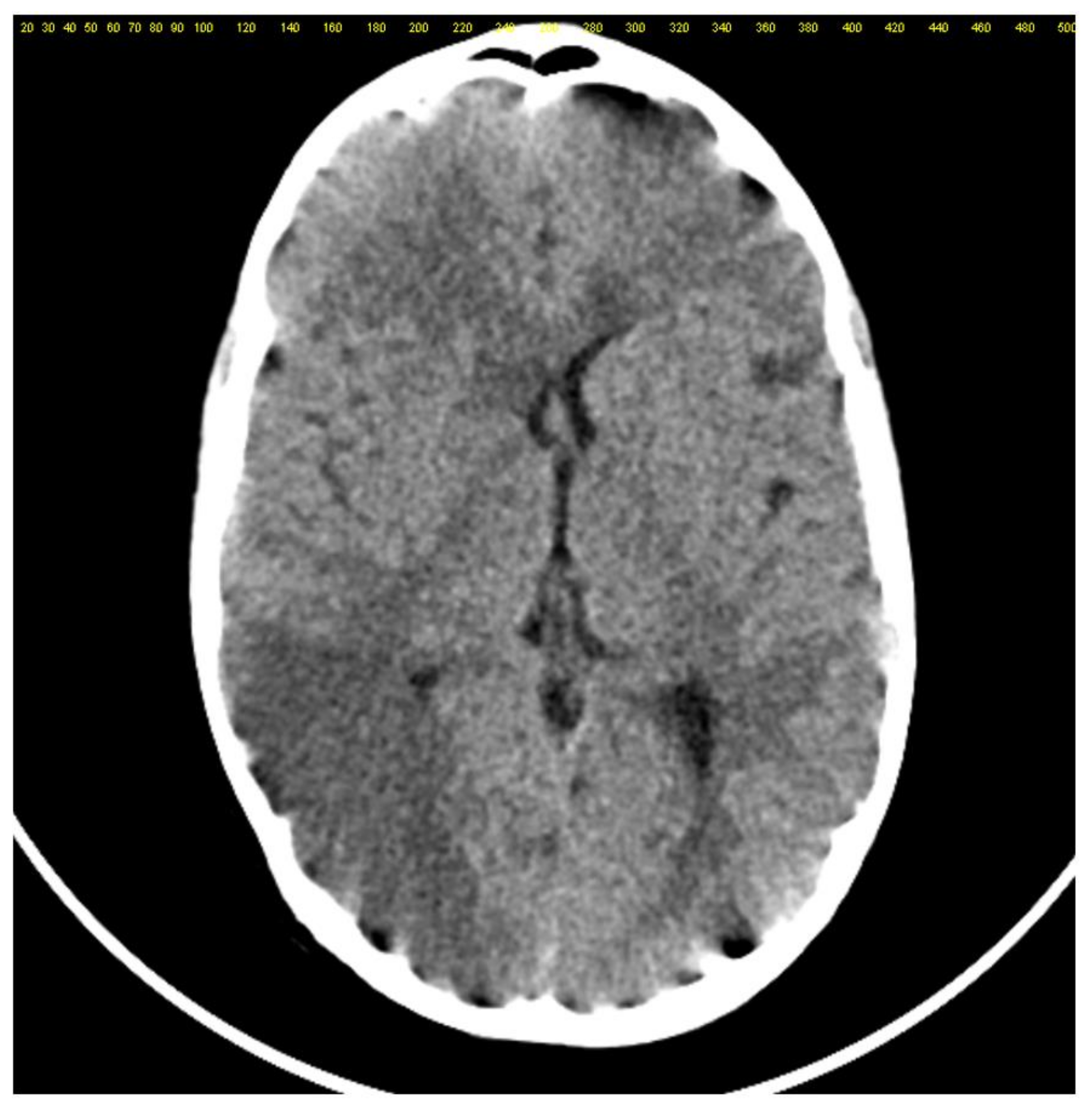
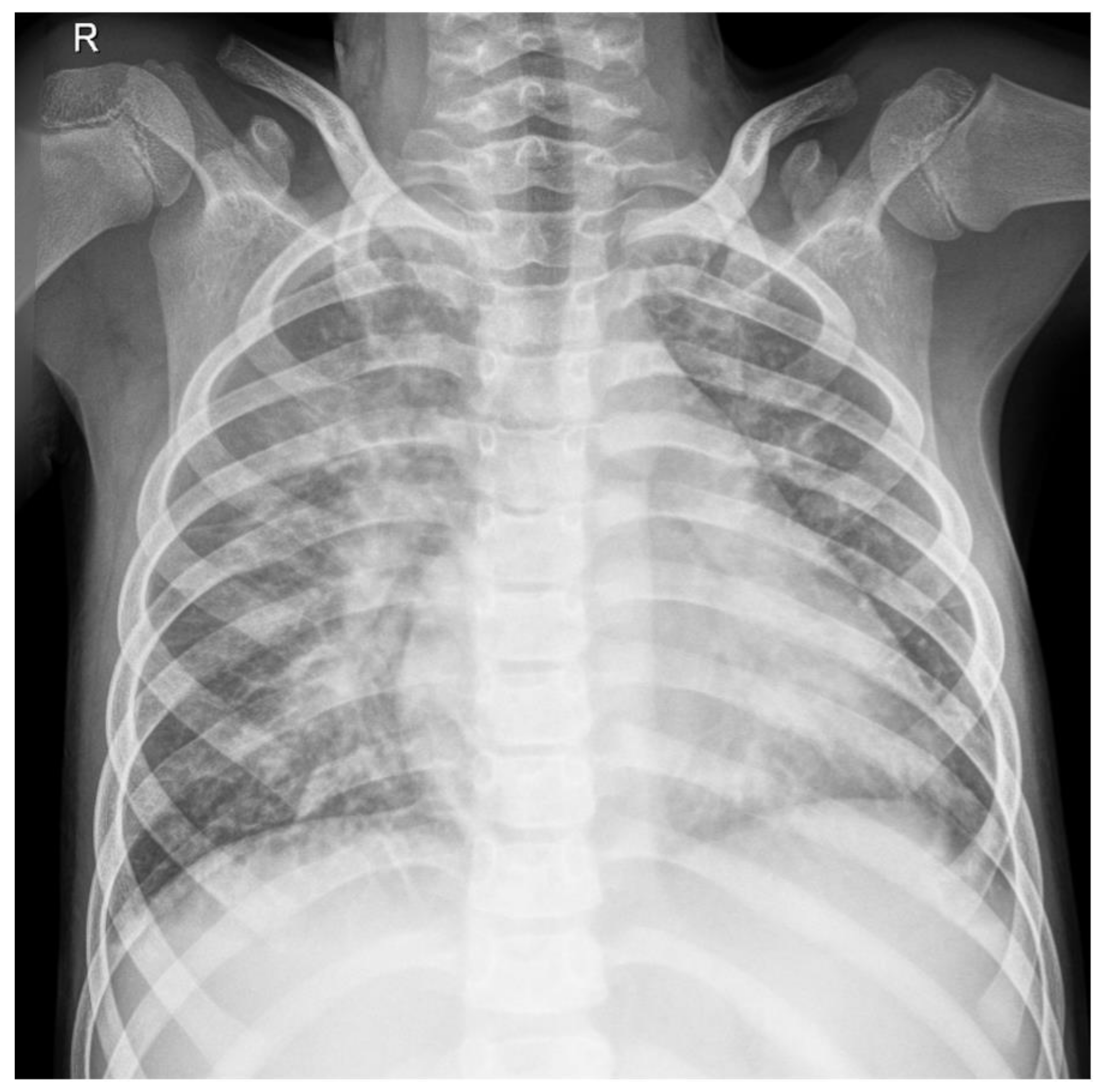
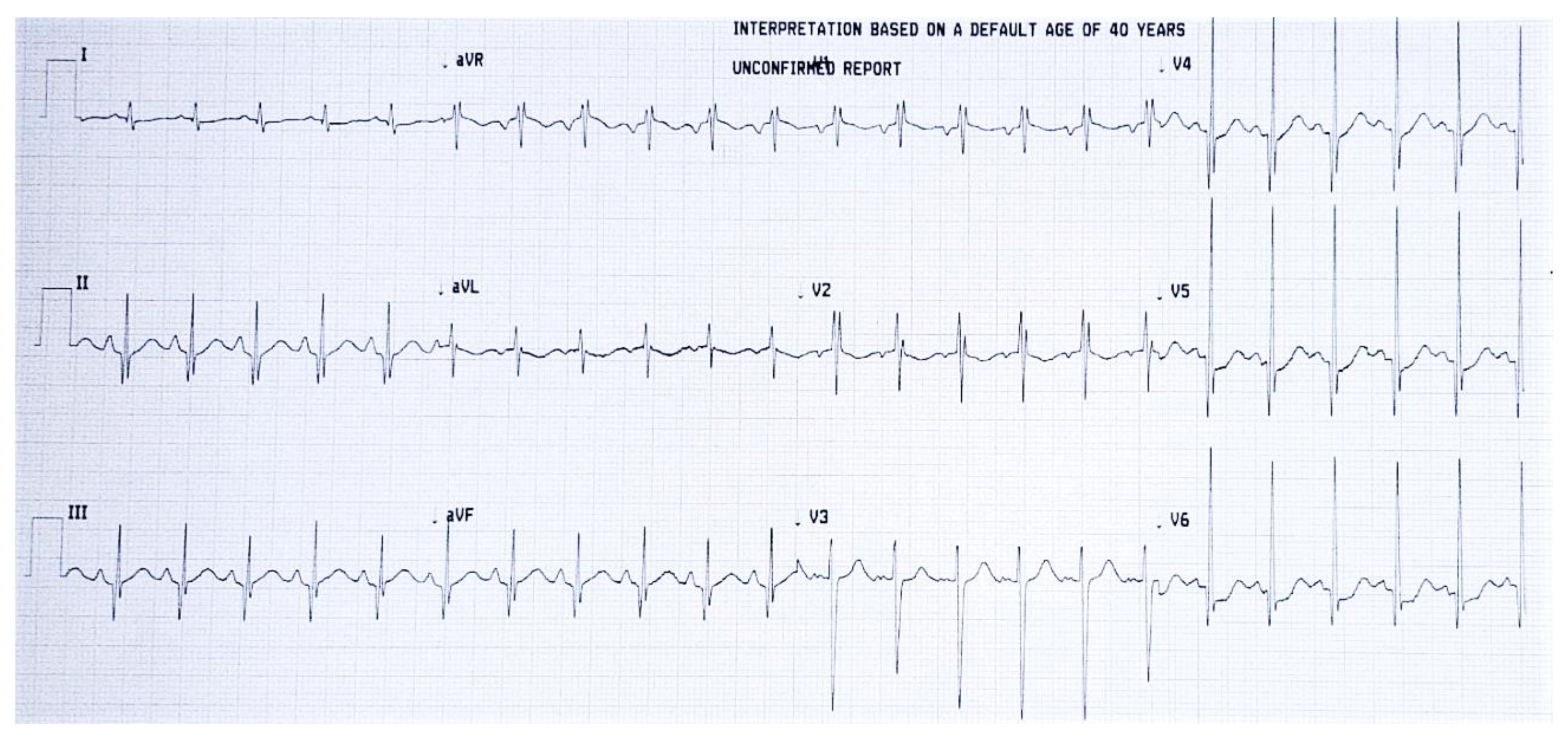
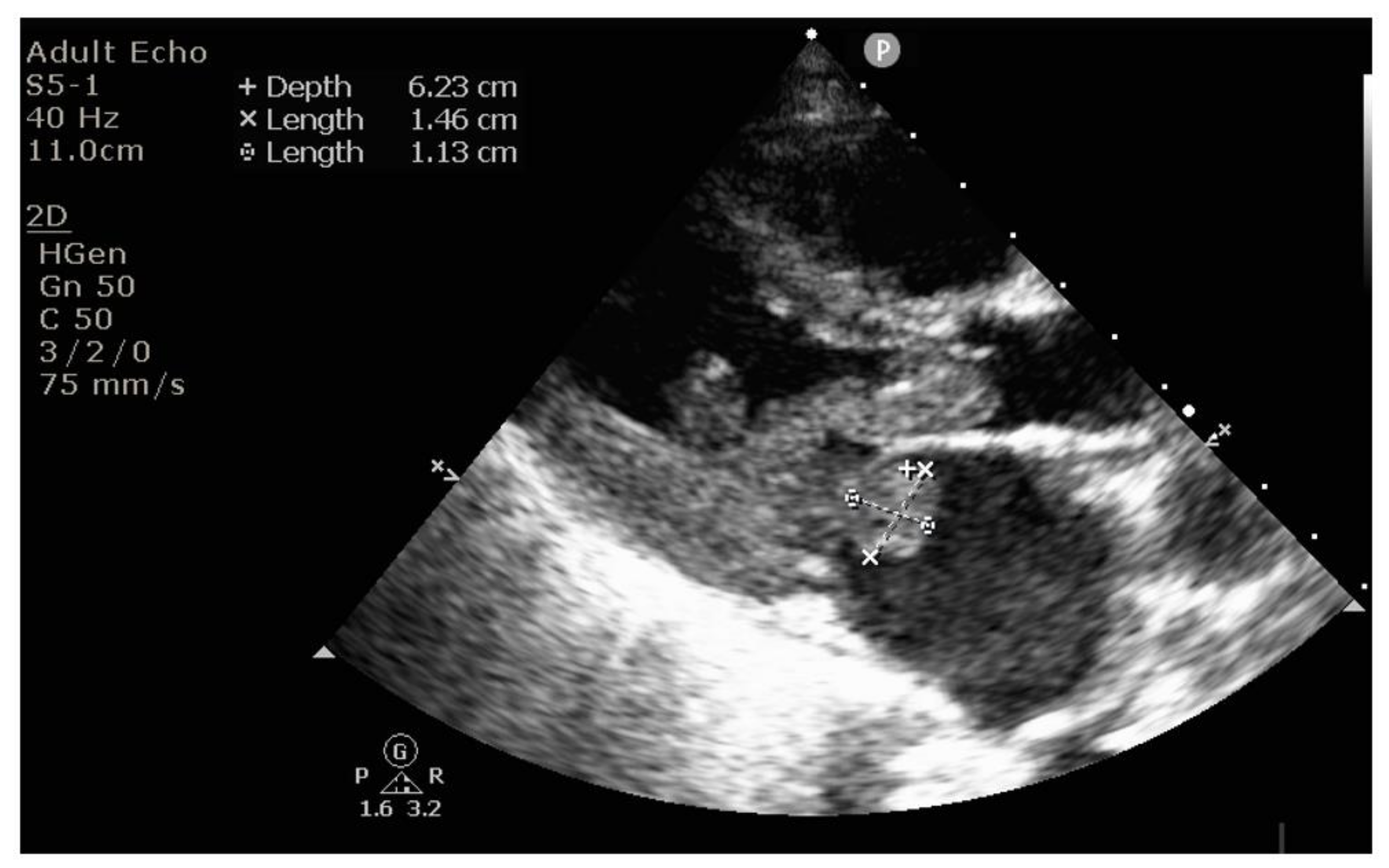
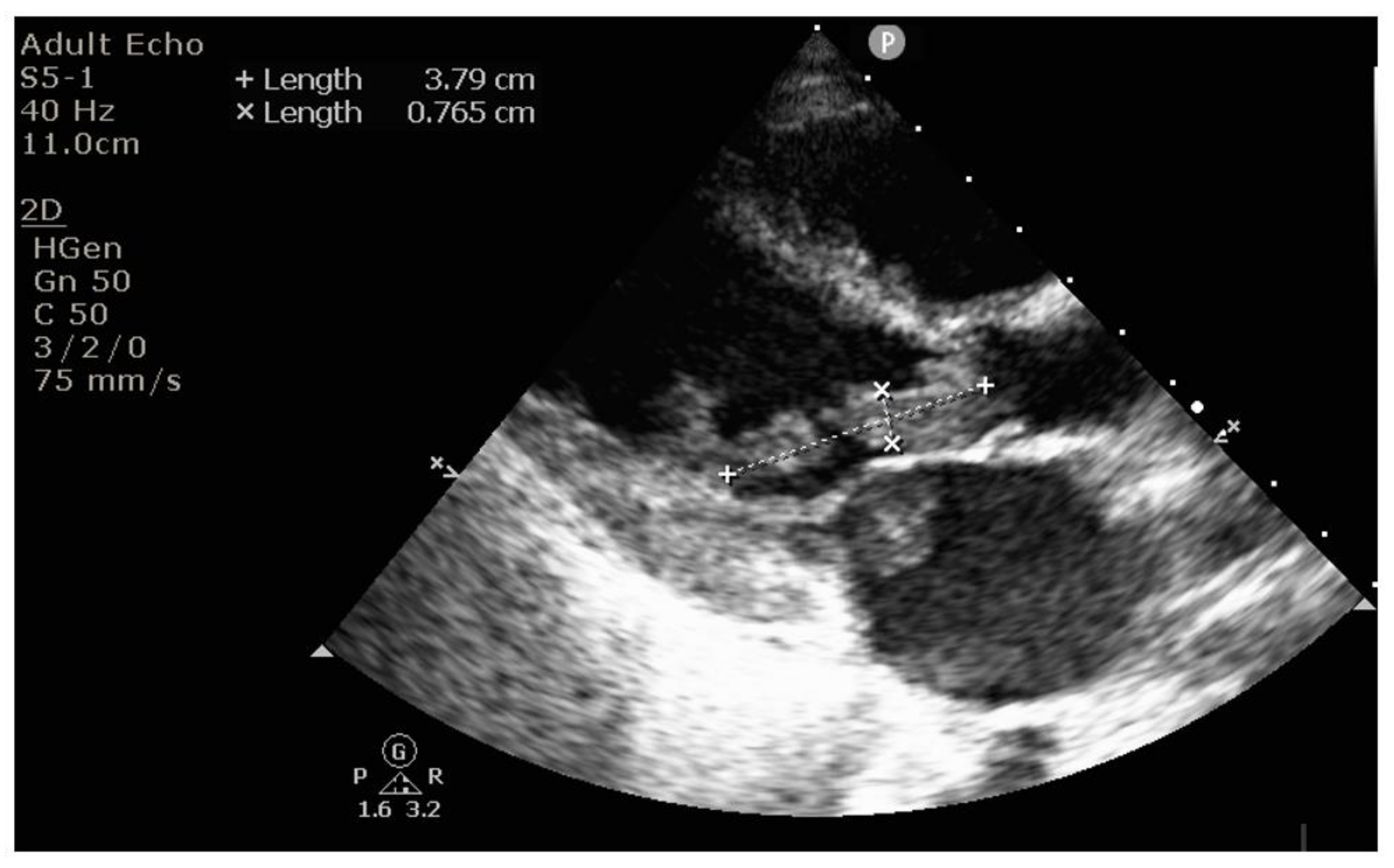
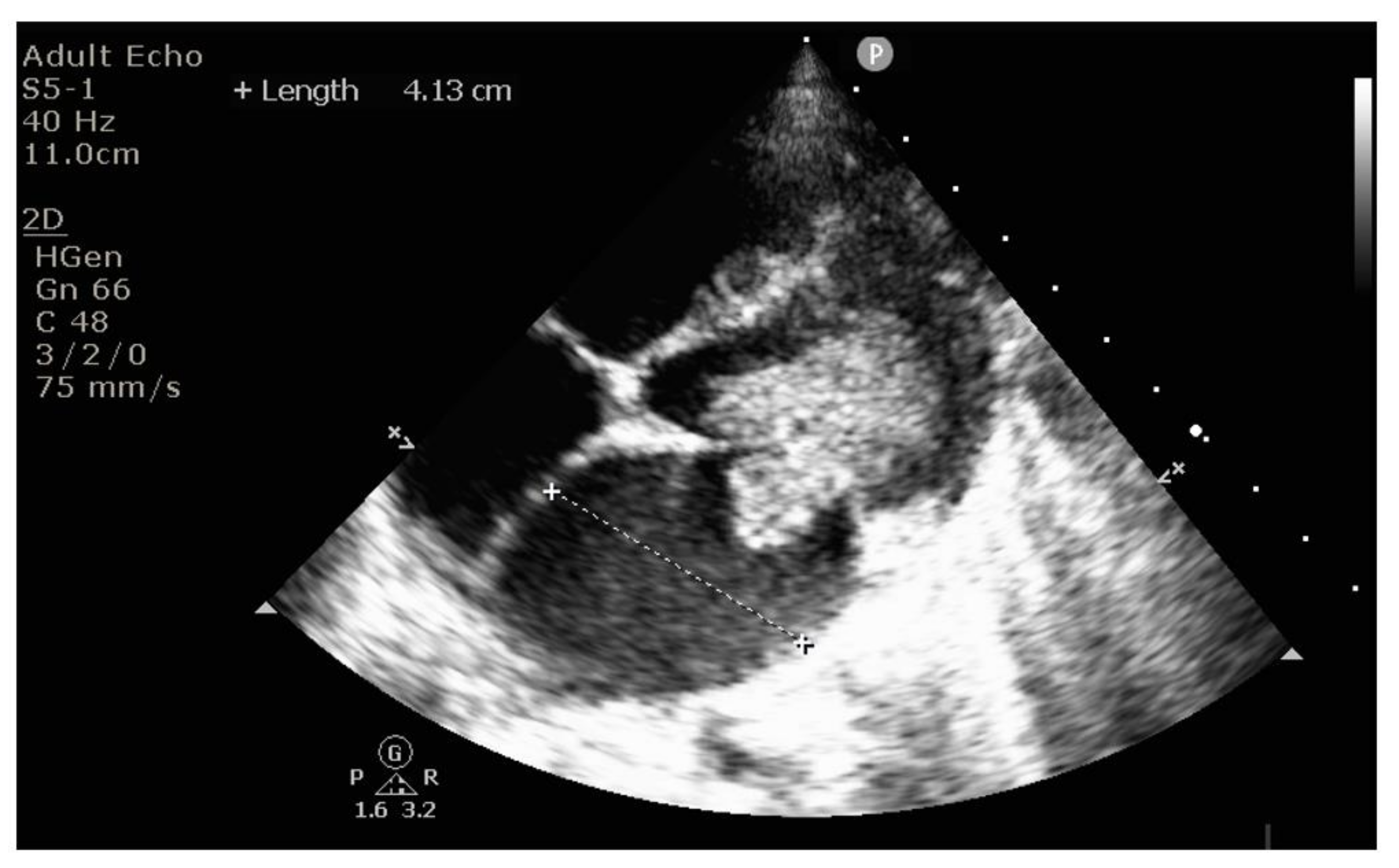
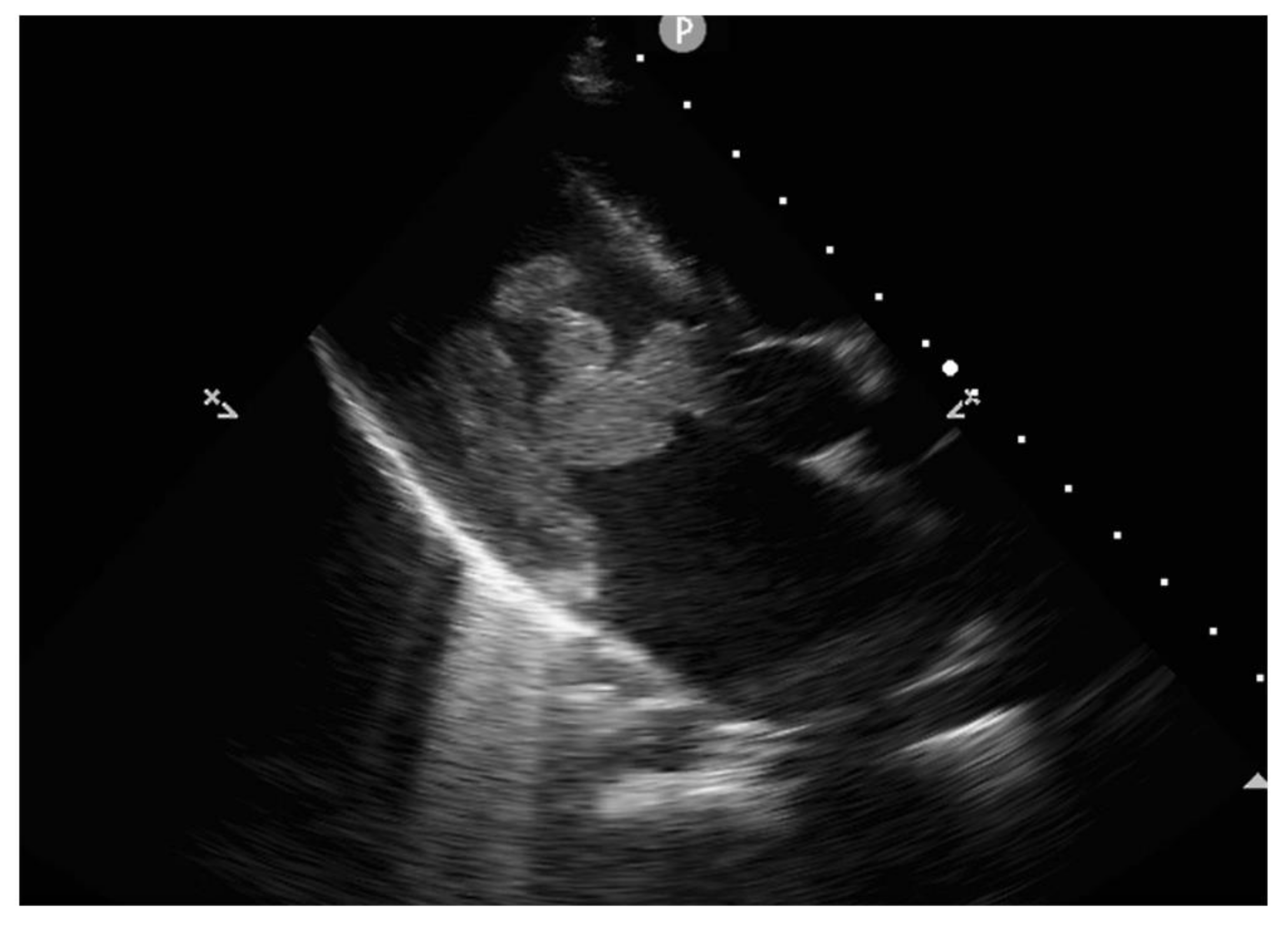
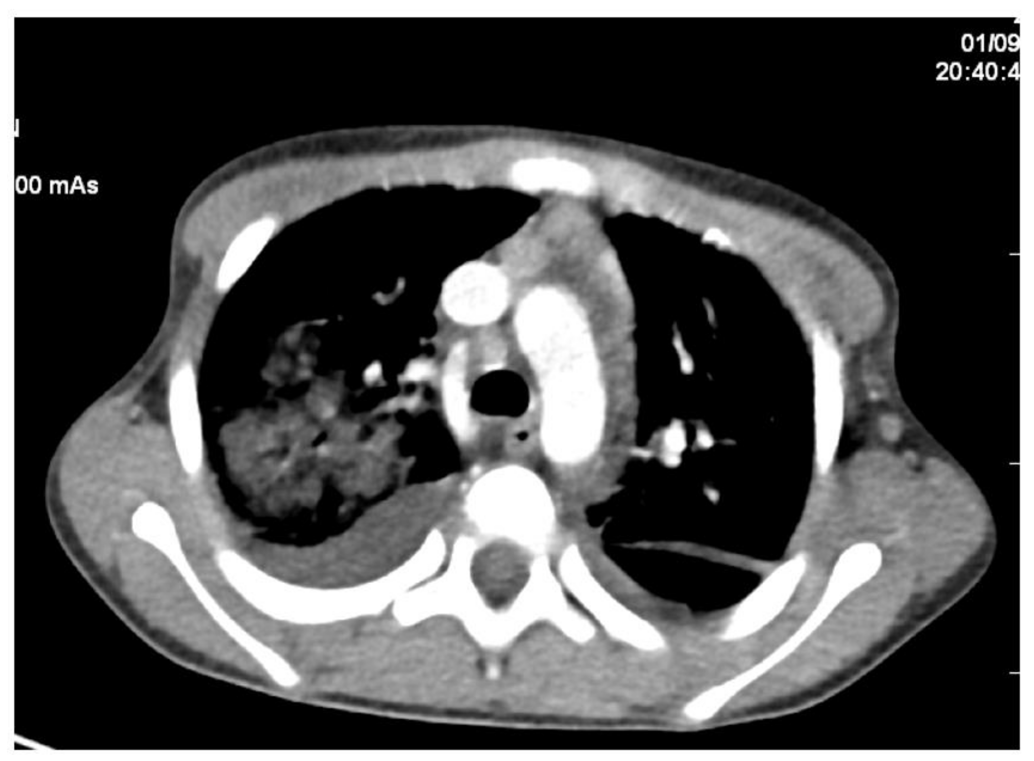
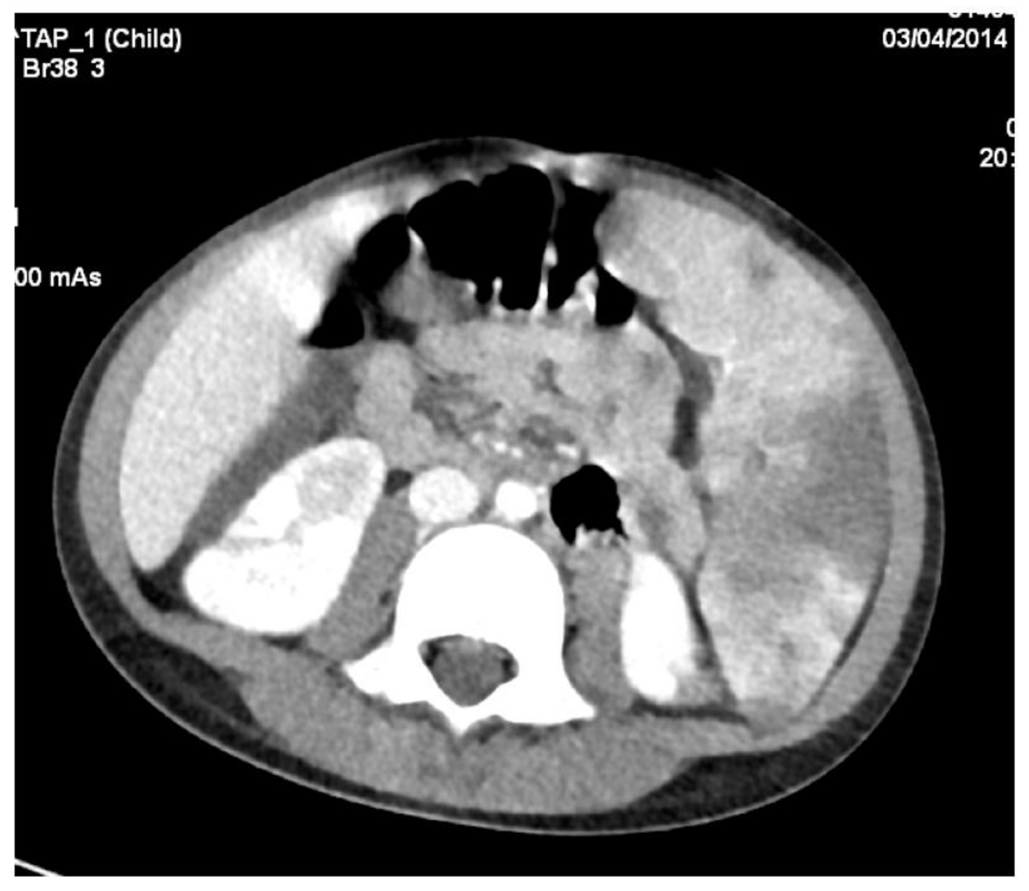
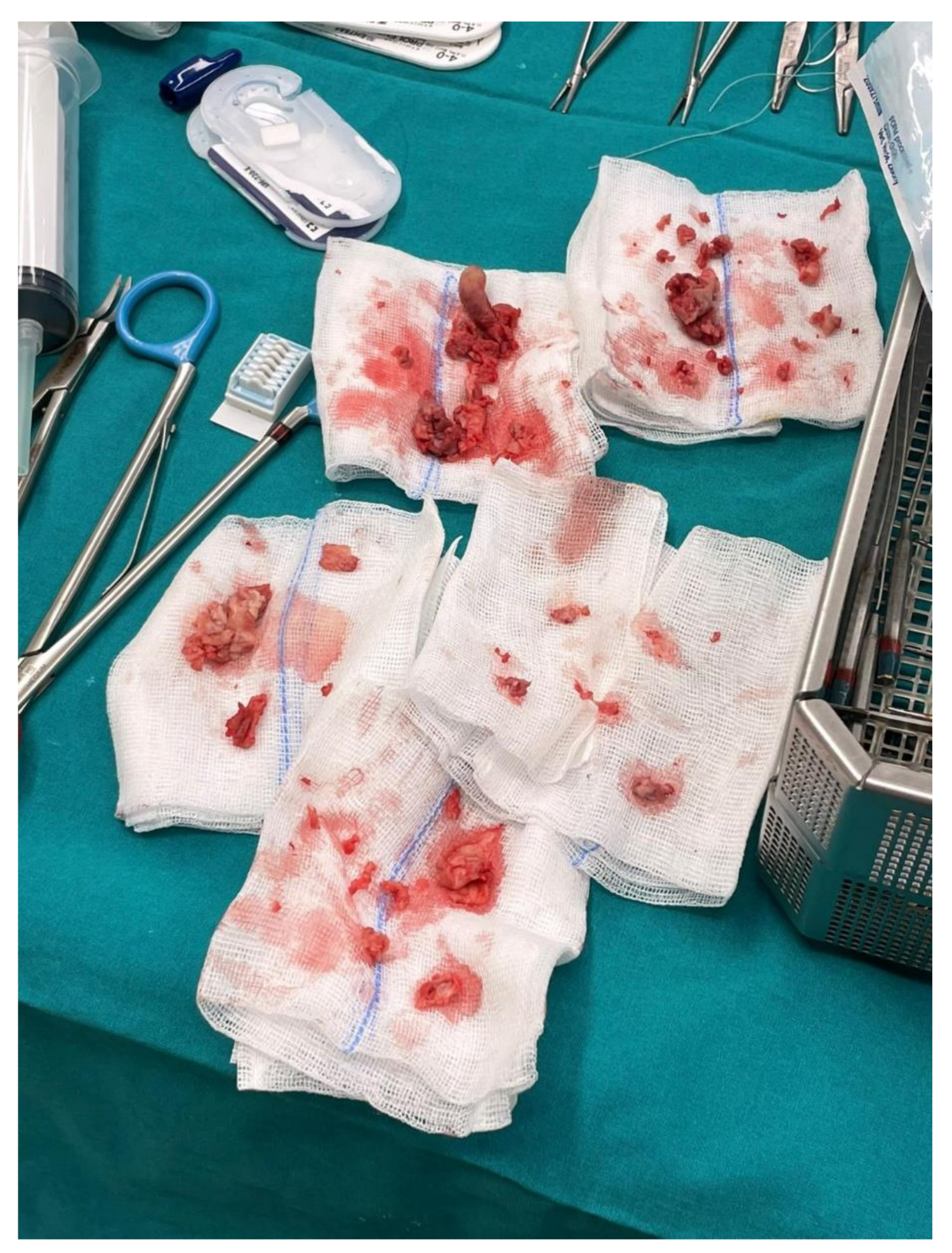
2. Case Report
3. Discussion
4. Conclusions
Author Contributions
Funding
Institutional Review Board Statement
Informed Consent Statement
Data Availability Statement
Conflicts of Interest
References
- Von Lilienfeld-Toal, M.; Wagener, J.; Einsele, H.; Cornely, O.A.; Kurzai, O. Invasive fungal infection—New treatments to meet new challenges. Dtsch. Arztebl. Int. 2019, 116, 271–278. [Google Scholar] [CrossRef] [PubMed]
- Pana, Z.D.; Seidel, D.; Skiada, A.; Groll, A.H.; Petrikkos, G.; Roilides, E. Invasive mucormycosis in children: An epidemiologic study in European and non-European countries based on two registries. BMC Infect. Dis. 2016, 16, 667. [Google Scholar] [CrossRef] [PubMed] [Green Version]
- Farmakiotis, D.; Kontoyiannis, D.P. Mucormycoses. Infect. Dis. Clin. N. Am. 2016, 30, 143. [Google Scholar] [CrossRef] [PubMed]
- Lanternier, F.; Dannaoui, E.; Morizot, G.; Elie, C.; Garcia-Hermoso, D.; Huerre, M.A. Global Analysis of Mucormycosis in France: The RetroZygo Study (2005–2007). Clin. Infect. Dis. 2012, 54 (Suppl. S1), S35–S43. [Google Scholar] [CrossRef] [Green Version]
- Petrikkos, G.; Skiada, A.; Lortholary, O. Epidemiology and clinical manifestations of mucormycosis. Clin. Infect. Dis. 2012, 54 (Suppl. S1), S23. [Google Scholar] [CrossRef] [PubMed]
- Roden, M.M.; Zaoutis, T.E.; Buchanan, W.L.; Knudsen, T.A.; Sarkisova, T.A.; Schaufele, R.L.; Sein, M.; Sein, T.; Chiou, C.C.; Chu, J.H.; et al. Epidemiology and outcome of zygomycosis: A review of 929 reported cases. Clin. Infect. Dis. 2005, 41, 634. [Google Scholar] [CrossRef] [Green Version]
- Ribes, J.A.; Vanover-Sams, C.L.; Baker, D.J. Zygomycetes in Human Disease. Clin. Microbiol. Rev. 2000, 13, 236–301. [Google Scholar] [CrossRef]
- Hallur, V.; Prakash, H.; Sable, M. Cunninghamella arunalokei a New Species of Cunninghamella from India Causing Disease in an Immunocompetent Individual. J. Fungi 2021, 7, 670. [Google Scholar] [CrossRef]
- Kontoyiannis, D.P.; Lionakis, M.S.; Lewis, R.E.; Chamilos, G.; Healy, M.; Perego, C.; Safdar, A.; Kantarjian, H.; Champlin, R.; Walsh, T.J.; et al. Zygomycosis in a tertiary-care cancer center in the era of Aspergillus-active antifungal therapy: A case-control observational study of 27 recent cases. J. Infect. Dis. 2005, 191, 1350. [Google Scholar] [CrossRef] [Green Version]
- Mahalaxmi, I.; Jayaramayya, K.; Venkatesan, D.; Subramaniam, M.D.; Renu, K.; Vijayakumar, P.; Vellingiri, B. Mucormycosis: An opportunistic pathogen during COVID-19. Environ. Res. 2021, 201, 111643. [Google Scholar] [CrossRef]
- Lanternier, F. Disease Entities in Mucormycosis. J. Fungi 2019, 5, 23. [Google Scholar] [CrossRef] [Green Version]
- Singh, A.K.; Singh, R.; Joshi, S.R.; Misra, A. Mucormycosis in COVID-19: A systematic review of cases reported worldwide and in India. Diabetes Metab. Syndr. 2021, 15, 102146. [Google Scholar] [CrossRef] [PubMed]
- Ibrahim, A.S. Host cell invasion in mucormycosis: Role of iron. Curr. Opin. Microbiol. 2011, 14, 406–411. [Google Scholar] [CrossRef] [PubMed] [Green Version]
- Van Well, G.T.; van Groeningen, I.; Debets-Ossenkopp, Y.J.; van Furth, A.M.; Zwaan, C.M. Zygomycete infection following voriconazole prophylaxis. Lancet Infect. Dis. 2005, 5, 594. [Google Scholar] [CrossRef]
- Boelaert, J.R.; Van Cutsem, J.; de Locht, M.; Schneider, Y.J.; Crichton, R.R. Deferoxamine augments growth and pathogenicity of Rhizopus, while hydroxypyridinone chelators have no effect. Kidney Int. 1994, 45, 667. [Google Scholar] [CrossRef] [Green Version]
- Ibrahim, A.S.; Spellberg, B.; Walsh, T.J.; Kontoyiannis, D.P. Pathogenesis of mucormycosis. Clin. Infect. Dis. 2012, 54 (Suppl. S1), S16. [Google Scholar] [CrossRef]
- Cherchi, G.B.; Pacifico, L.; Cossellu, S.; Gallisai, D.; Zanetti, S.; Fadda, G.; Chiesa, C. Prospective study of Yersinia enterocolitica infection in thalassemic patients. Pediatr. Infect. Dis. J. 1995, 14, 579–583. [Google Scholar] [CrossRef]
- Roilides, E.; Zaoutis, T.E.; Walsh, T.J. Invasive zygomycosis in neonates and children. Clin. Microbiol. Infect. 2009, 15, 50–54. [Google Scholar] [CrossRef] [Green Version]
- Virmani, R.; Connor, D.H.; Mcallister, H.A. Cardiac Mucormycosis: A Report of Five Patients and Review of 14 Previously Reported Cases. Am. J. Clin. Pathol. 1982, 78, 42–47. [Google Scholar] [CrossRef] [Green Version]
- Chen, L.; Xiao, Y.; Wang, X. Successful Treatment of Mucormycosis in the Pulmonary Artery After Cardiac Surgery. J. Card. Surg. 2005, 20, 186–188. [Google Scholar] [CrossRef]
- Chinen, K.; Matsumoto, H.; Fujioka, Y. Cardiac Mucormycosis Presenting as a “Fungus Ball” in the Left Atrium. Intern. Med. 2009, 48, 1781–1782. [Google Scholar] [CrossRef] [PubMed] [Green Version]
- Mehta, N. Native aortic valve vegetative endocarditis with Cunninghamella. Eur. J. Echocardiogr. 2004, 5, 156–158. [Google Scholar] [CrossRef] [PubMed] [Green Version]
- Ellis, M.E.; Al-Abdely, H.; Sandridge, A.; Greer, W.; Ventura, W. Fungal Endocarditis: Evidence in the World Literature, 1965–1995. Clin. Infect. Dis. 2001, 32, 50–62. [Google Scholar] [CrossRef] [PubMed] [Green Version]
- Van de Glind, G.J.; Gidding, C.E.M.; Verlaat, C.M.W.; Duthoi, K.; Backx, A.P.C.; Verweij, P.E.; Warris, A. Acute Cardiac Failure due to Intra-Atrial Mass Caused by Zygomycetes in an Immunocompromised Paediatric Patient. Case Rep. Med. 2010, 2010, 241791. [Google Scholar] [CrossRef] [PubMed]
- Gubarev, N.; Separovic, J.; Gasparovic, V.; Jelic, I. Successful Treatment of Mucormycosis Endocarditis Complicated by Pulmonary Involvement. Thorac. Cardiovasc. Surg. 2007, 55, 257–258. [Google Scholar] [CrossRef]
- Jackman, J.D.; Simonsen, R.L. The Clinical Manifestations of Cardiac Mucormycosis. Chest 1992, 101, 1733–1736. [Google Scholar] [CrossRef] [PubMed]
- Misumi, T.; Kudo, M.; Ito, T.; Matsubara, T.; Kumamaru, H. Floating ball thrombus in the left atrium with mitral stenosis. Jpn. J. Thorac. Cardiovasc. Surg. 2003, 51, 387–389. [Google Scholar] [CrossRef] [PubMed]
- Seelig, M.S.; Goldberg, P.; Kozinn, P.J.; Berger, A.R. Fungal endocarditis: Patients at risk and their treatment. Postgrad. Med. J. 1979, 55, 632–641. [Google Scholar] [CrossRef] [Green Version]
- Shah, S.; Suresh, P.V.; Maheshwari, S.; Rao, S. Cardiac mucormycosis with T-cell immunodeficiency. Indian Pediatr. 2009, 46, 257–259. [Google Scholar]
- Soliman, M.; Harding, C.; El Haddad, H.; Mansour, A.; Anstead, M. Disseminated Mucormycosis with Extensive Cardiac Involvement. Cureus 2019, 11, e4760. [Google Scholar] [CrossRef] [Green Version]
- Benbow, E.W.; McMahon, R.F. Myocardial infarction caused by cardiac disease in disseminated zygomycosis. J. Clin. Pathol. 1987, 40, 70–74. [Google Scholar] [CrossRef] [PubMed] [Green Version]
- Kontoghiorghes, G.J.; Kolnagou, A.; Skiada, A.; Petrikkos, G. The Role of Iron and Chelators on Infections in Iron Overload and Non Iron Loaded Conditions: Prospects for the Design of New Antimicrobial Therapies. Hemoglobin 2010, 34, 227–239. [Google Scholar] [CrossRef] [PubMed]
- Sepaskhah, M.; Moezzi, I.; Davarpanah, M.A.; Sari Aslani, F. Primary Cutaneous Mucormycosis in a Beta-Thalassemia Patient. Indian J. Hematol. Blood Transfus. 2018, 34, 776–777. [Google Scholar] [CrossRef] [PubMed]
- Georgiadou, S.P.; Sipsas, N.V.; Marom, E.M.; Kontoyiannis, D.P. The diagnostic value of halo and reversed halo signs for invasive mold infections in compromised hosts. Clin. Infect. Dis. 2011, 52, 1144–1155. [Google Scholar] [CrossRef] [PubMed] [Green Version]
- Spellberg, B.; Walsh, T.J.; Kontoyiannis, D.P.; Edwards, J.; Ibrahim, A.S. Recent advances in the management of mucormycosis: From bench to bedside. Clin. Infect. Dis. 2009, 48, 1743. [Google Scholar] [CrossRef]
- Hata, D.J.; Buckwalter, S.P.; Pritt, B.S.; Roberts, G.D.; Wengenack, N.L. Real-Time PCR Method for Detection of Zygomycetes. J. Clin. Microbiol. 2008, 46, 2353–2358. [Google Scholar] [CrossRef] [Green Version]
- Millon, L.; Caillot, D.; Berceanu, A.; Bretagne, S.; Lanternier, F.; Morio, F.; Letscher-Bru, V.; Dalle, F.; Denis, B.; Alanio, A.; et al. Evaluation of serum Mucorales PCR for the diagnosis of Mucormycoses: The MODIMUCOR prospective trial. Clin. Infect. Dis. 2022, ciab1066. [Google Scholar] [CrossRef]


Publisher’s Note: MDPI stays neutral with regard to jurisdictional claims in published maps and institutional affiliations. |
© 2022 by the authors. Licensee MDPI, Basel, Switzerland. This article is an open access article distributed under the terms and conditions of the Creative Commons Attribution (CC BY) license (https://creativecommons.org/licenses/by/4.0/).
Share and Cite
Cinteza, E.; Nicolescu, A.; Ciomartan, T.; Gavriliu, L.-C.; Voicu, C.; Carabas, A.; Popescu, M.; Margarint, I. Disseminated Cunninghamella spp. Endocarditis in a Beta-Thalassemia Patient after Asymptomatic COVID-19 Infection. Diagnostics 2022, 12, 657. https://0-doi-org.brum.beds.ac.uk/10.3390/diagnostics12030657
Cinteza E, Nicolescu A, Ciomartan T, Gavriliu L-C, Voicu C, Carabas A, Popescu M, Margarint I. Disseminated Cunninghamella spp. Endocarditis in a Beta-Thalassemia Patient after Asymptomatic COVID-19 Infection. Diagnostics. 2022; 12(3):657. https://0-doi-org.brum.beds.ac.uk/10.3390/diagnostics12030657
Chicago/Turabian StyleCinteza, Eliza, Alin Nicolescu, Tatiana Ciomartan, Liana-Cătălina Gavriliu, Cristiana Voicu, Adelina Carabas, Monica Popescu, and Irina Margarint. 2022. "Disseminated Cunninghamella spp. Endocarditis in a Beta-Thalassemia Patient after Asymptomatic COVID-19 Infection" Diagnostics 12, no. 3: 657. https://0-doi-org.brum.beds.ac.uk/10.3390/diagnostics12030657