Article

Acute Adverse Effects of Metallic Nanomaterials on Cardiac and Behavioral Changes in Daphnia magna

1
Chemical Safety Management Group, Nakdong River Basin Environmental Office, Ministry of Environment, Changwon 51439, Korea
2
Environmental Safety Group, Korea Institute of Science and Technology Europe Forschungsgesellschaft mbH, 66123 Saarbrücken, Germany
3
Smart Convergence Group, Korea Institute of Science and Technology Europe Forschungsgesellschaft mbH, 66123 Saarbrücken, Germany
4
Division of Energy & Environment Technology, University of Science & Technology, Daejeon 34113, Korea
*
Author to whom correspondence should be addressed.
Submission received: 7 January 2022 / Revised: 6 February 2022 / Accepted: 7 February 2022 / Published: 10 February 2022

Abstract

:
Nanomaterials are widely believed to induce toxic effects on organisms by evoking oxidative stress. We evaluated the toxic effects of nanomaterials on the cardiac and behavioral changes in Daphnia magna under varying exposure conditions. Titanium dioxide nanoparticles (TiO2 NPs), silver nanoparticles (AgNPs), and silver nitrate (AgNO3) were selected for the acute toxicity tests. The adverse effects of the substances on the neonates including heart rate, swimming speed, and oxidative stress were measured. The heart rate level decreased as the concentration of both NPs and silver ions (Ag+) increased. The average swimming speed was measured to be approximately 15 mm/min for the control group. The swimming speed generally increased with a longer exposure to both NPs although it reached a plateau at the lowest concentration of AgNPs. A similar but less clear trend was observed for Ag+. For all substances, the overall swimming speed exhibited no correlation or weak negative correlations with the exposure concentration. The oxidative stress levels increased after exposure compared with the control group. We conclude that aquatic nanotoxicity tests should consider multilevel physicochemical, physiological, and behavioral parameters for the official guidelines to quantify more robust adverse outcomes.

1. Introduction

Nanomaterials are used in a wide range of sectors due to their specific physicochemical properties compared with their bulk materials that have the same chemical composition [1,2,3]. To ensure safe use, it is important to identify and manage the effects of exposure to nanomaterials on humans and environmental health more effectively. In particular, the aquatic phase is considered to be a key starting point of potential entry and diffusion into the environment for understanding the environmental fate and behavior. The aquatic phase creates connections with other environmental compartments such as the soil, sediment, and air [4].
Several studies have indicated that a few nanomaterials are toxic to aquatic organisms such as fish and crustaceans [5,6,7]. Of these, fish have a direct impact on human health but it is difficult to obtain information on the exact lethal concentration of nanomaterials from fish. Therefore, the impact of nanomaterials on non-target organisms plays an important role [8]. Aquatic invertebrates are a large group of animals and a few have over 1000 different species. Among them, arthropods are the largest invertebrate and consist of the two largest groups: insects and crustaceans [9]. Crustaceans are the most important group of invertebrates, of which Daphnia magna (D. magna, here and after) is commonly used to assess the risk of nanomaterials [10]. Algae and crustaceans exposed to nanomaterials can poison and cause death to aquatic organisms such as fish through the food chain [5,11].
Nanomaterials are generally known to have a higher toxicity than bulk materials. It has been reported from in vitro studies that nanomaterials can induce biological effects such as cell inflammation and apoptosis by oxidative stress [12,13,14,15]. The preliminary factors causing toxicity are size, shape, and surface area/charge [16]. Nanomaterials are more reactive due to their large surface area and easy penetration into cells [16,17]. Depending on the surface properties, nanomaterials may exhibit enhanced interactions with cell membranes [18,19]. Here, we focus on two representative types of metallic nanoparticles, titanium dioxide (TiO2, here and after) and silver nanoparticles (AgNPs, here and after). TiO2 is generally known to be non-toxic and can be applied in various ways such as through cosmetics and catalysts [20,21]. AgNPs are also used in a wide range of consumer products as antibacterial agents due to their high cytotoxicity [22,23,24]. One of the most fundamental sources of AgNP toxicity has been attributed to the release of silver ions (Ag+, here and after) in tissue environments.
D. magna is a standard organism for ecotoxicity tests using toxic chemicals in the standardized protocols suggested by the Organization for Economic Cooperation and Development (OECD), International Organization for Standardization (ISO), and the United States Environmental Protection Agency (USEPA) [25,26]. As an organism in the bottom stage of the food chain, D. magna plays a key role in aquatic ecosystems because it is sensitive to environmental stresses and population changes can ultimately affect the population of upper predators [25,27]. Previous studies have reported the biological effects of hazardous chemicals on physiological factors such as cardiac and behavioral characteristics in D. magna [28,29,30,31,32]. Fekete-Kertész et al. evaluated changes in heart rates using D. magna and found that the heart rate was influenced by several factors: the chemical exposure level (triclosan); test medium; organism age; and exposure time [33]. Chung et al. also reported that the heart rate and swimming distance were linearly changed by the TiO2 nanoparticle concentration [34]. They adopted a video tracking method, which has recently been used to evaluate the behavioral characteristics of organisms such as zebrafish and D. magna. Therefore, we aimed to evaluate the toxic effects on the physiological factors of D. magna using nanomaterials and to identify the relationships between the physiological factors according to the exposure conditions in this study.

2. Materials and Methods

2.1. Experimental Scheme

This study consisted of three steps to examine the toxicity of nanomaterials using D. magna. The procedures included the sample preparation (test organism, test substances), treatment (exposure), and analytical tests using instruments. One-day-old neonates that were newly born within 24 h were prepared and certain exposure levels of nanomaterials were treated for different exposure times. After an exposure duration at each concentration level, the physiological effects on the neonates including the cardiac effects and behavioral changes were characterized. The test procedures for each process are described in the following sections in detail.

2.2. D. magna Culture

D. magna cultured in a laboratory were used in a series of experiments. The test organism was cultured in 5 L beakers in a 21 ± 1 °C thermostatic incubator under a constant light cycle for 24 h. The light conditions were maintained in the light for 16 h and then the light was turned off for 8 h (12 W, 356 mm lamp, 672 Lumen, 4200 K). To maintain the breeding conditions, the D. magna adults were cultivated in an OECD M4 medium and daily feeding of the organisms was conducted using an algae containing a mixture of Chlorella vulgaris and additional nutrition (yeast, cerophyll, and trout chow; YCT) [35]. One-day-old neonates of D. magna were used for the exposure test. To check the sensitivity of the D. magna culture, an acute immobilization toxicity test was performed with potassium dichromate (K2Cr2O7, here and after) as a reference. The sensitivity of the D. magna culture to K2Cr2O7 ranged within the limits (half-maximal effective dose, EC50: 0.6–2.1 mg/L for 24 h) as suggested by OECD Guideline 202. For each test concentration and control, five neonates each (<15 h old) were placed in 6-well plates (VWR Tissue Culture Plates, VWR, Darmstadt, Germany) containing 10 mL of either a test solution or suspension. The neonates (<24 h old) obtained from the fifth generation were used in the toxicity tests to minimize the variability. After incubation for 3 and 48 h, D. magna was used to measure the heart rate and swimming speed.

2.3. Preparation of the Nanomaterials

A total of three commercially available materials including two metallic nanoparticles (TiO2 and AgNPs) and silver nitrate (AgNO3, here and after) were used for the tests in this study. The metallic nanomaterials were purchased from different manufacturers (TiO2: Aeroxide® TiO2 P25, Evonik Industries, Essen, Germany; AgNP: NanoXactTM, NanoComposix Inc., San Diego, CA, USA) and the particle size of each nanomaterial was measured using a transmission electron microscope (TEM, here and after). Individual TiO2 particles are normally 21 nm in size and their aggregates are distributed hundreds of nanometers in size. Spherical AgNPs were provided at 0.02 mg/mL in a 2 mM sodium citrate solution and the primary particle size was 31 ± 3 nm. AgNO3 (ACS reagent, purity ≥ 99.0%; Sigma-Aldrich, St. Louis, MO, USA) was used as a source of Ag+. It is the most common silver salt and dissolves easily in water; it is also easy to produce a desired Ag+ concentration. All the chemicals were used without further purification.

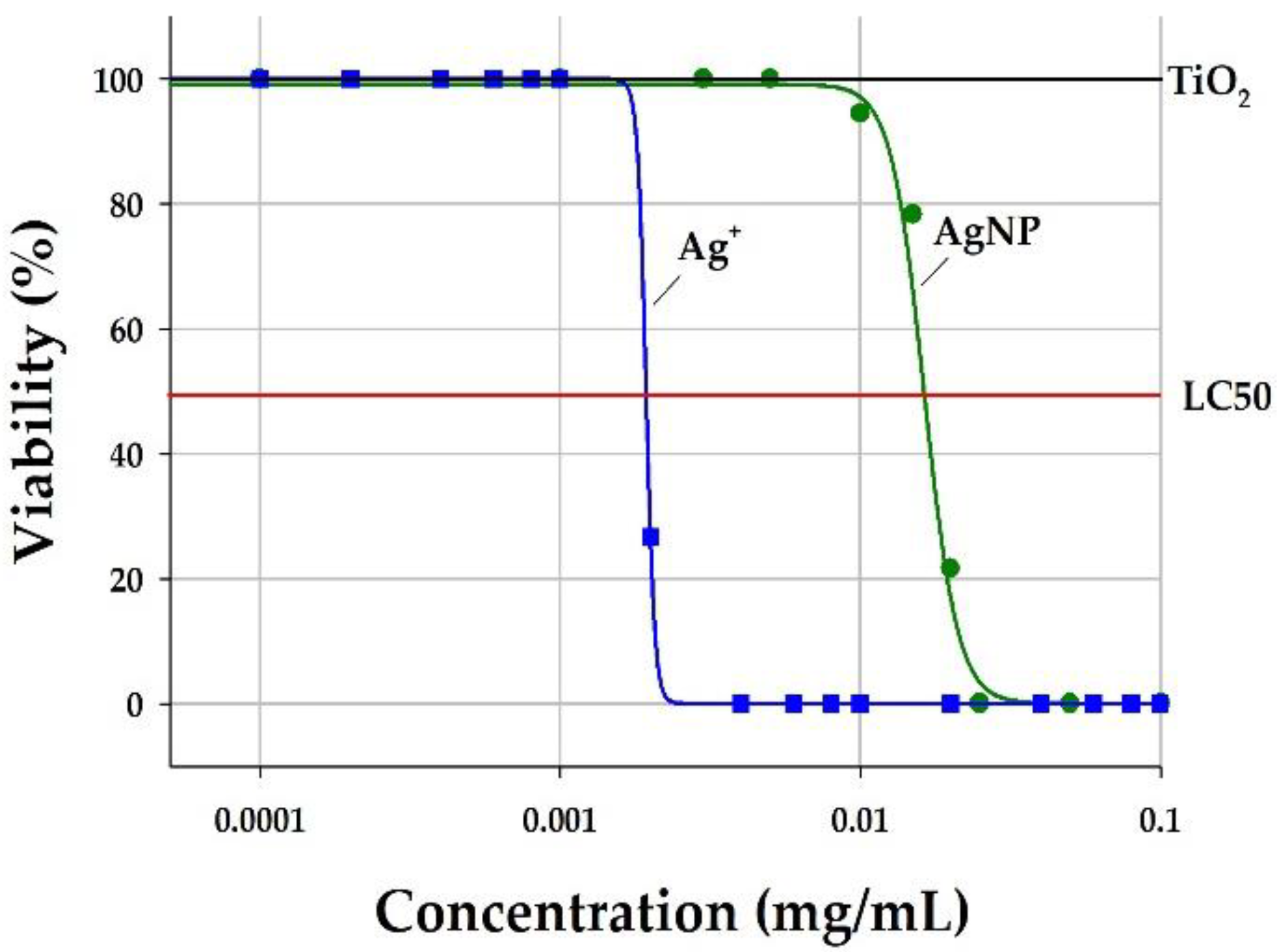
2.4. Determination of the Exposure Levels

The TiO2 NP stock solutions were made by dispersing 1 mg in 1 mL of distilled water and then sonicating for 30 min. Different suspended concentrations were prepared using an ISO medium for each material. The OECD-recommended ISO medium consisted of a mixture of 294 mg/L CaCl2·2H2O, 123.25 mg/L MgSO4·7H2O, 64.75 mg/L NaHCO3, 5.75 mg/L KCl, and 2 μg/L Na2SeO3. The diluted solutions of TiO2 NPs were prepared with concentrations of 0.1, 1.0, and 10 μg/mL. The concentration levels of AgNPs and silver ions were 10−4, 10−3, and 10−2 μg/mL and 10−5, 10−4, and 10−3 μg/mL, respectively. All diluted suspensions were immediately homogenized after vortexing. Each concentration range was determined with a reference to the EC50 from previous studies [25,36,37,38,39,40,41,42,43,44,45,46].
After each material was dispersed in the medium, the size distribution of each material was measured using a TEM (JEM-2100 LaB6, JEOL, Tokyo, Japan) to identify whether the particle size was changed by the medium type (distilled water vs. the ISO medium) (Figure 1). The characterization of the NP suspensions was conducted under a 200 kV accelerating voltage. An aliquot of each suspension was rinsed over a holey carbon TEM grid (Type S147-4, Plano, Wetzlar, Germany) and then dried at room temperature. More than 100 particles were taken at three magnifications due to the different particle sizes. The images were analyzed for the average length (diameter) using the pixel ruler in Image J software (version 1.52, National Institute of Health, Bethesda, MD, USA). The primary particle size of TiO2 was 24 ± 5 nm and AgNP was 31 ± 3 nm (Figure 1a,c). However, we found that the primary particles aggregated in the medium over 48 h (Figure 1b,d). Thus, we confirmed that D. magna could be exposed to primary and aggregated particles simultaneously.

2.5. Heart Rate Counting

The most commonly used endpoint of toxicity is to measure the death rates of D. magna. The immobilization of D. magna was observed at each concentration before counting the heart rate. A median lethal concentration (LC50, here and after) is defined as the concentration of toxic substances that kills 50% of the test organism within a certain exposure period. The survival data were plotted and the LC50 values were calculated using logistic three-parameter curve fitting with Sigmaplot 13.0 software (Systat Software Inc., San Jose, CA, USA). The heart rate was measured to evaluate the influences of exposure to each nanomaterial in triplicate. Neonates hatched within 24 h from the fifth generation of D. magna adults were newly prepared on the day of the experiment. Every five neonates were exposed to each concentration level for 3 and 48 h. The control group was also prepared for a comparison with the exposure groups under the same conditions without a chemical treatment. After the exposure, an aliquot of a methyl cellulose solution (4% V/W, Lot No. SLCC9072, Sigma-Aldrich Corp., St. Louis, MO, USA) was used to fix individual neonates onto a glass plate. We observed the heart rate conditions for one minute using an optical microscope at 4× magnification (Model CKX41, Olympus Inc., Tokyo, Japan) and recorded them with video files. The heart rate was finally counted manually in the play condition at a low speed (×0.3). The heart rate counting was conducted by three observers to minimize any individual bias. The average values for the counting data were used. In addition, a blind condition was maintained to ensure each observer was unaware of the treatment condition.

2.6. Swimming Performance Monitoring

As a behavioral index, the swimming performance of D. magna after exposure to each concentration level of nanomaterials was measured using a direct-reading instrument (Model Zebrabox, View Point Life Science Inc., Lyon, France). Individual neonates exposed to each concentration were separately transferred to each well of a 96-well plate. Each well plate batch was placed into the Zebrabox and all exposure groups were kept stable in entirely dark conditions for 30 min before the measurement to minimize the effects of sudden environmental changes. After stabilization for 30 min, the swimming speed (distance per minute) and moving route were measured under a tracking mode every minute for 80 min. The real-time data were analyzed using automated observation software (Zebralab-2, View Point Life Science Inc., Lyon, France).

2.7. Measurement of Oxidative Stress

The measurement of reactive oxygen species (ROS, here and after), which are byproducts of aerobic metabolism in D. magna, was performed using a 2,7-dichlorofluorescein diacetate (DCFDA, here and after) cellular ROS detection assay kit (ab113851, Abcam, Berlin, Germany). After nanoparticle exposure for 3 and 48 h, a total of 10 D. magna in each concentration were washed in a beaker with pure water and transferred to Eppendorf tubes with 200 μL of phosphate-buffered saline (PBS). The D. magna was homogenized by a VWR® Disposable Pellet Mixer and Cordless Motor (VWR, Darmstadt, Germany). The homogenates were then centrifuged at 13,000× g for 20 min and the supernatant of the samples was collected. The samples were kept at −80 °C until the assay was performed. A total of 20 μL of each collected supernatant and 80 μL of the assay buffer was placed on a black 96-well microplate (Thermo-Scientific, Karlsruhe, Germany) and treated with 100 μL of 10 μM DCFDA. Fluorescence measurements were then immediately conducted after incubation for 30 min in the dark using a Spark® Multimode Microplate Reader (Tecan Trading AG, Männedorf, Switzerland). The wavelength was maintained at 485 nm for the excitation and at 535 nm for the emission state, respectively. Each concentration was measured five times and the mean value was obtained. The total protein content of each sample was quantified by a BCA protein assay for the normalization of the samples. The DCFDA levels in the D. magna were also visualized using a Zeiss SteREO Discovery V8 microscope with a Plan S 1.0 × FWD 81 mm objective (Carl Zeiss NTS, Ltd., Jena, Germany). The captured images were then analyzed using ZEN imaging software (Carl Zeiss NTS Ltd., Jena, Germany).

2.8. Data Analysis

Descriptive statistics were conducted to identify the representative levels of heart rate, swimming performance, and oxidative stress (ROS) in D. magna using all data obtained from each test. An arithmetic mean and standard deviation (mean ± SD) was provided as a representative value for each index. A one-way analysis of variance (ANOVA) was also conducted to compare the heart rate and swimming performance between the control and the exposure group. The data analyses were performed using SAS 9.4 software (SAS Institute, Cary, NC, USA).

3. Results

3.1. Effects of the Nanomaterials on the Heart Rate

Immobilization and heart rate were measured after an acute exposure. The acute immobilization test was performed in accordance with OECD Guideline 202. The LC50 value of TiO2 in D. magna after 48 h was determined to be more than 100 mg/mL and the LC50 values of AgNPs and Ag+ were 0.0134 and 0.0016 μg/mL, respectively (Figure 2).
The heart rate decreased as the concentration of all substances (TiO2, AgNPs, and Ag+) increased. The heart rate of the control group was observed to be 364 ± 28 BPM (beats per minute). At the lowest concentration level of each substance after exposure for 3 and 48 h (355–385 BPM), the heart rate was not significantly different from that of the control level but it decreased by approximately 7.3% (TiO2, 345 ± 17 BPM), 4.3% (AgNP, 347 ± 26 BPM), and 15.0% (Ag+, 302 ± 15 BPM) at the highest concentration level after exposure for 3 h, respectively (Figure 3a). After 48 h of exposure, the heart rate recovered at the lowest and medium concentration levels compared with the control level. At the lowest concentration of each substance, the heart rate was 378 ± 25 BPM for TiO2, 385 ± 14 BPM for AgNPs, and 375 ± 17 BPM for Ag+ but it gradually decreased with an increasing concentration level for all substances (Figure 3b). The reduction rate with a higher concentration was relatively lower than that after 3 h of exposure (TiO2, 5.8%; AgNPs, 2.4%; Ag+, 3.3%).

3.2. Effects on the Behavioral Performance

The behavioral performance was measured as the swimming speed, which was an averaged moving distance per minute. The speed level of the control group was approximately 15 mm/min. Real-time tracking paths were recorded according to the instantaneous swimming speed, as shown in Figure 4 with examples of 10 min exposure. The observed behavioral change upon a substance exposure was limited to locomotive movements. The swimming speed generally increased with a longer exposure to both NPs although it reached a plateau at the lowest concentration of AgNPs. A similar but less clear trend was observed for Ag+. The swimming behavior appeared to be more activated at the lowest concentration level of each substance compared with the higher concentration levels. For the neonates that were exposed to 0.1 μg/mL TiO2, the swimming speed increased proportionally with the exposure duration (from 8.2 ± 0.9 mm/min for 3 h to 21.6 ± 2.8 mm/min for 48 h). In addition, the swimming speed increased with an increasing exposure duration although the swimming speed was lower at higher concentrations (1.0 and 10 μg/mL) than at the lowest level (0.1 μg/mL) (Figure 5a). In the case of AgNPs, the swimming speed showed irregular patterns from the concentration level and exposure duration. It significantly increased up to the 3 h exposure duration from 7.4 to 17.2 mm/min and decreased slightly after 24 h exposure at 0.0001 μg/mL. It continuously increased in proportion to the exposure duration at 10−3 μg/mL from 6.7 to 16.0 mm/min but this trend was weakened at the highest concentration (Figure 5b). At the lowest concentration of Ag+ (10−5 μg/mL), the speed increased proportionally up to 24 h of exposure (from 7.0 ± 0.8 mm/min for 3 h to 21.7 ± 4.1 mm/min for 24 h). There was no obvious pattern at higher concentration levels but the speed peaked for the neonates exposed for 24 h (Figure 5c). The real-time temporal variations in the swimming speed for each material are available in the Supplementary Materials (Figures S1–S3).

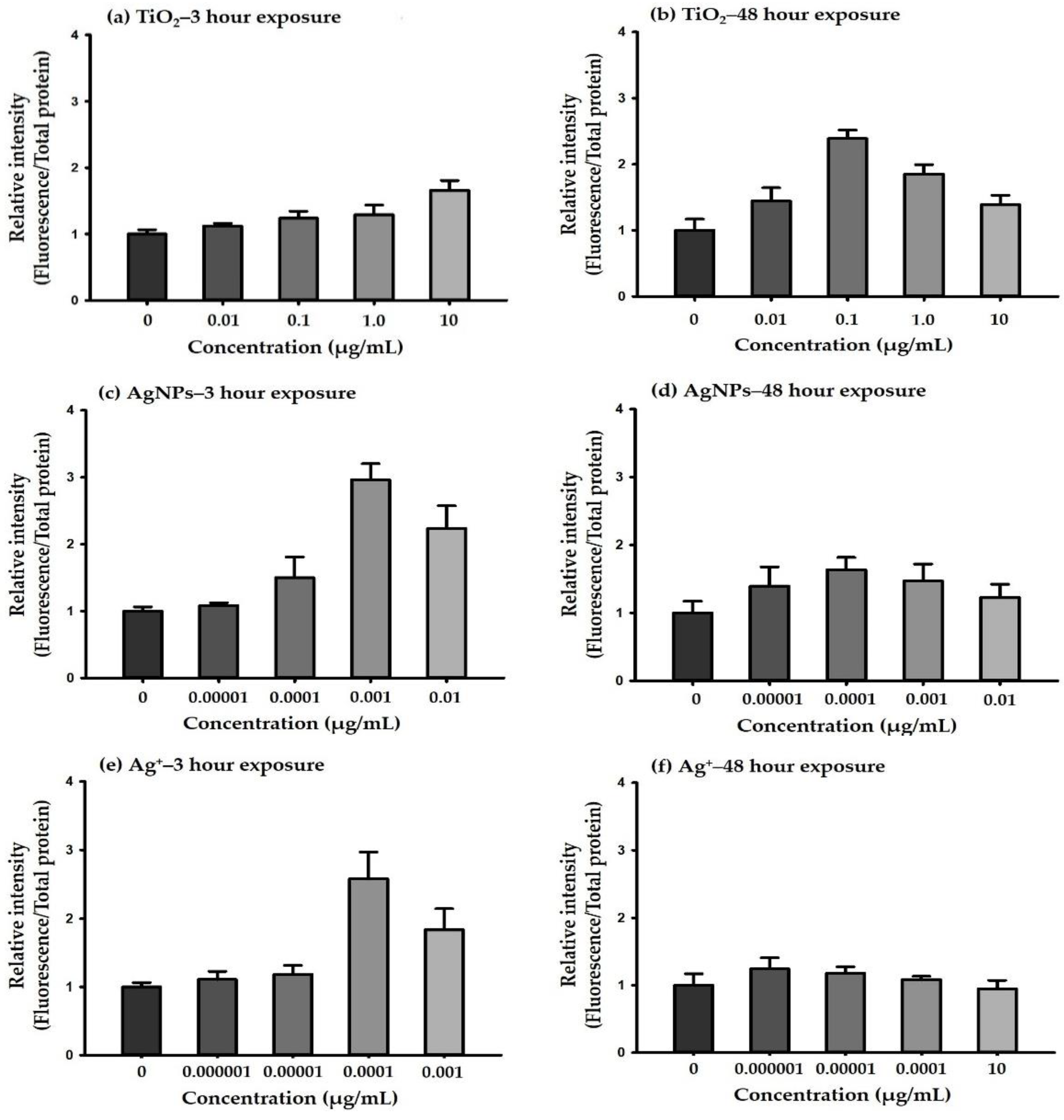
3.3. Quantification of the Oxidative Stress

The ROS levels were measured by a DCFDA assay. Figure 6 and Figure 7 show the results of the ROS measurements in the homogenized D. magna. All three materials including TiO2, AgNPs, and Ag+ exhibited stronger intensities than the control group after 3 h of exposure. The ROS intensity of TiO2 significantly increased in proportion to the concentration (n = 3) but the AgNPs and their ions showed concentration-dependent tendencies and then decreased at the highest concentration (10−2 μg/mL for AgNPs and 10−3 μg/mL for Ag+) (Figure 7). The results indicated that exposure to all substances for 3 h significantly affected the ROS levels whereas exposure for 48 h decreased the ROS levels at a higher concentration. This was likely due to the alteration of the effective concentration after 48 h of exposure.

4. Discussion

The toxicity test using D. magna is well-documented in OECD Guideline 202 and it is determined by immobilization at 24 and 48 h after exposure to nanoparticles. In most cases, it is difficult to determine accurate toxic effects based on this method because the data vary depending on the different groups. Considering this limitation, we attempted to test the acute toxicity on D. magna using influential indices including immobilization, heart rate, swimming performance, and ROS level.
We determined the LC50 values for each material as the first step in the exposure test using the organisms. The LC50 value of TiO2 in D. magna after 48 h was determined to be greater than 100 mg/mL and the values of AgNPs and Ag+ were 0.0134 and 0.0016 μg/mL, respectively. Ribeiro et al. determined the LC50 of AgNPs to be 11.02 μg/L after 48 h of exposure and 1.05 μg/L for AgNO3 using D. magna [46]. Shen et al. also calculated LC50 values of 0.58–2.51 μg/L for AgNO3, which were lower than our results [47]. The results can depend on the strain and water physicochemical parameters, suggesting that even if we measured the LC50 considering the experimental conditions, the outcomes might be different between laboratories.
The change in the heart rate as an index of the toxic effects on D. magna is widely used in acute toxicity tests. The heart rate was measured repeatedly using more than 10 neonates exposed to the materials and a cross-check for all measurements was also performed to minimize bias by observers. The results showed that the heart rate decreased with an increasing concentration for all substances. It was possible to obtain a similar pattern even for 3 h of exposure with the OECD guidelines (48 h exposure) [48]. Thus, we confirmed that an exposure duration of 3 h was sufficient to test the acute toxicity. In addition, TiO2 (considered to be non-toxic) also reduced the heart rate when the exposure level increased. This showed that TiO2 affected the heart rate of D. magna in a similar manner to other toxic NPs (e.g., AgNPs) but it was limited to conclude that TiO2 influenced the immobilization (behavioral performances).
Locomotion-based behavior is a highly sensitive index for identifying the toxic effects of chemicals [28,49]. Thus, immobilization was characterized by measuring the swimming speed (video tracking) in addition to the heart rate counting. In particular, the behavioral change was measured at each exposure duration (3, 6, 12, 24, and 48 h); thus, we could identify the temporal effects in detail compared with most previous studies that examined tests after 48 h of exposure according to OECD Guideline 202 (Figure S1–S3 in the Supplementary Materials).
To date, information on not only the temporal variations in immobilization but also the relationship between the heart rate and behavioral responses to the toxic effects of nanoparticles remains limited. For example, Lovern et al. examined the behavioral and physiological changes in D. magna using TiO2, fullerenes, and fullerene derivatives [31]. The authors found that an increased exposure level influenced a low heart rate and movement. The effect was not statistically significant for D. magna exposed to TiO2 but the temporal variation was not characterized. In our study, the heart rate increased after 48 h of exposure compared with that after 3 h of exposure whereas it decreased in proportion to the exposure level (Figure 3). The swimming speed at an earlier exposure duration (3 h) was low for all materials and increased as time elapsed (Figure 5). Therefore, it could be inferred that the exposure duration had a greater influence on the cardiac and behavioral effects at earlier periods and D. magna were more active after a certain stabilization period.
The ROS level was additionally measured to support the outcomes in this study. The ROS levels of the target materials increased with the exposure level but they gradually decreased from a certain level after 48 h of exposure. The relative intensities of the ROS were observed with inverted U-shaped patterns after 48 h of exposure. This could be interpreted as the ROS levels recovered by enhanced antioxidative responses at high concentrations [50,51]; the overall fluorescence was enhanced by the ROS production from the nanoparticles. Excessive ROS generation can cause oxidative stress [52]. Previous studies have also reported that oxidative stress can cause toxic effects on the heart rate, swimming speed, and reproduction in D. magna [53,54,55]. In this study, it was observed that the nanoparticles enhanced the ROS levels compared with the control group, which was consistent with previous studies [13,56].

5. Conclusions

There have been significant differences between the results on immobilization, cardiac effects, and behavioral changes in previous studies. In this study, the heart rate level decreased as the concentration of NPs and Ag+ increased. The swimming speed generally increased with a longer exposure to both NPs although it reached a plateau at the lowest concentration of AgNPs. We also found the overall swimming speed exhibited no correlation or weak negative correlations with the exposure concentration for all substances and the oxidative stress levels increased after exposure compared with the control group. Therefore, a comprehensive test including these indices would be necessary to improve the quality of the results on the acute toxicity. Furthermore, the effects of the nanomaterial mixtures on aquatic toxicity need to be explored in terms of invertebrate heart physiology and swimming patterns.

Supplementary Materials

The following are available online at https://0-www-mdpi-com.brum.beds.ac.uk/article/10.3390/environments9020026/s1. Figure S1: Temporal variation of swimming speed by titanium dioxide (TiO2) level and exposure duration; Figure S2: Temporal variation of swimming speed by silver nanoparticle (AgNP) level and exposure duration; Figure S3: Temporal variation of swimming speed by silver ion (Ag+) level and exposure duration.

Author Contributions

Conceptualization, C.R., Y.L. and Y.K.; methodology, J.P. (Jihoon Park) and J.P. (Jayoung Park); software, J.P. (Jihoon Park) and C.P.; validation, C.R. and Y.L.; formal analysis, J.P. (Jihoon Park), C.P., C.R. and J.P. (Jayoung Park); investigation, J.P. (Jihoon Park) and J.P. (Jayoung Park); data curation, J.P. (Jihoon Park), C.P. and J.P. (Jayoung Park); writing—original draft preparation, J.P. (Jihoon Park) and J.P. (Jayoung Park); writing—review and editing, J.P. (Jihoon Park), C.R., Y.L. and J.P. (Jayoung Park); visualization, J.P. (Jihoon Park), C.P. and J.P. (Jayoung Park); supervision, J.P. (Jayoung Park); project administration, J.P. (Jayoung Park) and Y.K.; funding acquisition, Y.K. All authors have read and agreed to the published version of the manuscript.

Funding

This research was funded by the National Research Fund (Basic Science Research Program of the Ministry of Education), grant number 2019R1A6A3A03033990, the Ministry of Science & ICT (Nanomaterial Technology Development Program), grant number 2017M3A7B6052455, and the Ministry of Trade, Industry & Energy (the Technology Innovation Program), grant number 20011630.

Data Availability Statement

Not applicable.

Acknowledgments

All authors appreciate Baeckkyoung Sung (KIST Europe) and Marcus Koch (INM, Saarbrücken, Germany) for providing technical aids used in this study.

Conflicts of Interest

The authors declare no conflict of interest. The funders had no role in the design of the study; in the collection, analyses, or interpretation of data; in the writing of the manuscript, or in the decision to publish the results.

References

  1. European Commission Joint Research Centre Institute for Health and Consumer Protection. Considerations on Information Needs for Nanomaterials in Consumer Products. Available online: https://iacmcolor.org/wp-content/uploads/2014/08/JRC-Consumer-Product-Labeling-for-nano-Policy-Report-0414.pdf (accessed on 30 September 2021).
  2. Boverhof, D.R.; David, R.M. Nanomaterial characterization: Considerations and needs for hazard assessment and safety evaluation. Anal. Bioanal. Chem. 2010, 396, 953–961. [Google Scholar] [CrossRef]
  3. Oomen, A.; Bennink, M.; van Engelen, J.; Sips, A. Nanomaterial in Consumer Products: Detection, Characterisation and Interpretation. Available online: https://rivm.openrepository.com/handle/10029/257129 (accessed on 30 September 2021).
  4. Hartmann, N.I.B.; Skjolding, L.M.; Hansen, S.F.; Baun, A.; Kjølholt, J.; Gottschalk, F. Environmental Fate and Behaviour of Nanomaterials: New Knowledge on Important Transfomation Processes; Environmental Progect No. 1594; The Danish Environmental Protection Agency: Kopenhagen, Denmark, 2014. [Google Scholar]
  5. Cedervall, T.; Hansson, L.-A.; Lard, M.; Frohm, B.; Linse, S. Food chain transport of nanoparticles affects behaviour and fat metabolism in fish. PLoS ONE 2012, 7, e32254. [Google Scholar] [CrossRef] [Green Version]
  6. Hou, J.; Liu, H.; Wang, L.; Duan, L.; Li, S.; Wang, X. Molecular toxicity of metal oxide nanoparticles in Danio rerio. Environ. Sci. Technol. 2018, 52, 7996–8004. [Google Scholar] [CrossRef] [PubMed]
  7. Lu, G.; Yang, H.; Xia, J.; Zong, Y.; Liu, J. Toxicity of Cu and Cr nanoparticles to Daphnia magna. Water Air Soil Pollut. 2017, 228, 18. [Google Scholar] [CrossRef]
  8. De Marchi, L.; Neto, V.; Pretti, C.; Figueira, E.; Chiellini, F.; Morelli, A.; Soares, A.M.; Freitas, R. The impacts of seawater acidification on Ruditapes philippinarum sensitivity to carbon nanoparticles. Environ. Sci. Nano 2017, 4, 1692–1704. [Google Scholar] [CrossRef]
  9. Ruppert, E.E.; Barnes, R.D.; Fox, R.S. Invertebrate Zoology: A Functional Evolutionary Approach, 7th ed.; Cengage Learning Acquisition Inc.: Boston, MA, USA, 2003; pp. 154–196. [Google Scholar]
  10. European Chemicals Bureau. Technical Guidance Document on Risk Assessment—Part 1. Available online: https://echa.europa.eu/documents/10162/16960216/tgdpart1_2ed_en.pdf (accessed on 1 October 2021).
  11. Tangaa, S.R.; Selck, H.; Winther-Nielsen, M.; Khan, F.R. Trophic transfer of metal-based nanoparticles in aquatic environments: A review and recommendations for future research focus. Environ. Sci. Nano 2016, 3, 966–981. [Google Scholar] [CrossRef] [Green Version]
  12. Fu, P.P.; Xia, Q.; Hwang, H.-M.; Ray, P.C.; Yu, H. Mechanisms of nanotoxicity: Generation of reactive oxygen species. J. Food Drug Anal. 2014, 22, 64–75. [Google Scholar] [CrossRef] [Green Version]
  13. Kim, T.H.; Kim, M.; Park, H.S.; Shin, U.S.; Gong, M.S.; Kim, H.W. Size-dependent cellular toxicity of silver nanoparticles. J. Biomed. Mater. Res. A 2012, 100, 1033–1043. [Google Scholar] [CrossRef]
  14. Miller, M.R. Oxidative stress and the cardiovascular effects of air pollution. Free Radical Biol. Med. 2020, 151, 69–87. [Google Scholar] [CrossRef]
  15. Xia, T.; Li, N.; Nel, A.E. Potential health impact of nanoparticles. Annu. Rev. Publ. Health 2009, 30, 137–150. [Google Scholar] [CrossRef] [Green Version]
  16. Pietroiusti, A.; Campagnolo, L.; Fadeel, B. Interactions of engineered nanoparticles with organs protected by internal biological barriers. Small 2013, 9, 1557–1572. [Google Scholar] [CrossRef] [Green Version]
  17. Pietroiusti, A.; Stockmann-Juvala, H.; Lucaroni, F.; Savolainen, K. Nanomaterial exposure, toxicity, and impact on human health. Wiley Interdiscip. Rev. Nanomed. Nanobiotechnol. 2018, 10, e1513. [Google Scholar] [CrossRef]
  18. Gatoo, M.A.; Naseem, S.; Arfat, M.Y.; Mahmood Dar, A.; Qasim, K.; Zubair, S. Physicochemical properties of nanomaterials: Implication in associated toxic manifestations. BioMed Res. Int. 2014, 2014, 498420. [Google Scholar] [CrossRef] [PubMed]
  19. Navya, P.; Daima, H.K. Rational engineering of physicochemical properties of nanomaterials for biomedical applications with nanotoxicological perspectives. Nano Converg. 2016, 3, 1–14. [Google Scholar] [CrossRef] [PubMed] [Green Version]
  20. Hou, J.; Wang, L.; Wang, C.; Zhang, S.; Liu, H.; Li, S.; Wang, X. Toxicity and mechanisms of action of titanium dioxide nanoparticles in living organisms. J. Environ. Sci. 2019, 75, 40–53. [Google Scholar] [CrossRef] [PubMed]
  21. Rizk, M.Z.; Ali, S.A.; Hamed, M.A.; El-Rigal, N.S.; Aly, H.F.; Salah, H.H. Toxicity of titanium dioxide nanoparticles: Effect of dose and time on biochemical disturbance, oxidative stress and genotoxicity in mice. Biomed. Pharmacother. 2017, 90, 466–472. [Google Scholar] [CrossRef] [PubMed]
  22. Nomiya, K.; Yoshizawa, A.; Tsukagoshi, K.; Kasuga, N.C.; Hirakawa, S.; Watanabe, J. Synthesis and structural characterization of silver (I), aluminium (III) and cobalt (II) complexes with 4-isopropyltropolone (hinokitiol) showing noteworthy biological activities. Action of silver (I)-oxygen bonding complexes on the antimicrobial activities. J. Inorg. Biochem. 2004, 98, 46–60. [Google Scholar]
  23. Sarmast, M.; Salehi, H.; Khosh-Khui, M. Nano silver treatment is effective in reducing bacterial contaminations of Araucaria excelsa R. Br. var. glauca explants. Acta Biol. Hung. 2011, 62, 477–484. [Google Scholar] [CrossRef] [PubMed]
  24. Seitz, F.; Rosenfeldt, R.R.; Storm, K.; Metreveli, G.; Schaumann, G.E.; Schulz, R.; Bundschuh, M. Effects of silver nanoparticle properties, media pH and dissolved organic matter on toxicity to Daphnia magna. Ecotoxicol. Environ. Safe 2015, 111, 263–270. [Google Scholar] [CrossRef]
  25. Asghari, S.; Johari, S.A.; Lee, J.H.; Kim, Y.S.; Jeon, Y.B.; Choi, H.J.; Moon, M.C.; Yu, I.J. Toxicity of various silver nanoparticles compared to silver ions in Daphnia magna. J. Nanobiotechnol. 2012, 10, 14. [Google Scholar] [CrossRef] [Green Version]
  26. Hu, Y.; Chen, X.; Yang, K.; Lin, D. Distinct toxicity of silver nanoparticles and silver nitrate to Daphnia magna in M4 medium and surface water. Sci. Total Environ. 2018, 618, 838–846. [Google Scholar] [CrossRef] [PubMed]
  27. Martins, J.; Teles, L.O.; Vasconcelos, V. Assays with Daphnia magna and Danio rerio as alert systems in aquatic toxicology. Environ. Int. 2007, 33, 414–425. [Google Scholar] [CrossRef]
  28. Bownik, A. Daphnia swimming behaviour as a biomarker in toxicity assessment: A review. Sci. Total Environ. 2017, 601, 194–205. [Google Scholar] [CrossRef]
  29. Kunze, J.; Hartmann, S.; Witte, K.; Kuhnert, K.D. Daphnia magna as biosensor for Ag-nanoparticles in water systems: Development of a computer vision system for the detection of behavioral changes. In Proceedings of the 23rd International Conference on Pattern Recognition (ICPR 2016), Cancun, Mexico, 4–8 December 2016; Available online: http://homepages.inf.ed.ac.uk/rbf/VAIB16PAPERS/vaibkunze.pdf (accessed on 1 October 2021).
  30. Lovern, S.B.; Klaper, R. Daphnia magna mortality when exposed to titanium dioxide and fullerene (C60) nanoparticles. Environ. Toxicol. Chem. 2006, 25, 1132–1137. [Google Scholar] [CrossRef]
  31. Lovern, S.B.; Strickler, J.R.; Klaper, R. Behavioral and physiological changes in Daphnia magna when exposed to nanoparticle suspensions (titanium dioxide, nano-C60, and C60HxC70Hx). Environ. Sci. Technol. 2007, 41, 4465–4470. [Google Scholar] [CrossRef] [Green Version]
  32. Simão, F.C.; Martínez-Jerónimo, F.; Blasco, V.; Moreno, F.; Porta, J.M.; Pestana, J.L.; Soares, A.M.; Raldúa, D.; Barata, C. Using a new high-throughput video-tracking platform to assess behavioural changes in Daphnia magna exposed to neuro-active drugs. Sci. Total Environ. 2019, 662, 160–167. [Google Scholar] [CrossRef] [Green Version]
  33. Fekete-Kertész, I.; Stirling, T.; Ullmann, O.; Farkas, É.; Kirchkeszner, C.; Feigl, V.; Molnár, M. How Does Experimental Design Modify the Result of Daphnia magna Heartbeat Rate Test?–Analyses of Factors Affecting the Sensitivity of the Test System. Period. Polytech-Chem. 2018, 62, 257–264. [Google Scholar] [CrossRef] [Green Version]
  34. Chung, W.; Song, J.M.; Lee, J. The Evaluation of Titanium Dioxide Nanoparticle Effects on Cardiac and Swimming Performance of Daphnia magna. Int. J. Appl. Environ. Sci. 2016, 11, 1375–1385. [Google Scholar]
  35. National Institute of Environmental Research. Operational Guideline for Ecotoxicity test Procedures. Document No. NIER-GP2015-094. Available online: https://ecolibrary.me.go.kr/nier/#/search/detail/5595086 (accessed on 1 October 2021). (In Korean).
  36. Baumann, J.; Sakka, Y.; Bertrand, C.; Köser, J.; Filser, J. Adaptation of the Daphnia sp. acute toxicity test: Miniaturization and prolongation for the testing of nanomaterials. Environ. Sci. Pollut. Res. 2014, 21, 2201–2213. [Google Scholar] [CrossRef]
  37. Cupi, D.; Hartmann, N.B.; Baun, A. Influence of pH and media composition on suspension stability of silver, zinc oxide, and titanium dioxide nanoparticles and immobilization of Daphnia magna under guideline testing conditions. Ecotoxicol. Environ. Safe 2016, 127, 144–152. [Google Scholar] [CrossRef] [PubMed]
  38. Georgantzopoulou, A.; Balachandran, Y.L.; Rosenkranz, P.; Dusinska, M.; Lankoff, A.; Wojewodzka, M.; Kruszewski, M.; Guignard, C.; Audinot, J.-N.; Girija, S. Ag nanoparticles: Size-and surface-dependent effects on model aquatic organisms and uptake evaluation with NanoSIMS. Nanotoxicology 2012, 7, 1168–1178. [Google Scholar] [CrossRef]
  39. Heinlaan, M.; Ivask, A.; Blinova, I.; Dubourguier, H.-C.; Kahru, A. Toxicity of nanosized and bulk ZnO, CuO and TiO2 to bacteria Vibrio fischeri and crustaceans Daphnia magna and Thamnocephalus platyurus. Chemosphere 2008, 71, 1308–1316. [Google Scholar] [CrossRef]
  40. Hlavkova, D.; Havelkova, B.; Kopel, P.; Beklova, M. Evaluation of nanosilver ecotoxicity using representatives of distinct trophic levels. Fresenius Environ. Bull. 2019, 28, 745–749. [Google Scholar]
  41. Hurel, C.; Bignon, C.; Said-Mohamed, C.; Amigoni, S.; Devers, T.; Guittard, F. Functionalized and grafted TiO2, CeO2, and SiO2 nanoparticles—Ecotoxicity on Daphnia magna and relevance of ecofriendly polymeric networks. Environ. Sci. Pollut. Res. 2018, 25, 21216–21223. [Google Scholar] [CrossRef] [PubMed]
  42. Jemec, A.; Kahru, A.; Potthoff, A.; Drobne, D.; Heinlaan, M.; Böhme, S.; Geppert, M.; Novak, S.; Schirmer, K.; Rekulapally, R. An interlaboratory comparison of nanosilver characterisation and hazard identification: Harmonising techniques for high quality data. Environ. Int. 2016, 87, 20–32. [Google Scholar] [CrossRef]
  43. Kim, J.; Kim, S.; Lee, S. Differentiation of the toxicities of silver nanoparticles and silver ions to the Japanese medaka (Oryzias latipes) and the cladoceran Daphnia magna. Nanotoxicology 2011, 5, 208–214. [Google Scholar] [CrossRef] [PubMed]
  44. Marcone, G.P.; Oliveira, Á.C.; Almeida, G.; Umbuzeiro, G.A.; Jardim, W.F. Ecotoxicity of TiO2 to Daphnia similis under irradiation. J. Hazard. Mater. 2012, 211, 436–442. [Google Scholar] [CrossRef]
  45. Picado, A.; Paixão, S.M.; Moita, L.; Silva, L.; Diniz, M.S.; Lourenço, J.; Peres, I.; Castro, L.; Correia, J.B.; Pereira, J. A multi-integrated approach on toxicity effects of engineered TiO2 nanoparticles. Front. Environ. Sci. Eng. 2015, 9, 793–803. [Google Scholar] [CrossRef] [Green Version]
  46. Ribeiro, F.; Gallego-Urrea, J.A.; Jurkschat, K.; Crossley, A.; Hassellöv, M.; Taylor, C.; Soares, A.M.; Loureiro, S. Silver nanoparticles and silver nitrate induce high toxicity to Pseudokirchneriella subcapitata, Daphnia magna and Danio rerio. Sci. Total Environ. 2014, 466, 232–241. [Google Scholar] [CrossRef]
  47. Shen, M.-H.; Zhou, X.-X.; Yang, X.-Y.; Chao, J.-B.; Liu, R.; Liu, J.-F. Exposure medium: Key in identifying free Ag+ as the exclusive species of silver nanoparticles with acute toxicity to Daphnia magna. Sci. Rep. 2015, 5, 9674. [Google Scholar] [CrossRef] [Green Version]
  48. Organization for Economic Cooperation and Development (OECD). OECD Guideline for Testing of Chemicals—Daphnia sp., Acute Immobilization Test. Available online: https://0-www-oecd--ilibrary-org.brum.beds.ac.uk/environment/test-no-202-daphnia-sp-acute-immobilisation-test_9789264069947-en (accessed on 1 October 2020).
  49. Mora-Zamorano, F.X.; Larson, J.K.; Carvan, M.J. Neurobehavioral Analysis Methods for Adverse Outcome Pathway (AOP) Models and Risk Assessment. In A Systems Biology Approach to Advancing Adverse Outcome Pathways for Risk Assessment; Springer: New York, NY, USA, 2018; pp. 149–175. [Google Scholar]
  50. Galdiero, E.; Siciliano, A.; Maselli, V.; Gesuele, R.; Guida, M.; Fulgione, D.; Galdiero, S.; Lombardi, L.; Falanga, A. An integrated study on antimicrobial activity and ecotoxicity of quantum dots and quantum dots coated with the antimicrobial peptide indolicidin. Int. J. Nanomed. 2016, 11, 4199. [Google Scholar] [CrossRef] [Green Version]
  51. Zhou, H.; Wang, J.; Hu, S.; Zhu, H.; Toan, S.; Ren, J. BI1 alleviates cardiac microvascular ischemia-reperfusion injury via modifying mitochondrial fission and inhibiting XO/ROS/F-actin pathways. J. Cell. Physiol. 2019, 234, 5056–5069. [Google Scholar] [CrossRef] [PubMed]
  52. Klaper, R.; Crago, J.; Barr, J.; Arndt, D.; Setyowati, K.; Chen, J. Toxicity biomarker expression in daphnids exposed to manufactured nanoparticles: Changes in toxicity with functionalization. Environ. Pollut. 2009, 157, 1152–1156. [Google Scholar] [CrossRef] [PubMed]
  53. Bownik, A.; Stępniewska, Z. Protective effects of ectoine on behavioral, physiological and biochemical parameters of Daphnia magna subjected to hydrogen peroxide. Com. Biochem. Phys. C 2015, 170, 38–49. [Google Scholar] [CrossRef] [PubMed]
  54. De Felice, B.; Salgueiro-González, N.; Castiglioni, S.; Saino, N.; Parolini, M. Biochemical and behavioral effects induced by cocaine exposure to Daphnia magna. Sci. Total Environ. 2019, 689, 141–148. [Google Scholar] [CrossRef]
  55. Liu, Z.; Cai, M.; Wu, D.; Yu, P.; Jiao, Y.; Jiang, Q.; Zhao, Y. Effects of nanoplastics at predicted environmental concentration on Daphnia pulex after exposure through multiple generations. Environ. Pollut. 2020, 256, 113506. [Google Scholar] [CrossRef] [PubMed]
  56. Lu, H.; Fan, W.; Dong, H.; Liu, L. Dependence of the irradiation conditions and crystalline phases of TiO2 nanoparticles on their toxicity to Daphnia magna. Environ. Sci. Nano 2017, 4, 406–414. [Google Scholar] [CrossRef]
Figure 1. TEM images of the nanomaterials: (a) TiO2 in distilled water; (b) TiO2 in ISO medium; (c) AgNPs in distilled water; (d) AgNPs in ISO medium. Scale bars: 200 nm.
Environments 09 00026 g001
Figure 2. Viability of D. magna as a function of TiO2 and Ag nanoparticles (NPs) and Ag+.
Environments 09 00026 g002
Figure 3. The changes of heart rate in D. magna after 3 and 48 h of exposure to TiO2, AgNPs, and Ag+ at varying concentrations. The asterisk (*) above the histogram indicates statistically significant differences in heart rate changes compared with the control group (p < 0.05).
Environments 09 00026 g003
Figure 4. Representative images of the D. magna swimming paths for the initial 10 min as a function of the concentration of TiO2, AgNPs, and Ag+.
Environments 09 00026 g004
Figure 5. Swimming speed of D. magna by the concentrations of TiO2, AgNPs, and Ag+ and the exposure time (up to 48 h). The asterisk (*) above the histogram indicates statistically significant differences in heart rate changes compared with the control group (p < 0.0001).
Environments 09 00026 g005
Figure 6. Representative optical microscopic images of D. magna exposed to TiO2, AgNPs, and Ag+ for 3 h. Each image set represents bright field, fluorescence, and merged images, from left to right. Scale bars indicate 200 µm.
Environments 09 00026 g006
Figure 7. Effects of TiO2, AgNPs, and Ag+ on the ROS levels in homogenized D. magna after 3 and 48 h of exposure.
Environments 09 00026 g007
Publisher’s Note: MDPI stays neutral with regard to jurisdictional claims in published maps and institutional affiliations.

