Article

The Effects of Copper and Silver Nanoparticles on Container-Grown Scots Pine (Pinus sylvestris L.) and Pedunculate Oak (Quercus robur L.) Seedlings

by
Marta Aleksandrowicz-Trzcińska
1,*,
Magdalena Bederska-Błaszczyk
2,
Adam Szaniawski
1,
Jacek Olchowik
3 and
Marcin Studnicki
4
1
Department of Forest Protection and Ecology, Faculty of Forestry, Warsaw University of Life Sciences, Nowoursynowska 159, 02-776 Warsaw, Poland
2
Department of Botany, Faculty of Agriculture and Biology, Warsaw University of Life Sciences, Nowoursynowska 159, 02-776 Warsaw, Poland
3
Department of Plant Pathology, Faculty of Horticulture, Biotechnology and Landscape Architecture, Warsaw University of Life Sciences, Nowoursynowska 159, 02-776 Warsaw, Poland
4
Department of Experimental Design and Bioinformatics, Faculty of Agriculture and Biology, Warsaw University of Life Sciences, Nowoursynowska 159, 02-776 Warsaw, Poland
*
Author to whom correspondence should be addressed.
Submission received: 13 February 2019 / Revised: 12 March 2019 / Accepted: 14 March 2019 / Published: 17 March 2019
(This article belongs to the Section Forest Ecophysiology and Biology)

Abstract

:
Metal nanoparticles (NPs) are finding ever-wider applications in plant production (agricultural and forestry-related) as fertilisers, pesticides and growth stimulators. This makes it essential to examine their impact on a variety of plants, including trees. In the study detailed here, we investigated the effects of nanoparticles of silver and copper (i.e., AgNPs and CuNPs) on growth, and chlorophyll fluorescence, in the seedlings of Scots pine and pedunculate oak. We also compared the ultrastructure of needles, leaves, shoots and roots of treated and untreated plants, under transmission electron microscopy. Seedlings were grown in containers in a peat substrate, prior to the foliar application of NPs four times in the course of the growing season, at the four concentrations of 0, 5, 25 and 50 ppm. We were able to detect species-specific activity of the two types of NP. Among seedling pines, the impact of both types of NP at the concentrations supplied limited growth slightly. In contrast, no such effect was observed for the oaks grown in the trial. Equally, it was not possible to find ultrastructural changes in stems and roots associated with the applications of NPs. Cell organelles apparently sensitive to the action of both NPs (albeit only at the highest applied concentration of 50 ppm) were chloroplasts. The CuNP-treated oaks contained large plastoglobules, whereas those dosed with AgNP contained large starch granules. The NP-treated pines likewise exhibited large numbers of plastoglobules, while the chloroplasts of NP-treated plants in general presented shapes that changed from lenticular to round. In addition, large osmophilic globules were present in the cytoplasm. Reference to maximum quantum yields from photosystem II (Fv/Fm)—on the basis of chlorophyll a fluorescence measurements—revealed a slight debilitation of oak seedlings following the application of both kinds of NP at higher concentrations. In contrast, in pines, this variable revealed no influence of AgNPs, as well as a favourable effect due to the CuNPs applied at a concentration of 5 ppm. Our research also showed that any toxic impact on pine or oak seedlings due to the NPs was limited and only present with higher concentrations.

1. Introduction

Nanoparticles (NPs) are defined as particles with at least one dimension of less than 100 nm. They have existed in the environment naturally since the beginning of Earth’s history, in ash from volcanoes and forest fires; as sea salt aerosols; and as oxides of iron or other metals in soils, rivers and oceans [1]. A second group of “incidental NPs” are produced unintentionally by human activity and are also defined as waste or anthropogenic particles. They appear through desertification, biomass burning and coal combustion, as well as in engine exhaust fumes and thanks to mining and other similar activities [2].
A third group comprises the engineered NPs. They have gained wide application around the world in the last few decades, thanks to their unique electronic, optical, mechanical, magnetic and chemical properties that ensure a wide range of applications in medicine, pharmacy, cosmetic manufacture, electronics, construction, jewelry-making and photography [3,4]. Today, more than 1000 products containing nanomaterials are available commercially [1].
Nanotechnology has also been applied in plant production (agriculture and forestry), with nanomaterials used as fertilisers to increase plant growth and yields, pesticides to achieve pest and disease management, and sensors by which to monitor soil quality and plant health [5].
NPs can be taken up by plants and transported to other plant tissues [6]. Some studies have reported the direct uptake of NPs (e.g., those of Ag) by roots, in which they accumulate [7] or are translocated to other organs [8]. NPs can enter plant tissues through above-ground organs, tissues and cellular structures, such as the cuticles, epidermis, stomata, lenticels, hydathodes, trichomes and stigmata, as well as through root tissues, i.e., the rhizodermis/cortex and through root tips and lateral root junctions [9]. NP uptake across the cell wall is shown to depend on the sizes of particles and cell-wall pores, with NPs crossing the latter usually limited to diameters of 5–20 nm. Both the apoplastic and symplastic pathways have been proven to be traversed by NPs [10]. Long-distance transport in turn takes place through the phloem and xylem in the plant stem [6,11,12].
Some NPs, such as AgNPs, have been reported to induce the formation of new and larger pores in cell walls and cuticles, thereby facilitating the internalisation of larger NPs [6].
Thus far, the results of studies document positive, negative and neutral effects on plants at the morphological, physiological, biochemical and molecular levels [13,14].
A positive influence of NPs on plants is manifested in the stimulated germination of seeds; faster plant growth rate; higher biomass of plants; larger numbers of flowers, fruits and seeds; and higher gluten or starch content [15,16,17,18]. In turn, a negative influence is to be seen in inhibited seed germination or reduced shoot and root length, as well as amounts of fresh and dry matter [19,20,21,22]. An indirectly beneficial or negative effect of nanoparticles on plant growth may in turn reflect alterations in numbers of microorganisms in the rhizosphere and/or in their metabolic activity [14,23,24].
Stimulation or inhibition of plant growth often results from the (favourable or unfavourable) effect of NPs on chloroplasts, chlorophyll content (higher or lower) and rate of photosynthesis (faster or slower) [25,26]. Low concentrations of NPs may have a positive influence on plant growth by sustaining increases in the numbers of chloroplasts, as well as tighter packing of chloroplast grana [27]. The negative impact of NPs may in turn be manifested in swollen chloroplasts with a dilated thylakoid system [7]; the presence of a large number of starch granules within chloroplasts; and the destruction of grana [12], or smaller, ruptured chloroplasts not manifesting changes in the thylakoid system [28,29]. Following NP treatment, chloroplasts may change shape and become either spherical or flatter [25,30].
NPs in plants may cause changes in the content or activity of plant hormones, generate oxidative stress, or modify plant gene expression [14,31].
The main factors influencing the effects of NPs on plants reflect the characteristics of the nanomaterials themselves (size, shape, chemical composition, surface structure and coating, solubility, aggregation and concentration), as well as the plant species involved, the growth medium, the growth stage and the length of the period of exposure [31,32].
Certain authors consider that NPs have a toxic impact when taken up by plants, but exert a positive influence when merely present in the environment [26].
From among the methods by which the impacts on plants of both biotic and abiotic stress factors are researched, it is the chlorophyll a fluorescence measurement in vivo that has gained wide application [33]. This allows for a rapid, precise and non-invasive measurement of the efficiency of photosystem II (PSII) to photosynthesis, with plants’ molecular-level responses to stress factors, in essence, being indicated in this way [34]. Higher values for such chlorophyll a fluorescence measurements in Brassica juncea, following the application of AgNPs, were considered to reflect a higher chlorophyll content, and hence favourable changes in enzyme activity from a plant’s perspective, capable of stimulating further growth [35]. Other studies point to impaired chlorophyll fluorescence parameters as a result of the application of NPs [12,26,36,37].
Although plants must have been exposed to naturally-occurring NPs since they first evolved, the impact is likely to be infinitely greater now, given the increased ambient concentrations of incidental and engineered NPs as the result of industrial processes and the increasing overt demand for NP-based products, for example, as pesticides, fertilisers, growth stimulators or promoters of plant resistance [2,5]. However, notwithstanding the rather large number of studies that have been carried out, a full picture of the influence they exert remains elusive. This is particularly the case for woody plants, given the clear emphasis on crop plants thus far [14].
In these circumstances, the work detailed here sought: (i) to determine the influence of CuNPs and AgNPs on growth in one-year-old seedlings of Scots pine and oak; (ii) to compare the TEM-ultrastructure of leaves/needles, roots and stems of pines and oaks treated with NPs or left untreated; and (iii) to assess the condition of plants following NP application, by reference to chlorophyll a fluorescence measurements. We hypothesised that NPs would have a slightly negative effect on the plants tested, albeit with these effects differing between species.

2. Materials and Methods

2.1. Plant Material

The study was conducted at the nursery of the Forest Experimental Station of Warsaw University of Life Sciences (Rogów, Poland, 51°40′ N, 19°55′ E, 195 m a.s.l). One-year-old seedlings were grown in plastic container trays: Scots pine—V-120 (40 pots per tray with a capacity of 120 cm3) and pedunculate oak—V-360 (15 pots per tray with a capacity of 360 cm3). The substrate used was a peat–perlite mix purchased from the container nursery in Nędza, Rudy Raciborskie Forest District. With a pH of 4.5, the composition was 85% Sphagnum peat from Estonia and 15% coarse-grained perlite (No. 3), regarded as improving aeration. Seedlings were treated with a mixture of Osmocote Exact Standard controlled-release fertilisers of different release characteristics, i.e., 3–4 M, 5–6 M and 8–9 M (1:1:1). Fertiliser was added at the same time as seeds were sown. The seeds of both species were of local origin, from managed forest. At the beginning of May, two seeds were planted in each pot. After germination, one plant per pot was selected at random and the others were removed. The containers were placed in a foil-covered greenhouse (with a height of 2.8 m, width of 6 m and length 30 m) and watered regularly when necessary. Temperatures in the greenhouse at the time of the experiment were in the 25–32 °C range.

2.2. Nanoparticles and Treatments

The two types of metal nanoparticle selected for trials were silver (AgNPs) and copper (CuNPs). Samples of commercially-available solutions of AgNPs and CuNPs were purchased from Nano-Koloid Sp. z o. o, Warsaw, Poland as a licensee of the Nano Technologies Group, Inc. (USA), manufactured under European Patent EP2081672 A2. As the producer notes, these are generated in a physical process, consist of around 100 atoms (about 5 nm) and are suspended in demineralised water. The concentration of nanoparticles in the commercially-available product is 50 ppm.
There were four spray treatments of seedlings’ aerial parts using 1000 L/ha (=100 mL/m2) aqueous solutions of NPs (AgNPs, CuNPs) in four concentrations: 0 ppm, 5 ppm, 25 ppm and 50 ppm. Previously, the NPs had been suspended in deionised water by means of vigorous shaking for at least 10 min. Applications were foliar, with the first on May 27th, followed by three more at two-week intervals. Spraying was done in the early morning, with a manual compressed-air sprayer (Kisan Kraft, model KK-PS5000). For each species, the experimental design comprised two factors (types of metal nanoparticle—Ag and Cu, at the four different concentrations of 0, 5, 25 and 50 ppm). The experiment was performed in a randomised complete block design of four blocks.

2.3. Chlorophyll a Fluorescence Measurements

Leaves were subject to this kind of measurement with the aid of a Hansatech FMS-2 fluorometer (Hansatech Ltd., Kings Lynn, UK), 11 times in total, with nine readings taken in the July-August period and two final ones separated by a two-week interval in September. The total number of leaves subject to measurement was 10, selected at random from among the youngest that were fully developed in the young oak seedlings. Ten needles from the pines in each variant were also selected. With the aid of clips, leaves and needles were given the chance to adapt to darkness for some 20 min. Such shaded leaves were then connected to the optical fibre, with the parameters referred to in measuring the fluorescence of chlorophyll a being: Fo—initial (zero) fluorescence following adaptation to darkness; Fm—maximal fluorescence after adaptation to the dark; and Fv/Fm—maximal efficiency of photosystem II (PSII), where Fv is the variable fluorescence Fm − Fo.

2.4. Samplings and Biometric Parameters

At the end of October, 40 seedlings (10 plants × 4 blocks) were harvested at random from each treatment. A total of 320 Scots pine and 320 pedunculate oak seedlings were sampled. The shoot-growth parameters measured length [cm] and root-collar diameter [mm]. To determine the total root length [cm], the root systems were analysed using the Epson Perfection 4990 Photo scanner integrated with WinRhizo software (Regent Instruments Inc., Quebec City, QC, Canada). Ultimately, values for the dry mass of the above-ground part and root [g] were determined after drying at 105 °C for 24 h.

2.5. Microscopic Investigations

The transmission electron microscopic imaging (TEM) work was based on ten seedlings chosen at random from each treatment. The adsorption of NPs and ultrastructure of studied organs were observed under TEM eight weeks after the last treatment. Tissue samples were collected from the middle parts of needles or leaves, shoots and roots of the pine and oak seedlings. Obtained plant material was fixed in 2% (v/v) glutaraldehyde and 2% paraformaldehyde (v/w) in 0.05 M cacodylate buffer (pH 7.2) after Karnovsky [38], for 4 h at room temperature. The material was then rinsed four times in cacodylate buffer, prior to samples being contrasted in 1% OsO4 for 2 h at 4 °C, and then rinsed four times with the same buffer. Material was next dehydrated in a series of aqueous solutions of ethanol, and subsequently in propylene oxide. Finally, the material was gradually saturated with resin (Epon, Fluka) and polymerised for 24 h at 60 °C. The Epon blocks with plant material were cut for TEM, with an ultramicrotome (Leica) into ultrathin sections (~90 nm thick), which were collected on Formvar-coated slot-grids and contrasted with 1% lead citrate and 2% uranyl acetate. Thereafter, the material was examined using a Morgagni 268D (FEI) electron microscope. Photographic documentation was prepared with a Morada (SIS) digital camera and the iTEM (SIS) computer programme.

2.6. Data Analysis

We tested relationships among biometric and fluorescence parameters as set against different concentrations of AgNPs and CuNPs, using a two-way Analysis of Variance ANOVA for the randomised complete block design. The Tukey Test was used in pairwise comparisons (as a post-hoc test) of different concentrations of AgNPs and CuNPs. The statistical analysis was performed using R version 3.5.1 (The R Foundation for Statistical Computing, Vienna, Austria) [39], with the accepted level of significance set at p < 0.05.

3. Results

3.1. Biometric Parameters of Seedlings

Pine seedlings exposed to the 50 ppm dosage of AgNPs had impaired growth in comparison with the control seedlings, in the sense that stems were shorter and the root dry mass was lower. Shorter average lengths of stems were also to be noted among pines sprayed with AgNPs at a concentration of 5 ppm. The application of CuNPs had an apparent impact on growth in pines, with the comparisons with the control revealing a smaller root collar diameter (in the presence of all concentrations of the nanoparticles), a shorter length of shoots (at 50 ppm), and a lower dry mass of roots (at 5 ppm). Treatment with 5 ppm of CuNPs had the effect of increasing the tendency for side-shoots to be produced, with this ensuring a greater dry mass of above-ground parts than in control seedlings (Table 1).
The apparent influence of NP applications was more limited in the case of oaks than pines, where most of the measured variables were concerned. Oaks treated with AgNPs at 25 ppm were characterised by a lower total root length than control plants. The dry mass of roots in oak seedlings supplied with CuNPs at the highest (25 and 50 ppm) concentrations was in turn significantly greater than that noted for the seedlings not treated with NPs (Table 1).

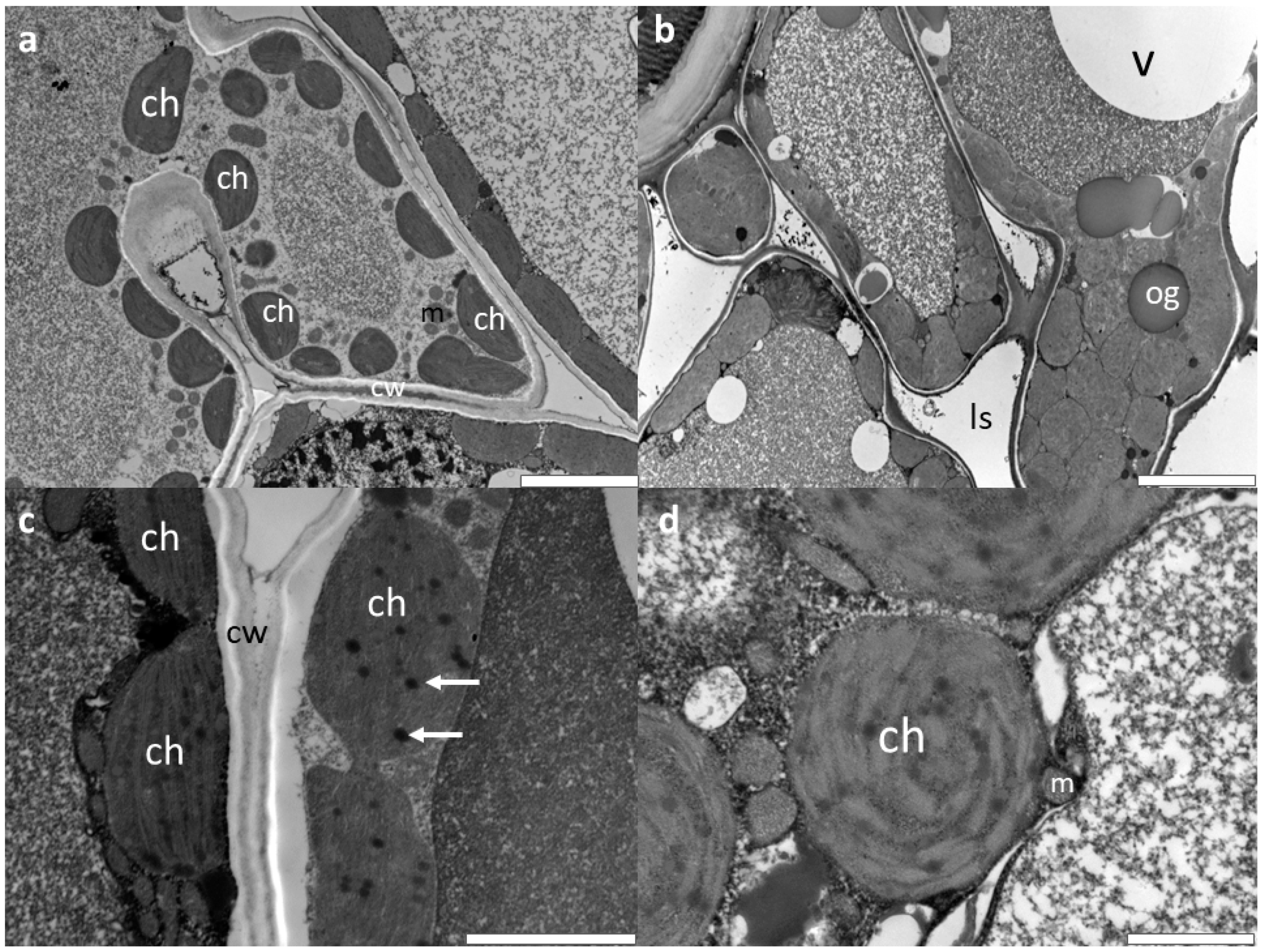
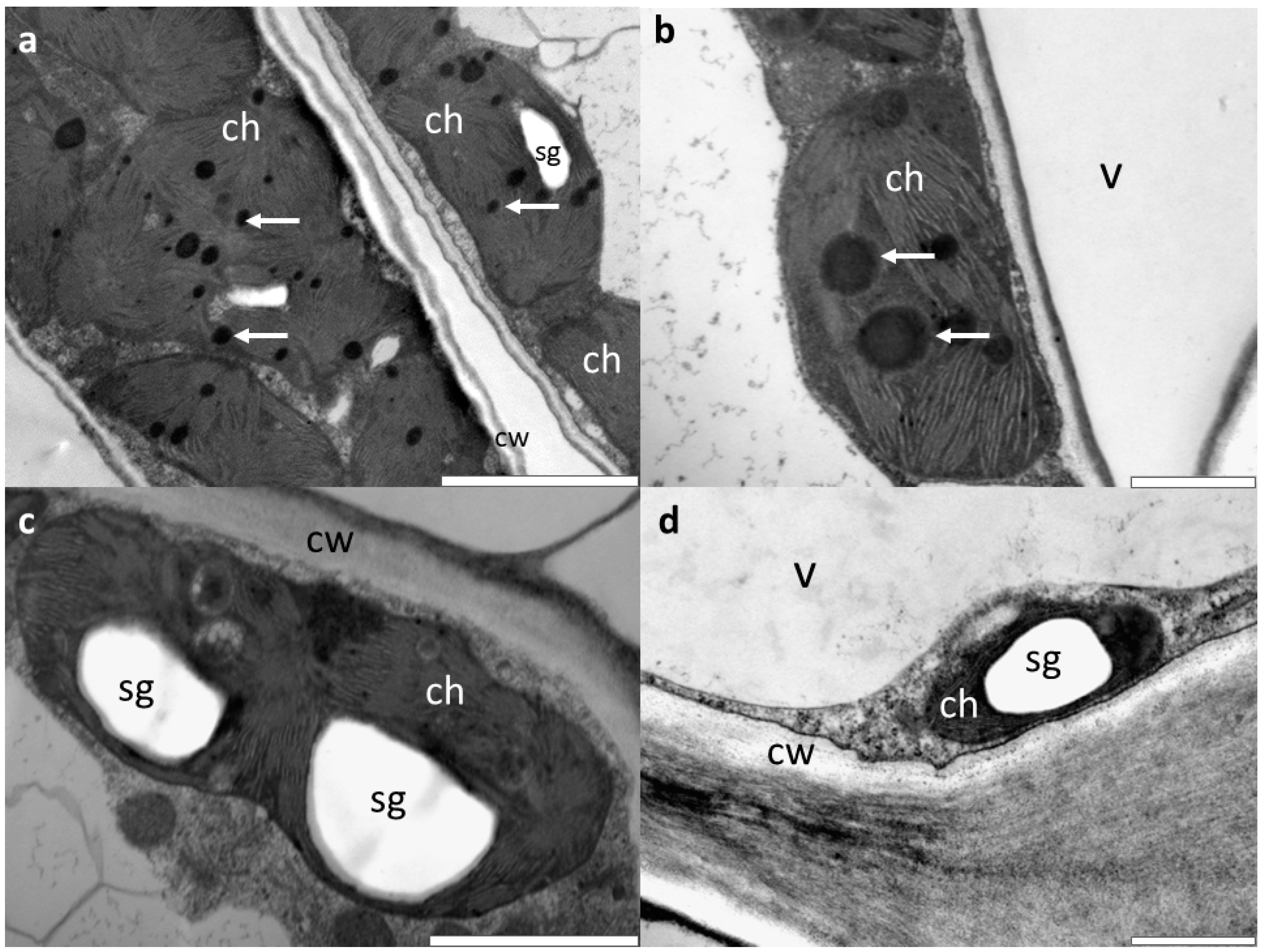
3.2. Microscopic Investigations

The ultrastructure of shoots and roots in the nanoparticle-treated pines and oaks is similar to that in control plants (data not shown). Seedlings treated with CuNPs and AgNPs, but only at the highest (50 ppm) concentrations, exhibit a disturbed ultrastructure in needles and leaves, especially in the photosynthetic apparatus. In the control pines (no AgNP and CuNP), the chloroplasts contain single plastoglobules, whereas the NP-treated plants exhibit large numbers of plastoglobules, while the chloroplasts of NP-treated plants are of a shape modified from lenticular to round. In addition, large, osmophilic globules are present in the cytoplasm of NP-treated needle cells. Application of CuNPs, as compared with AgNPs, only resulted in more limited disturbance to the ultrastructure of mesophyll cells in the pine needles (Figure 1).
Changes of ultrastructure were more limited in the leaves of oaks than in pine needles, only therefore being observed where seedlings had received the highest (50 ppm) concentrations of NPs, and being differentiated, depending on whether CuNPs or AgNPs had been applied. While the CuNP-treated oaks have chloroplasts containing large plastoglobules (compared with the small ones present in control seedlings), those supplied with AgNPs have large starch granules (Figure 2). Other organelles (e.g., mitochondria and vacuoles) in both species remained unchanged, as did the structure of cell walls.
At 5 nm, the sizes of the AgNPs and CuNPs combined with limits to the microscopic resolution prevented direct observation of the NPs. Because of this, the work carried out could neither confirm nor preclude the presence of NPs in the tissues and cells of leaves or needles, or else stems and roots.

3.3. Chlorophyll a Fluorescence

The spraying of pine seedlings with AgNPs was not associated with any emerging differences when it came to mean values for the maximum efficiency of photosytem II (expressed as Fv/Fm and noted in different experimental variants; F = 1.204; p = 0.471). In contrast, where the application was of CuNPs, higher values for Fv/Fm were noted in pines receiving the nanoparticles at a 5 ppm concentration (0.8119), as opposed to those in the control (0.8011). Where young oaks were concerned, both NPs applied caused lower values of the chlorophyll fluorescence parameter (Fv/Fm) in comparison to untreated seedlings. This was true for the highest concentrations of CuNPs (25 and 50 ppm) and the 50 ppm concentration of AgNPs (Table 2).

4. Discussion

The results obtained point to a toxic, though limited, impact of both kinds of tested nanoparticle on one-year old pine and oak seedlings. The presence and magnitude of the influence exerted were found to depend on the features analysed (be these growth parameters; ultrastructural features of stems, roots and leaves; or chlorophyll fluorescence), as well as the types and concentrations of NPs and the species of plant concerned.
Previously, the influence of NPs has mainly been researched by reference to crop plants, with only a few studies devoted to woody species. The results of the latter have furthermore been very varied—encompassing stimulation of growth [30] and the lack of any influence [40,41,42], through to inhibition [43]. In contrast, our work points to a differentiated influence of NPs, depending on both plant species and type of nanomaterial. Oaks were not found to have been affected negatively in terms of their growth, while pines experienced a curtailment of growth under the influence of NPs of both kinds studied. However, a linear decline in the magnitude of the effect was only obtained for the root collar diameter, following the application of CuNPs. There was a more marked restriction on growth in seedlings treated with either of the NPs at the highest concentration tested (50 ppm), and this was particularly clear in respect of shoot length and the dry mass of roots. Equally, CuNPs, even at a concentration of 5 ppm, induced a change of habit in pine seedlings, given the large number of side stems and consequent significant increase in the dry mass of above-ground parts.
Stimulation or inhibition of plant growth often results from the effect of NPs on chloroplasts, chlorophyll content and photosynthesis rate [25,26]. In both the pines and oaks studied, we were able to show changes in chloroplasts (see below) that were capable of producing the observed impacts on seedling growth parameters. Given that changes in chloroplasts in oak were very minor, this may have translated into a lack of differences in plant growth between specimens treated or not treated with NPs. In turn, as the unfavourable changes were greater in young pines, this may account for the curtailment of growth observed with both kinds of NP. Nevertheless, the results obtained as regards chlorophyll a fluorescence do not allow for this kind of unambiguous interpretation regarding the influence that nanoparticles are able to exert in the two species.
Many studies have noted the application of various different types of NP corresponding with a linear decline (accompanying increased dosages or concentrations of NPs) in chlorophyll fluorescence as expressed in terms of the Fv/Fm index. At the same time, a lowered content of chlorophyll, changes in chloroplasts, a reduction in the level of activity of enzymes associated with photosynthesis and, in consequence, restrictions on plant growth, are noted [12,26,36,37]. In our research into the maximum value, the efficiency of photosystem II (as Fv/Fm) was found to depend on the type of NP, the concentration and the plant species. In the oak seedlings studied, the level of fluorescence of chlorophyll was lower than in the control variant, where both NPs were applied at the highest concentrations, though this was not reflected in any curtailment of seedling growth. Chlorophyll a fluorescence is not strictly proportional to the intensity of photosynthesis (expressed via the assimilation of CO2 or emission of O2; [33]), so the results we obtained point to a weakening of seedlings that does not have to denote an influence on their actual growth. In turn, a significantly higher value for the Fv/Fm index in pines treated with CuNPs at a 5 ppm concentration may link up with a specific kind of “bushy” form of seedlings, with numerous side stems, and hence a significantly greater above-ground dry mass.
The inhibition of the growth of pine seedlings may be an effect of the indirect action of NPs, and, drawing on the work of other authors, it is possible to invoke two possible mechanisms underpinning such an unfavourable indirect influence. The presence of NPs on the root surface can change the surface chemistry there and consequently affect the uptake of nutrients [8]. NPs can also clog root transport channels for water and ions, inhibiting root hydraulic conductivity and nutrient absorption [44]. The indirectly negative effect of NPs on plant growth may also be the result of some alteration in the numbers of microorganisms present in the rhizosphere, and/or their metabolic activity [14,23,24,45]. In fact, it would appear that the first of these mechanisms does not apply to our experiment. We noted no ultrastructural change in either roots or stems. What is more, even if NPs did make it into the substratum, that would be in such small amounts and concentrations that any influence in disturbing the uptake of water and/or nutrients looks improbable. A more marked influence in modifying growth parameters in seedlings would seem likely to have been exerted by the second aforementioned mechanism. The results of the research cited above make it clear that microbiological change in soil may have been the consequence of NP application.
The assimilatory organs of plants in the genera Pinus and Quercus are colonised by endophytic bacteria inter alia of genera Bacillus and Pseudomonas [46,47]. These bacteria may exert a positive influence on plant growth by way of the synthesis of biologically active compounds and growth hormones, a curbing of ethylene synthesis, some kind of binding of nitrogen or an antagonistic action as regards pathogens [48]. Regarding this, there is probably an indirect influence of NPs (following foliar application) on plant growth, due to quantitative and qualitative changes in endophytic bacteria colonising leaves and needles.
In oak and pine seedlings, we have not observed any changes in the ultrastructure of stems and roots in plants capable of being attributed to treatment with NPs. There are two possible explanations to account for this situation: the NPs are delivered into the roots without causing damaging effects, or the NPs only accumulate in leaves. Most studies show that NPs 5 nm in size are taken up by both roots and above-ground parts of plants, and transported via both phloem and xylem [6,8,9,12]. However, work by Peharec Štefanić et al. [7] failed to confirm this kind of transport. AgNPs proved not to be present in the leaves of tobacco seedlings, even though changes in the ultrastructure of chloroplasts and the presence of the nanoparticles in roots were both detected. It was likewise demonstrated that the exposure of maize plants to either an aerosol or a suspension of cerium dioxide nanoparticles resulted in cerium being adsorbed onto or incorporated into maize leaves. However, no translocation into newly-growing leaves was found as maize plants were cultivated following airborne particle exposure [49].
While changes in the ultrastructure of leaves reflecting the application of NPs have already been documented in quite a large number of studies [7,25,27,28,29], work involving the ultrastructure of stems and roots has been rare. AgNPs taken in via cells of tobacco-plant roots were most often accumulated in vacuoles. They did not induce more major change in cell-ultrastructure, other than in terms of the enlarged size of these vacuoles. It likewise proved impossible to confirm that transport to leaves had taken place [7]. In turn, FeNPs taken in through the roots of Capsicum annuum did prove damaging to vascular bundles where the concentrations involved were greater, while only serving to regulate the development of the bundles where concentrations were lower [27]. Results pointing to vascular bundles being damaged by CeO2 NPs in cotton were also obtained by Nhan et al. [28]. In our work, no changes in stem or root ultrastructure were noted, in particular in the vascular bundles of these organs. However, unlike with the results concerning the transport of NPs as cited above, where this did take place, it only involved phloem transport—from leaves to roots.
The results of the ultrastructural study of needles and leaves confirm the species-specific action of NPs, as well as different influences being exerted by AgNPs as opposed to CuNPs. Ultrastructural changes in the leaves of oak were less well-marked than those involving pine needles. In the former species, each of the NPs tested gave rise to its own form of change in chloroplasts. The result of the application of CuNPs was the appearance of large plastoglobules, while AgNPs led to the presence of large grains of starch. In turn, in pine needles, changes in the mesophyll layer were similar, with larger numbers of plastoglobules appearing in chloroplasts, and with a change in the shape of the latter from lenticular to spherical; with osmophilic globules also being present. These changes were less tangible where the treatment was with CuNPs, as opposed to AgNPs. Our work thus shows that, in woody plants, the cellular organelles most prone to the action of NPs are indeed chloroplasts. Changes of a similar nature have been observed in herbaceous plants as a result of the action of different kinds of NP [7,25,27,28,29]. Some of the observed changes do not represent a specific reaction to the action of NPs on the part of plants. Plastoglobules are sub-compartments of thylakoids, containing enzymes that participate in lipid metabolic pathways. It is well-documented that under biotic and abiotic stress conditions, the size, as well as the number, of plastoglobules increases [50,51]. In our study, at 5 nm, the sizes of the AgNPs and CuNPs combined with limits to the microscopic resolution prevented the direct observation of NPs. Because of this, the work carried out could neither confirm nor preclude the presence of NPs in the tissues and cells of leaves or needles, or else stems and roots. It should also be noted that some studies indicate changes in the ultrastructure of cells without the presence of nanoparticles [7]. The results obtained show that changes in the ultrastructure are in fact confined to the assimilatory apparatus, are not major, and only arise with the highest of the concentrations studied (50 ppm), albeit pointing unambiguously to the phytotoxic action of both of the studied NPs.
The results obtained confirm species-specific activity of the two types of NP, of a kind also pointed to by many other authors [8]. Work by Nhan et al. [28] showed that even minor genetic differences may generate disparate reactions to NPs on the part of plants. Bt-transgenic and non-transgenic cotton thus displayed differences in growth, nutrient content and hormone concentrations, following the application of Fe2O3 NPs. It should also be noted that both pine and oak (grown in containers on peat substratum) react in different ways to applied treatments, e.g., artificial mycorrhization combined with the application of plant protection products [52,53].
Our work’s indication of low toxicity or in some cases, a lack of toxicity, due to AgNPs and CuNPs may indicate that plants evolved defence mechanisms to deal with naturally-occurring NPs long before they were exposed to engineered NPs [10].

5. Conclusions

AgNPs and CuNPs at higher concentrations of 25 and 50 ppm produced effects of a certain toxicity to one-year-old seedlings of Scots pine and pedunculate oak. With both kinds of nanoparticles, these effects were manifested (by comparison with the control) in terms of the curtailed growth of pine seedlings (though not those of oak), presenting significantly lower values for the maximum efficiency of photosystem II Fv/Fm (F = 2.1174; p = 0.021). In oaks only, changes at the ultrastructural level in both needles and leaves were demonstrated. Chloroplasts were the organelles seen to be vulnerable to the action of the NPs. Overall, the results obtained point to species-specific impacts of nanoparticles.

Author Contributions

M.A.-T. conceptualised, conceived and designed the methodological approach, and wrote the paper; J.O. and A.S. performed the experiments; M.B.-B. performed the microscopic investigation; M.S. was responsible for formal analysis.

Acknowledgments

This work was supported by the Rector of Warsaw University of Life Sciences—SGGW—within the framework of research projects 505-10-030400-L00373-99 and 505-10-030400-L00374-99. The editorial help of James R.A. Richards, is also gratefully acknowledged.

Conflicts of Interest

The authors declare no conflict of interest.

References

  1. Bernhardt, E.S.; Colman, B.P.; Hochella, M.F.; Cardinale, B.J.; Nisbet, M.; Richardson, C.J.; Yin, L. An ecological perspective on nanomaterial impacts in the environment. J. Environ. Qual. 2010, 39, 1–12. [Google Scholar] [CrossRef]
  2. Ruffini Castiglione, M.; Cremonini, R. Nanoparticles and higher plants. Caryologia 2009, 62, 161–165. [Google Scholar] [Green Version]
  3. Klaine, S.; Alvarez, P.J.J.; Batley, G.E.; Fernandes, T.F.; Handy, R.D.; Lyon, D.Y.; Mahendra, S.; McLaughlin, M.J.; Lead, J.R. Nanomaterials in the environment: Behavior, fate, bioavailability, and effects. Environ. Toxicol. Chem. 2008, 27, 1825–1851. [Google Scholar] [CrossRef]
  4. Masarovičá, E.; Kráľová, K. Metal nanoparticles and plants. Ecol. Chem. Eng. S 2013, 20, 9–22. [Google Scholar] [CrossRef]
  5. Duhan, J.S.; Kumar, R.; Kumar, N.; Nehra, K.; Duhan, S. Nanotechnology: The new perspective in precision agriculture. Biotechnol. Rep. 2017, 15, 11–23. [Google Scholar] [CrossRef] [PubMed] [Green Version]
  6. Wang, P.; Lombi, E.; Zhao, F.-J.; Kopittke, P.M. Nanotechnology: A new opportunity in plant sciences. Trends Plant Sci. 2016, 21, 699–712. [Google Scholar] [CrossRef]
  7. Peharec Štefanić, P.; Cvjetko, P.; Biba, R.; Domijan, A.-M.; Letofsky-Papst, I.; Tkalec, M.; Šikić, S.; Cindrić, M.; Balen, B. Physiological, ultrastructural and proteomic responses of tobacco seedlings exposed to silver nanoparticles and silver nitrate. Chemosphere 2018, 209, 640–655. [Google Scholar] [CrossRef]
  8. Ruttkay-Nedecky, B.; Krystofova, O.; Nejdl, L.; Adam, V. Nanoparticles based on essential metals and their phytotoxicity. J. Nanobiotechnol. 2017, 15, 33. [Google Scholar] [CrossRef] [PubMed]
  9. Dietz, K.-J.; Herth, S. Plant nanotoxicology. Trends Plant Sci. 2011, 16, 582–589. [Google Scholar] [CrossRef]
  10. Schwab, F.; Zhai, G.; Kern, M.; Turner, A.; Schnoor, J.L.; Wiesner, M.R. Barriers, pathways and processes for uptake, translocation and accumulation of nanomaterials in plants—Critical review. Nanotoxicology 2016, 10, 257–278. [Google Scholar] [CrossRef]
  11. Zhai, G.; Walters, K.S.; Peate, D.W.; Alvarez, P.J.J.; Schnoor, J.L. Transport of gold nanoparticles through plasmodesmata and precipitation of gold ions on woody poplar. Environ. Sci. Technol. Lett. 2014, 1, 146–151. [Google Scholar] [CrossRef] [PubMed]
  12. Li, J.; Song, Y.; Wu, K.; Tao, Q.; Liang, Y.; Li, T. Effects of Cr2O3 nanoparticles on the chlorophyll fluorescence and chloroplast ultrastructure of soybean (Glycine max). Environ. Sci. Pollut. Res. 2018, 25, 19446–19457. [Google Scholar] [CrossRef] [PubMed]
  13. Servin, A.; Elmer, W.; Mukherjee, A.; De la Torre-Roche, R.; Hamidi, H.; White, J.C.; Bindraban, P.; Dimkpa, C. A review of the use of engineered nanomaterials to suppress plant disease and enhance crop yield. J. Nanopart. Res. 2015, 17, 92. [Google Scholar] [CrossRef]
  14. Rizwan, M.; Ali, S.; Qayyum, M.F.; Ok, Y.S.; Adrees, M.; Ibrahim, M.; Zia-ur-Rehman, M.; Farid, M.; Abbas, F. Effect of metal and metal oxide nanoparticles on growth and physiology of globally important food crops: A critical review. J. Hazard. Mat. 2017, 322, 2–16. [Google Scholar] [CrossRef]
  15. Linglan, M.; Chao, L.; Chunxiang, Q.; Sitao, Y.; Jie, L.; Fengqing, G.; Fashui, H. Rubisco activase mRNA expression in spinach: Modulation by nanoanatase treatment. Biol. Trace Elem. Res. 2008, 122, 168–178. [Google Scholar] [CrossRef]
  16. Prasad, T.N.V.K.V.; Sudhakar, P.; Sreenivasulu, Y.; Latha, P.; Munaswamy, V.; Raja Reddy, K.; Sreeprasad, T.S.; Sajanlal, P.R.; Pradeep, T. Effect of nanoscale zinc oxide particles on the germination, growth and yield of peanut. J. Plant Nutr. 2012, 35, 905–927. [Google Scholar] [CrossRef]
  17. Jaberzadeh, A.; Moaveni, P.; Tohidi Moghadam, H.R.; Zahedi, H. Influence of bulk and nanoparticles titanium foliar application on some agronomic traits, seed gluten and starch contents of wheat subjected to water deficit stress. Not. Bot. Horti Agrobo. 2013, 41, 201–207. [Google Scholar] [CrossRef]
  18. Khodakovskaya, M.V.; Kim, B.S.; Kim, J.N.; Alimohammadi, M.; Dervishi, E.; Mustafa, T.; Cernigla, C.E. Carbon nanotubes as plant growth regulators: Effects on tomato growth, reproductive system, and soil microbial community. Small 2013, 9, 115–123. [Google Scholar] [CrossRef]
  19. Shaw, A.K.; Hossain, Z. Impact of nano-CuO stress on rice (Oryza sativa L.) seedlings. Chemosphere 2013, 93, 906–915. [Google Scholar] [CrossRef]
  20. Dimkpa, C.O.; McLean, J.E.; Latta, D.E.; Manangón, E.; Britt, W.D.; Johnson, W.P.; Boyanov, M.I.; Anderson, A.L. CuO and ZnO nanoparticles: Phytotoxicity, metal speciation, and induction of oxidative stress in sand-grown wheat. J. Nanopart. Res. 2012, 14, 1125. [Google Scholar] [CrossRef]
  21. Nair, P.M.G.; Chung, I.M. A mechanistic study on the toxic effect of copper oxide nanoparticles in soybean (Glycine max L.) root development and lignification of root cells. Biol. Trace Elem. Res. 2014, 162, 342–352. [Google Scholar] [CrossRef] [PubMed]
  22. Vannini, C.; Domingo, G.; Onelli, E.; De Mattia, F.; Bruni, I.; Marsoni, M.; Bracale, M. Phytotoxic and genotoxic effects of silver nanoparticles exposure on germinating wheat seedlings. J. Plant Physiol. 2014, 171, 1142–1148. [Google Scholar] [CrossRef] [PubMed]
  23. Raliya, R.; Tarafdar, J.C. ZnO nanoparticle biosynthesis and its effect on phosphorous-mobilizing enzyme secretion and gum contents in clusterbean (Cyamopsis tetragonoloba L.). Agric. Res. 2013, 2, 48–57. [Google Scholar] [CrossRef]
  24. Tarafdar, J.C.; Raliya, R.; Mahawar, H.; Rathore, I. Development of zinc nanofertilizer to enhance crop production in pearl millet (Pennisetum americanum). Agric. Res. 2014, 3, 257–262. [Google Scholar] [CrossRef]
  25. Qian, H.; Peng, X.; Han, X.; Ren, J.; Sun, L.; Fu, Z. Comparison of the toxicity of silver nanoparticles and silver ions on the growth of terrestrial plant model Arabidopsis thaliana. J. Environ. Sci. 2013, 25, 1947–1955. [Google Scholar] [CrossRef]
  26. Rastogi, A.; Zivcak, M.; Sytar, O.; Kalaji, H.M.; He, X.; Mbarki, S.; Brestic, M. Impact of metal and metal oxide nanoparticles on plant: A critical Review. Front. Chem. 2017, 5, 78. [Google Scholar] [CrossRef]
  27. Yuan, J.; Chen, Y.; Li, H.; Lu, J.; Zhao, H.; Liu, M.; Nechitaylo, G.S.; Glushchenko, N.N. New insights in to the cellular responses to iron nanoparticles in Capsicum annuum. Sci. Rep. 2018, 8, 3228. [Google Scholar] [CrossRef]
  28. Nhan, L.V.; Ma, C.; Rui, Y.; Liu, S.; Li, X.; Xing, B.; Liu, L. Phytotoxic mechanism of nanoparticles: Destruction of chloroplast and vascular bundles and alteration of nutrient absorption. Sci. Rep. 2015, 5, 11618. [Google Scholar] [CrossRef] [PubMed]
  29. Cvjetko, P.; Zovko, M.; Peharec Štefanić, P.; Biba, R.; Tkalec, M.; Domijan, A.-M.; Vinković Vrček, I.; Letofsky-Papst, I.; Šikić, S.; Balen, B. Phytotoxic effects of silver nanoparticles in tobacco plants. Environ. Sci. Pollut. Res. 2018, 25, 5590–5602. [Google Scholar] [CrossRef] [PubMed]
  30. Aleksandrowicz-Trzcińska, M.; Szaniawski, A.; Studnicki, M.; Bederska-Błaszczyk, M.; Urban, A. The effect of silver and copper nanoparticles on the growth and mycorrhizal colonisation of Scots pine (Pinus sylvestris L.) in a container nursery experiment. iForest 2018, 11, 690–697. [Google Scholar] [CrossRef]
  31. Yang, J.; Cao, W.; Rui, Y. Interactions between nanoparticles and plants: Phytotoxicity and defense mechanisms. J. Plant Interact. 2017, 12, 158–169. [Google Scholar] [CrossRef]
  32. Nel, A.; Xia, T.; Mädler, L.; Li, N. Toxic potential of materials at the nanolevel. Science 2006, 311, 622–627. [Google Scholar] [CrossRef] [PubMed]
  33. Maxwell, K.; Johnson, N.G. Chlorophyll fluorescence—A practical guide. J. Exp. Bot. 2000, 51, 659–666. [Google Scholar] [CrossRef] [PubMed]
  34. Baker, N.R.; Rosenqvist, E. Applications of chlorophyll fluorescence can improve crop production strategies: An examination of future possibilities. J. Exp. Bot. 2004, 55, 1607–1621. [Google Scholar] [CrossRef] [PubMed]
  35. Sharma, P.; Bhatt, D.; Zaidi, M.G.; Saradhi, P.P.; Khanna, P.K.; Arora, S. Silver nanoparticle-mediated enhancement in growth and antioxidant status of Brassica juncea. Appl. Biochem. Biotechnol. 2012, 167, 2225–2233. [Google Scholar] [CrossRef]
  36. Tripathi, D.K.; Singh, S.; Singh, S.; Srivastava, P.K.; Singh, V.P.; Singh, S.; Prasad, S.M.; Singh, P.K.; Dubey, N.K.; Pandey, A.C.; et al. Nitric oxide alleviates silver nanoparticles (AgNps)-induced phytotoxicity in Pisum sativum seedlings. Plant Physiol. Biochem. 2017, 110, 167–177. [Google Scholar] [CrossRef] [PubMed]
  37. Vishwakarma, K.; Shweta, U.N.; Singh, J.; Liu, S.; Singh, V.P.; Prasad, S.M.; Chauhan, D.K.; Tripathi, D.K.; Sharma, S. Differential phytotoxic impact of plant mediated silver nanoparticles (AgNPs) and silver nitrate (AgNO3) on Brassica sp. Front. Plant Sci. 2017, 8, 1501. [Google Scholar] [CrossRef]
  38. Karnovsky, M.J. A formaldehyde-glutaraldehyde fixative of high osmolality for use in electron microscopy. J. Cell Biol. 1965, 27, 137. [Google Scholar]
  39. R Core Team. R. A Language and Environment for Statistical Computing. R Foundation for Statistical Computing: Vienna, Austria, 2012. Available online: http://www.R-project.org/ (accessed on 7 February 2019).
  40. Seeger, E.M.; Baun, A.; Kästner, M.; Trapp, S. Insignificant acute toxicity of TiO2 nanoparticles to willow trees. J. Soils Sediments 2009, 9, 46–53. [Google Scholar] [CrossRef]
  41. Zarafshar, M.; Akbarinia, M.; Askari, H.; Hossein, S.M.; Rahaie, M.; Struve, D. Toxicity Assessment of SiO2 nanoparticles to pear seedlings. Int. J. Nanosci. Nanotechnol. 2015, 11, 13–22. [Google Scholar]
  42. Olchowik, J.; Bzdyk, R.; Studnicki, M.; Bednarska-Błaszczyk, M.; Urban, A.; Aleksandrowicz-Trzcińska, M. The effects of silver and copper nanoparticles on the condition of English oak (Quercus robur L.) seedlings in a container nursery experiment. Forests 2017, 8, 310. [Google Scholar] [CrossRef]
  43. Sweet, M.J.; Singleton, I. Soil contamination with silver nanoparticles reduces Bishop pine growth and ectomycorrhizal diversity on pine roots. J. Nanopart. Res. 2015, 17, 448. [Google Scholar] [CrossRef] [PubMed]
  44. Adani, F.; Papa, G.; Schievano, A.; Cardinale, G.; D’Imporzano, G.; Tambone, F. Nanoscale structure of the cell wall protecting cellulose from enzyme attack. Environ. Sci. Technol. 2010, 45, 1107–1113. [Google Scholar] [CrossRef]
  45. Rajput, V.D.; Minkina, T.; Sushkova, S.; Tsitsishvili, V.; Mandzhieva, S.; Gorovtsov, A.; Nevidomskyaya, D.; Gromakova, N. Effect of nanoparticles on crops and soil microbial communities. J. Soils Sediments 2018, 18, 2179–2187. [Google Scholar] [CrossRef]
  46. Izumi, H.; Anderson, I.C.; Killham, K. Diversity of predominant endophytic bacteria in European deciduous and coniferous trees. Can. J. Microbiol. 2008, 54, 173–179. [Google Scholar] [CrossRef]
  47. Tashi-Oshnoei, F.; Harighi, B.; Abdollahzadeh, J. Isolation and identification of endophytic bacteria with plant growth promoting and biocontrol potential from oak trees. For. Pathol. 2017, 47, e12360. [Google Scholar] [CrossRef]
  48. Ryan, R.P.; Germaine, K.; Franks, A.; Ryan, D.J.; Dowling, D.N. Bacterial endophytes: Recent developments and applications. FEMS Microbiol. Lett. 2008, 278, 1–9. [Google Scholar] [CrossRef]
  49. Birbaum, K.; Brogioli, R.; Schellenberg, M.; Martinoia, E.; Stark, W.J.; Gunter, D.; Limbach, L.K. No evidence for cerium dioxide NP translocation in maize plants. Environ. Sci. Technol. 2010, 44, 8718–8723. [Google Scholar] [CrossRef] [PubMed]
  50. Selga, T.; Selga, M. Response of Pinus silvestris L. needles to electromagnetic fields. Cytological and ultrastructural aspects. Sci. Total Environ. 1996, 180, 65–73. [Google Scholar] [CrossRef]
  51. Piller, L.E.; Abraham, M.; Dormann, P.; Kessler, F.; Besagni, C. Plastid lipid droplets at the crossroads of prenylquinone metabolism. J. Exp. Bot. 2012, 63, 1609–1618. [Google Scholar] [CrossRef] [Green Version]
  52. Kuc, T.; Aleksandrowicz-Trzcińska, M. Sterowana mikoryzacja i doglebowa aplikacja fungicydów w hodowli dębu szypułkowego. I. Kolonizacja mikoryzowa i wzrost sadzonek z zakrytym systemem korzeniowym w szkółce. Sylwan 2012, 156, 765–775. [Google Scholar] [CrossRef]
  53. Aleksandrowicz-Trzcińska, M.; Hamera-Dzierżanowska, A.; Żybura, H.; Drozdowski, S. Wpływ mikoryzacji i chitozanu na wzrost sosny zwyczajnej (Pinus sylvestris L.) w szkółce i na uprawie. Sylwan 2013, 157, 899–908. [Google Scholar] [CrossRef]
Figure 1. Ultrastructure of pine needles under TEM. (a) Cross-section of mesophyll cells in control needle. (b) Plant treated with 50 ppm AgNPs—osmophilic globules in cytoplasm. (c) Plant treated with 50 ppm CuNP—chloroplast with disturbed ultrastructure and many plastoglobules. (d) Chloroplasts in seedlings treated with 50 ppm AgNPs are of shape modified from lenticular to round. Abbreviations: cw—cell wall; m—mitochondrion; ch—chloroplast; is—intercellular space, v—vacuole; og—osmophilic globule; white arrow—plastoglobule. Scale bars: a = 5 µm, b = 10 µm, c = 2 µm, d = 1 µm.
Forests 10 00269 g001
Figure 2. Ultrastructure of oak leaves under TEM. (a) Cross-section of palisade mesophyll cells in control oak leaf. (b) Plant treated with 50 ppm CuNPs—chloroplast with disturbed ultrastructure, large plastoglobules. (c,d) Chloroplasts from plants treated with 50 ppm AgNPs containing large starch granules. Abbreviations: cw—cell wall; ch—chloroplast; sg—starch granule; v—vacuole; white arrow—plastoglobule. Scale bars: a = 5 µm, b = 1 µm, c = 2 µm, d = 1 µm.
Forests 10 00269 g002
Table 1. Biometric parameters (mean ± SD) describing one-year-old Scots pine and pedunculate oak seedlings treated with different concentrations of AgNPs and CuNPs. Ag0, Cu0—0 ppm; Ag5, Cu5—5 ppm; Ag25, Cu25—25 ppm; Ag50, Cu50—50 ppm. Different letters indicate significant differences between treatments with different applied concentrations of AgNPs and CuNPs as assessed using Tukey´s Test (α = 0.05).
Treatment (n = 40)Length of Shoot [cm]Root Collar Diameter [mm]Total Root Length [cm]Dry Mass of Aboveground part [g]Dry Mass of Root [g]
Scots pine
Ag09.4 ± 2.70 b2.43 ± 0.451 a283.4 ± 71.3 a0.788 ± 0.369 a0.421 ± 0.101 b
Ag57.8 ± 1.75 a2.29 ± 0.363 a293.4 ± 76.8 a0.719 ± 0.332 a0.348 ± 0.165 ab
Ag259.7 ± 2.41 b2.27 ± 0.463 a295.0 ± 80.5 a0.883 ± 0.355 a0.338 ± 0.147 ab
Ag507.6 ± 2.57 a2.36 ± 0.542 a287.5 ± 74.4 a0.876 ± 0.326 a0.325 ± 0.116 a
Cu09.5 ± 2.17 bc2.63 ± 0.375 b269.6 ± 80.9 a0.986 ± 0.371 ab0.431 ± 0.159 b
Cu58.7 ± 1.86 ab2.25 ± 0.469 a290.4 ± 87.9 a1.314 ± 0.443 c0.294 ± 0.113 a
Cu2510.7 ± 2.14 c2.22 ± 0.547 a269.3 ± 89.3 a0.944 ± 0.381 b0.332 ± 0.162 ab
Cu508.0 ± 1.59 a2.03 ± 0.453 a296.0 ± 84.9 a0.730 ± 0.323 a0.404 ± 0.125 b
Pedunculate oak
Ag028.7 ± 4.33 a5.28 ± 0.622 a408.8 ± 122.3 b3.145 ± 1.082 a1.633 ± 1.194 a
Ag527.1 ± 5.64 a4.62 ± 0.989 a400.1 ± 115.2 b3.010 ± 1.444 a2.415 ± 1.249 a
Ag2527.4 ± 6.42 a4.98 ± 0.906 a366.2 ± 127.6 a2.761 ± 1.133 a2.365 ± 1.199 a
Ag5025.9 ± 7.92 a5.06 ± 1.012 a410.9 ± 102.8 b2.988 ± 1.530 a1.753 ± 1.210 a
Cu028.0 ± 4.68 ab4.77 ± 0.786 a401.3 ± 122.5 ab2.690 ± 0.968 a1.607 ± 1.133 a
Cu525.3 ± 7.37 ab5.06 ± 0.962 a371.0 ± 113.5 a2.660 ± 1.245 a1.763 ± 1.272 ab
Cu2528.5 ± 5.61 b5.11 ± 1.110 a379.2 ± 123.6 a3.063 ± 1.481 a2.543 ± 1.155 b
Cu5025.0 ± 6.02 a5.05 ± 0.911 a432.8 ± 124.0 b2.543 ± 1.079 a2.072 ± 1.167 b
Table 2. Maximum efficiency of photosystem II (Fv/Fm), mean ± SE, among one-year-old Scots pine and pedunculate oak seedlings dosed with different concentrations of AgNPs and CuNPs. Ag0, Cu0—0 ppm; Ag5, Cu5—5 ppm; Ag25, Cu25—25 ppm; Ag50, Cu50—50 ppm. Different letters indicate significant differences between treatments with different applied concentrations of AgNPs and CuNPs, as assessed using Tukey´s Test (α = 0.05).
TreatmentScots PinePedunculate Oak
Ag00.8026 ± 0.0276 a0.7905 ± 0.0467 b
Ag50.8086 ± 0.0339 a0.7803 ± 0.0533 ab
Ag250.8049 ± 0.0360 a0.7798 ± 0.0630 ab
Ag500.8074 ± 0.0282 a0.7629 ± 0.0901 a
Cu00.8011 ± 0.0391 a0.7905 ± 0.0425 b
Cu50.8119 ± 0.0263 b0.7780 ± 0.0679 ab
Cu250.8013 ± 0.0352 a0.7628 ± 0.0634 a
Cu500.8075 ± 0.0355 ab0.7651 ± 0.0611 a

