Article

Towards the Biological Control of Devastating Forest Pathogens from the Genus Armillaria

1
Department of Microbiology, Faculty of Science and Informatics, University of Szeged, Szeged, Közép fasor 52, H-6726 Szeged, Hungary
2
Functional Genomics and Bioinformatics Group, Research Center for Forestry and Wood Industry, University of Sopron, Bajcsy-Zsilinszky str. 4., H-9400 Sopron, Hungary
3
Bakonyerdő Ltd., Jókai Mór u. 46., H-8500 Pápa, Hungary
4
Swiss Federal Research Institute WSL, Zürcherstrasse 111, CH-8903 Birmensdorf, Switzerland
*
Author to whom correspondence should be addressed.
Submission received: 18 October 2019 / Revised: 31 October 2019 / Accepted: 9 November 2019 / Published: 13 November 2019

Abstract

:
Research Highlights: A large scale effort to screen, characterize, and select Trichoderma strains with the potential to antagonize Armillaria species revealed promising candidates for field applications. Background and Objectives: Armillaria species are among the economically most relevant soilborne tree pathogens causing devastating root diseases worldwide. Biocontrol agents are environment-friendly alternatives to chemicals in restraining the spread of Armillaria in forest soils. Trichoderma species may efficiently employ diverse antagonistic mechanisms against fungal plant pathogens. The aim of this paper is to isolate indigenous Trichoderma strains from healthy and Armillaria-damaged forests, characterize them, screen their biocontrol properties, and test selected strains under field conditions. Materials and Methods: Armillaria and Trichoderma isolates were collected from soil samples of a damaged Hungarian oak and healthy Austrian spruce forests and identified to the species level. In vitro antagonism experiments were performed to determine the potential of the Trichoderma isolates to control Armillaria species. Selected biocontrol candidates were screened for extracellular enzyme production and plant growth-promoting traits. A field experiment was carried out by applying two selected Trichoderma strains on two-year-old European Turkey oak seedlings planted in a forest area heavily overtaken by the rhizomorphs of numerous Armillaria colonies. Results: Although A. cepistipes and A. ostoyae were found in the Austrian spruce forests, A. mellea and A. gallica clones dominated the Hungarian oak stand. A total of 64 Trichoderma isolates belonging to 14 species were recovered. Several Trichoderma strains exhibited in vitro antagonistic abilities towards Armillaria species and produced siderophores and indole-3-acetic acid. Oak seedlings treated with T. virens and T. atrobrunneum displayed better survival under harsh soil conditions than the untreated controls. Conclusions: Selected native Trichoderma strains, associated with Armillaria rhizomorphs, which may also have plant growth promoting properties, are potential antagonists of Armillaria spp., and such abilities can be exploited in the biological control of Armillaria root rot.

1. Introduction

Armillaria and Desarmillaria species (Physalacriaceae and Basidiomycota) are globally distributed fungal plant pathogens varying in host range and pathogenicity [1,2]. They cause white rot, a severe destructive disease (also known as Armillaria root rot) on a wide range of woody hosts growing in managed plantations, natural forests, orchards, and amenity plantings in urban areas, and their impact often leads to devastating forest damages and immense economic losses [3]. Armillaria colonies are spread in the soil by root-like rhizomorphs, which can attack host trees through root contacts, and then the penetrating hyphae colonize heartwood and invade the cambium as mycelial fans [4]. In general, Armillaria root disease results in reduced forest productivity due to direct mortality or permanent non-lethal infections affecting the health and growth of the trees [5].
It is well known that most Armillaria species exhibit specialization towards either coniferous or broadleaf hosts. Although native coniferous forests in the Northern hemisphere are predominantly inhabited by A. ostoyae and A. cepistipes, various oak and other broadleaf species are most exposed to A. mellea, A. gallica, and Desarmillaria tabescens [6,7,8].
The use of naturally occurring antagonistic fungi (e.g., certain Trichoderma species) and bacteria (e.g., Bacillus and Pseudomonas species) has uncovered great potential to successfully reduce the pathogenic activities of Armillaria. Particularly, native microorganisms isolated from soil, rhizosphere, or directly from plant roots usually have a better adaptation to that specific soil and plant environment, and thus can display more efficient control of diseases, than introduced exotic microorganisms [9]. Species of the soilborne genus Trichoderma have been widely used as biocontrol agents. Results of field trials showed that mycelia and conidia of five combined isolates of Trichoderma species significantly reduced the root colonization of A. luteobubalina and may have had additional inhibitory effects on fruiting body development as well [10]. Application of T. harzianum to the soil surrounding the wood-borne inoculum of Armillaria caused a significant reduction in the viability of the pathogen [11]. Armillaria failed to invade the stem sections colonized by T. harzianum and had low viability in the plant materials inoculated with Trichoderma [12]. The use of air-spading combined with T. harzianum inoculation also proved to be a potential joint cultural/biocontrol strategy against A. mellea in a forest [13].
The biocontrol abilities of Trichoderma strains are based on a wide arsenal of various antagonistic mechanisms [14]. Trichoderma species are excellent competitors for space and nutrients. Their extracellular enzyme systems, including cellulases (e.g., endocellulases, cellobiohydrolases, and β-glucosidases) and xylanases (e.g., endoxylanases and β-xylosidases), enable the efficient utilization of plant polysaccharides, while they can successfully compete for iron by the production of siderophores [14]. Antifungal secondary metabolites including pyrones, polyketides and non-ribosomal peptides play important roles in their antibiotic effects against fungal plant pathogens [14]. Direct mycoparasitism is closely associated with the production of extracellular cell wall-degrading enzymes (CWDE)—such as glucanases, chitinases, and proteases—playing an important role in the degradation of the cell wall and the penetration into the host hyphae [14,15]. Besides the above-mentioned direct mechanisms of antagonism, the ability of Trichoderma to promote plant growth via mechanisms including phosphorous mobilization, by extracellular phosphatases and the production of indole-3-acetic acid derivatives [14], and induce systemic resistance in the host plant can also be considered when screening for biocontrol agents. The antagonistic behavior of some Trichoderma strains may result from the interaction with plant roots, promotion of plant growth, and improving tolerance to abiotic stresses as well as plant resistance to diseases [13,16].
The presence of Armillaria root rot disease in the forests of the Northern hemisphere, and its economic consequences have consumed a lot of environmentally harmful and polluting fungicides. Woody plants, beyond their commercial values, provide essential components of wildlife habitats worldwide. However, Armillaria species often seem to dominate in the forests and may cause serious diseases leading to compromised seedlings. Commercial products based on Trichoderma used to protect plants have been available on the market. However, isolating and screening for antagonistic Trichoderma strains from diverse populations distributed at different geographic regions may be more helpful for developing efficient biocontrol agents against a broad range of pathogens from the genus Armillaria. The aim of this study is to select and characterize Trichoderma strains with the potential to control Armillaria and examine their performance during application in the field.

2. Materials and Methods

2.1. Isolation of Armillaria and Trichoderma

Samples of bulk soil (soil outside the rhizosphere), upper rhizospheric soil, Armillaria rhizomorphs and their surrounding soil, as well as Armillaria fruiting bodies were collected from a heavily Armillaria-damaged oak stand (Keszthely Hills, Hungary) and healthy native spruce forests (Rosalia, Austria). The rhizomorph samples were taken as aliquots of the soil pools associated with the collected rhizomorphs. The Roth and Shaw medium [17] supplemented with 15 mg/L benomyl and 250 mg/L streptomycin was applied for Armillaria isolation from the field samples. For Trichoderma isolation, 1 g of fresh soil per sample was suspended in sterile 0.9% NaCl solution, diluted serially (the 10−1, 10−2, and 10−3 dilution) and spread on Trichoderma selective media. The composition of the media for selectively isolating Trichoderma strains was 10 g/L glucose, 5 g/L peptone, 1 g/L KH2PO4, 0.5 g/L MgSO4 × 7H2O, 20 g/L agar, amended with 0.25 mL/L 5% Rose-Bengal in water, 0.5 mL/L 0.2% dichloran in ethanol, 0.01% streptomycin, 0.01% oxytetracycline, and 0.01% chloramphenicol [18]. After 3 days of incubation at 25 ± 0.5 °C, fungal colonies including Trichoderma were detected and transferred onto potato dextrose agar (PDA).

2.2. Identification of Armillaria and Trichoderma Isolates

One-hundred mg of fresh mycelia from each fungal isolate was collected for DNA extraction following the manufacturer’s instructions of the E.Z.N.A.® Fungal DNA Mini Kit (Omega Bio-tek, USA). The Internal Transcribed Spacer (ITS) region of the nuclear ribosomal RNA gene cluster was amplified using the ITS4 (5′-TCCTCCGCTTATTGATATGC-3′) and ITS5 (5′-GGAAGTAAAAGTCGTAACAAGG-3′) universal primers for fungi [19]. The PCR reactions were carried out in a final volume of 25.1 μL, consisting of 2.5 μL 10× DreamTaq Buffer with 20 mM MgCl2, 2.5 μL of 2 mM dNTP mix, 0.1 μL of 5 U/μL DreamTaq DNA Polymerase (Thermo Scientific), 0.5 μL of each primer (10 μM), 18 μL bidistilled water, and 1 μL template DNA. Amplifications were performed in a Doppio Thermal Cycler (VWR, Hungary). Thermal cycling parameters were as follows; initial denaturation at 94 °C for 5 min, 35 cycles of DNA denaturation at 94 °C for 30 s, primer annealing at 50 °C for 30 s, elongation at 72 °C for 50 s, and a final elongation at 72 °C for 7 min. For amplification of the translation elongation factor 1-alpha (tef1α) gene fragment, reaction mixtures were the same as described above, but with universal primers TEF-LLErev (5′-AACTTGCAGGCAATGTGG-3′) and EF1-728F: (5′-CATCGAGAAGTTCGAGAAGG-3′) [20] and the thermal cycling program, with an initial denaturation at 94 °C for 5 min, 40 cycles of DNA denaturation at 94 °C for 45 s, primer annealing at 57 °C for 30 s, elongation at 72 °C for 90 s, and a final elongation at 72 °C for 7 min. The amplicon quality was detected by 2% agarose gel electrophoresis of 4 µL samples from the reaction mixtures. Direct sequencing of the unpurified PCR products was performed by the sequencing platform of the Biological Research Centre, Szeged. The resulting sequences were analyzed by TrichOkey 2.0 [21], TrichoBLAST [22] and NCBI Nucleotide BLAST. The isolated and identified Armillaria and Trichoderma strains were deposited in the Szeged Microbiology Collection (SZMC, www.szmc.hu), Szeged, Hungary, whereas the sequences were submitted to the GenBank Nucleotide database (ncbi.nlm.nih.gov) under the accession numbers listed in Table 1.

2.3. Antagonistic Activity Assessment In Vitro by Dual Culture Assay

Trichoderma isolates were screened for their antagonistic abilities against Armillaria isolates in vitro using dual-culture confrontation test. During the experiments, 25 Armillaria isolates were confronted with 62 Trichoderma isolates on PDA plates. Armillaria strains were inoculated with agar plugs (5 mm in diameter, cut from the edge of 14-day-old colonies) 1.5 cm from the center of PDA plates. After 14 days, the Trichoderma isolates were inoculated in a similar way, 1.5 cm from the center of PDA plates in the opposite direction, resulting in a distance of 3 cm between the two inoculation positions. After a further 5 days of incubation, image analysis of plate photographs was performed by ImageJ. Biocontrol Index (BCI) values were calculated with Microsoft Excel 2010 according to the formula BCI = (area of Trichoderma colony/total area occupied by the colonies of both Trichoderma and the plant pathogenic fungus) × 100 [23]. All confrontation tests were repeated three times under the same experimental conditions. Values were recorded as the means with standard deviations for triplicate experiments.

2.4. Extracellular Enzyme Activity Measurements

Conidiospores (2 × 10⁵/plate) of Trichoderma strains were transferred into Petri plates (9 cm in diameter), each containing 3 g spelt bran and 10 mL distilled water. After 9 days of incubation at room temperature, the enzyme extraction was carried out in 25 mL distilled water at 5 °C for 3 h, followed by filtering through gauze, to remove fungal hyphae and spelt bran, and centrifugation of the crude extract in a Heraeus Multifuge 3SR (Thermo Fisher Scientific, Hungary) at 4300 g for 10 min. One mg/mL stock solutions were prepared from chromogenic substrates in distilled water. β-Glucosidase, cellobiohydrolase, β-xylosidase, and phosphatase enzyme activities were measured with p-nitrophenyl-β-d-glucopyranoside, p-nitrophenyl-β-d-cellobioside, p-nitrophenyl phosphate, p-nitrophenyl-β-d-xylopyranoside, and disodium salt hexahydrate (all from Sigma-Aldrich, Hungary), respectively. One-hundred microliters of substrate solution, 25 µL 10-fold diluted culture supernatant, and 75 µL distilled water were mixed in the wells of a microtiter plate. After 1 h of incubation at room temperature, 50 µL 10% Na2CO3 was added to stop the reaction. The optical densities were measured with a Spectrostar Nano microplate reader (BMG Biotech) at 405 nm. Background values of the crude extract and the value resulting from the self-degradation of the substrate were subtracted from the optical density of the enzymatic reactions. The U/mL values were calculated according to the formula ((A/ε × l) × 106)/60, where “A” is the absorbance of the solution at 405 nm, “ε” is the molar extinction coefficient (for p-nitrophenol: 1.75 × 10⁴ M−1 cm−1) and “l” is the pathlength of the light in the solution. All measurements were carried out in 3 biological replicates.

2.5. Quantitative Analysis of Indole-3-Acetic Acid Production

The indole-3-acetic acid (IAA) production of Trichoderma isolates was analyzed by colorimetric analysis using Salkowsky’s reagent [24] with some modification. The isolates were inoculated into 20 mL tryptone soy broth (TSB) (15 g/L tryptone, 5 g/L peptone from soy, 5 g/L NaCl, 1 mg/mL tryptophan) and incubated for 7 days at 25 °C with shaking at 150 rpm. After the incubation period, 2 mL of each culture was centrifuged in a Heraeus Fresco 17 Microcentrifuge (Thermo Fisher Scientific, Hungary) at 8000 rpm for 15 min. The supernatant was preserved and 100 µL was mixed with 200 µL of Salkowski’s reagent (300 mL H2SO4 (98%), 15 mL FeCl3 (0.5 M), and 500 mL distilled water) and incubated at room temperature in the dark for 1 h. The optical density (OD) was measured at 530 nm with a Spectrostar Nano microplate reader (BMG Labtech, Germany) after 30 min. The IAA concentration was determined using a calibration curve of standard IAA solutions. All measurements were carried out in 3 biological replicates.

2.6. Siderophore Production

Siderophore production of Trichoderma isolates was determined by using a modified chrome azurol S (CAS) agar test [25]. One half was CAS blue agar and the other half was an iron-free medium in 9 cm diameter Petri plates. The CAS agar was prepared according to Schwyn and Neilands [26]. The iron-free medium was MEA agar medium (10 g/L glucose, 12.5 g/L yeast extract, 5 g/L malt extract, and 20 g/L agar). A fungal mycelial disc (4 mm) of active culture was transferred to the plates with iron-free medium. Orange and purple halos around the colonies on the blue medium were indicative of siderophore production. All measurements were carried out in 3 biological replicates.

2.7. Field Study in the Keszthely Hills

A field study was set up on the 13 April 2017 in the Keszthely Hills, in a forest clearing surrounded by a 2-meter-high fence, located in the central part of a heavily Armillaria-damaged Turkey oak (Quercus cerris) stand. A total of 235 two-year-old, bare rooted seedlings of Q. cerris from the nursery of the Bakonyerdő Ltd. forestry company, with a stem length of 10–52 cm, a main root length of minimum 25 cm, and a stem base diameter of minimum 6 mm, were planted. Before planting, 10 L plastic buckets were used to soak the roots of 115 seedlings for at least 2 h in tap water (control group), whereas the roots of the other 120 seedlings were soaked in tap water containing conidia of T. virens SZMC 24205 and T. atrobrunneum SZMC 24206, both at a concentration of 106 conidia per mL (treated group) for at least 2 h. The seedlings were planted in groups of 40 into parcels of 6.4 × 6.3 m resulting in a density of 1 seedling per m2. The allocation of the parcels was random in a block design of 3 × 4 parcels, with 3 parcels treated, 3 parcels untreated (control), and 6 parcels left empty, to cover a larger area for balancing the eventual differences in soil quality and distribution of potential Armillaria inoculum in the soil. Seedlings were planted into 20 cm deep holes made with 10 cm wide drain spades. Seedling stem height (in mm) and stem base diameter (in mm with one decimal precision) were recorded individually for each tree with measuring tape and slide calipers, respectively. From the recorded values a biomass index (BMI) was calculated for each seedling according to the following formula: (BD/2)2 × π × L, where BD is the stem base diameter and L is the stem length. The area received no further treatment. Half a year later, on the 17 October 2017, the seedlings were evaluated for survival, their L and BD values recorded again, and the BMI values calculated. A seedling was recorded as “dead” if it was degraded or showed a dry brown appearance without any leaves, and it was not possible to excoriate the surface around the stem base with the orifice of a 1 mL plastic pipette tip. Stem height extensions (dL), stem base diameter extensions (dBD), and BMI changes (dBMI) were calculated. Seedlings with green leaves and an increase in biomass production were taken as “growing” live plants. All other seedlings without a significant biomass extension but with stems still green under the bark and slightly damp to the touch were considered “surviving” ones. In the end, after the second round of the measurements, size values lower than the ones measured directly after planting were considered as the result of measurement error and were removed from the total pools. The percentage of dead and surviving seedlings was calculated for each parcel, and their total numbers were compared between the control and treated groups by testing “independence” with the aid of the χ2 test with Yates’s correction.

3. Results

3.1. Diversity of the Genera Armillaria and Trichoderma in Healthy and Armillaria-Damaged Forests

Armillaria and Trichoderma strains were isolated from different locations of healthy and Armillaria-infested forests. The sampling sites were two different regions, one in Northwest Hungary (Keszthely-hills) and one in Northeast Austria (Rosalia Mountains) (Table 1). Four Armillaria species could be identified by the sequence analysis of a fragment of the tef1α gene: the conifer-specific species A. cepistipes (2 isolates) and A. ostoyae (3 isolates) were abundant in the neighboring Rosalia spruce forest stands, whereas the presence of A. mellea (18 isolates) and A. gallica (4 isolates) was revealed in the Keszthely oak stand (Table 1).
A total of 64 strains showing typical morphology of Trichoderma were also isolated from soil, rhizosphere or Armillaria rhizomorph-associated samples collected in the two examined forest areas (Table 1). Forty-two and 22 isolates were collected from the oak stand near Keszthely (Hungary) and the spruce forest at Rosalia (Austria), respectively. As the ITS sequences did not enable an exact, species-level identification in the case of many Trichoderma isolates, the species identification was set up based on the sequence of a tef1α gene fragment. The isolates proved to represent 14 Trichoderma species: T. simmonsii (17), T. koningii (11), T. virens (8), T. atroviride (8), T. atrobrunneum (7), T. asperellum (3), T. hamatum (2), T. citrinoviride (2), T. tomentosum (1), T. paratroviride (1), T. crassum (1), T. guizhouense (1), T. paraviridescens (1), and T. longipile (1) (Table 1). The diversity of Trichoderma species showed a difference between the two forests. Only two species—T. atroviride and T. simmonsii—were isolated from both locations. The species T. virens, T. atrobrunneum, T. citrinoviride, T. hamatum, T. tomentosum, T. paratroviride, and T. crassum were only isolated from the oak stand near Keszthely (Hungary), whereas T. koningii, T. asperellum, T. guizhouense, T. paraviridesens, and T. longipile were only found in the spruce forest at Rosalia (Austria) (Table 1). Eleven samples revealed isolates from a single species. A frequent species pair detected in Rosalia samples was T. koningii–T. asperellum, whereas in the Keszthely samples, the co-occurrence of T. simmonsii–T. virens and T. virensT. atrobrunneum. Communities consisting of more than 2 species in the same sample were T. guizhouenseT. paraviridescensT. simmonsii (Rosalia), T. paratrovirideT. citrinovirieT. simmonsii (Keszthely), and T. atrobrunneumT. simmonsiiT. crissumT. virens (Keszthely).

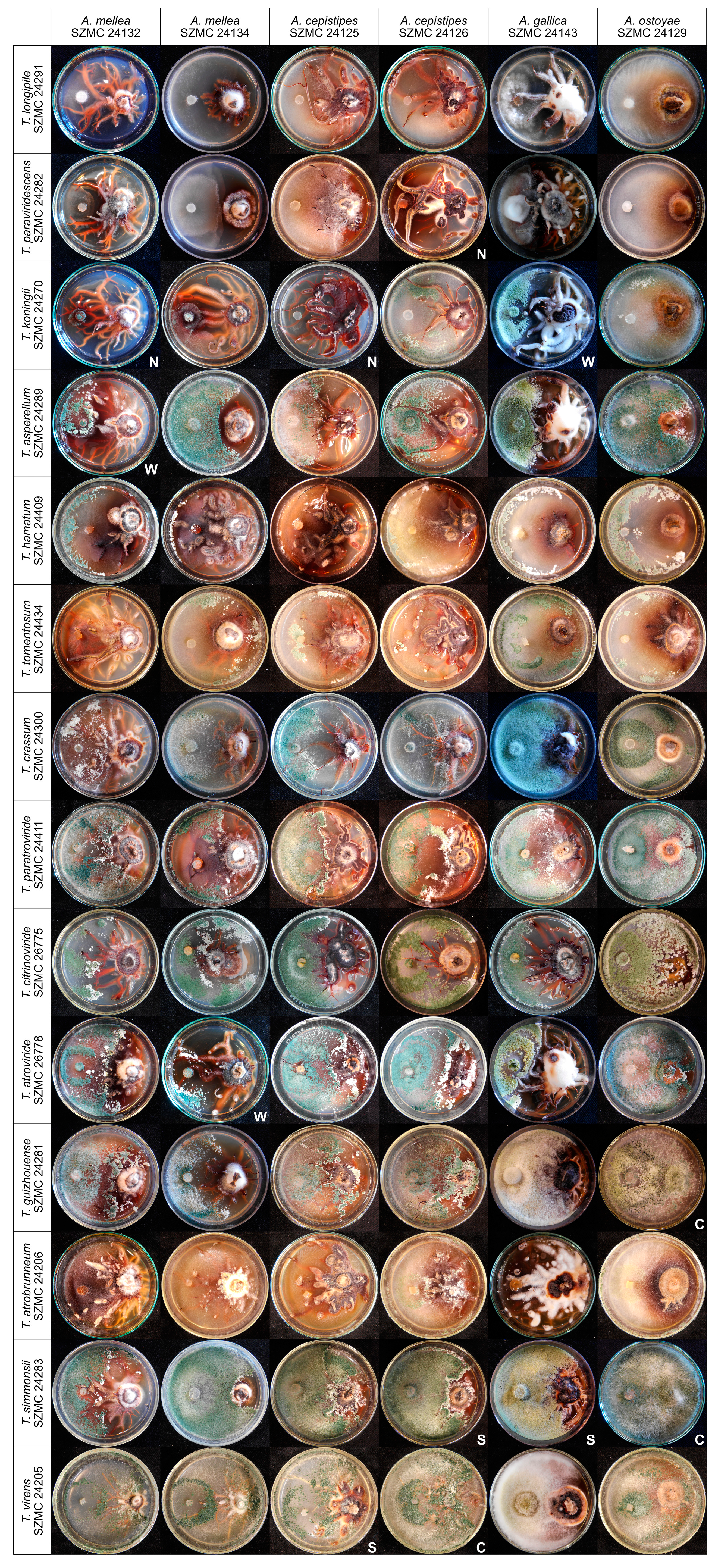
3.2. In Vitro Antagonism of the Isolated Trichoderma Strains Towards Armillaria Species

All 8 isolates of T. virens, and some isolates of T. simmonsii, T. atrobrunneum, T. guizhouense, T. atroviride, T. citrinoviride, T. paratroviride, T. hamatum, and T. tomentosum, proved to be highly effective against the 25 examined Armillaria isolates. Figure 1 shows a representative set of plate photographs taken during the in vitro antagonism experiments and reflecting all species combinations of Trichoderma and Armillaria. In many cases, antagonistic Trichoderma isolates were able to overgrow Armillaria colonies and intensely produce conidia on their surface, thereby potentially restricting Armillaria growth. Isolates of T. virens, such as SZMC 24205, SZMC 24294, SZMC 24303, SZMC 24293, and SZMC 26774, proved to be the best in vitro antagonists with BCI values above 80 for more than 17 out of 25 Armillaria strains (Table 2). Isolates of T. simmonsii showed high in vitro antagonistic abilities with BCI values above 80 for more than 15 out of the 25 tested Armillaria isolates, whereas the T. koningii, T. asperellum, T. paraviridescens, and T. longipile isolates had the lowest BCI values against almost all of the tested Armillaria isolates. The distribution of antagonistic Trichoderma species with higher BCI values showed a geographical pattern. Except for the two species—T. simmonsii and T. atroviride, isolated from both the oak stand in Keszthely—and the spruce forest in Rosalia, having relatively high antagonistic activities, species that were only isolated from the Keszthely Hills, including T. virens, T. atrobrunneum, T. hamatum, T. citrinoviride, T. paratroviride, and T. tomentosum, exhibited good in vitro antagonistic abilities against most of the tested Armillaria isolates. All isolates of T. koningii, T. asperellum, T. paraviridesens, and T. longipile, which seem to dominate in the soil of the Rosalia forest, showed lower BCI values against most Armillaria isolates.

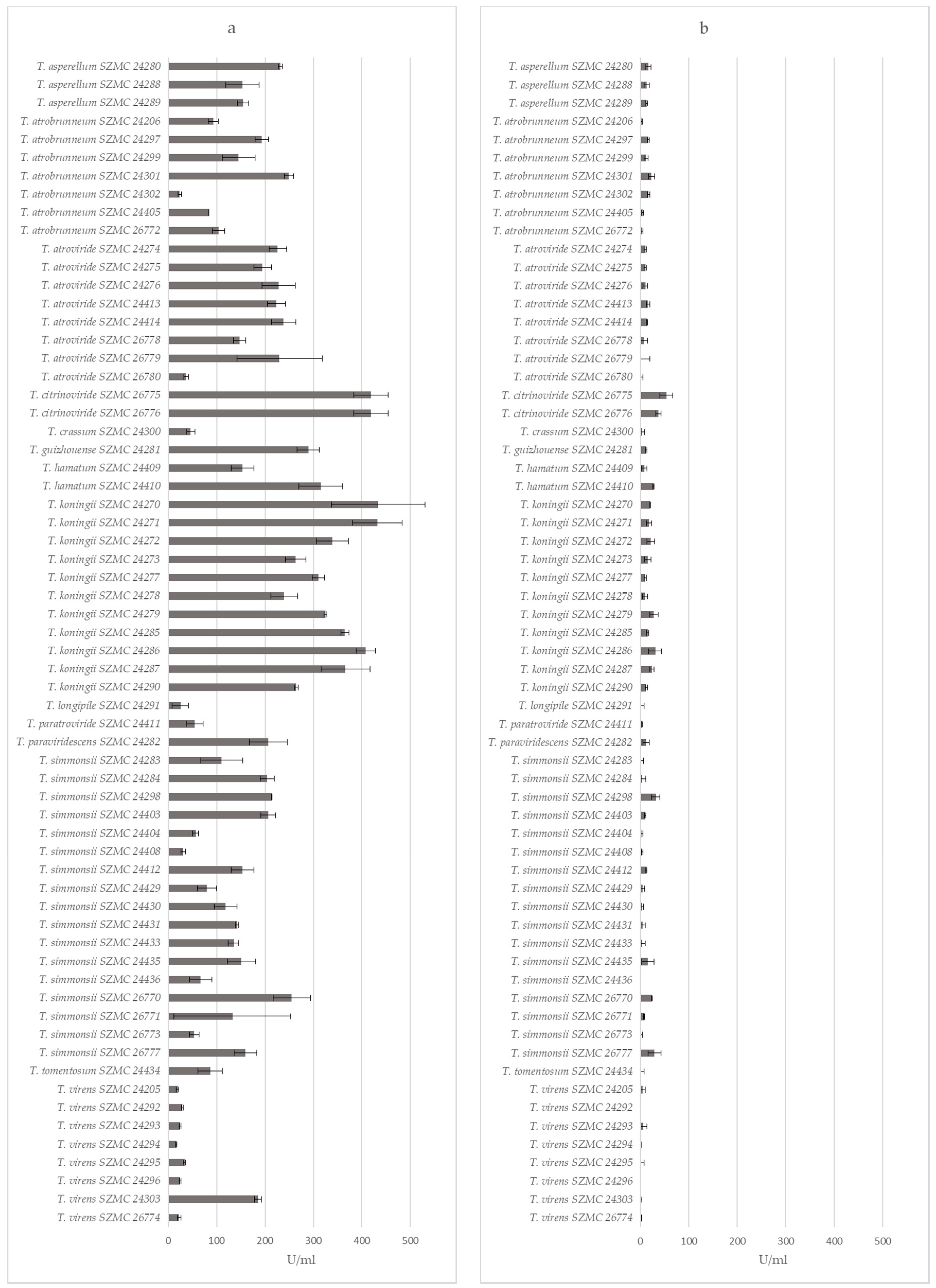
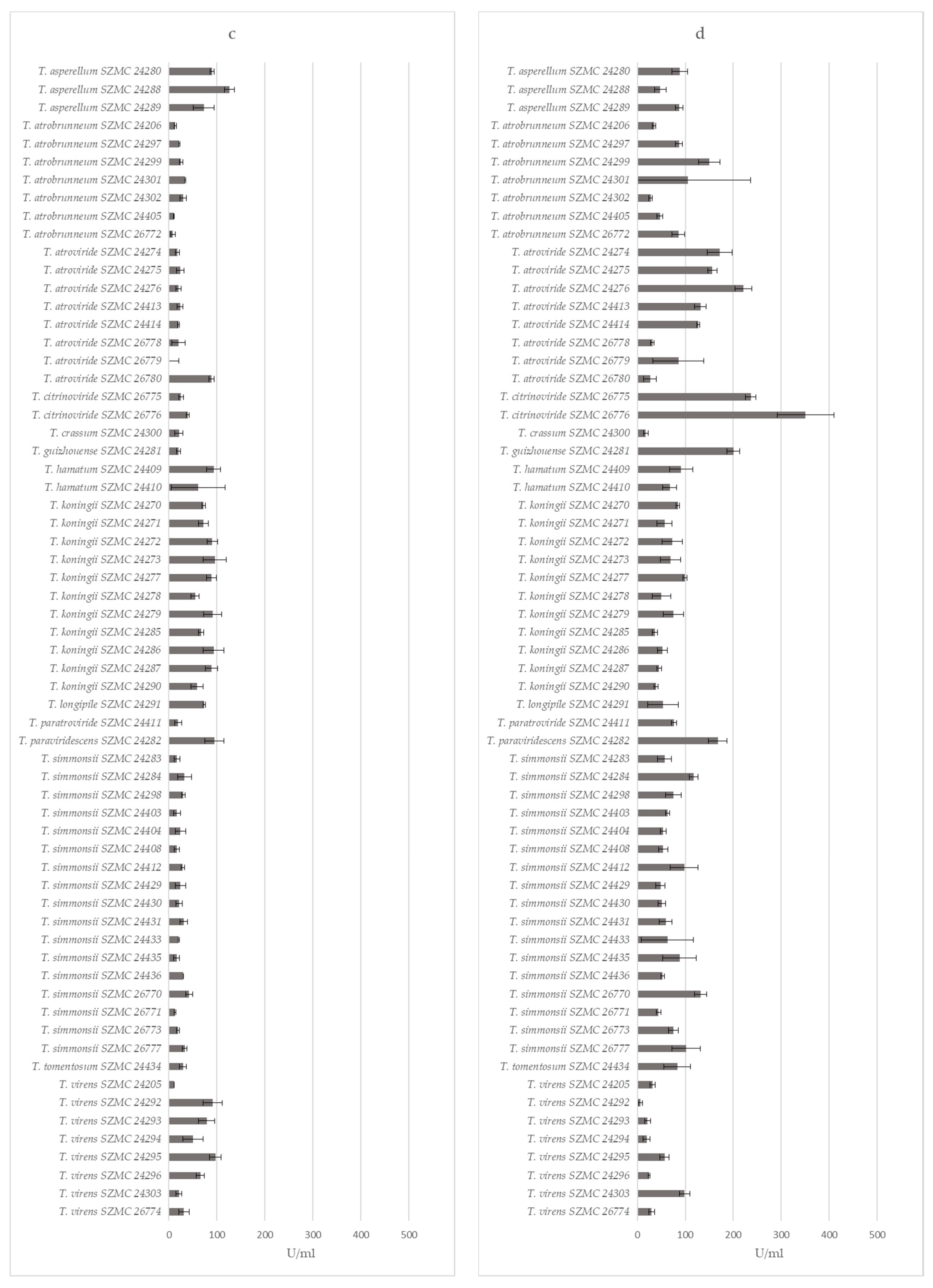
3.3. Extracellular Enzyme Production of the Trichoderma Isolates

The extracellular enzyme measurements revealed that isolates of the same Trichoderma species have similar enzyme activity values (Figure 2). Altogether, most of the isolates could be characterized by high β-glucosidase (Figure 2a) and phosphatase (Figure 2d), but lower cellobiohydrolase (Figure 2b) and β-xylosidase (Figure 2c) activities. The 11 T. koningii isolates, along with T. asperellum SZMC 24288, SZMC 24280, and T. paraviridescens SZMC 24282, showed good β-glucosidase and β-xylosidase activities. Among them, T. paraviridescens SZMC 24282 had good phosphatase, whereas T. koningii SZMC 24286 and SZMC 24270 good cellobiohydrolase activities as well. The examined T. virens, T. atrobrunneum, T. simmonsii, and T. atroviride isolates showed lower activity levels for all enzymes tested, except for T. atroviride SZMC 26780, which had a very high β-xylosidase activity.

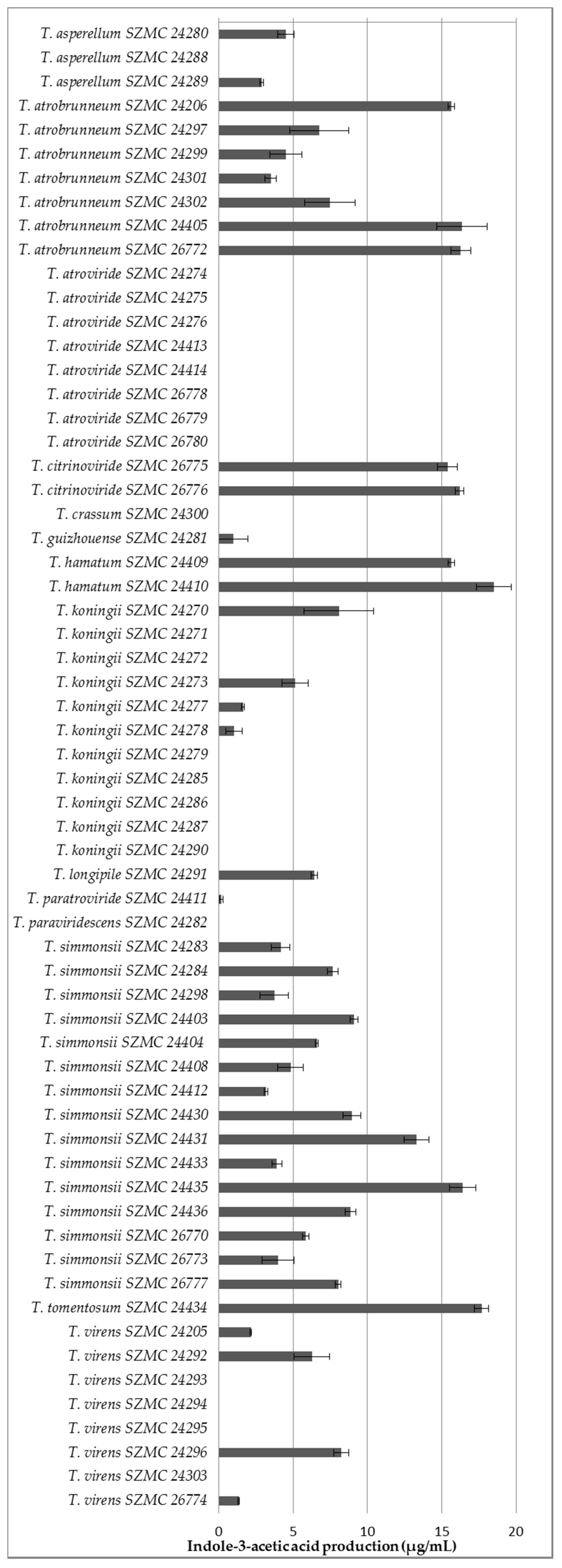
3.4. Potential Plant Growth-Promoting Traits of the Isolated Trichoderma Strains

All of the examined isolates from T. atrobrunneum, T. simmonsii, T. hamatum, and T. citrinoviride, along with the single isolates of T. tomentosum, T. longipile, T. paratroviride, and T. guizhouense, proved to be IAA producers, whereas the T. atroviride isolates, as well as the examined single isolates of T. paraviridescens and T. crissum, were unable to produce this metabolite (Figure 3). Both producers and non-producers were found among the examined isolates of T. virens, T. koningii, and T. asperellum. The highest amounts of IAA were detected in the case of the isolates of T. tomentosum, T. citrinoviride, T. hamatum, as well as certain isolates of T. atrobrunneum, T. simmonsii, and T. koningii.
All the Trichoderma isolates tested were able to produce siderophores, which was indicated by the change of the color of blue medium to orange or purple (Figure 4). The different colors of the medium suggested that the produced siderophores were structurally different. There are two major groups of siderophores, known as catechol-type and hydroxamate-type [25]. In the case of catechol-type siderophores the medium turns to purple, which was detected in the case of the T. atroviride, T. paraviridescens, and T. koningii isolates, whereas the hydroxamate-type siderophores result in an orange color, as it was the case for all other examined species (Figure 4). The isolates of the species T. asperellum seemed to produce both types of siderophores (see plate 8 on Figure 4).

3.5. Field Experiment in a Heavily Armillaria-Damaged Forest in the Keszthely Hills

Two Trichoderma isolates—T. virens SZMC 24205 and T. atrobrunneum SZMC 24206—were selected for a field experiment. Both strains were isolated from a Keszthely soil sample associated with decaying Armillaria rhizomorphs, which have not revealed any Armillaria growth upon isolation attempts; furthermore, both exerted very good in vitro antagonistic abilities towards the tested Armillaria isolates and were able to produce hydroxamate-type siderophores. The isolates were applied to Turkey oak seedlings as a root treatment before planting in the form of a conidial suspension (106 conidia per mL for both). The total survival rates calculated after 6 months for 120 treated and 115 control trees were 84.3% and 54.7%, respectively (Table 3), indicating that the applied treatment had a beneficial effect on the survival of oak seedlings planted into the soil of an Armillaria-infested forest area.

4. Discussion

Armillaria fruiting bodies, rhizomorphs, and soil samples were collected at previously established study sites from both spruce and oak stands; all of them with abundant rhizomorph and mushroom production. The conifer sampling sites selected in Rosalia represented a native environment for Norway spruce with single clones of A. ostoyae and A. cepistipes colonies appearing only around relatively freshly cut trunks. All identified genets appeared non-damaging and tolerable by the surrounding live trees. In contrast, the Turkey oak stand from Keszthely, Hungary was a heavily infested area with multiple A. mellea and A. gallica clones merged to form a continuous coverage of the whole stand. All remaining standing trees were showing symptoms of Armillaria infections. The same bulk, rhizospheric and rhizomorph-associated soil samples were also subjected to Trichoderma isolation. The reported diversity of Trichoderma had expanded to ~75 species in temperate Europe [27,28]. All the Trichoderma species collected in this study from Keszthely and Rosalia (T. tomentosum, T. paratroviride, T. crassum, T. hamatum, T. citrinoviride, T. atrobrunneum, T. virens, T. simmonsii, T. atroviride, T. koningii, T. asperellum, T. guizhouense, T. paraviridescens, and T. longipile) had already been reported from Southern Europe [29]. Among them, the species T. citrinoviride, T. atroviride, T. koningii, T. paraviridescens, and T. longipile were also identified from Central Europe [30]. The T. harzianum species complex (also known as T. harzianum sensu lato) from the Harzianum clade of the genus Trichoderma was supposed to comprise at least 14 species [31], including the more recently described, biocontrol-relevant species of T. atrobrunneum, T. guizhouense, and T. simmonsii that were also found at both locations of our current investigation.
The application of biocontrol agents as alternatives to chemical fungicides reduces the impacts and risks on human health as well as on the environment [32]. Trichoderma species as effective biocontrol agents against diverse genera of pathogenic fungi can be used for plant disease management, especially in the case of soilborne diseases. Strains of the T. koningii, T. asperellum, T. atroviride, T. hamatum, T. virens, and T. harzianum species complex and other Trichoderma taxa have been officially registered and commercialized as crop protection products and microbial fungicides throughout the world including the European countries [33].
Antagonistic activity assessment in vitro by dual culture assay has demonstrated in this study that, besides T. virens, T. atroviride, and T. hamatum and the members of the T. harzianum species complex (T. simmonsii, T. atrobrunneum, and T. guizhouense), strains of T. citrinoviride, T. paratroviride, and T. tomentosum also proved to be effective in vitro antagonists of Armillaria species with the potential to be used as biocontrol agents against Armillaria root rot. On the other hand, certain species and strains of Trichoderma showed weak antagonistic abilities against Armillaria strains reflected by low BCI values. For example, all the tested isolates of T. koningii and T. asperellum had lower BCI values than the isolates of T. virens, T. simmonsii, T. atrobrunneum, T. atroviride, and T. hamatum. Previously, T. koningii and T. asperellum showed excellent antagonistic activities during the application against other plant pathogens. For instance, T. koningii showed the highest growth inhibition of Rhizoctonia solani causing root rot in cotton, followed by T. viride, T. harzianum, and T. virens [34]. Similarly, T. asperellum showed effective antagonistic activity against the white-rot fungus Phellinus noxius, the causal agent of an epidemic brown root rot disease of various coniferous and broad-leaved tree species [35].
The Trichoderma isolates collected during this study were characterized for their abilities to produce polysaccharide-degrading enzymes of the cellulolytic (β-glucosidase and cellobiohydrolase) and xylanolytic (β-xylosidase) enzyme systems that are important for efficient competition in habitats rich of plant-derived polysaccharides, as well as acidic phosphatase playing a role in phosphorus mobilization. Interestingly, the isolates of species with the best in vitro antagonistic abilities against Armillaria (T. virens, T. atrobrunneum, T. simmonsii, and T. atroviride) were among the worst producers of these extracellular enzymes and vice versa, suggesting that the main antagonistic mechanism of these Trichoderma species against Armillaria may be mycoparasitism of hyphae and rhizomorphs rather than competition for polysaccharides or increasing phosphorous availability to the tree roots. Certain Trichoderma species (e.g., T. reesei) can be characterized with a predominantly saprophytic behavior, while others (e.g., T. virens and T. atroviride and members of the Harzianum clade) are described as successful mycoparasitic species [14]. Extracellular hydrolytic enzymes are known as key players of both the saprophytic and the mycoparasitic behavior: the former is relying on the production of plant polysaccharide-degrading enzyme systems like cellulases or xylanases, whereas the latter is based on CWDEs targeting the cell wall of the fungal host (glucanases, chitinases, and proteases).
The competition for iron may also contribute to the anti-Armillaria activity of the examined Trichoderma iolates, as the production of siderophores proved to be a general feature among them. Previous studies reported that certain strains of T. asperellum, T. atrobrunneum, T. atroviride, T. gamsii, T. hamatum, T. harzianum, T. polysporum, T. reesei, T. virens, T. paratroviride, T. pyramidale, T. rufobrunneum, T. thermophilum, T. viridulum, T. guizhouense, and T. simmonsii were mainly used as biocontrol agents due to their siderophore producing abilities [36,37,38,39,40]. Wang and Zhuang [40] firstly reported the siderophore-producing ability of T. guizhouense and T. simmonsii. To the best of our knowledge, the production of siderophores by T. citrinoviride, T. koningii, T. crassum, T. longipile, and T. paraviridescens strains is firstly demonstrated in the present study.
From the forest-derived Trichoderma isolates of our study, 40 were able to produce IAA with T. hamatum SZMC 24410, T. citrinoviride SZMC 26776, and T. atrobrunneum SZMC 24206 producing the highest quantities (18.49, 16.198, and 15.64 µg/mL, respectively). Data in the literature about the IAA-producing ability of Trichoderma strains is limited. Chagas et al. [41] investigated the IAA production of T. harzianum, T. pinnatum, T. longibrachiatum, and T. asperelloides, as well as two strains of T. virens, and recorded production values of 2.9–3.2 µg/mL. In the present study, the detected values were in a wider concentration range (1.349–8.248 µg/mL). A previous study used a similar method to show that strains of T. atrobrunneum, T. guizhouense, T. paratroviride, and T. simmonsii produce IAA at concentrations of 6.6, 10.3–21.8, 4.1–8.5, and 6.0–7.2 µg/mL [40]. In our study, the examined T. guizhouense and T. paratroviride isolates produced lower amounts of IAA. We also present the first data about the IAA production of T. koningii, T. longipile, T. tomentosum, T. hamatum, and T. citrinoviride.
A comparison of the data about the in vitro antagonism and the production of indole-3-acetic acid, siderophores as well as extracellular β-glucosidase, cellobiohydrolase, β-xylosidase, and phosphatase enzymes among the Trichoderma isolates mostly revealed very similar values for isolates deriving from the same sample and belonging to the same species, suggesting that the respective isolates are clonal and represent the same strain, which is, in many cases, also supported by identical sequences of the tef1α fragment used for species-level identification (Table 1). Examples for probable clonality are the isolate groups T. koningii SZMC 24277/24278/24279, T. atroviride SZMC 24274/24275/24276 and SZMC 24413/24414, T. simmonsii SZMC 24435/26771/24436/24403/24404 and SZMC 26777/24412, or T. atrobrunneum SZMC 26772/24405. On the other hand, in certain cases, the differences in the physiological parameters or tef1α sequences clearly revealed the presence of multiple strains from the same species in the same sample, e.g., T. koningii SZMC 2470/2471 vs. T. koningii SZMC 2472/2473, T. simmonsii SZMC 26770 vs. SZMC 24429/24430/24431/24433, T. hamatum SZMC 24409 vs. SZMC 24410, or T. simmonsii SZMC 26773 vs. SZMC 24408—the difference of the latter two isolates is also supported by a series of single nucleotide polymorphisms in the analyzed tef1α gene fragment (Table 1).
Only limited information is available in the literature about field studies evaluating the applicability of Trichoderma strains against Armillaria root rot of trees. Otieno et al. [12] screened Trichoderma isolates for antagonism to Armillaria in tea stem sections buried in the soil and selected a T. harzianum strain, the wheat bran culture of which significantly reduced the viability of Armillaria in woody blocks of inoculum. The selected strain also exhibited high efficiency in the biocontrol of the destructive tree and bush pathogens from the genus Armillaria [13]. Schnabel et al. [42] applied biannual drenches of T. asperellum and T. gamsii, formulated as Remedier WP, onto peach trees planted in spots where a tree had declined from Armillaria root rot during the previous season, but did not find any statistical significance in survival between the treated and control trees. However, the surviving Remedier WP-treated trees were found to have significantly larger tree trunks compared to control trees three and four years after planting at one of the two replant sites involved in the study. In another study, spraying a combination of T. harzianum and T. koningii at concentrations of 2 × 10⁷ CFU/mL and 3 × 10⁷ CFU/mL, respectively, into holes made in an avocado orchard previously infested with A. mellea did not increase the survival rates of grafted peach (Prunus persica) saplings [43]. The lack of Trichoderma effect on the survival of peach trees in the above studies may partly be due to the Trichoderma species applied: our study demonstrated that isolates of T. asperellum and T. koningii are not among the good in vitro antagonists of Armillaria species. Furthermore, the success of a tested control strategy may also rely on the thorough selection of the Trichoderma strains, which should also consider the origin of the isolates. The two strains involved in the field test of our study were derived from a soil sample associated with Armillaria rhizomorphs which have not revealed any Armillaria growth, suggesting that the isolation of Trichoderma strains from naturally decaying Armillaria rhizomorphs and the soil surrounding them may increase the chances to find promising candidates for the successful biocontrol of Armillaria root rot. A similar strain isolation strategy from Armillaria rhizomorphs and soil samples around Armillaria-infected roots of cherry and almond trees revealed isolates of T. virens and T. harzianum sensu lato efficiently inhibiting both colony growth and rhizomorph formation of A. mellea [44].

5. Conclusions

One of the possible reasons behind the so-far limited success of Armillaria biocontrol by the application of Trichoderma strains in forests may lie in the selection strategy of the biocontrol strains. The results of our study suggest that certain Trichoderma species are known as successful biocontrol agents and components of commercially formulated products, e.g., T. asperellum or T. koningii, may not be the agents of choice for this purpose, as many isolates from these species were shown to lack good in vitro antagonistic abilities against Armillaria isolates. On the other hand, this study demonstrates that decaying rhizomorphs are potential sources of antagonistic Trichoderma strains with the potential to control Armillaria species and increase the survival rate of seedlings planted in the Armillaria-infested forest areas.
Another limitation of Trichoderma application in forest stands is arising from the difficulties of delivery, as the regular treatment of large forest areas with a biocontrol product is not economically feasible. An obvious time point of intervention is the planting time of the seedlings, as their roots can be easily treated with microbial products by soaking. A further promising strategy could be the conditioning of the seedlings with microbial products before planting, which could be performed in nurseries under more controlled circumstances than the ones allowed by field conditions.
A further crucial point is the exact, species-level molecular identification of the candidate Trichoderma strains for Armillaria biocontrol, which should be performed by the sequence analysis of a fragment of the tef1α gene fragment, as, according to our recent knowledge about the taxonomy of the genus Trichoderma, ITS sequence analysis is not allowing an exact diagnosis in many cases.
The interactions of introduced Trichoderma strains with other beneficial microorganisms such as mycorrhizal fungi need further investigations, as they may represent both advantages and disadvantages to the host plant [45]. Trichoderma may act negatively on mycorrhizal fungi via competition for the colonization sites and nutrients [46], or via direct mycoparasitic attack, which, however, may also increase the uptake of phosphorous by the mycorrhizal fungus as the result of stress reaction [47]. Using beneficial fungi in forestry therefore requires the adjustment of Trichoderma–mycorrhizal fungus combinations to the host tree, as well as the optimization of the inoculation methods and the applied sylvicultural practices.

Author Contributions

Conceptualization, L.C., L.K., C.V., and G.S.; sample collection: G.S., R.R., B.I., S.C., and T.H.; strain isolation and identification: L.C., B.B., T.H., and L.K.; in vitro antagonism experiments: L.C., B.B., and C.V.; extracellular enzyme activity measurements: V.D.N.; indole-3-acetic acid and siderophore tests: O.K.; field experiment—design: Z.P. and G.S.; field experiment—realization: Z.P., G.S., L.K., O.K., and B.B.; data curation: A.S., S.C., B.I., and M.M.; writing—original draft preparation: L.C., B.B., O.K., V.D.N., T.H., and L.K.; writing—review and editing: L.K., G.S., and C.V.; funding acquisition: G.S and C.V.

Funding

This research was funded by the Hungarian Government and the European Union within the frames of the Széchenyi 2020 Programme (GINOP-2.3.2-15-2016-00052). LK is a grantee of the János Bolyai (Hungarian Academy of Sciences) and Bolyai Plus (New National Excellence Programme, Hungary) research scholarships. M.M. is granted from the “EFOP-3.6.1-16-2016- 560 00018 - Improving the role of research + development + innovation in the higher education through institutional developments assisting intelligent specialization in Sopron and Szombathely.

Acknowledgments

The authors are grateful for the help of Eszter Tóth, Csilla Szebenyi, Mónika Vörös, Ottó Bencsik (University of Szeged), and Jenő Fliszár (Bakonyerdő Ltd.) in the realization of the field experiment, the expert suggestions of László Szalay (University of Sopron) regarding the statistical evaluation of the results deriving from the field experiment, and Sándor Kocsubé for the preparation of the final layout of Figure 1. L.C. thanks for the grant from the Chinese CSC Scholarship Program.

Conflicts of Interest

The authors declare no conflicts of interest.

References

  1. Baumgartner, K.; Coetzee, M.P.; Hoffmeister, D. Secrets of the subterranean pathosystem of Armillaria. Mol. Plant Pathol. 2011, 12, 515–534. [Google Scholar] [CrossRef] [PubMed]
  2. Heinzelmann, R.; Dutech, C.; Tsykun, T.; Labbé, F.; Soularue, J.-P.; Prospero, S. Latest advances and future perspectives in Armillaria research. Can. J. Plant Pathol. 2019, 41, 1–23. [Google Scholar] [CrossRef]
  3. Sipos, G.; Anderson, J.B.; Nagy, L.G. Armillaria. Curr. Biol. 2018, 28, R297–R298. [Google Scholar] [CrossRef] [PubMed]
  4. Sipos, G.; Prasanna, A.N.; Walter, M.C.; O’Connor, E.; Bálint, B.; Krizsán, K.; Kiss, B.; Hess, J.; Varga, T.; Slot, J.; et al. Genome expansion and lineage-specific genetic innovations in the forest pathogenic fungi Armillaria. Nat. Ecol. Evol. 2017, 12, 1931–1941. [Google Scholar] [CrossRef] [PubMed]
  5. Ross-Davis, A.L.; Stewart, J.E.; Hanna, J.W.; Kim, M.S.; Knaus, B.J.; Cronn, R.; Rai, H.; Richardson, B.A.; Mcdonald, G.I.; Klopfenstein, N.B. Transcriptome of an Armillaria root disease pathogen reveals candidate genes involved in host substrate utilization at the host–pathogen interface. For. Pathol. 2013, 43, 468–477. [Google Scholar] [CrossRef]
  6. Morrison, D. Epidemiology of Armillaria root disease in Douglas-fir plantations in the cedar-hemlock zone of the southern interior of British Columbia. For. Pathol. 2009, 41, 31–40. [Google Scholar] [CrossRef]
  7. Kromroy, K.; Blanchette, R.A.; Grigal, D. Armillaria species on small woody plants, small woody debris, and root fragments in red pine stands. Can. J. For. Res. 2005, 35, 1487–1495. [Google Scholar] [CrossRef]
  8. Lee, C.; Dey, D.; Muzika, R.-M. Oak stump-sprout vigor and Armillaria infection after clearcutting in southeastern Missouri, USA. For. Ecol. Manag. 2016, 374, 211–219. [Google Scholar] [CrossRef]
  9. Cook, R.J. Making greater use of introduced microorganisms for biological control of plant pathogens. Annu. Rev. Phytopathol. 1993, 31, 53–80. [Google Scholar] [CrossRef]
  10. Nelson, E.E.; Pearce, M.H.; Malajczuk, N. Effects of Trichoderma spp. and ammonium sulphamate on establishment of Armillaria luteobubalina on stumps of Eucalyptus diversicolor. Mycol. Res. 1995, 99, 957–962. [Google Scholar] [CrossRef]
  11. Otieno, W.; Jeger, M.; Termorshuizen, A. Effect of infesting soil with Trichoderma harzianum and amendment with coffee pulp on survival of Armillaria. Biol. Control. 2003, 26, 293–301. [Google Scholar] [CrossRef]
  12. Otieno, W.; Termorshuizen, A.; Jeger, M.; Othieno, C. Efficacy of soil solarization, Trichoderma harzianum, and coffee pulp amendment against Armillaria sp. Crop. Prot. 2003, 22, 325–331. [Google Scholar] [CrossRef]
  13. Percival, G.; Smiley, E.; Fox, R. Root collar excavation with Trichoderma inoculations as a potential management strategy for honey fungus (Armillaria mellea). Arboric. J. 2011, 33, 267–280. [Google Scholar] [CrossRef]
  14. Olmedo Monfil, V.; Casas-Flores, S. Molecular mechanisms of biocontrol in Trichoderma spp. and their applications in agriculture. In Biotechnology and Biology of Trichoderma; Gupta, V.K., Schmoll, M., Herrera-Estrella, A., Upadhyay, R.S., Druzhinina, I., Tuohy, M.G., Eds.; Elsevier: Oxford, UK, 2014; pp. 429–453. [Google Scholar] [CrossRef]
  15. Sharma, V.; Salwan, R.; Sharma, P. Differential response of extracellular proteases of Trichoderma harzianum against fungal phytopathogens. Curr. Microbiol. 2016, 73, 419–425. [Google Scholar] [CrossRef] [PubMed]
  16. Contreras-Cornejo, H.A.; Macías-Rodríguez, L.; Alfaro-Cuevas, R.; López-Bucio, J. Trichoderma spp. improve growth of Arabidopsis seedlings under salt stress through enhanced root development, osmolite production, and Na+ elimination through root exudates. Mol. Plant Microbe Interact. 2014, 27, 503–514. [Google Scholar] [CrossRef] [PubMed]
  17. Shaw, C.G.; Roth, L.F. Persistence and distribution of a clone of Armillaria mellea in a ponderosa pine forest. Phytopathology 1976, 66, 1210–1213. [Google Scholar] [CrossRef]
  18. King, A.D.; Hocking, A.D.; Pitt, J.I. Dichloran-rose bengal medium for enumeration and isolation of molds from foods. Appl. Environ. Microbiol. 1979, 37, 959–964. [Google Scholar]
  19. White, T.J.; Bruns, T.; Lee, S.; Taylor, J.; Innis, M.; Gelfand, D.; Sninsky, J. Amplification and direct sequencing of fungal ribosomal RNA genes for phylogenetics. In PCR Protocols: A Guide to Methods and Applications; Innis, M.A., Gelfand, D.H., Sninsky, J.J., White, T.J., Eds.; Academic Press Inc.: San Diego, CA, USA, 1990; pp. 315–322. [Google Scholar]
  20. Oskiera, M.; Szczech, M.; Bartoszewski, G. Molecular identification of Trichoderma strains collected to develop plant growth-promoting and biocontrol agents. J. Hortic. Res. 2015, 23, 75–86. [Google Scholar] [CrossRef]
  21. Druzhinina, I.; Kopchinskiy, A.; Komoń, M.; Szakacs, G.; Kubicek, C. An oligonucleotide barcode for species identification in Trichoderma and Hypocrea. Fungal Genet. Biol. 2005, 42, 813–828. [Google Scholar] [CrossRef]
  22. Kopchinskiy, A.; Komoń, M.; Kubicek, C.P.; Druzhinina, I.S. TrichoBLAST: A multilocus database for Trichoderma and Hypocrea identifications. Mycol. Res. 2005, 109, 658–660. [Google Scholar] [CrossRef]
  23. Szekeres, A.; Leitgeb, B.; Kredics, L.; Manczinger, L.; Vágvölgyi, C. A novel, image analysis-based method for the evaluation of in vitro antagonism. J. Microbiol. Meth. 2006, 65, 619–622. [Google Scholar] [CrossRef] [PubMed]
  24. Gordon, S.A.; Weber, R.P. Colorimetric estimation of indoleacetic acid. Plant Physiol. 1951, 26, 192–195. [Google Scholar] [CrossRef] [PubMed]
  25. Milagres, A.M.; Machuca, A.; Napoleao, D. Detection of siderophore production from several fungi and bacteria by a modification of chrome azurol S (CAS) agar plate assay. J. Microbiol. Meth. 1999, 37, 1–6. [Google Scholar] [CrossRef]
  26. Schwyn, B.; Neilands, J.B. Universal chemical assay for the detection and determination of siderophores. Anal. Biochem. 1987, 160, 47–56. [Google Scholar] [CrossRef]
  27. Jaklitsch, W. European species of Hypocrea part II: Species with hyaline ascospores. Fungal Divers. 2011, 48, 1–250. [Google Scholar] [CrossRef]
  28. Jaklitsch, W.M. European species of Hypocrea Part I. The green-spored species. Stud. Mycol. 2009, 63, 1–91. [Google Scholar] [CrossRef]
  29. Jaklitsch, W.M.; Voglmayr, H. Biodiversity of Trichoderma (Hypocreaceae) in Southern Europe and Macaronesia. Stud. Mycol. 2015, 80, 1–87. [Google Scholar] [CrossRef]
  30. Błaszczyk, L.; Strakowska, J.; Chełkowski, J.; Gąbka-Buszek, A.; Kaczmarek, J. Trichoderma species occurring on wood with decay symptoms in mountain forests in Central Europe: Genetic and enzymatic characterization. J. Appl. Genet. 2016, 57, 397–407. [Google Scholar] [CrossRef]
  31. Chaverri, P.; Branco-Rocha, F.; Jaklitsch, W.; Gazis, R.; Degenkolb, T.; Samuels, G.J. Systematics of the Trichoderma harzianum species complex and the re-identification of commercial biocontrol strains. Mycologia 2015, 107, 558–590. [Google Scholar] [CrossRef]
  32. Newitt, J.T.; Prudence, S.M.M. Biocontrol of cereal crop diseases using Streptomycetes. Pathogens 2019, 8, 78. [Google Scholar] [CrossRef]
  33. Woo, S.; Ruocco, M.; Vinale, F.; Nigro, M.; Marra, R.; Lombardi, N.; Pascale, A.; Lanzuise, S.; Manganiello, G.; Lorito, M. Trichoderma-based products and their widespread use in agriculture. Open Mycol. J. 2014, 8, 71–126. [Google Scholar] [CrossRef] [Green Version]
  34. Gajera, H.P.; Hirpara, D.G.; Katakpara, Z.A.; Patel, S.V.; Golakiya, B.A. Molecular evolution and phylogenetic analysis of biocontrol genes acquired from SCoT polymorphism of mycoparasitic Trichoderma koningii inhibiting phytopathogen Rhizoctonia solani Kuhn. Infect. Genet. Evol. 2016, 45, 383–392. [Google Scholar] [CrossRef] [PubMed]
  35. Chou, H.; Xiao, Y.T.; Tsai, J.N.; Li, T.T.; Wu, H.Y.; Liu, L.D.; Tzeng, D.S.; Chung, C.L. In vitro and in planta evaluation of Trichoderma asperellum TA as a biocontrol agent against Phellinus noxius, the cause of brown root rot disease of trees. Plant Dis. 2019, 103, 2733–2741. [Google Scholar] [CrossRef] [PubMed]
  36. Mukherjee, P.K.; Hurley, J.F.; Taylor, J.T.; Puckhaber, L.; Lehner, S.; Druzhinina, I.; Schumacher, R.; Kenerley, C.M. Ferricrocin, the intracellular siderophore of Trichoderma virens, is involved in growth, conidiation, gliotoxin biosynthesis and induction of systemic resistance in maize. Biochem. Biophys. Res. Commun. 2018, 505, 606–611. [Google Scholar] [CrossRef] [PubMed]
  37. Lehner, S.M.; Atanasova, L.; Neumann, N.K.; Krska, R.; Lemmens, M.; Druzhinina, I.S.; Schuhmacher, R. Isotope-assisted screening for iron-containing metabolites reveals a high degree of diversity among known and unknown siderophores produced by Trichoderma spp. Appl. Environ. Microbiol. 2013, 79, 18–31. [Google Scholar] [CrossRef] [Green Version]
  38. Segarra, G.; Casanova, E.; Avilés, M.; Trillas, I. Trichoderma asperellum strain T34 controls Fusarium wilt disease in tomato plants in soilless culture through competition for iron. Microb. Ecol. 2010, 59, 141–149. [Google Scholar] [CrossRef] [PubMed]
  39. Anke, H.; Kinn, J.; Bergquist, K.-E.; Sterner, O. Production of siderophores by strains of the genus Trichoderma. Biol. Met. 1991, 4, 176–180. [Google Scholar] [CrossRef]
  40. Wang, C.; Zhuang, W.-Y. Evaluating effective Trichoderma isolates for biocontrol of Rhizoctonia solani causing root rot of Vigna unguiculata. J. Integr. Agric. 2019, 18, 2072–2079. [Google Scholar] [CrossRef]
  41. Chagas, L.F.B.; De Castro, H.G.; Colonia, B.S.O.; De Carvalho Filho, M.R.; De Oliveira Miller, L.; Chagas, A.F.J. Efficiency of Trichoderma spp. as a growth promoter of cowpea (Vigna unguiculata) and analysis of phosphate solubilization and indole acetic acid synthesis. Braz. J. Bot. 2016, 39, 437–445. [Google Scholar] [CrossRef]
  42. Schnabel, G.; Rollins, A.P.; Henderson, G.W. Field evaluation of Trichoderma spp. for control of Armillaria root rot of peach. Plant Health Prog. 2011, 10, 3. [Google Scholar] [CrossRef] [Green Version]
  43. Downer, J.; Faber, B. Non-chemical control of Armillaria mellea infection of Prunus persica. J. Plant Sci. Phytopathol. 2019, 3, 050–055. [Google Scholar] [CrossRef] [Green Version]
  44. Reza Asef, M.; Goltapeh, E.M.; Danesh, Y.R. Antagonistic effects of Trichoderma species in biocontrol of Armillaria mellea in fruit trees in Iran. J. Plant Prot. Res. 2008, 48, 213–222. [Google Scholar] [CrossRef]
  45. Szczałba, M.; Kopta, T.; Gąstoł, M.; Sękara, A. Comprehensive insight into arbuscular mycorrhizal fungi, Trichoderma spp. and plant multilevel interactions with emphasis on biostimulation of horticultural crops. J. Appl. Microbiol. 2019, 127, 630–647. [Google Scholar] [CrossRef] [PubMed]
  46. Martínez-Medina, A.; Roldán, A.; Pascual, J.A. Performance of a Trichoderma harzianum bentonite-vermiculite formulation against Fusarium wilt in seedling nursery melon plants. Hortic. Sci. 2009, 44, 2025–2027. [Google Scholar] [CrossRef] [Green Version]
  47. De Jaeger, N.; de la Providencia, I.; de Boulois, D.H.; Declerck, S. Trichoderma harzianum might impact phosphorus transport by arbuscular mycorrhizal fungi. FEMS Microbiol. Ecol. 2011, 77, 558–567. [Google Scholar] [CrossRef] [PubMed] [Green Version]
Figure 1. In vitro antagonism of Trichoderma strains from different species against Armillaria mellea, A. cepistipes, A. gallica, and A. ostoyae. Example plates are marked with N: no inhibition; W: weak inhibition; S: strong inhibition; C: complete overgrowth of Armillaria by Trichoderma.
Forests 10 01013 g001
Figure 2. Extracellular enzyme activities of Trichoderma isolates derived from forest soil samples: (a) β-glucosidase, (b) cellobiohydrolase, (c) β-xylosidase, and (d) phosphatase.
Forests 10 01013 g002aForests 10 01013 g002b
Figure 3. Indole-3-acetic acid production of Trichoderma isolates derived from forest soil samples.
Forests 10 01013 g003
Figure 4. Production of siderophores on modified CAS agar medium by forest-derived Trichoderma isolates belonging to different species. (1) T. atroviride SZMC 24275, (2) T. virens SZMC 24205, (3) T. hamatum SZMC 24410, (4) T. paraviridescens SZMC 24282, (5) T. koningii SZMC 24287, (6) T. citrinoviride SZMC 26776, (7) T. simmonsii SZMC 24431, (8) T. asperellum SZMC 24280, (9) T. atrobrunneum SZMC 24206, (10) T. guizhouense SZMC 24281, (11) T. tomentosum SZMC 24434, and (12) T. crassum SZMC 24300.
Forests 10 01013 g004
Table 1. Armillaria and Trichoderma isolates collected during the study.
LocationGPS-NGPS-ECollection DateSampleIsolate IdentifierDiagnosisITS (GenBank)tef1α (GenBank)
Armillaria Isolates
Rosalia, Austria47° 41.64916° 17.94028.10.2016fruiting bodySZMC 24125Armillaria cepistipes-MN580140
47° 41.64016° 17.93728.10.2016fruiting bodySZMC 24126Armillaria cepistipes-MN580151
47° 41.62816° 17.92928.10.2016rhizomorphSZMC 24128Armillaria ostoyae-MN580144
47° 41.62916° 17.96428.10.2016fruiting bodySZMC 24129Armillaria ostoyae-MN580139
47° 41.62116° 17.94828.10.2016fruiting bodySZMC 24130Armillaria ostoyae-MN580142
Keszthely, Hungary46° 48.72817° 16.99220.07.2016bulk soilSZMC 24095Armillaria gallica-MN580162
46° 48.71217° 16.99420.07.2016bulk soilSZMC 24098Armillaria gallica-MN580163
46° 48.70217° 16.98729.10.2016bulk soilSZMC 24099Armillaria gallica-MN580160
46° 48.65717° 16.95429.10.2016fruiting bodySZMC 24131Armillaria melleaMN585779MN580137
46° 48.67117° 16.95929.10.2016fruiting bodySZMC 24132Armillaria melleaMN585780MN580159
46° 48.70617° 16.94929.10.2016fruiting bodySZMC 24133Armillaria melleaMN585781MN580138
46° 48.72317° 16.97429.10.2016fruiting bodySZMC 24134Armillaria melleaMN585777MN580152
46° 48.71217° 16.97829.10.2016fruiting bodySZMC 24135Armillaria mellea-MN580145
46° 48.73617° 16.99229.10.2016fruiting bodySZMC 24651Armillaria mellea-MN580153
46° 48.77217° 16.99229.10.2016fruiting bodySZMC 24136Armillaria melleaMN585778MN580155
46° 48.76017° 16.98229.10.2016fruiting bodySZMC 24137Armillaria melleaMN585776MN580154
46° 48.74917° 16.93629.10.2016fruiting bodySZMC 24139Armillaria melleaMN585782MN580158
46° 48.72017° 17.00929.10.2016fruiting bodySZMC 24140Armillaria melleaMN585783MN580146
46° 48.73617° 16.99203.11.2016fruiting bodySZMC 24141Armillaria mellea-MN580150
46° 48.66517° 16.99303.11.2016fruiting bodySZMC 24142Armillaria mellea-MN580143
46° 48.73817° 16.95603.11.2016fruiting bodySZMC 24143Armillaria gallica-MN580141
46° 48.87717° 17.14303.11.2016fruiting bodySZMC 24144Armillaria mellea-MN580148
46° 48.88317° 17.15303.11.2016fruiting bodySZMC 24145Armillaria mellea-MN580149
46° 48.91717° 17.06203.11.2016fruiting bodySZMC 24146Armillaria mellea-MN580161
46° 48.89217° 16.94103.11.2016fruiting bodySZMC 24147Armillaria mellea-MN580157
46° 48.88317° 16.93703.11.2016fruiting bodySZMC 24148Armillaria mellea-MN580156
46° 47.93517° 16.95803.11.2016fruiting bodySZMC 24149Armillaria mellea-MN580147
Trichoderma Isolates
Rosalia, Austria47° 41.64916° 17.94028.10.2016bulk soilSZMC 24270Trichoderma koningiiMN516459MN520036
SZMC 24271Trichoderma koningiiMN516460MN520038
SZMC 24272Trichoderma koningiiMN516461MN520042
SZMC 24273Trichoderma koningiiMN516462MN520041
47° 41.64016° 17.93728.10.2016bulk soilSZMC 24274Trichoderma atrovirideMN516463MN520048
SZMC 24275Trichoderma atrovirideMN516464MN520049
SZMC 24276Trichoderma atrovirideMN516465MN520050
47° 41.61816° 17.87328.10.2016bulk soilSZMC 24277Trichoderma koningiiMN516466MN520033
SZMC 24278Trichoderma koningiiMN516467MN520040
SZMC 24279Trichoderma koningiiMN516468MN520039
SZMC 24280Trichoderma asperellumMN516469MN520031
47° 41.62916° 17.96428.10.2016bulk soilSZMC 24285Trichoderma koningiiMN516474MN520034
SZMC 24286Trichoderma koningiiMN516475MN520037
SZMC 24287Trichoderma koningiiMN516476MN520035
SZMC 24288Trichoderma asperellumMN516477MN520032
47° 41.62116° 17.94828.10.2016bulk soilSZMC 24289Trichoderma asperellumMN516478MN520030
SZMC 24290Trichoderma koningiiMN516479MN520043
47° 40.89616° 17.21128.10.2016bulk soilSZMC 24291Trichoderma longipileMN516480MN520056
47° 41.62816° 17.92928.10.2016rhizomorphSZMC 24281Trichoderma guizhouenseMN516470MN520084
SZMC 24282Trichoderma paraviridescensMN516471MN520044
SZMC 24283Trichoderma simmonsiiMN516472MN520079
SZMC 24284Trichoderma simmonsiiMN516473MN520078
Keszthely, Hungary46° 48.65717° 16.95429.10.2016bulk soilSZMC 24429Trichoderma simmonsii-MN520069
SZMC 24430Trichoderma simmonsii-MN520071
SZMC 24431Trichoderma simmonsii-MN520077
SZMC 26770Trichoderma simmonsii-MN520072
SZMC 24433Trichoderma simmonsii-MN520081
46° 48.67117° 16.95929.10.2016bulk soilSZMC 24434Trichoderma tomentosum-MN520066
46° 48.70617° 16.94929.10.2016bulk soilSZMC 24435Trichoderma simmonsii-MN520075
SZMC 26771Trichoderma simmonsii-MN520080
SZMC 24436Trichoderma simmonsii-MN520068
SZMC 24403Trichoderma simmonsii-MN520076
SZMC 24404Trichoderma simmonsii-MN520082
46° 48.72317° 16.97429.10.2016bulk soilSZMC 26772Trichoderma atrobrunneum-MN520090
SZMC 24405Trichoderma atrobrunneum-MN520091
46° 48.71217° 16.97829.10.2016bulk soilSZMC 26773Trichoderma simmonsii-MN520083
SZMC 26774Trichoderma virens-MN520058
SZMC 24408Trichoderma simmonsii-MN520067
46° 48.73617° 16.99229.10.2016bulk soilSZMC 24409Trichoderma hamatum-MN520028
SZMC 24410Trichoderma hamatum-MN520029
46° 48.77217° 16.99229.10.2016rhizosphereSZMC 26778Trichoderma atrovirideMN516444MN520052
SZMC 26779Trichoderma atrovirideMN516445MN520051
SZMC 26780Trichoderma atrovirideMN516446MN520053
46° 48.76917° 16.96129.10.2016bulk soilSZMC 24411Trichoderma paratroviride-MN520045
SZMC 26775Trichoderma citrinoviride-MN520054
SZMC 26776Trichoderma citrinoviride-MN520055
SZMC 26777Trichoderma simmonsii-MN520070
SZMC 24412Trichoderma simmonsii-MN520074
46° 48.74917° 16.93629.10.2016rhizosphereSZMC 24413Trichoderma atroviride MN520047
SZMC 24414Trichoderma atroviride MN520046
46° 48.73817° 16.95620.07.2016rhizomorphSZMC 24292Trichoderma virensMN516447MN520059
SZMC 24293Trichoderma virensMN516448MN520061
SZMC 24294Trichoderma virensMN516449MN520062
SZMC 24295Trichoderma virensMN516450MN520060
SZMC 24296Trichoderma virensMN516451MN520064
46° 48.75817° 16.95920.07.2016rhizomorphSZMC 24297Trichoderma atrobrunneumMN516452MN520087
SZMC 24298Trichoderma simmonsiiMN516453MN520073
SZMC 24299Trichoderma atrobrunneumMN516454MN520086
SZMC 24300Trichoderma crassumMN516455MN520057
SZMC 24301Trichoderma atrobrunneumMN516456MN520088
SZMC 24302Trichoderma atrobrunneumMN516457MN520089
SZMC 24303Trichoderma virensMN516458MN520063
46° 48.72217° 16.99320.07.2016rhizomorphSZMC 24205Trichoderma virens-MN520065
SZMC 24206Trichoderma atrobrunneumMN516443MN520085
Table 2. In vitro antagonistic abilities of Trichoderma isolates towards Armillaria isolates.
SpeciesIsolateNumber of Tested Armillaria IsolatesBCI Values ≥ 80Percentage of BCI ≥ 80 (%)
T. tomentosumSZMC 24434251560.00
T. paratrovirideSZMC 24411251976.00
T. crassumSZMC 24300251352.00
T. hamatumSZMC 2440925936.00
T. hamatumSZMC 24410251664.00
T. citrinovirideSZMC 26775251248.00
T. citrinovirideSZMC 26776251352.00
T. virensSZMC 26774252496.00
T. virensSZMC 24205251768.00
T. virensSZMC 24292252288.00
T. virensSZMC 24293252288.00
T. virensSZMC 24294252392.00
T. virensSZMC 24295252288.00
T. virensSZMC 24296252288.00
T. virensSZMC 24303252496.00
T. atrobrunneumSZMC 26772251976.00
T. atrobrunneumSZMC 24405251560.00
T. atrobrunneumSZMC 2420625624.00
T. atrobrunneumSZMC 24297251040.00
T. atrobrunneumSZMC 2429925624.00
T. atrobrunneumSZMC 24301251144.00
T. atrobrunneumSZMC 24302251040.00
T. simmonsiiSZMC 24430251560.00
T. simmonsiiSZMC 24431251768.00
T. simmonsiiSZMC 26770252184.00
T. simmonsiiSZMC 24433251664.00
T. simmonsiiSZMC 24435251976.00
T. simmonsiiSZMC 24436251872.00
T. simmonsiiSZMC 24403251768.00
T. simmonsiiSZMC 24404251872.00
T. simmonsiiSZMC 26773251976.00
T. simmonsiiSZMC 24408251040.00
T. simmonsiiSZMC 26777251768.00
T. simmonsiiSZMC 24412251664.00
T. simmonsiiSZMC 24298251352.00
T. simmonsiiSZMC 24283251872.00
T. simmonsiiSZMC 24284251664.00
T. atrovirideSZMC 26778251248.00
T. atrovirideSZMC 2677925832.00
T. atrovirideSZMC 26780251040.00
T. atrovirideSZMC 24413251664.00
T. atrovirideSZMC 24414251456.00
T. atrovirideSZMC 24274251456.00
T. atrovirideSZMC 24275251248.00
T. atrovirideSZMC 24276251040.00
T. koningiiSZMC 2427025936.00
T. koningiiSZMC 2427125936.00
T. koningiiSZMC 2427225936.00
T. koningiiSZMC 2427325728.00
T. koningiiSZMC 2427725936.00
T. koningiiSZMC 2427825728.00
T. koningiiSZMC 24279251040.00
T. koningiiSZMC 2428525728.00
T. koningiiSZMC 2428625832.00
T. koningiiSZMC 2428725728.00
T. koningiiSZMC 24290251040.00
T. asperellumSZMC 2428025936.00
T. asperellumSZMC 2428825728.00
T. asperellumSZMC 2428925832.00
T. guizhouenseSZMC 24281251664.00
T. paraviridescensSZMC 2428225624.00
T. longipileSZMC 2429125624.00
Table 3. Survival of Trichoderma-treated and control trees 6 months after planting into heavily Armillaria-infested soil.
ParcelTotal
No.
1 Dead
No.
2 Growing
No.
3 Survivor
No.
4 FM
No.
5 Corrected
Total No.
Dead
%
Growing
%
Growing + Survivor %
1 (treated)40524383215.675.084.4
2 (treated)40527533713.573.086.5
3 (treated)40728413917.971.882.1
Total1201779121210815.773.184.3
4 (untreated)402352103076.716.723.3
5 (untreated)391319253438.255.961.8
6 (untreated)36724053122.677.477.4
Total11543484209545.350.554.7
1 already degraded/disappeared and dry seedlings; 2 positive biomass production; 3 still alive but no biomass production; 4 failed measurement (size values after 6 month lower than the ones measured directly after planting); 5 Total—FM.

