Dual Extraction of Crustacean and Fungal Chitosan from a Single Mucor circinelloides Fermentation


School of Chemical and Biomedical Engineering, Nanyang Technological University, 62 Nanyang Drive, Singapore 637459, Singapore
*
Author to whom correspondence should be addressed.
Fermentation 2020, 6(2), 40; https://0-doi-org.brum.beds.ac.uk/10.3390/fermentation6020040
Submission received: 31 January 2020
/
Revised: 26 March 2020
/
Accepted: 9 April 2020
/
Published: 12 April 2020
Abstract
:Mucor circinelloides is a fungus that has been reported to produce ethanol, oil, protein, phosphate and glucosamine, depending on the available nutrients and cultivation conditions. Due to its ability to produce extracellular proteases, it is able to ferment polypeptides and amino acids broken down from various protein sources. In this study, we attempted to culture the Mucor circinelloides on waste substrates to deproteinize prawn shells for the extraction of chitin and subsequently extract chitosan from its fungal cell wall in a concurrent fermentation. The physio-chemical properties of the extracted crustacean chitin and fungal chitosan were determined by Fourier Transform Infrared Spectroscopy (FTIR) and Elemental Analysis (EA). We found that Mucor circinelloides grown on okara and coffee waste behaved as an excellent protease producer and successfully extracted chitin from prawn shells with a degree of deacetylation of 69.94% and 68.82%, respectively, comparable to commercial chitin (70.46%). The fungal chitosan extracted from the fermentation of Mucor circinelloides on red grape pomace substrate showed a degree of deacetylation of 61.05%, comparable to commercial chitosan (64.00%). Our results suggested feasibility of extracting chitosan from seafood waste-streams using cost-effective microbial fermentation.
1. Introduction
Chitin (β-(1-4)-poly-N-acetyl-d-glucosamine), the second most abundant polysaccharide occurring in nature after cellulose, is found in the exoskeletons of crustaceans, such as crayfish, crabs and shrimps, as well as in the cell walls of fungi, such as mushrooms [1]. Due to its high biocompatibility, predictable biodegradability, anti-microbial activity and non-toxicity to cells, chitin and its deacetylated derivative chitosan have been deployed in a wide range of emerging biomedical applications [2]. Prominent examples include the successful fabrication of chitin and chitosan into hydrogels and scaffolds for tissue repair and regeneration, the development of chitosan derivatives into vaccine adjuvants for enhanced immune response against pathogens, the use of chitin-based nanoparticles for drug encapsulation and delivery as well as the coating of chitosan on semiconductor nanocrystals or quantum dots for bioimaging in cancer diagnosis [3].
Crustacean chitin exists in three different crystalline allomorphs—α, β and γ, classified by the orientation of the microfibrils [4]. While α-chitin is made up of two N-N’-diacetyl-glucosamine units forming two polymer chains in an anti-parallel and stable arrangement, β-chitin enjoys a more flexible structure composed of only one poly-N-acetylglucosamine unit arranged in parallel polymer chains with no inter-sheet H bonds [5]. γ-chitin, however, is composed of mixed anti-parallel and parallel arrangements [6]. Varying degrees of deacetylation have been reported in both crustaceans and fungi, with a continuum of structure between fully acetylated chitin and fully deacetylated chitosan [7]. Chitin is highly insoluble, and the N-deacetylation of chitin to form chitosan enables its solubility in diluted acetic and formic acids, increasing its versatility [8]. The degree of deacetylation indicates the degree of transformation of chitosan from chitin, and chitin is classified as chitosan when the degree of deacetylation rises above 50% [9].
Industrial extraction of chitin from seafood waste requires the demineralization of minerals such as calcium carbonate and calcium phosphate as well as the deproteinization of proteins contained in crustacean shells [10]. Demineralization is usually achieved by hydrochloric acid (HCl) treatment to dissolve the calcite minerals while deproteinization is generally obtained after sodium hydroxide (NaOH) processing to remove the proteins [11]. Both processes require high concentrations of acids and bases as well as elevated temperature conditions, leading to high energy consumption and increased production costs, as well as posing a hazardous threat to the environment [12]. Hence scientific interest is piqued by the use of biotechnological fermentation to perform economical and environmentally friendly microbial extraction of chitin and chitosan on low-cost industrial by-products and wastes [13]. Co-fermentation of lactic acid-producing bacteria, such as Lactobacillus plantarum, protease-producing bacteria, such as Bacillus subtilis and Pseudomonas aeruginosa, as well as fungi, such as Aspergillus niger and Rhizopus oligosporus, have been reported to facilitate the demineralization and deproteinization of shrimp shells through the secretion of microbial proteases and organic acids [14].
The fungal species Mucor circinelloides of the Zygomycetes class has ignited much scientific interest due to the high chitin and chitosan content in its mycelia, which is around 35% of cell wall dry weight, comparable with the 20–30% chitin content found in crustacean shells [15]. Unlike crustacean chitin, which is a hard composite of highly mineralized chitin protein, the fungal cell wall is a polysaccharide-based three-dimensional network whereby chitin is linked with branched β-1,3- and β-1,6-glucan via a β-1,4 linkage, and hence does not require harsh demineralization [16]. While the main commercial sources of chitin remain crab and shrimp shells with 80,000 tons of chitin extracted from marine by-products annually, the use of seafood waste is subject to seasonal variations and discontinuous supply [17]. The ease of cultivation of fungi in the laboratory on cheap substrates can supplement the constant quality and supply of chitosan in the commercial market [18].
Mucor circinelloides has been reported to be a dimorphic fungus that can be cultured on a wide range of lignocellulosic sugars, including pentoses and hexoses, to produce valuable biochemical products, such as bioethanol, polyunsaturated fatty acids, such as gamma-linolenic acid (omega 6) and essential lipids, such as oleic acids, for biodiesel synthesis [19]. However, few studies have been performed on the efficacy of Mucor circinelloides in releasing proteolytic enzymes for the extraction of chitin from shrimp shell waste [20]. In this paper, we explore a variety of waste substrates for the simultaneous production of chitin from prawn shells and cultivation of fungal chitosan from Mucor circinelloides by placing prawn shell waste in direct contact with the fermentation of filamentous fungi. The protease secreted from the Mucor circinelloides is expected to hydrolyze the proteins in the prawn shells, with the released amino acids serving as a nitrogen source for fungal growth and lowering the pH of the fermentation medium, thereby resulting in further demineralization of the prawn shells. The objective of this work is to examine the yield and quality of the extracted crustacean chitin and fungal chitosan in a concurrent fermentation by profile characterization using Fourier Transform Infrared Spectroscopy (FTIR) and Elemental Analysis (EA).
2. Materials and Methods
2.1. Fermentation of Prawn Shell Waste using Mucor circinelloides Van Tieghem ATCC 24905
Mucor circinelloides van Tieghem ATCC 24905 was purchased from the American Type Culture Collection (ATCC). The freeze-dried culture was rehydrated with sterile deionized (DI) water and stock cultures were generated by cultivation of the rehydrated fungal cells on a potato dextrose agar (PDA) plate incubated at 30 °C for 1 day. A total of 5 g of prawn shells was mixed with 10 g of waste as a carbon source. Five types of waste substrates were tested: okara, coffee residues, discarded durian and avocado seeds as well as red grape pomace. The waste residues were collected from food processing industrial factories in Singapore, milled into a fine powder using a blender and dried in a vacuum oven at 60 °C for 1 day before being used for fermentation. The experimental setups were autoclaved at 121 °C for sterilization before 100 mL of sterile DI water was added into each fermentation flask.
A total of 10 mL of sterile DI water was added to the PDA plate and the entire mycelial growth on the agar surface was scraped using a sterile cell spreader to obtain a homogeneous spore suspension. Then 1 mL of the fungal spore suspension was inoculated into each fermentation flask and the mixture was gently shaken to suspend the fungal spores in water. The flasks were then placed in the incubator to ferment at 30 °C, 200 rpm, for 7 days. After 7 days, the deproteinated and demineralized prawn shells as well as the cultured Mucor circinelloides fungal biomass were removed from the fermentation flasks and separated manually with a laboratory metal spoon, washed with DI water and sprayed with ethanol for sterilization before being placed in a 60 °C vacuum oven to dry for 24 h. The dried crustacean chitin was immediately analyzed whereas the dried fungal biomass underwent further chitosan extraction from its cell walls.
Lactobacillus plantarum subsp. plantarum ATCC 14917 and Bacillus subtilis subsp. subtilis ATCC 6051 were inoculated in 5 mL of De Man, Rogosa and Sharpe (MRS) broth and Luria broth (LB), respectively, and incubated overnight at 200 rev · min−1 at 30 °C. The dried fungal biomass was then dropped into a 50 mL Lactobacillus plantarum and Bacillus subtilis growing culture (i.e., 25 mL of MRS broth and 25 mL of LB broth) and left to ferment at 30 °C, 200 rpm, for 5 days. After 5 days, the demineralized and deproteinized fungal mycelia were removed, washed with DI water and sprayed with ethanol for sterilization. They were then dried in the 60 °C vacuum oven for 24 h before analysis.
2.2. Fourier Transform Infrared Spectroscopy (FTIR) Analysis
A PerkinElmer Spectrum One Fourier Transform Infrared Spectroscopy (FTIR) was used to characterize the crustacean chitin and fungal chitosan samples. The samples were ground with dried potassium bromide (KBr) and a pressure of 10 tons was applied for 2 min to form a disc. Analysis was performed across the range 4000–500 cm−1. The degree of deacetylation (DD%) was calculated using the formula:
where “A” represents the absorbance of the respective wavenumbers 1650 and 3450 cm−1. The amide-I band (1650 cm−1) was used as the analytical band and the hydroxyl band (3450 cm−1) as the internal reference band. The factor “1.33” denotes the value of the ratio of A1650/A3450 for fully N-acetylated chitin [21].
DD% = (A1650/A3450)/1.33 × 100
2.3. Elemental Analysis (EA)
An Elementar Vario EL III Elemental Analyzer was used to determine the carbon/nitrogen ratio of the crustacean chitin and fungal chitosan extracted. A total of 5 mg of each sample was added to a tin foil boat, which was folded into a pellet and loaded into a sample carousel. The samples were then dropped inside the instrument, where catalytic tube combustion occurred in an oxygenated, high temperature CO2 atmosphere. The carbon and nitrogen components were carried by helium through specific adsorption columns to separate them, after which their respective concentrations were determined using a thermal conductivity detector.
2.4. Statistical Analysis
All measurements were taken in triplicate and the results were expressed as the mean ± standard deviation.
3. Results and Discussion
3.1. Dry Weights of Crustacean Chitin and Fungal Chitosan Extracted
The dry weights of crustacean chitin extracted from prawn shell waste and fungal chitosan cultured from Mucor circinelloides on various fermentation substrates are recorded in Table 1. Out of 5 g of prawn shells, extracted crustacean chitin ranged from 0.6 g to 1.1 g, translating to an average chitin extraction yield of 10% to 20% from seafood waste. Cultivated fungal chitosan demineralized by Lactobacillus plantarum and deproteinized by Bacillus subtilis ranged from 0.5 g to 1 g, which translated to an average yield of 5% to 10% per gram of biomass waste substrate used. Fungal growth could not be measured for okara as the Mucor circinelloides cultured on it was enmeshed in the soft soybean processing residues and could not be separated without chemical methods. The remaining waste substrates were harder in nature and yielded fungal growth that floated on top of the fermentation, which was easily extracted from the rest of the fermentation.
3.2. FTIR Spectrums and Degree of Deacetylation of Extracted Crustacean Chitin and Fungal Chitosan
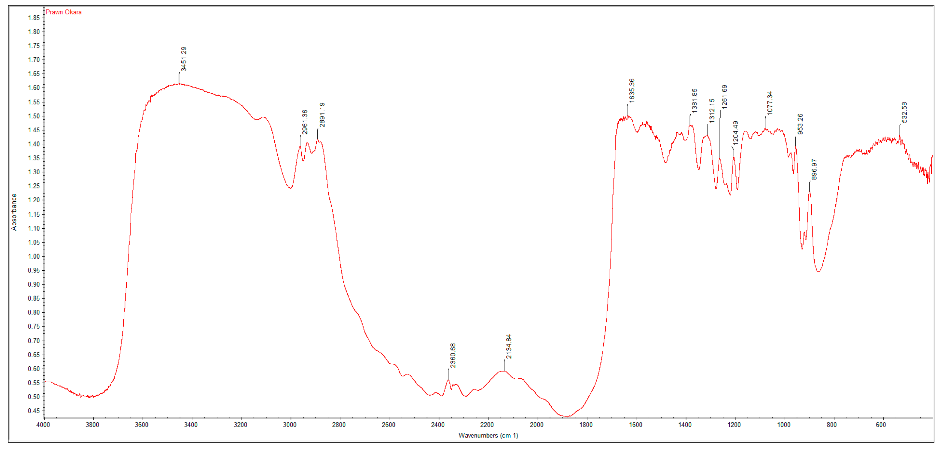
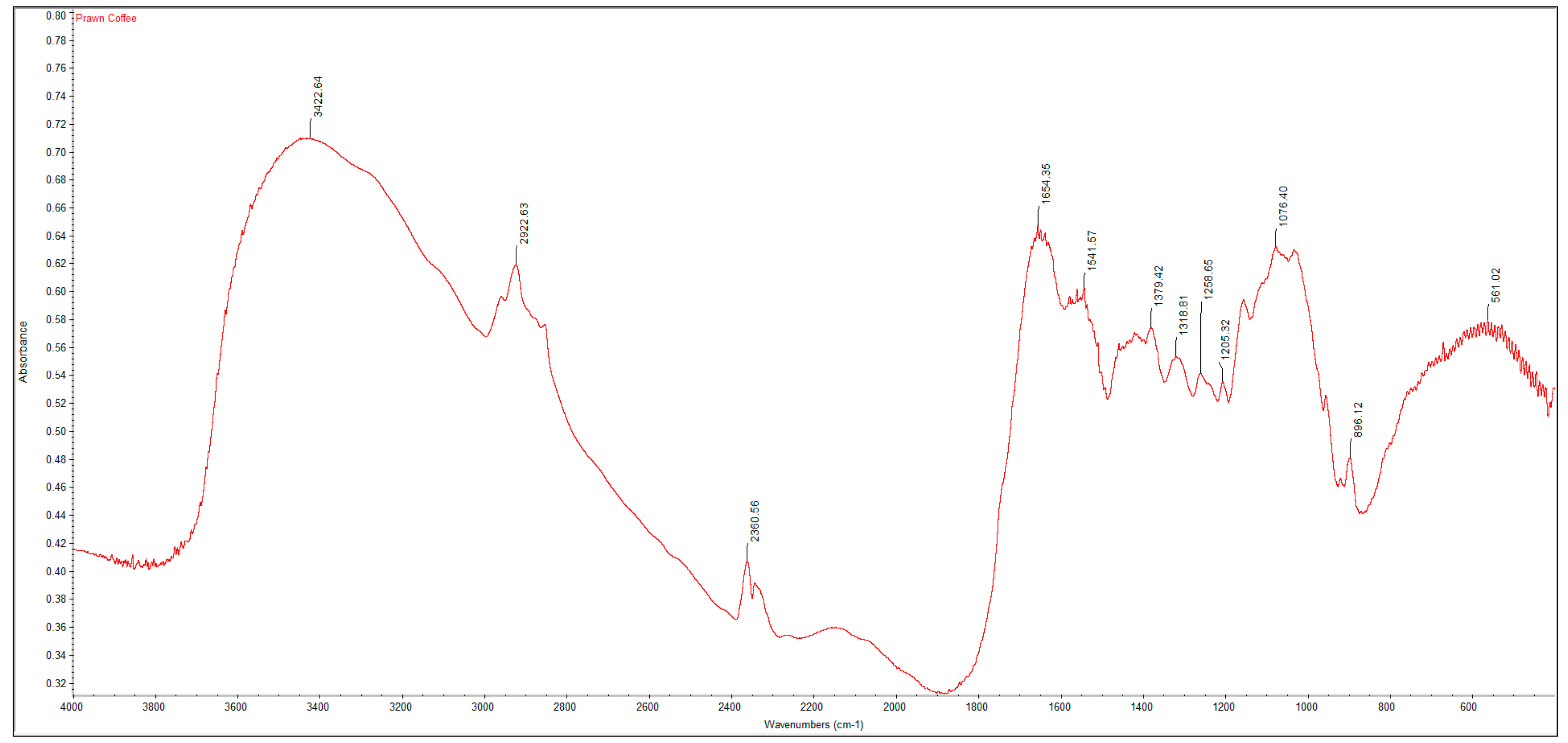
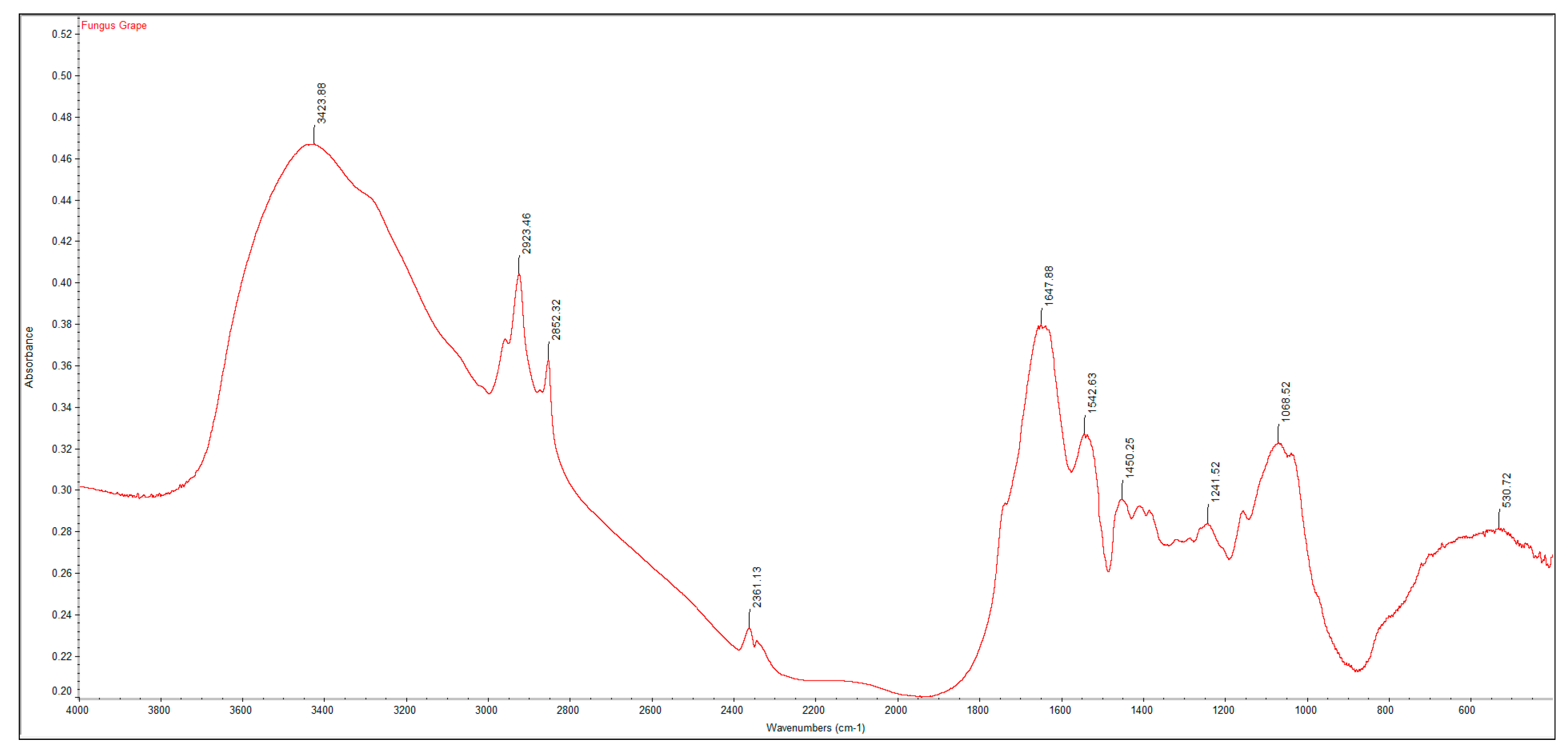
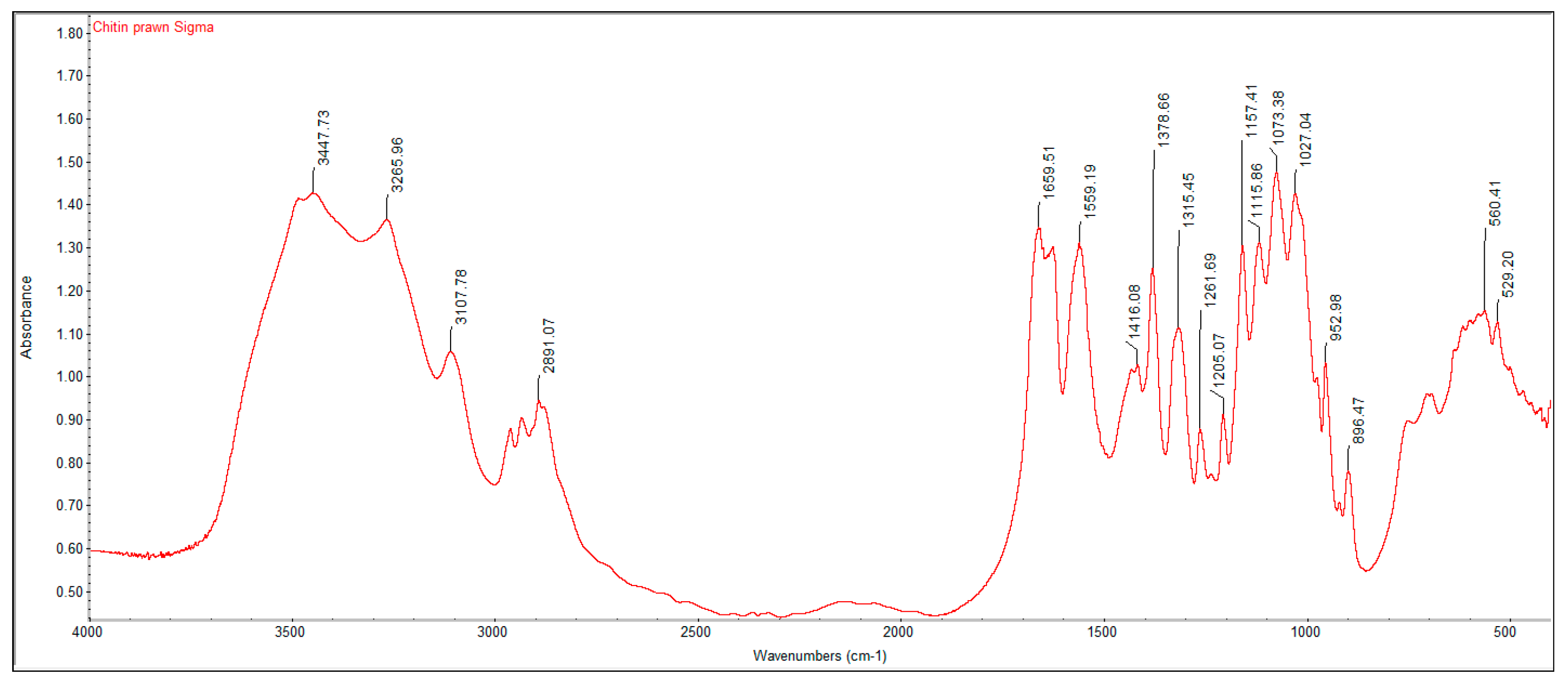
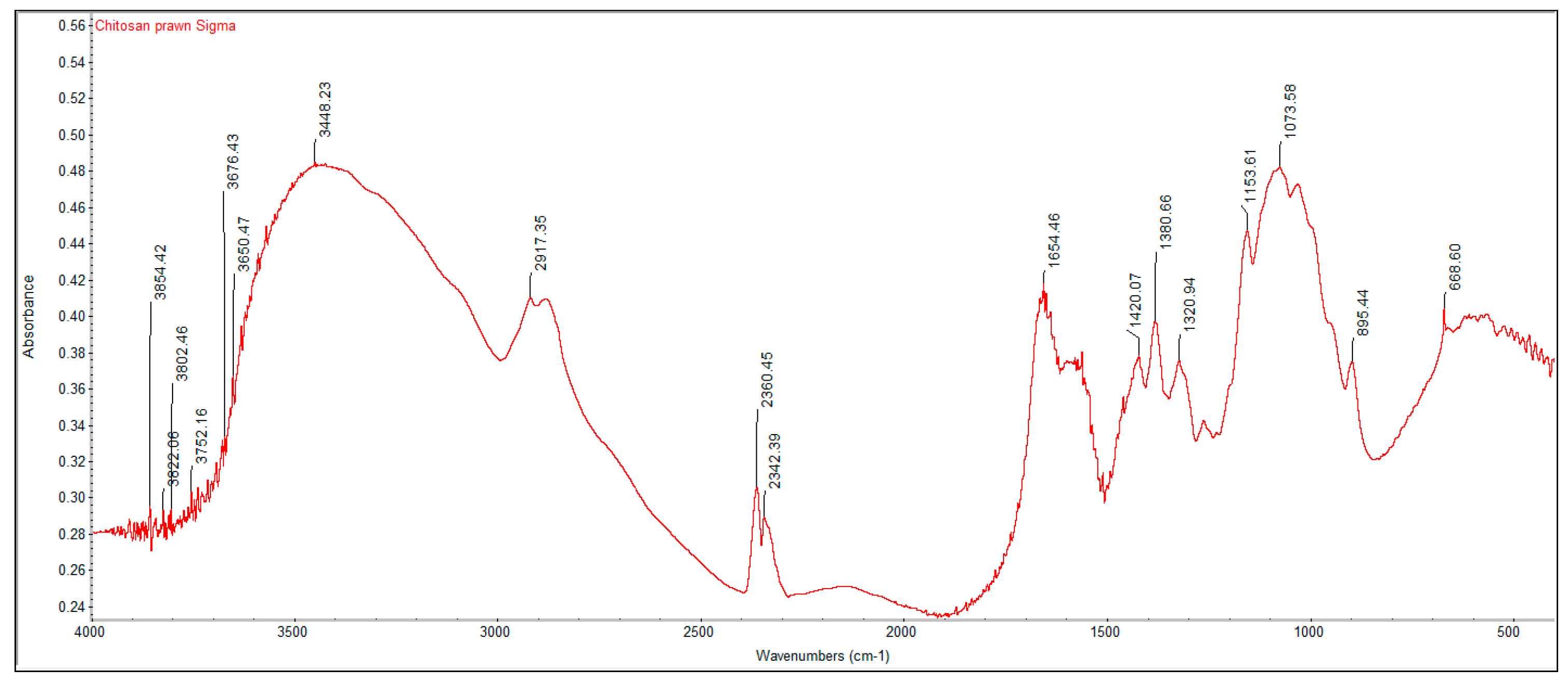
The characteristic FTIR bands for crustacean chitin extracted from using okara residues and coffee waste (Figure 1 and Figure 2) as well as fungal chitosan obtained from Mucor circinelloides using red grape pomace as substrates (Figure 3) were similar to commercial chitin and chitosan purchased from Sigma Aldrich (Figure 4 and Figure 5). The spectra of commercial chitin and commercial chitosan showed amide bands at 1660, 1559 and 1315 cm−1 and 1655, 1540 and 1321 cm−1, respectively, which were assigned to the C=O stretching, N–H bending in the CONH plane and the CN bond stretching plus CH2 wagging. Similarly, the spectra for crustacean chitin extracted from coffee waste fermentation as well as fungal chitosan from Mucor circinelloides extracted from red grape pomace exhibited bands in the amide region, at 1654, 1542 and 1319 cm−1 and 1648, 1543 and 1242 cm−1, respectively (Table 2). The spectra data are in agreement with the fact that the structure of the chitin and chitosan chains contain two types of amide groups, stabilized by intermolecular hydrogen bonds between two N-acetyl groups (C=O-N-H) and between one N-acetyl [22] and CH2OH group [23]. The deacetylation and regeneration processes have been reported to induce disturbance in the initial crystalline reticulum of chitin, causing a reordering of the hydrogen linkages of chitosan [24]. This can be observed in the distinct broad bands for crustacean chitin and fungal chitosan at approximately 3423 cm−1 and 3424 cm−1, in the region of the axial deformation of OH, which appears as overlapping the band of axial deformation of NH, indicating the intermolecular hydrogen linking formation, and at the displacement of the higher frequency band, denoting an increase in the structural order [25]. The data are in accordance with those reported in the literature when comparing both chitin and chitosan infrared spectra obtained by microbiological methods [26].
The degree of deacetylation (DD%) for crustacean chitin and fungal chitosan were calculated and tabulated in Table 3. Based on the experimental results, the highest DD% was observed in crustacean chitin extracted from okara and coffee waste fermentation (69.94% and 68.82%) and the highest DD% of fungal chitosan obtained from Mucor circinelloides was from red grape pomace fermentation (61.05%). These values are comparable to commercial chitin and chitosan purchased from Sigma Aldrich (70.46% and 64.00%).
The degree of deacetylation (DD%) is a critical parameter related to the physical and chemical properties of chitosan as it influences its cationic properties [27]. Chitosan with a high DD% has increasing positive charges, therefore it is suitable for use in food applications as a clarifying agent, a chelating or coagulating agent, or an antibacterial agent [28]. In our study, a DD% of above 60% was set as a criteria indicative of successful deacetylation for crustacean chitin [29]. Extracted crustacean chitin from fermentation using coffee waste, red grape pomace, durian seed and avocado displayed a DD% above 60% (68.82%, 63.06%, 62.74% and 61.34%, respectively), suggesting a good quality chitin was produced. With a higher DD%, there is increased solubility of chitosan and better application use [30]. A similar criterion of DD% above 60% was chosen to demonstrate efficient deacetylation of chitosan from fungal mycelia. Other studies also reported a high DD% of more than 60% obtained for fungal chitosan [31]. Fungal chitosan extracted from fermentation using red grape pomace (61.05%) displayed a DD% above 60%.
The experimental results suggest that the degree of deacetylation is dependent on the type of substrate. Red grape pomace used a waste substrate resulted in a high DD% for both crustacean chitin and fungal chitosan. This may be due to the high levels of soluble monosaccharides, such as glucose, that are present in red grapes pomace, favoring the fermentation process [32]. Durian seed (9.08% protein, 72.49% carbohydrate), [33] avocado seed (15.55% protein, 49.03% carbohydrate) [34] and coffee waste (17.44% protein, 24.08% glucose) [35] contains high compositions of sugars and protein. The rich source of nitrogen content provides an optimal condition for growth of Mucor circinelloides, [36], which produces chitinase to hydrolyze chitin into chitosan [37].
3.3. Elemental Analysis of Extracted Crustacean Chitin and Fungal Chitosan
The carbon, hydrogen and nitrogen content of the extracted crustacean chitin and fungal chitosan from fermentation on various waste substrates are tabulated in Table 4; Table 5. The fungal chitosan from Sigma-Aldrich showed a higher nitrogen (7.75%) and lower carbon (43.45%) content than the crustacean chitin from Sigma-Aldrich (6.65% of N, 46.12% of C), as the nitrogen content of fungal chitosan increases with a more efficient deacetylation process [38]. In addition, the carbon content of the fungal chitosan is lower than that of crustacean chitin due to the loss of acetamide groups during the deacetylation reaction [39]. In fungal chitosan, higher amounts of nitrogen were observed ranging from 5.03% to 8.46%, compared to crustacean chitin where lower amounts of nitrogen were observed, from 5.21% to 6.85%. This shows that the nitrogen content of chitosan increases with a more efficient deacetylation process for Mucor circinelloides due to its absence of mineral content compared to prawn shells [40]. Overall, the carbon/nitrogen ratio was the lowest for fungal chitosan extracted from the fermentation using coffee waste (C/N ratio of 5.11).
Fungi require an organic or inorganic source of nitrogen in their nutrition to synthetize nitrogen in their walls [41]. Ammonium ions may be a direct source of nitrogen in fungi nutrition. The inorganic form of nitrogen is reduced during the redox reaction to ammonium [42]. The source of nitrogen is one of the most crucial factors in production of fungal chitosan [43]. A cultivation medium with higher carbon and nitrogen contents will contain greater amounts of amino acids and carbohydrates, which will influence the growth conditions of the Mucoralean fungi, and this is reflected in the different carbon/nitrogen ratios of the fungal chitosan tabulated [44].
An overall summary and quantification of the fermentation parameters is presented below in Table 6. The extraction efficiency of crustacean chitin from prawn shell waste was calculated by dividing the dry weights of the crustacean chitin in Table 1 over 5 g of prawn shell waste used. The substrate conversion yield of the cultivated fungal chitosan was obtained by dividing the dry weights of the fungal chitosan in Table 1 over 10 g of waste substrate used.
4. Conclusions
In this study, deproteinization of prawn shells by proteolytic Mucor circinelloides has been shown to be successful, and the resulting acidic conditions enable further demineralization of the prawn shells. Using FTIR analysis, the degree of deacetylation of the isolated chitin from prawn shells and extracted chitosan from fungal mycelia have been proven to be comparable to commercial quality. By avoiding the use of expensive commercial protease enzymes, fungal fermentation serves as a simple and cost-effective approach to recover chitin from both crustacean shells and fungal mycelia. However, while preliminary results from this study at the shake-flask scale is promising, further experiments need to be performed at the bench-top scale to confirm its viability and cost-effectiveness.
Author Contributions
Conceptualization, W.N.C.; methodology, Y.N.T. and P.P.L.; software, Y.N.T. and P.P.L.; validation, Y.N.T. and P.P.L.; formal analysis, Y.N.T. and P.P.L.; investigation, Y.N.T. and P.P.L.; resources, W.N.C.; data curation, Y.N.T. and P.P.L.; writing—original draft preparation, Y.N.T. and P.P.L.; writing—review and editing, Y.N.T. and P.P.L.; visualization, W.N.C.; supervision, W.N.C.; project administration, W.N.C.; funding acquisition, W.N.C. All authors have read and agreed to the published version of the manuscript.
Funding
This research was funded by Nanyang Technological University, grant number M4062121.120.703012.
Acknowledgments
We thank Nanyang Technological University for support.
Conflicts of Interest
The authors declare no conflict of interest. The funders had no role in the design of the study; in the collection, analyses, or interpretation of data; in the writing of the manuscript, or in the decision to publish the results.
References
- Sitanggang, A.B.; Sophia, L.; Wu, H.S. Aspects of glucosamine production using microorganisms. Int. Food Res. J. 2012, 19, 393–404. [Google Scholar]
- Nwe, N.; Furuike, T.; Tamura, H. Chitosan from Aquatic and Terrestrial Organisms and Microorganisms: Production, Properties and Applications. In Biodegradable Materials, 1st ed.; Johnson, B.M., Berkel, Z.E., Eds.; Nova Science Pub., Inc.: Hauppauge, NY, USA, 2011; pp. 29–50. [Google Scholar]
- Elieh-Ali-Komi, D.; Hamblin, M.R. Chitin and Chitosan: Production and Application of Versatile Biomedical Nanomaterials. Int. J. Adv. Res. 2016, 4, 411–427. [Google Scholar]
- Zukiewicz-Sobczak, W.; Sobczak, P.; Zawislak, K.; Zagorski, J.; Wojtyla-Buciora, P.; Wojtyla, A. Physical and chemical properties comparison of fungal and crustaceous chitosan. J. Health Inequal. 2015, 1, 7–14. [Google Scholar] [CrossRef]
- Ghormade, V.; Pathan, E.; Desphande, M. Can fungi compete with marine sources for chitosan production? Int. J. Biol. Macromol. 2017, 104, 1415–1421. [Google Scholar] [CrossRef]
- Islam, S.; Rahman Buiyan, M.A.; Islam, M.N. Chitin and Chitosan: Structure, Properties and Applications in Biomedical Engineering. J. Polym. Environ. 2017, 25, 854–866. [Google Scholar] [CrossRef]
- Ferreira, J.A.; Lennartsson, P.R.; Edebo, L.; Taherzadeh, M.J. Zygomycetes-based biorefinery: Present status and future prospects. Bioresour. Technol. 2013, 135, 523–532. [Google Scholar] [CrossRef] [Green Version]
- Younes, I.; Hajji, S.; Frachet, V.; Rinaudo, M.; Jellouli, K.; Nasri, M. Chitin extraction from shrimp shell using enzymatic treatment. Antitumor, antioxidant and antimicrobial activities of chitosan. Int. J. Biol. Macromol. 2014, 69, 489–498. [Google Scholar] [CrossRef]
- Abo Elsoud, M.M.; El Kady, E.M. Current trends in fungal biosynthesis of chitin and chitosan. Bull. Natl. Res. Cent. 2019, 43, 1–12. [Google Scholar] [CrossRef] [Green Version]
- Kaur, S.; Dhillon, G.S. The versatile biopolymer chitosan: Potential sources, evaluation of extraction methods and applications. Crit. Rev. Microbiol. 2014, 40, 155–175. [Google Scholar] [CrossRef]
- Philibert, T.; Lee, B.H.; Fabien, N. Current Status and New Perspectives on Chitin and Chitosan as Functional Biopolymers. Appl. Biochem. Biotech. 2017, 181, 1314–1337. [Google Scholar] [CrossRef]
- Dhillon, G.S.; Kaur, S.; Brar, S.K.; Verma, M. Green synthesis approach: Extraction of chitosan from fungus mycelia. Crit. Rev. Biotech. 2013, 33, 379–403. [Google Scholar] [CrossRef] [PubMed]
- Ma, Q.; Gao, X. Categories and biomanufacturing methods of glucosamine. Appl. Microbiol. Biot. 2019, 103, 7883–7889. [Google Scholar] [CrossRef] [PubMed]
- Aranday-Garcia, R.; Guerrero, A.R.; Ifuku, S.; Shirai, K. Successive inoculation of Lactobacillus brevis and Rhizopus oligosporus on shrimp wastes for recovery of chitin and added-value products. Process Chem. 2017, 58, 17–24. [Google Scholar] [CrossRef]
- Razak, M.A.; Pinjari, A.B.; Begum, P.S.; Viswanath, B. Biotechnological Production of Fungal Biopolymers Chitin and Chitosan: Their Potential Biomedical and Industrial Applications. Curr. Biotech. 2018, 7, 214–230. [Google Scholar] [CrossRef]
- Chan, L.G.; Cohen, J.L.; Nobrega de Moura Bell, J.M.L. Conversion of Agricultural Streams and Food-Processing By-Products to Value-Added Compounds Using Filamentous Fungi. Annu. Rev. Food Sci. Technol. 2018, 9, 503–523. [Google Scholar] [CrossRef]
- Rodrigues Reis, C.E.; Bento, H.B.S.; Carvalho, A.K.F.; Rajendran, A.; Hu, B.; De Castro, H.F. Critical applications of Mucor circinelloides within a biorefinery context. Crit. Rev. Biotech. 2019, 39, 555–570. [Google Scholar] [CrossRef]
- Stamford, T.; Freitas Silva, M.; Berger, L.; Anjos, F.; Alcantara, S.; Stamford, N.; Campos-Takaki, G. Chitin and chitosan produced by Mucoralean fungi using a new economic medium corn steep. In Microorganisms in Industry and Environment, 1st ed.; Mendez-Vilas, A., Ed.; World Scientific: Singapore, 2010; pp. 579–583. [Google Scholar]
- Sharifyazd, S.; Karimi, K. Effects of fermentation conditions on valuable products of ethanolic fungus Mucor indicus. Electron. J. Biotechn. 2017, 30, 77–82. [Google Scholar] [CrossRef]
- Teng, W.L.; Khor, E.; Tan, T.K.; Lim, Y.L.; Tan, S.C. Concurrent production of chitin from shrimp shells and fungi. Carbohyd. Res. 2001, 332, 305–316. [Google Scholar] [CrossRef]
- Anandhi, K. Biological Extraction of Chitin and Chitosan from Marine Fungi, its Characterization, Anti-Microbial Activity, Anti-Textile Activity against MDR Pathogens and Anti-Cancer Activity. World J. Pharm. Res. 2017, 6, 844–863. [Google Scholar]
- Stamford, T.C.M.; Stamford, T.L.M.; Stamford, N.P.; Neto, B.D.B.; Campos-Takaki, G.M.D. Growth of Cunninghamella elegans UCP 542 and production of chitin and chitosan using yam bean medium. Electron. J. Biotechnol. 2007, 10, 61–68. [Google Scholar]
- Franca, E.F.; Lins, R.D.; Freitas, L.C.G.; Straatsma, T.P. Characterization of Chitin and Chitosan Molecular Structure in Aqueous Solution. J. Chem. Theory Comput. 2008, 4, 9. [Google Scholar] [CrossRef] [PubMed]
- Berger, L.R.R.; Cavalcante, H.M.D.M.; Stamford, T.C.M.; da Silva, M.C.D.F.; de Oliveira, C.E.V.; Sarmento, B.F.C.C.; de Campos-Takaki, G.M. Chitin and chitosan produced by Cunninghamella elegans using alternative medium–coconut water. In Microbes in Applied Research-Current Advances and Challenges, 1st ed.; Mendez-Vilas, A., Ed.; World Scientific: Malaga, Spain, 2012; Volume 1, pp. 377–381. [Google Scholar]
- Elizabeth, A.C.F.; Stamford, T.C.; Stamford-Arnaud, T.M.; D´Amorim Santa-Cruz, P.; Freitas da Silva, M.C.; Campos-Takaki, G.M.; Stamford, T.L. Physico-Chemical Characteristics and Functional Properties of Chitin and Chitosan Produced by Mucor circinelloides using Yam Bean as Substrate. Molecules 2011, 16, 10. [Google Scholar]
- Chatterjee, S.; Adhya, M.; Guha, A.; Chatterjee, B. Chitosan from Mucor circinelloides: Production and physico-chemical characterization. Process Chem. 2005, 40, 395–400. [Google Scholar]
- Patria, A. Production and characterization of Chitosan from shrimp shells waste. Aquac. Aquar. Conserv. Legis. Int. J. Bioflux Soc. 2013, 6, 339–344. [Google Scholar]
- Chatterjee, S.; Guha, A. A study on biochemical changes during cultivation of Rhizopus oryzae in deproteinized whey medium in relation to chitosan production. Lett. Appl. Microbiol. 2014, 59, 155–160. [Google Scholar] [CrossRef] [PubMed]
- Devikrishna, S.; Remya, R. Effect of Degree of Deacetylation and Molecular Weight of Chitosan Extracted From Various Marine Sources on Its Applications. Int. J. Innov. Res. Sci. Eng. Technol. 2015, 4, 6078–6083. [Google Scholar]
- Cheung, R.C.F.; Ng, T.B.; Wong, J.H.; Chan, W.Y. Chitosan: An Update on Potential Biomedical and Pharmaceutical Applications. Mar. Drugs 2015, 13, 30. [Google Scholar] [CrossRef]
- Pochanavanich, P.; Suntornsuk, W. Fungal chitosan production and its characterization. Lett. Appl. Microbiol. 2002, 35, 4. [Google Scholar] [CrossRef]
- Corbin, K.R.; Hsieh, Y.S.; Betts, N.S.; Byrt, C.S.; Henderson, M.; Stork, J.; DeBolt, S.; Fincher, G.B.; Burton, R.A. Grape marc as a source of carbohydrates for bioethanol: Chemical composition, pre-treatment and saccharification. Bioresour. Technol. 2015, 193, 8. [Google Scholar] [CrossRef]
- Mulyati, A.; Widiastuti, D.; Oktaviani, L. Characterization of Durian Seed Flour (Durio zibhetinuss l.) and Estimation of its Self Life with Accelerated Self Life Testing (ASLT) Moisture Critical Method. J. Phy. Conf. Ser. 2018, 1095, 7. [Google Scholar] [CrossRef]
- Ejiofor, N.C.; Ezeagu, I.E.; Ayoola, M.B.; Umera, E.A. Determination of the Chemical Composition of Avocado (Persea Americana) Seed. Adv. Food Technol. Nutr. Sci. 2018, 2, 5. [Google Scholar] [CrossRef]
- Ballesteros, L.F.; Teixeira, J.A.; Mussatto, S.I. Chemical, Functional, and Structural Properties of Spent Coffee Grounds and Coffee Silverskin. Food Bioprocess Technol. 2014, 7, 11. [Google Scholar] [CrossRef] [Green Version]
- Chatterjee, S.; Chatterjee, B.; Guha, A.K. Kinetics of Mucor circinelloides Fermentation in Relation to Chitosan Production. Res. J. Microbiol. 2006, 1, 5. [Google Scholar]
- Stoykov, Y.M.; Pavlov, A.I.; Krastanov, A.I. Chitinase Biotechnology: Production, Purification and Application. Eng. Life Sci. 2015, 15, 9. [Google Scholar] [CrossRef]
- Zamani, A.; Edebo, L.; Sjostrom, B.; Taherzadeh, M.J. Extraction and Precipitation of Chitosan from Cell Wall of Zygomycetes Fungi by Dilute Sulfuric Acid. Biomacromolecules 2007, 8, 3786–3790. [Google Scholar] [CrossRef] [PubMed]
- White, S.A.; Farina, P.R.; Fulton, I. Production and Isolation of Chitosan from Mucor circinelloides. Appl. Environ. Microbiol. 1979, 38, 323–328. [Google Scholar] [CrossRef] [Green Version]
- Berger, L.R.R.; Stamford, T.C.M.; Stamford-Arnaud, T.M.; de Oliveira Franco, L.; do Nascimento, A.E.; Cavalcante, H.M.D.M.; Macedo, R.O.; de Campos-Takaki, G.M. Effect of Corn Steep Liquor (CSL) and Cassava Wastewater (CW) on Chitin and Chitosan Production by Cunninghamella elegans and Their Physicochemical Characteristics and Cytotoxicity. Molecules 2014, 19, 2771–2792. [Google Scholar] [CrossRef] [Green Version]
- Abasian, L.; Alavijeh, R.S.; Satari, B.; Karimi, K. Sustainable and Effective Chitosan Production by Dimorphic Fungus Mucor circinelloides via Replacing Yeast Extract with Fungal Extract. Appl. Biochem. Biotechnol. 2019, in press. [Google Scholar] [CrossRef]
- Sousa Andrade, V.; de Barros Neto, B.; Fukushima, K.; de Campos-Takaki, G.M. Effect of medium components and time of cultivation on chitin production by Mucor circinelloides (Mucor javanicus IFO 4570) -- a factorial study. Revista Iberoamericana de Micología 2003, 20, 149–153. [Google Scholar]
- Andrade, V.S.; Sarubbo, L.A.; Fukushima, K.; Miyaji, M.; Nishimura, K.; de Campos-Takaki, G.M. Production of Extracellular Proteases by Mucor circinelloides using D-Glucose as Carbon Source Substrate. Braz. J. Microbiol. 2002, 33, 106–110. [Google Scholar] [CrossRef] [Green Version]
- Karimi, K.; Zamani, A. Mucor indicus: Biology and industrial application perspectives: A review. Biotechnol. Adv. 2013, 31, 466–481. [Google Scholar] [CrossRef] [PubMed]
Figure 1.
FTIR spectra of crustacean chitin extracted after fermentation on okara.

Figure 2.
FTIR spectra of crustacean chitin extracted after fermentation on coffee waste.

Figure 3.
FTIR spectra of fungal chitosan extracted after fermentation on red grape pomace.

Figure 4.
FTIR spectra of commercial chitin.

Figure 5.
FTIR spectra of commercial chitosan.


Table 1.
Dry weights of crustacean chitin and fungal chitosan extracted after fermentation on various waste substrates.
Table 1.
Dry weights of crustacean chitin and fungal chitosan extracted after fermentation on various waste substrates.
| Waste Substrate | Dry Weight of Crustacean Chitin (g) | Dry Weight of Fungal Chitosan (g) |
|---|---|---|
| 10 g of okara | 0.74 ± 0.01 | - |
| 10 g of coffee waste | 0.58 ± 0.01 | 1.0 ± 0.01 |
| 10 g of durian seed | 0.56 ± 0.01 | 0.77 ± 0.01 |
| 10 g of red grape pomace | 0.6 ± 0.01 | 0.76 ± 0.01 |
| 10 g of avocado seed | 1.13 ± 0.01 | 0.46 ± 0.01 |
Table 2.
FTIR bands (cm−1) for commercial chitin and chitosan versus extracted crustacean chitin from coffee waste fermentation and extracted fungal chitosan from red grape pomace fermentation.
Table 2.
FTIR bands (cm−1) for commercial chitin and chitosan versus extracted crustacean chitin from coffee waste fermentation and extracted fungal chitosan from red grape pomace fermentation.
| Functional Group | Commercial Chitin | Extracted Crustacean Chitin from Coffee Waste Fermentation | Commercial Chitosan | Extracted Fungal Chitosan from Red Grape Pomace Fermentation |
|---|---|---|---|---|
| O–H stretch | 3447 | 3423 | 3448 | 3424 |
| C–H stretch | 2891 | 2922 | 2917 | 2925 |
| C=O stretch of N-acetyl group (Amide I) | 1660 | 1654 | 1655 | 1648 |
| N–H bend, C–N stretch (Amide II) | 1559 | 1542 | 1540 | 1543 |
| CH3 in NHCOCH3 group | 1379 | 1379 | 1381 | 1450 |
| CH2 wagging (Amide III) | 1315 | 1319 | 1321 | 1242 |
| C–O–C stretch | 1073 | 1076 | 1074 | 1069 |
Table 3.
Degree of deacetylation of crustacean chitin and fungal chitosan extracted after fermentation on various waste substrates.
Table 3.
Degree of deacetylation of crustacean chitin and fungal chitosan extracted after fermentation on various waste substrates.
| Waste Substrate | Degree of Deacetylation (%) of Crustacean Chitin | Degree of Deacetylation (%) of Fungal Chitosan |
|---|---|---|
| Commercial Chitin | 70.5 ± 0.1 | - |
| Commercial Chitosan | - | 64.0 ± 0.1 |
| 10 g of okara | 69.9 ± 0.1 | - |
| 10 g of coffee waste | 68.8 ± 0.1 | 57.5 ± 0.1 |
| 10 g of durian seed | 63.1 ± 0.1 | 52.9 ± 0.1 |
| 10 g of red grape pomace | 62.7 ± 0.1 | 61.1 ± 0.1 |
| 10 g of avocado seed | 61.3 ± 0.1 | 42.1 ± 0.1 |
Table 4.
Elemental analysis of crustacean chitin extracted after fermentation on various waste substrates.
Table 4.
Elemental analysis of crustacean chitin extracted after fermentation on various waste substrates.
| Waste Substrate | %C | %H | %N | C/N Ratio of Crustacean Chitin |
|---|---|---|---|---|
| Commercial Chitin | 46.1 ± 0.1 | 7.3 ± 0.1 | 6.7 ± 0.1 | 6.9 ± 0.1 |
| 10 g of okara | 33.9 ± 0.1 | 10.5 ± 0.1 | 5.4 ± 0.1 | 6.3 ± 0.1 |
| 10 g of coffee waste | 43.1 ± 0.1 | 15.1 ± 0.1 | 5.2 ± 0.1 | 8.3 ± 0.1 |
| 10 g of durian seed | 38.8 ± 0.1 | 6.6 ± 0.1 | 5.7 ± 0.1 | 6.9 ± 0.1 |
| 10 g of red grape pomace | 43.8 ± 0.1 | 7.5 ± 0.1 | 6.9 ± 0.1 | 6.4 ± 0.1 |
| 10 g of avocado seed | 44.0 ± 0.1 | 5.8 ± 0.1 | 5.4 ± 0.1 | 8.1 ± 0.1 |
Table 5.
Elemental analysis of fungal chitosan extracted after fermentation on various waste substrates.
Table 5.
Elemental analysis of fungal chitosan extracted after fermentation on various waste substrates.
| Waste Substrate | %C | %H | %N | C/N Ratio of Fungal Chitosan |
|---|---|---|---|---|
| Commercial Chitosan | 43.5 ± 0.1 | 8.7 ± 0.1 | 7.8 ± 0.1 | 5.6 ± 0.1 |
| 10 g of coffee waste | 34.1 ± 0.1 | 6.6 ± 0.1 | 6.7 ± 0.1 | 5.1 ± 0.1 |
| 10 g of durian seed | 46.2 ± 0.1 | 6.8 ± 0.1 | 5.0 ± 0.1 | 9.2 ± 0.1 |
| 10 g of red grape pomace | 49.5 ± 0.1 | 7.0 ± 0.1 | 6.9 ± 0.1 | 7.2 ± 0.1 |
| 10 g of avocado seed | 43.4 ± 0.1 | 6.7 ± 0.1 | 8.5 ± 0.1 | 5.1 ± 0.1 |
Table 6.
Extraction efficiency of crustacean chitin from prawn shell waste and the substrate conversion efficiency for growth of fungal chitosan.
Table 6.
Extraction efficiency of crustacean chitin from prawn shell waste and the substrate conversion efficiency for growth of fungal chitosan.
| Waste Substrate | Extraction Efficiency of Crustacean Chitin | Substrate Conversion of Fungal Chitosan |
|---|---|---|
| 10 g of okara | 14.8% | - |
| 10 g of coffee waste | 11.6% | 10.0% |
| 10 g of durian seed | 11.2% | 7.7% |
| 10 g of red grape pomace | 12.0% | 7.6% |
| 10 g of avocado seed | 22.6% | 4.6% |
© 2020 by the authors. Licensee MDPI, Basel, Switzerland. This article is an open access article distributed under the terms and conditions of the Creative Commons Attribution (CC BY) license (http://creativecommons.org/licenses/by/4.0/).
Share and Cite
MDPI and ACS Style
Tan, Y.N.; Lee, P.P.; Chen, W.N. Dual Extraction of Crustacean and Fungal Chitosan from a Single Mucor circinelloides Fermentation. Fermentation 2020, 6, 40. https://0-doi-org.brum.beds.ac.uk/10.3390/fermentation6020040
AMA Style
Tan YN, Lee PP, Chen WN. Dual Extraction of Crustacean and Fungal Chitosan from a Single Mucor circinelloides Fermentation. Fermentation. 2020; 6(2):40. https://0-doi-org.brum.beds.ac.uk/10.3390/fermentation6020040
Chicago/Turabian StyleTan, Yun Nian, Pei Pei Lee, and Wei Ning Chen. 2020. "Dual Extraction of Crustacean and Fungal Chitosan from a Single Mucor circinelloides Fermentation" Fermentation 6, no. 2: 40. https://0-doi-org.brum.beds.ac.uk/10.3390/fermentation6020040
Note that from the first issue of 2016, this journal uses article numbers instead of page numbers. See further details here.