Article

Production of Proteins prM/M and E of Dengue Virus-3 in Pichia pastoris: Simplified Purification and Evaluation of Their Use as Antigens in Serological Diagnosis of Dengue

by
Michelle D. O. Teixeira
1,
Roberto S. Dias
1,
John W. O. Prates
1,
Juliana M. C. Monteiro
1,
Mariana F. Xisto
1,
Cynthia C. da Silva
2 and
Sérgio O. De Paula
1,*
1
Department of General Biology, Federal University of Viçosa, Viçosa, Minas Gerais 36570-900, Brazil
2
Department of Microbiology, Federal University of Viçosa, Viçosa, Minas Gerais 36570-900, Brazil
*
Author to whom correspondence should be addressed.
Submission received: 23 June 2020 / Revised: 9 September 2020 / Accepted: 10 September 2020 / Published: 14 September 2020
(This article belongs to the Special Issue Yeast Biotechnology 4.0)

Abstract

:
Dengue is a major arbovirus affecting humans today. With the growing number of cases, it is essential to have large-scale production of antigens for the development of diagnostic kits for the rapid detection of patients infected by the virus and consequent proper medical intervention for them. In this work, we express the prM/M and E proteins of dengue virus-3 in yeast Pichia pastoris KM71H. The proteins were produced in soluble form in the supernatant of the culture and were purified by precipitation with ammonium sulfate. The fraction of 80% of ammonium sulfate was used as an antigen in an indirect enzyme-linked immunosorbent assay (ELISA), providing a sensitivity of 82.61% and a specificity of 89.25%. Thus, the methodology proposed here showed promise for obtaining antigens of dengue viruses and creating quick and inexpensive diagnostic tests, which is of great value since large portions of the areas affected by this disease are economically neglected.

1. Introduction

Dengue is a major arbovirus affecting humans, with 2.5 billion people living in areas considered at risk, about 50 to 100 million infections annually, and 500,000 among these evolving to more severe forms of the disease, including dengue shock syndrome and dengue hemorrhagic fever. In 2016, 2.1 million cases were reported by the World Health Organization in the Americas alone [1].
The causative agent of this disease is the dengue virus, which consists of four antigenically related serotypes (DENV1–4) [2]. These are mosquito-borne viruses belonging to the Flaviviridae family, which includes other members of great medical importance, including yellow fever virus, Japanese encephalitis virus, West Nile virus, and tick-borne encephalitis virus [3]. The dengue virus bodies are spherical and icosahedral with an RNA genome of approximately 11kb having a single ORF that encodes a polyprotein processed by the host and viral proteases into three structural proteins (C, prM, and E), that form the virion components, and seven nonstructural (NS1, NS2A, NS2B, NS3, NS4A, NS4B, and NS5) proteins involved in the replication cycle of the virus [4,5].
The main target of the neutralizing antibodies against a flavivirus is the envelope protein (E). The native E protein is a homodimer with a molecular weight of about 55–60 kDa that covers the surface of the viral membrane and is responsible for important biological activities essential for viral infectivity, such as recognition of the cellular receptor. Due to the function of adsorption and membrane fusion performed by this protein, several neutralizing epitopes are located in the E protein, making it an important antigen for the development of vaccines and for diagnostic use [5,6,7].
Interaction between the prM and E proteins is the main factor that drives the assembly of viruses [8]. Moreover, during the replicative cycle, the prM protein acts as a chaperone for the folding of the E glycoprotein. After cleavage, the pr peptide interacts with homodimers of E, stabilizing them and preventing their cleavage [9]. The structural protein of the envelope (E) is the target of several studies focusing on the importance of its glycosylation for the assembly of viral particles, infection, and interaction with the host receptors. Since it is the first target against which the immune system mounts a response, glycosylation is also important for antibody production [10].
Laboratory diagnosis of dengue can be accomplished by viral isolation from samples of patients, detection of the viral genome, detection of viral antigens, especially the NS1 protein, and serological studies. Basic serological tests have been used for the diagnosis of dengue infection, such as hemagglutination inhibition (HI), complement fixation (CF), the neutralization test (NT), capture and sandwich types of ELISA, and point-of-care (PoC) tests [6,11,12]. Despite the availability of different laboratory techniques for the diagnosis of dengue, some obstacles must be overcome for the development of techniques and new tools, such as obtaining antigens of dengue viruses on a large scale to capture specific antibodies present in suspected sera.
Traditionally, antigens used in diagnostic kits are those derived from cell culture or from the brain of an infected newborn mouse, which makes obtaining antigens on a large scale laborious and expensive [13,14]. The use of recombinant proteins in serological assays for the detection of dengue is a good alternative.
Several strategies have been used to verify the expression of dengue virus proteins in heterologous systems [15,16,17,18,19]. Among these is the production of viral proteins in the yeast Pichia pastoris. These methylotrophic microorganisms are capable of producing high levels of recombinant proteins. Additionally, P. pastoris can reach high cell densities and grow in inexpensive culture media [20,21]. Accordingly, P. pastoris is an important alternative for the production of recombinant proteins that require post-translational modifications not achieved in bacterial systems.
In this work, we express the prM and E proteins of dengue virus-3 in the yeast P. pastoris as an alternative for obtaining dengue virus antigen on a large scale. The proteins produced were good antigens for use in diagnostic tests, as shown by the results obtained through its use in indirect ELISA.

2. Materials and Methods

2.1. Yeast Strains and Plasmid

The host used in this study for expression of recombinant proteins was a Pichia pastoris strain KM71H (MutS), purchased from Invitrogen, San Diego, CA, USA. The expression plasmid used was the pPICZαA (Invitrogen, San Diego, CA, USA), which contains the zeocin-resistance marker, used for the selection of transformants in P. pastoris and Escherichia coli. Moreover, the plasmid has the AOX1 promoter and the pre-pro α factor secretory of Saccharomyces cerevisiae.

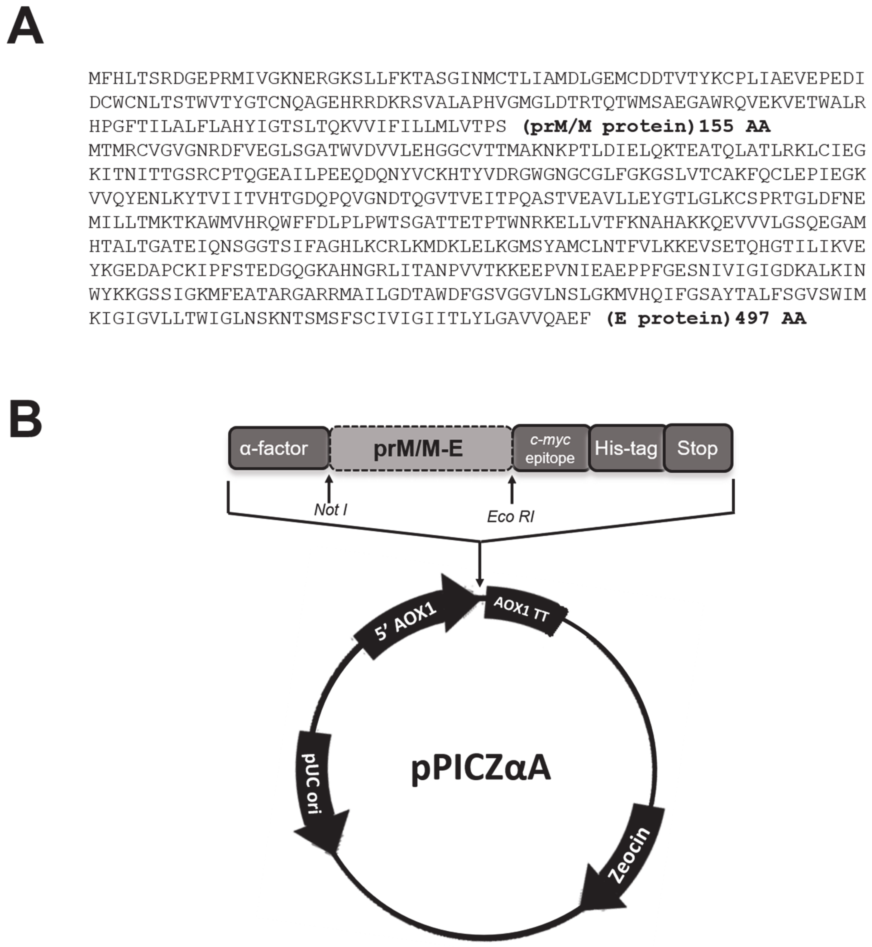
2.2. Cloning the Sequences of the prM/M and E Proteins in the Expression Vector pPICZαA

The sequences of the proteins prM/M and E (1986 bp) of dengue-3 (DENV3) virus were obtained by chemical synthesis due to need of codon optimization for expression in yeast, and the correct sequences were confirmed by sequencing (Genscript, Piscataway, NJ, USA). The sequences of interest were cloned into the expression vector pPICZαA after double digestion with restriction enzymes NotI and EcoRI. The vector was linearized with SacI enzyme and electroporated into P. pastoris KM71H. The transformed yeasts were selected on medium YPDS containing 500 µg/mL of Zeocin, as described in the Invitrogen Easy select TM Pichia expression kit manual (Invitrogen’s Easy Select Pichia Expression kit manual) and confirmed by PCR using the following primers for domain III of the dengue-3 virus E protein: 5′-TCACAAGAAGGTGCCATGCAC-3‘ and 5′-AGACAACTTCAAAGCCTTTTC-3′, resulting in a 408 bp amplicon. The correct integration of the genes of interest of the AOX1 locus in the genome of the yeast P. pastoris was confirmed by colony PCR using primers AOX1: 5′-GACTGGTTCCAATTGACAAGC-3′ and 5′-GCAAATGGCATTCTGACATCC-3′, resulting in a 2300 bp amplicon (referring to the prM/M and E sequence plus the AOX promoter sequence).

2.3. Expression of Recombinant Proteins

The recombinant yeast was grown in YPD medium for 24 h, followed by BMGY medium for biomass accumulation until an OD at 600 nm of 20. Thereafter, they were washed with water containing 1% yeast extract and 2% peptone and suspended in BMMY medium with induction of 1% methanol every 12 h for five days. The culture was then centrifuged at 3000× g for 20 min and the supernatant obtained was centrifuged again at 7000× g for 20 min. The supernatant was subjected to precipitation with 20%, 60%, and 80% of ammonium sulfate. The fractions were collected, suspended in water, and kept at 4 °C for further analysis.

2.4. Recombinant Protein Characterization

The proteins present in the fractions obtained from the precipitation with ammonium sulfate were separated on SDS-PAGE 12%. The bands corresponding to protein E in each fraction were quantified on the SDS-PAGE by densitometry with a standard curve of BSA. The samples were transferred to a nitrocellulose membrane to be analyzed by Western blot assay. After the transfer, the membrane was blocked for two hours with phosphate-buffered saline (PBS) (0.1 M NaCl, 2 mM KCl, 10 mM Na2HPO4, 1 mM KH2PO4 pH 7.4) at room temperature with 5% skim milk. After washing with PBS-0.5% Tween 20, the membrane was incubated with pooled sera from patients with confirmed dengue diluted 1:50 in PBS for 1 h at 37 °C. After washing as described, the membrane was incubated for one hour at 37 °C with anti-human IgM or IgG labeled with peroxidase (Sigma-Aldrich, Inc., St. Louis, MO, USA) diluted 1:10,000 in PBS. After washing with PBS-0.5% Tween 20, the membrane was developed with TMB (Sigma-Aldrich, Inc., St. Louis, MO, USA) according to the manufacturer’s specifications for the visualization of the bands of interest. A densitometry analysis was performed with the bands immunostained in the Western blot to confirm the yield of the differential precipitation using ammonium sulfate.
To confirm whether the recombinant protein E was glycosylated in yeast, a reaction was performed using periodic acid–Schiff (PAS). The E protein precipitated with 80% ammonium sulfate was electrophoresed (SDS-PAGE 12%) and transferred to nitrocellulose membrane. The membrane was washed with 12% trichloroacetic acid for 5 min. The next steps were carried out at 4 °C in the dark. The membrane was treated with 0.5% periodic acid for 15 min and then washed 3X with 15% acetic acid for 5 min. 50 mL of Schiff’s reagent were added and incubated for 30 min. Finally, six washes were performed with 7.5% acetic acid for 1 h, and the membrane was dried at room temperature.

2.5. Samples of Sera

Sera from 184 patients were provided by the Central Laboratory of Public Health of the State of Rondônia (LACEN/RO) and the Central Blood Bank of the State of Rondônia, Brazil (FHEMERON/RO). All sera were tested and used in accordance with protocols approved by the Central Blood Bank of the State of Rondônia, Brazil and all samples were kept anonymous. Of the 184 samples, 46 were IgG positive and 46 were IgG negative using Dengue Duo IgM and IgG ELISA Capture Kits; 92 were IgM positive and 46 were IgM negative using both kits MAC-ELISA IgM (Pan-Bio, Brisbane, Australia) and Dengue Duo IgM and IgG ELISA Capture Kits (Sanofi, Bridgewater, NJ, USA) (Table 1). The virus serotype could not be determined using these kits.

2.6. Indirect Enzyme-Linked Immunosorbent Assay (ELISA) for Detection of IgM and IgG

The fraction obtained by precipitation with 80% ammonium sulfate was used to sensitize polystyrene 96-well plates (Maxisorp, Nunc, Roskilde, Denmark). To these, 5 µg/well of proteins were transferred in a carbonate-bicarbonate buffer pH 9.6 and incubated at 4 °C overnight. Plates were blocked with PBS containing 5% fetal bovine serum for 30 min. Samples of sera were diluted 1:100 in PBS-5% fetal bovine serum and 100 µL of each dilution were added to wells in duplicate. After incubation at 37 °C for two hours, the plates were washed with PBS-0.5% Tween 20, and anti-IgG antibody or anti-IgM human conjugate with peroxidase (Sigma-Aldrich, Inc., St. Louis, MO, USA) diluted 1:2500 in PBS- fetal bovine serum 5% was added to them. The plates were incubated at 37 °C for 1 h. After five washes as previously described, the substrate TMB was added (Sigma-Aldrich, Inc., St. Louis, MO, USA), following the manufacturer’s recommendations. The reaction was stopped by adding 50 µL of 2M H2SO4. The absorbance was measured on a spectrophotometer at 450 nm (Multiskan, Thermo Scientific, Waltham, MA, USA).

2.7. Statistical Analysis

Receiver operating characteristic (ROC) curves were analyzed to estimate the diagnostic cutoff, sensitivity, and specificity (INSTAT software, GraphPad, San Diego, CA, USA).

3. Results

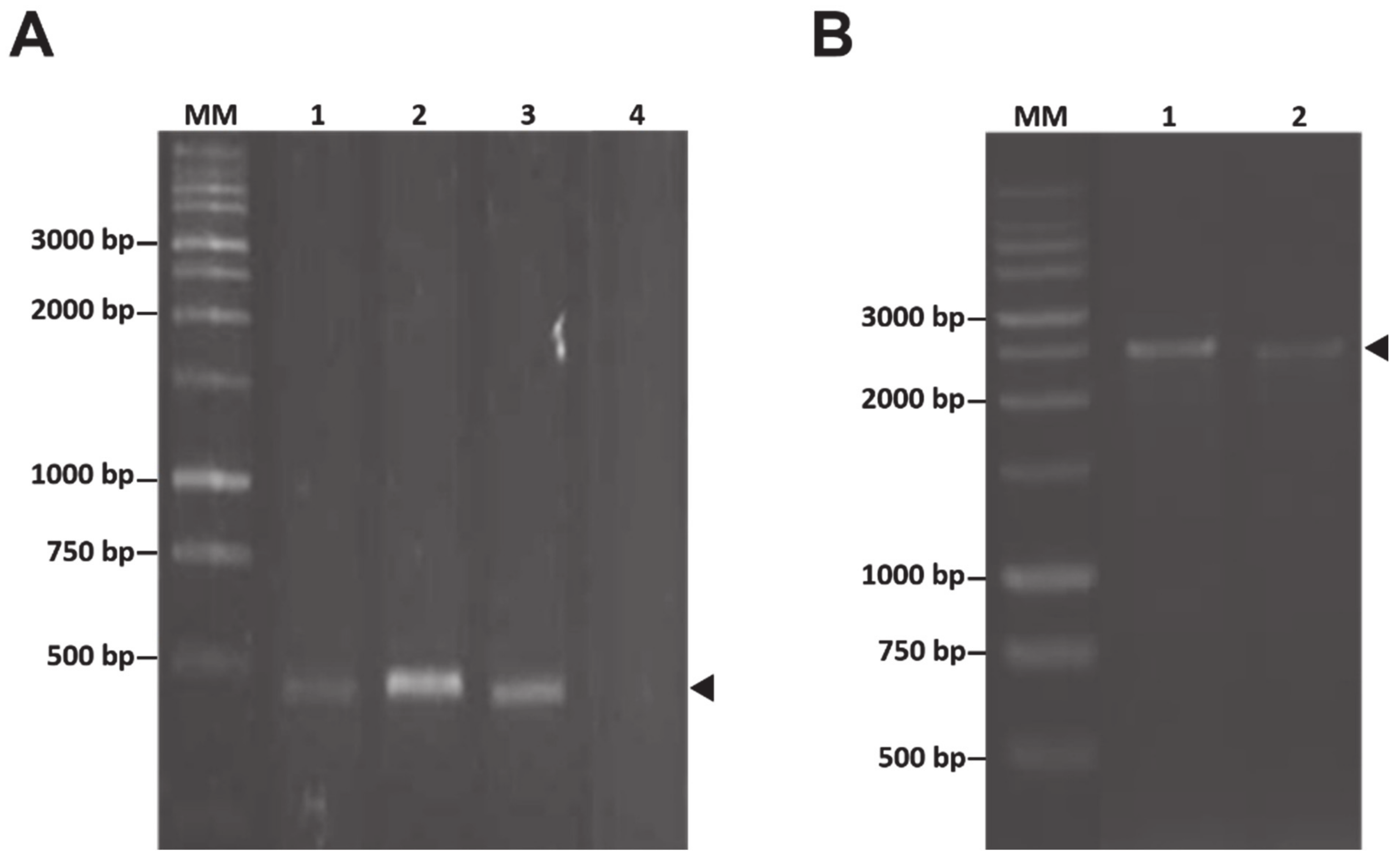
3.1. Construction of Recombinant Pichia pastoris Clones Expressing the prM and E Proteins of DENV3

To obtain yeast clones expressing the prM and E structural proteins of DENV3, the sequences of the codon-optimized genes were cloned into the expression vector pPICZαA, as shown in the scheme of Figure 1. The constructed pPICZαA-prM/M-E-DENV3 was used to transform yeast P. pastoris KM71H and this transformation was confirmed by PCR of yeast colonies using specific primers for domain III of the E protein of dengue. The correct insertion of the genes under study into the host genome was confirmed by colony PCR using primers AOX1 (Figure 2).

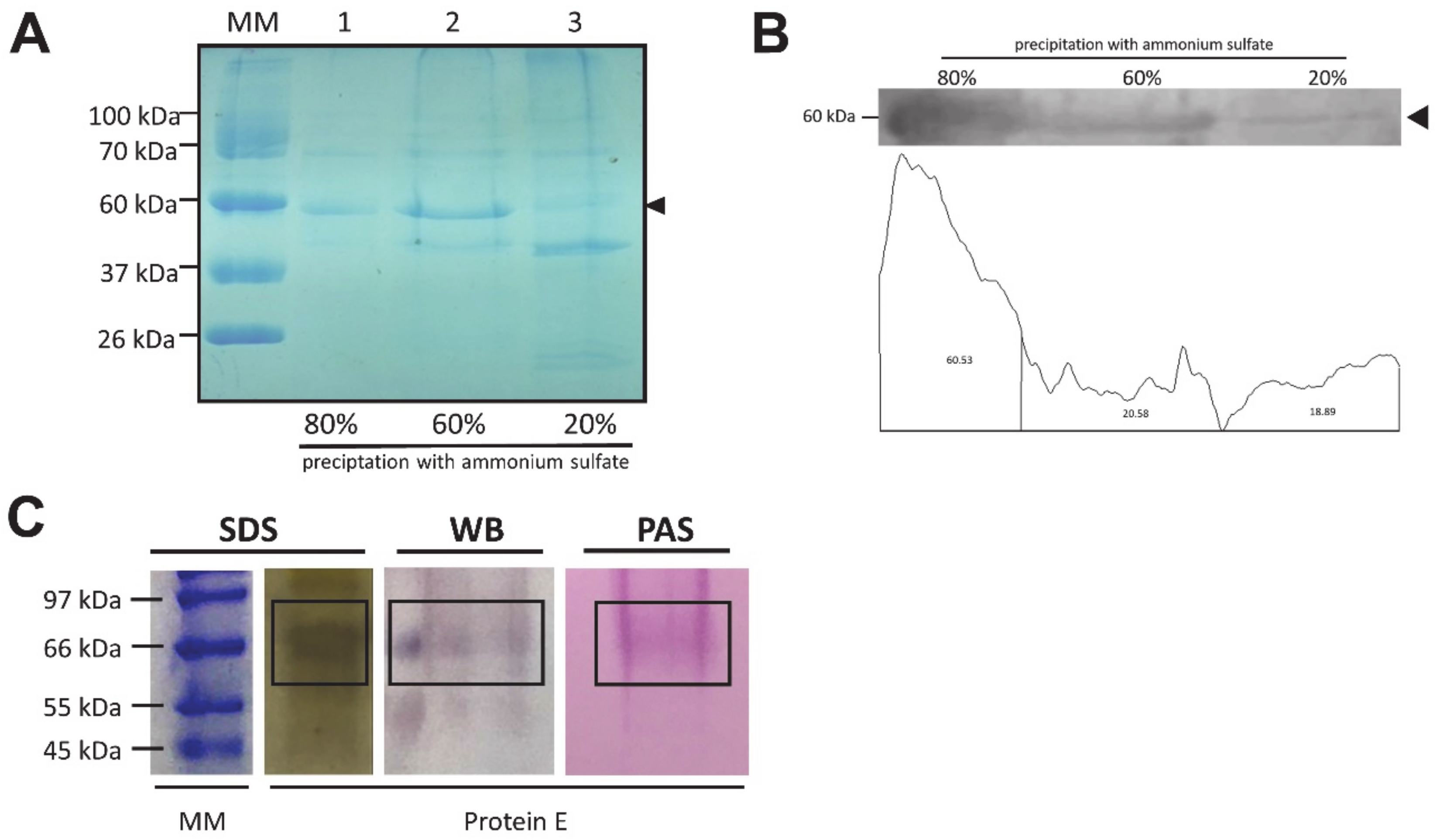
3.2. Expression of Recombinant Proteins
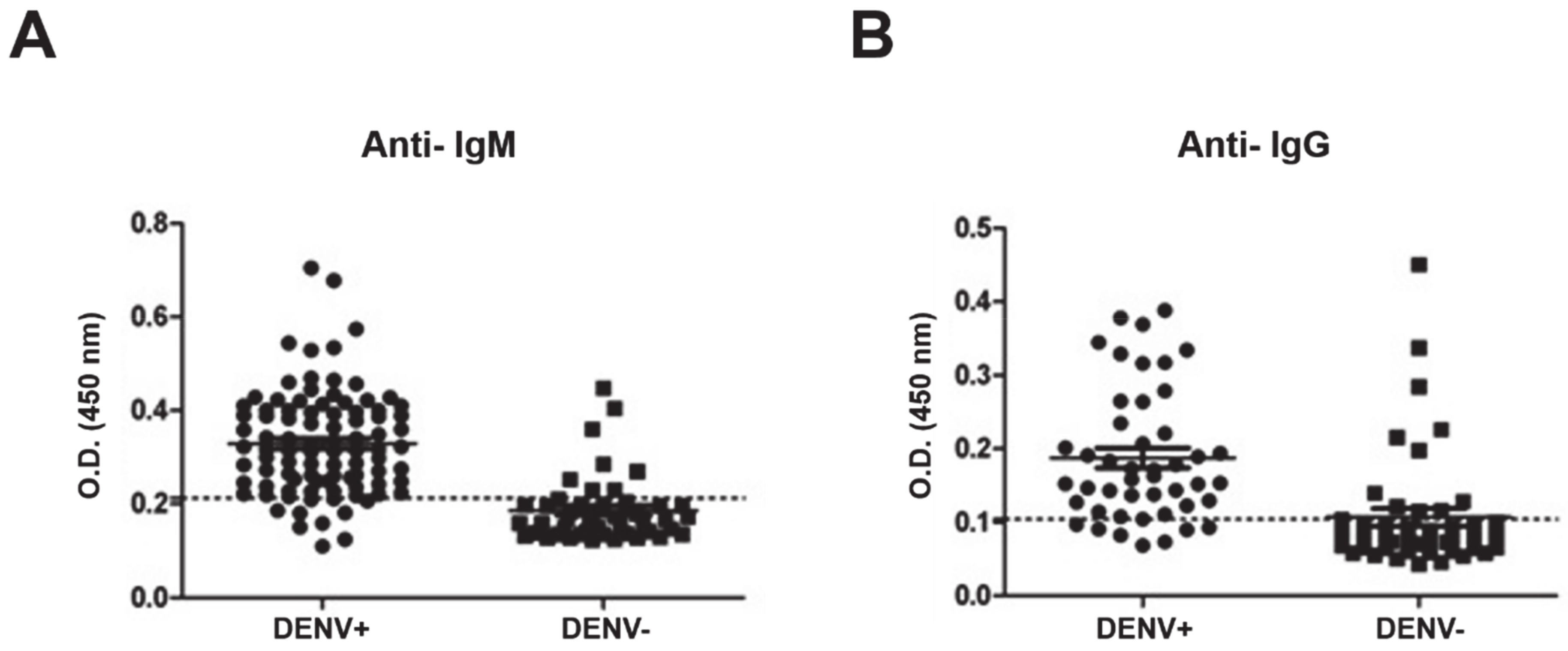
To obtain the prM/M and E proteins of DENV3, P. pastoris containing the genes of interest were grown in a selective medium and induced with 1% methanol every 12 h. The culture supernatant was subjected to precipitation with increasing concentrations of ammonium sulfate (20%, 60%, and 80%) and analyzed by electrophoresis, as shown in Figure 3A. The quantification of the bands by densitometry demonstrated that most of the protein E was precipitated from the fraction of 80% of ammonium sulfate (Table 2). The fractions were analyzed by Western blot using sera from patients with confirmed dengue, evidencing the presence of a band of 60 kDa in all fractions corresponding to the molecular weight of protein E. The densitometry graph of the bands marked with antibody confirms the presence of a greater amount of protein E in the precipitation fraction with 80% ammonium sulfate (Figure 3B). This fraction of protein E, precipitated with 80% ammonium sulfate, was analyzed separately and confirmed by the PAS reaction being a glycoprotein (Figure 3C). The same band observed on the SDS-PAGE and Western blot was stained at the height of 60 kDa on the PAS membrane.

3.3. ELISA

Fractions obtained from precipitation with ammonium sulfate were used as antigens to sensitize plates for indirect ELISA. The originating fraction from precipitation with 80% ammonium proved most satisfactory as an antigen for detection of dengue, with a sensitivity of 82.61% and specificity of 89.25%, and with the cutoff of 0.2138 for the test determining the presence of anti-dengue IgM in sera analyzed. For the assay of IgG, the results obtained using the same fraction was a sensitivity of 76.09% with a specificity of 84.78% and 0.1043 cutoff (Figure 4 and Table 3).

4. Discussion

Dengue fever affects a large portion of the world’s population, mainly those located in tropical and subtropical regions in Asia, Oceania, Africa, and the Americas [5,6,22]. Since there are no effective vaccines available, rapid and inexpensive diagnostic tests are essential. In this work, we produced the structural proteins prM/M and E of dengue-3 virus in soluble form using yeast P. pastoris as an alternative for obtaining antigens in large scale. DENV-3 was the most prevalent serotype for several years in the first decade of this century in Brazil [23].
The culture supernatant was subjected to precipitation with concentrations of 20% to 80% of ammonium sulfate. As seen in SDS-PAGE (Figure 3A), there is the presence of a band of 60 kDa corresponding to E protein in all fractions analyzed, which was confirmed by Western blot (Figure 3B). This is in agreement with results described by Sugrue and colleagues, in which the production of the E protein of DENV1 in P. pastoris was followed by degradation by proteases [24]. PAS reaction confirms that the fraction of protein E precipitated with 80% ammonium sulfate was glycosylated in Pichia pastoris (Figure 3C). Glycosylation is a very important factor for the correct folding of the protein and to obtain an antigen more similar to that expressed in the mammalian host, affecting the recognition of antibodies in diagnostic tests positively.
However, the antigenicity of the proteins produced was preserved as confirmed through a retrospective study using the indirect ELISA. The fraction obtained by precipitation with 80% of ammonium sulfate provided a sensitivity of 82.61% and a specificity of 89.25% for determining the presence of anti-IgM antibodies in patient sera. These values obtained are according to the different sensitivity of commercial diagnostic kits, as described in a comparative study in which the efficacy of commercially available tests for determination of IgM antibodies in sera from patients were analyzed, which obtained a range of 85% to 89% of sensitivity and 88% to 100% of specificity [25]. Thus, antigens produced here are promising for use in diagnostic tests for dengue, and an optimization step can be performed to improve sensibility and specificity.
Despite the other fractions from precipitation with ammonium sulfate also containing the E protein of DENV3 recombinant, as shown in the Western blot assay, it showed to be less effective as an antigen for the ELISA assay (data not shown). One reason for this result is that the fraction of 80% may have contained most immunogenic epitopes of the virus, and thus provided greater power of discrimination between samples during the test.
Rocha and colleagues used a mixture of E proteins of four serotypes produced in E. coli as antigens in a test IgG-ELISA. The results were 91% sensitivity and 98% specificity [15]. Here, however, the results for the IgG-ELISA were 76.09% of sensitivity with a specificity of 84.78%. However, the fact that dengue viruses have epitopes that are also present in other flaviviruses which can trigger cross-reactions should be taken into account. These occur more frequently in relation to IgG antibodies than IgM [26,27]. Moreover, usually the IgG-ELISA tests have reduced sensitivity among the flaviviruses [6]. It is interesting to note that the sera used in this study are from patients in northern Brazil, where other epidemics caused by different flaviviruses occur. In addition, other factors that can affect the effectiveness of serological tests are the infecting serotype and whether the infection is primary or not [28].
However, it is important to note that the results are promising and of great value since the proposed methodology to obtain the antigens does not require expensive and laborious methods of protein purification. Thus, the structural proteins of other serotypes of dengue virus could be produced using the same strategy employed here, seeking to create inexpensive and large-scale diagnostic tests, which makes the production of these antigens accessible to regions where dengue is endemic.

5. Conclusions

From the data obtained in this study, we can conclude that the prM/M and E proteins were produced correctly in yeast P. pastoris and were good antigens when used in ELISA tests for a retrospective study of dengue. Moreover, the methodology provides a good alternative for obtaining dengue virus antigens on a large scale, since expensive and elaborate techniques for protein purification are not required, reducing the costs related to antigen production for development of point-of-care devices.

Author Contributions

Conceptualization, M.D.O.T., M.F.X., and S.O.D.P.; methodology, M.D.O.T., M.F.X., and J.W.O.P.; validation, M.F.X., J.W.O.P., and J.M.C.M.; formal analysis, M.F.X. and R.S.D.; investigation, M.D.O.T., M.F.X., J.W.O.P., J.M.C.M, and R.S.D.; resources, C.C.d.S. and S.O.D.P.; data curation, R.S.D., C.C.d.S., and S.O.D.P.; writing—original draft preparation, M.F.X., R.S.D., and S.O.D.P.; writing—review and editing, M.F.X., R.S.D., and S.O.D.P.; visualization, M.F.X., R.S.D., C.C.d.S., and S.O.D.P.; supervision, S.O.D.P.; project administration, S.O.d.P.; funding acquisition, S.O.D.P. All authors have read and agreed to the published version of the manuscript.

Funding

This research received funding of the FAPEMIG and CNPq.

Acknowledgments

This work was supported by CNPq, INCT Dengue and FAPEMIG. M.D.O.T. was supported by a scholarship from the CAPES.

Conflicts of Interest

The authors declare no conflict of interest.

References

  1. World Health Organization. Dengue and Severe Dengue. 2020. Available online: https://www.who.int/news-room/fact-sheets/detail/dengue-and-severe-dengue (accessed on 10 September 2020).
  2. Halstead, S.B. Dengue. Lancet 2007, 370, 1644–1652. [Google Scholar] [CrossRef]
  3. Gubler, D.J.; Kuno, G.; Markoff, L. Flaviviruses. In Fields Virology, 5th ed.; Knipe, D.M., Howley, P.M., Eds.; Wolters Kluwer and Lippincott Williams: Philadelphia, PA, USA, 2007; pp. 1153–1252. [Google Scholar]
  4. Perera, R.; Kuhn, R.J. Structural proteomics of dengue virus. Curr. Opin. Microbiol. 2008, 11, 369–377. [Google Scholar] [CrossRef] [PubMed] [Green Version]
  5. Whitehead, S.S.; Blaney, J.E.; Durbin, A.P.; Murphy, B.R. Prospects for a dengue virus vaccine. Nat. Rev. Genet. 2007, 5, 518–528. [Google Scholar] [CrossRef] [PubMed]
  6. Guzman, M.G.; Halstead, S.B.; Artsob, H.; Buchy, P.; Farrar, J.; Gubler, D.J.; Hunsperger, E.; Kroeger, A.; Margolis, H.S.; Martinez, E.; et al. Dengue: A continuing global threat. Nat. Rev. Genet. 2010, 8, S7–S16. [Google Scholar] [CrossRef] [PubMed] [Green Version]
  7. Heinz, F.X.; Stiasny, K. Flaviviruses and flavivirus vaccines. Vaccine 2012, 30, 4301–4306. [Google Scholar] [CrossRef] [PubMed]
  8. Tan, T.T.T.; Bhuvanakantham, R.; Li, J.; Howe, J.; Ng, M.-L. Tyrosine 78 of premembrane protein is essential for assembly of West Nile virus. J. Gen. Virol. 2009, 90, 1081–1092. [Google Scholar] [CrossRef]
  9. Zheng, A.; Umashankar, M.; Kielian, M. In Vitro and In Vivo Studies Identify Important Features of Dengue Virus pr-E Protein Interactions. PLoS Pathog. 2010, 6, e1001157. [Google Scholar] [CrossRef]
  10. Rey, F.A. Dengue virus envelope glycoprotein structure: New insight into its interactions during viral entry. Proc. Natl. Acad. Sci. USA 2003, 100, 6899–6901. [Google Scholar] [CrossRef] [Green Version]
  11. De Paula, S.O.; Fonseca, B.A.L. Dengue: A review of the laboratory tests a clinican must know to achieve a correct diagnosis. Braz. J. Infect. Dis. 2004, 8, 390–398. [Google Scholar] [CrossRef] [Green Version]
  12. Le, R.D.; Melanson, S.E.F. What’s New in Point-of-Care Testing? Point Care J. Near-Patient Test. Technol. 2014, 13, 152–155. [Google Scholar] [CrossRef]
  13. Holmes, D.A.; Purdy, D.E.; Chao, D.-Y.; Noga, A.J.; Chang, G.-J.J. Comparative Analysis of Immunoglobulin M (IgM) Capture Enzyme-Linked Immunosorbent Assay Using Virus-Like Particles or Virus-Infected Mouse Brain Antigens To Detect IgM Antibody in Sera from Patients with Evident Flaviviral Infections. J. Clin. Microbiol. 2005, 43, 3227–3236. [Google Scholar] [CrossRef] [PubMed] [Green Version]
  14. De Paula, S.O.; Dutra, N.R.; De Paula, M.B.; De Oliveira, M.D.; De Oliveira, L.L. The laboratorial diagnosis of dengue: Applications and implications. J. Glob. Infect. Dis. 2009, 1, 38–44. [Google Scholar] [CrossRef] [PubMed]
  15. Batra, G.; Gurramkonda, C.; Nemani, S.K.; Jain, S.K.; Swaminathan, S.; Khanna, N. Optimization of conditions for secretion of dengue virus type 2 envelope domain III using Pichia pastoris. J. Biosci. Bioeng. 2010, 110, 408–414. [Google Scholar] [CrossRef] [PubMed]
  16. Liu, W.; Jiang, H.; Zhou, J.; Yang, X.; Tang, Y.; Fang, D.; Jiang, L. Recombinant dengue virus-like particles from Pichiapastoris: Efficient production and immunological properties. Virus Genes 2010, 40, 53–59. [Google Scholar] [CrossRef]
  17. Rocha, E.S.O.; Oliveira, J.G.; Santos, J.R.; Rodrigues, G.O.L.; Figueiredo, L.B.; Pessanha, E.M.; Proietti, F.A.; Fonseca, F.G.; Bonjardim, C.A.; Ferreira, P.C.P.; et al. Recombinant envelope protein-based enzyme immunoassay for IgG antibodies is comparable to netralization tests for epidemiological studies of dengue infection. J. Virol. Methods 2013, 187, 114–120. [Google Scholar] [CrossRef]
  18. Álvarez-Rodríguez, L.M.; Ramos-Ligonio, A.; Rosales-Encina, J.L.; Martínez-Cázares, M.T.; Parissi-Crivelli, A.; López-Monteoni, A. Expression, Purification, and evaluation of Diagnostic Potential and Immunogenicity of Recombinant NS3 Protein from All Serotypes of Degue Virus. J. Trop. Med. 2012. [Google Scholar] [CrossRef]
  19. Cardoso, S.A.; Paixão, V.F.; Oliveira, M.D.; Honda, E.R.; Oliveira, L.L.; da Silva, C.C.; De Paula, S.O. Dengue-1 envelope protein domain III produced in Pichiapastoris: potential use for serological diagnosis. Protein Expr. Purif. 2013, 92, 9–13. [Google Scholar] [CrossRef] [Green Version]
  20. Daly, R.; Hearn, M.T.W. Expression of heterologous proteins in Pichiapastoris: a useful experimental tool in protein engineering and production. J. Mol. Recognit. 2005, 18, 119–138. [Google Scholar] [CrossRef]
  21. Krainer, F.W.; Dietzsch, C.; Hajek, T.; Herwig, C.; Spadiut, O.; Glieder, A. Recombinant protein expression in Pichiapastoris strains with an engineered methanol utilization pathway. Microb. Cell Factories 2012, 11. [Google Scholar] [CrossRef] [Green Version]
  22. Gubler, D.J. Dengue and Dengue Hemorrhagic Fever. Clin. Microbiol. Rev. 1998, 11, 480–496. [Google Scholar] [CrossRef] [Green Version]
  23. Bavia, L.; Melanda, F.N.; Arruda, T.B.; Mosimann, A.L.P.; Silveira, G.F.; Aoki, M.N.; Kuczera, D.; Sarzi, M.L., Jr.; Conchon-Costa, I.; Pavanelli, W.R.; et al. Epidemiological study on dengue in southern Brazil under the perspective of climate and poverty. Sci. Rep. 2020, 10, 2127. [Google Scholar] [CrossRef] [PubMed]
  24. Sugrue, R.J.; Cui, T.; Xu, Q.; Fu, J.; Chan, Y.C. The production of recombinant dengue vírus E protein using Escherichia coli and Pichiapastoris. J. Virol. Methods 1997, 69, 159–169. [Google Scholar] [CrossRef]
  25. Blacksell, S.D.; Jarman, R.G.; Gibbons, R.V.; Tanganuchitcharnchai, A.; Mammen, M.P.; Nisalak, A.; Kalayanarooj, S.; Bailey, M.; Premaratna, R.; De Silva, H.J.; et al. Comparison of Seven Commercial Antigen and Antibody Enzyme-Linked Immunosorbent Assays for Detection of Acute Dengue Infection. Clin. Vaccine Immunol. 2012, 19, 804–810. [Google Scholar] [CrossRef] [Green Version]
  26. Cuzzubbo, A.J.; Vaughn, D.W.; Nisalak, A.; Solomon, T.; Kalayanarooj, S.; Aaskov, J.; Dung, N.M.; Devine, P.L. Comparison of PanBio Dengue Duo Enzyme-Linked Immunosorbent Assay (ELISA) and MRL Dengue Fever Virus Immunoglobulin M Capture ELISA for Diagnosis of Dengue Virus Infections in Southeast Asia. Clin. Diagn. Lab. Immunol. 1999, 6, 705–712. [Google Scholar] [CrossRef] [PubMed] [Green Version]
  27. Koraka, P.; Zeller, H.; Niedrig, M.; Osterhaus, A.D.; Groen, J. Reactivity of serum 473 samples from patients with a flavivirus infection measured by immunofluores- 474 cence assay and ELISA. Microbes Infect. 2002, 4, 1209–1215. [Google Scholar] [CrossRef]
  28. Fry, S.R.; Meyer, M.; Semple, M.G.; Simmons, C.P.; Sekaran, S.D.; Huang, J.X.; McElnea, C.; Huang, C.; Valks, A.; Cooper, M.A. The diagnostic sensivity of dengue rapid test assays is significantly enhanced by using a combined antigen and antibody testing approach. PLoS Negl. Trop. Dis. 2011, 5, 1–8. [Google Scholar] [CrossRef] [Green Version]
Figure 1. The prM/M and E sequences used for expression in P. pastoris. (A) Sequence of prM/M and E proteins of dengue-3 virus that was codon optimized for expression in yeast; (B) scheme of the construction of plasmid pPICZαA-prM/M-E-DENV3.
Fermentation 06 00088 g001
Figure 2. Construction of Pichia pastoris expressing recombinant prM/M and E proteins. (A) Amplification by PCR with primers for the domain III of the E protein of dengue-3 virus; lines (1–3) show the band of 408 bp related to domain III of dengue virus-3; line 4 shows the negative control and MM line, the molecular marker. (B) PCR with primers for AOX1 were used. Lines 1 and 2 show the band of 2300 bp; line MM, molecular marker.
Fermentation 06 00088 g002
Figure 3. Molecular characterization of recombinant protein. (A) SDS-PAGE; MM—protein molecular maker; (1–3) fractions from precipitation of the supernatant of culture from the yeast with 80%, 60%, and 20% ammonium sulfate; arrow head indicates the bands corresponding to the E protein DENV3. (B) Western blot; MM—molecular weight marker; fractions from precipitation with concentrations of 80%, 60%, and 20% of ammonium sulfate using sera from patients with anti-dengue positive. Bands of 60 kDa protein corresponding to E protein are observed in all fractions. (C) Protein E (fraction 80% ammonium sulfate): SDS-PAGE silver-stained; Western blot and PAS assays, respectively; MM—molecular marker.
Fermentation 06 00088 g003
Figure 4. Indirect ELISA using protein E-DENV3 produced in P. pastoris. Results of dengue-positive and negative (healthy individuals) samples in IgM (A) and IgG (B) detection. The horizontal line indicates the cutoff value of the assay.
Fermentation 06 00088 g004
Table 1. Serological status of the serum used in this study and test type used.
PositiveNegativeTest
Anti-IgM9246Dengue Duo IgM and IgG ELISA Capture Kits
+
MAC-ELISA IgM
Anti-IgG4646Dengue Duo IgM and IgG ELISA Capture Kits
Table 2. Quantification of E protein by band densitometry.
Ammonium sulfateDensitometry (Area)[] µg/mLYield
80%46,031131.21353.52 %
60%36,070102.773141.92 %
20%399711.201714.56 %
Standard curve equation: y = 350.25x + 74,009; R² = 0.9933.
Table 3. Sensitivity and specificity of the anti-dengue IgG indirect ELISA.
% Sensitivity% SpecificityCut-Off
Anti-IgM82.6189.250.2138
Anti-IgG76.0984.780.1043

