Article

The Effect of Electrospun Polycaprolactone Nonwovens Containing Chitosan and Propolis Extracts on Fresh Pork Packaged in Linear Low-Density Polyethylene Films

by
Emeli Vargas Romero
1,
Loong-Tak Lim
2,
Héctor Suárez Mahecha
1 and
Benjamin M. Bohrer
3,*
1
Instituto de Ciencia y Tecnología de Alimentos, Universidad Nacional de Colombia, Bogotá 111321, Colombia
2
Department of Food Science, University of Guelph, Guelph, ON N1G-2W1, Canada
3
Department of Animal Sciences, The Ohio State University, Columbus, OH 43210, USA
*
Author to whom correspondence should be addressed.
Submission received: 10 April 2021 / Revised: 10 May 2021 / Accepted: 13 May 2021 / Published: 17 May 2021
(This article belongs to the Special Issue Plant Bioactive Compounds in Foods and Food Packages)

Abstract

:
Fresh meat products are highly perishable and require optimal packaging conditions to maintain and potentially extend shelf-life. Recently, researchers have developed functional, active packaging systems that are capable of interacting with food products, package headspace, and/or the environment to enhance product shelf-life. Among these systems, antimicrobial/antioxidant active packaging has gained considerable interest for delaying/preventing microbial growth and deteriorative oxidation reactions. This study evaluated the effectiveness of active linear low-density polyethylene (LLDPE) films coated with a polycaprolactone/chitosan nonwoven (Film 1) or LLDPE films coated with a polycaprolactone/chitosan nonwoven fortified with Colombian propolis extract (Film 2). The active LLDPE films were evaluated for the preservation of fresh pork loin (longissimus dorsi) chops during refrigerated storage at 4 °C for up to 20 d. The meat samples were analyzed for pH, instrumental color, purge loss, thiobarbituric acid reactive substances (TBARS), and microbial stability (aerobic mesophilic and psychrophilic bacteria). The incorporation of the propolis-containing nonwoven layer provided antioxidant and antimicrobial properties to LLDPE film, as evidenced by improved color stability, no differences in lipid oxidation, and a delay of 4 d for the onset of bacteria growth of pork chops during the refrigerated storage period.

1. Introduction

Fresh meat is highly perishable and therefore requires optimal processing, packaging, and storage systems. The rapid deterioration of fresh meat quality is mainly attributed to the activation of biological reactions such as the oxidation of myoglobin to metmyoglobin, which causes meat to become brown and discolored, and lipid oxidation, which leads to off-flavors and off-aromas that are generally described as rancid [1]. Other intrinsic factors of meat such as water activity and pH favor the growth of microorganisms that cause microbial spoilage, which can lead to economic losses for the meat industry and health risks to the human population [2,3].
A number of innovative packaging strategies have been developed in recent years to extend the shelf-life of minimally processed food products. For example, researchers have developed active packaging systems that are capable of interacting with the food product, package headspace, and/or the environment to enhance product shelf-life [4,5,6,7,8,9,10]. Among these systems, antimicrobial active packaging has gained considerable interest for delaying/preventing microbial growth in the product via controlled delivery of antimicrobial agents from the package structure or add-on carrier components (e.g., label, sachet, insert) [4,11,12,13]. By virtue of their large surface area, electrospun nonwoven fibers are promising materials for the development of antimicrobial active packaging. They are versatile carriers of bioactive substances (e.g., antimicrobials, antioxidants) for controlling their release.
Polycaprolactone is a biodegradable, hydrophobic aliphatic polyester, widely used in controlled release systems of active substances [14], such as tea polyphenols, carnosic acid, and silver chloride [15,16,17]. Chitosan is a chitin-derived polysaccharide with a broad spectrum of useful attributes including antibacterial activity, film-forming capacity, and biodegradability [18]. Propolis is a natural resinous substance collected by honeybees, formed with a wide variety of chemical compounds such as polyphenols, flavonoids, and sesquiterpenes, which exhibit a wide-range of biological activities [19,20,21]. Ethanol propolis extract (PE) derived in Colombia has stood out as a natural food preservative due to its significant antioxidant, antimicrobial, and antifungal properties [22,23,24]. Moreover, the successful addition of PE as an antimicrobial has been well documented. For instance, PE has been used in the development of different antimicrobial nonwovens derived from other polymers, such as polyvinylpyrrolidone, polyurethane, poly (lactic acid), and zein. Nonwovens developed with PE have been mostly used for biomedical applications [25,26,27,28,29,30] but also for several food applications [31,32,33].
To our knowledge, linear low-density polyethylene (LLDPE) films coated with polycaprolactone nonwoven blended with either chitosan or chitosan and PE have not been reported before. Thus, the objective of the current study was to evaluate the antimicrobial and antioxidant properties of these active packaging films for the preservation of perishable meat products, using pork loin as a model system.

2. Materials and Methods

2.1. Propolis Extraction

A quantity of 60 g of raw propolis from Santander, Colombia, was mixed with 600 mL of an ether/ethanol solution (at a ratio of 60:40 v/v), placed in an ultrasound bath for 1 h, and centrifuged at 10,000 rpm for 5 min. The supernatant was filtered under vacuum conditions. The extract was then refrigerated (4 °C) for 2 h, followed by a second centrifugation at 10,000 rpm for 5 min. Finally, the extract was evaporated at 60 °C using a rotary evaporator (IKA, digital RV 10 V, Ciudad de México, CDMX, México).
The minimum inhibitory concentration of the Colombian PE was determined using the micro-dilution technique described by Bonou et al. [34]. Briefly, 317 µL of PE was dissolved in 3664 µL of Mueller Hinton broth (BD Difco, Fisher Scientific, Waltham, MA, USA) and 16 µL of Tween 80 to reach a final concentration of 80 mg/mL. Microplates (96-well) were used for the visual determination of microbial growth. Successive dilutions (two dilutions) were performed with the stock solution, and the dilutions were subsequently inoculated with 106 CFU of bacteria. A positive control (bacterium + medium) and a negative control (PE + medium) were performed. The microplates were covered and incubated at 37 °C for 24 h. The bacterial strains used were Staphylococcus aureus ATCC 29737, Escherichia coli ATCC 25922 and Salmonella enterica subsp. enterica serovar Enteritidis ATCC 13076 (Thermo Fisher Scientific Culti-Loops, Waltham, MA, USA).
The total phenolic content of PE was determined according to methods described by Singleton et al. [35] and adapted by Siripatrawan and Vitchayakitti [36]. Briefly, 0.1 mL of PE was dissolved in 9.9 mL of 96% ethanol, and subsequently, 0.1 mL of the solution was added to 7 mL of Milli-Q water and 0.5 mL of Folin–Ciocalteu reagent. The mixture was allowed to equilibrate for 2 min. An aliquot of 1.5 mL of 20% sodium carbonate and 0.9 mL of Milli-Q water were added to the mixture, homogenized, and left to rest for 2 h. The reaction was read at 765 nm using a spectrophotometer (Evolution 60s, Thermo Fisher Scientific, Waltham, MA, USA). The total phenol contents in the extracts were expressed as gallic acid equivalents (mg GAE/g PE).
The method described by Moon and Shibamoto [37] was adapted to determine the antioxidant activity. A stock solution of 38.41 mg of ABTS diammonium salt (7.00 mM) and 6.63 mg of potassium persulfate (2.45 mM) was used. The absorbance was read at 734 nm using a spectrophotometer (Evolution 60s, Thermo Fisher Scientific, Waltham, MA, USA). The results were expressed as equivalent of µmol Trolox/g of PE.

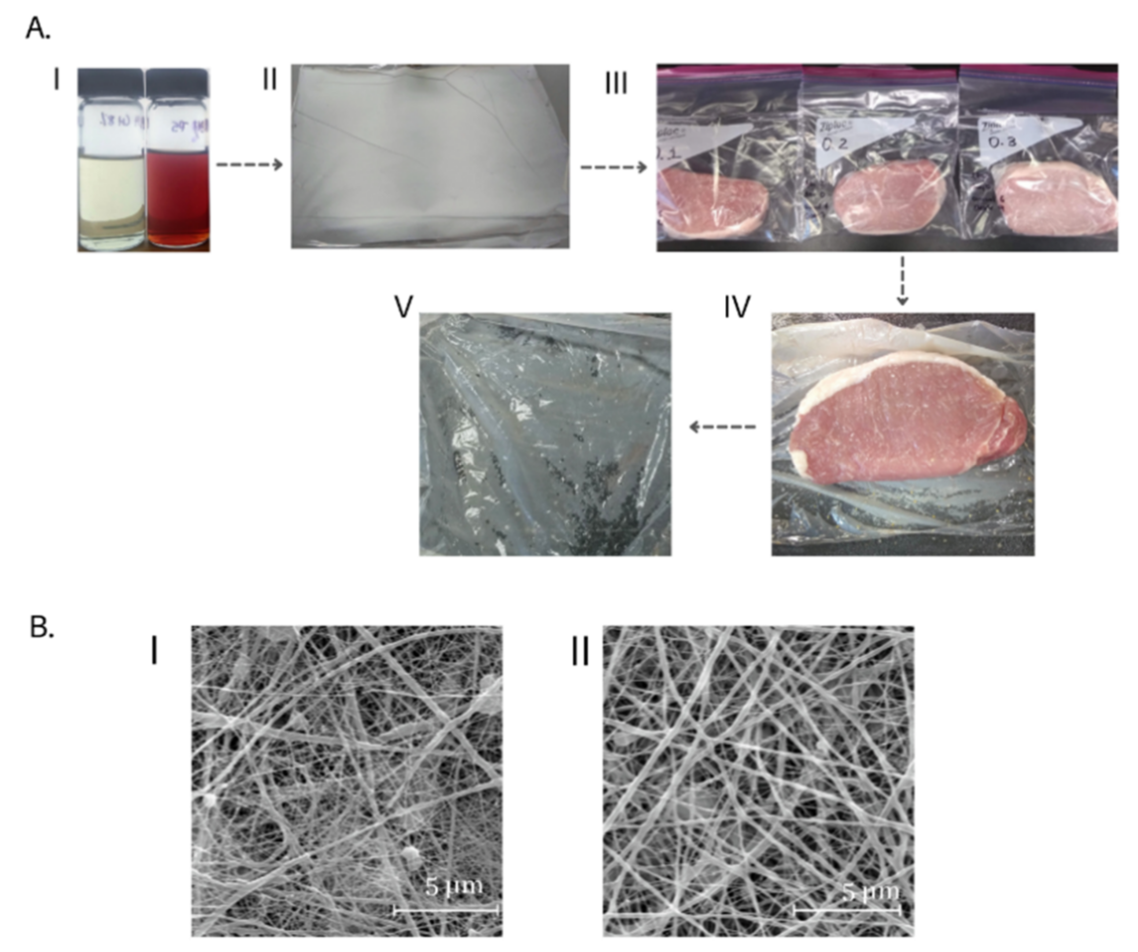
2.2. Preparation of Solutions and Electrospinning Process

The spin dope solutions for electrospinning were comprised of polycaprolactone (MW = 80,000 Da, Sigma-Aldrich, St. Louis, Missouri, USA) and chitosan (50,000–190,000 Da, Sigma-Aldrich, St. Louis, Missouri, USA; degree of deacetylation 75–85%). Briefly, a binary solvent of formic acid (88%, #A118P-500, Fisher Scientific, Waltham, MA, USA) and glacial acetic acid (≥99.7%, #LC1010036, Fisher Scientific) at a ratio of 60:40 (v/v) was made. Then, polycaprolactone granules were added at 14% (w/v) of the binary solvent to make the polycaprolactone solution. Chitosan powder was added to the polycaprolactone solution at 8% of the polycaprolactone granules (w/w) to generate the spin dope solution for Solution 1. Propolis extract was added at 5% of the weight of polycaprolactone granules and chitosan powder (w/w) to generate Solution 2. Both solutions were mixed with a magnetic stirring bar for 18 h at 21 °C to form homogeneous spin dope solutions.
The properties of the spin dope solutions for both solutions were analyzed (in triplicate) based on methods described previously by Zaitoon and Lim [38]. Apparent viscosity of the solutions at a shear rate of 100 s−1 was 9.24 ± 0.01 × 10⁻³ Pa·s for Solution 1 and 7.45 ± 0.01 × 10⁻³ Pa·s for Solution 2. Electrical conductivity was 639.9 ± 5.55 µS/cm for Solution 1 and 592.9 ± 6.45 µS/cm for Solution 2. Surface tension was 56.4 ± 0.5 mN/m for Solution 1 and 52.2 ± 0.37 mN/m for Solution 2.
These spin dope solutions were electrospun into nonwovens using free surface electrospinning equipment (Nanospider, Elmarco, Czech Republic) at a constant voltage of 80 kV, a carriage speed of 50 mm/s, and an electrode-collector distance of 180 mm. Linear low-density polyethylene film (LLDPE; Glad Food Pro, Oakland, CA, USA) was used as a collection substrate for the deposition of the electrospun nonwoven, forming Film 1 (LLDPE film deposited with polycaprolactone/chitosan nonwoven; Solution 1) and Film 2 (LLDPE film deposited with polycaprolactone/chitosan nonwoven fortified with Colombian PE; Solution 2). The electrospinning process was conducted at 21 ± 2 °C and 45% RH for 35 min. The developed films were stored at 25 °C in an environmental test chamber (Sanyo, Osaka, Japan) for 24 h before further testing.

2.3. Pork Loin Chops

Six boneless pork loins (longissimus dorsi), with the same lot/origination number, were commercially procured from a supermarket in Guelph, Ontario, Canada. All of the pork was purchased on a common day and the common origination date suggested that all samples were of common post-mortem aging time. Nine 2.54-cm thick chops (same shape: untrimmed height and width) were cut and portioned from each loin. A total of 54 chops were weighed and randomly distributed for assignment to three different treatments as follows: (1) Treatment 1—pork chops were wrapped in a commercial LLDPE film without a functional nonwoven component (CON); (2) Treatment 2—pork chops were wrapped in Film 1; and (3) Treatment 3—pork chops were wrapped in Film 2. The wrapped and packaged pork chops were then placed inside reusable/re-sealable plastic storage bags (Ziploc; S.C. Johnson & Son, Racine, WI, USA). The bags were properly sealed and stored in a dark refrigerated room at a temperature of 4 ± 1 °C. Samples (three samples per treatment on each day) were collected for analyses on 0, 4, 8, 12, 16, and 20 days following packaging. Samples for day 0 analysis were collected at 4 h post-packaging. Following sample collection, weights for purge loss during storage, instrumental color, and pH were collected immediately. Samples were then divided into two subsamples with one subsample frozen at −20 °C for future determination of lipid oxidation and one subsample frozen at −20 °C for future microbiological analysis.

2.4. Purge Loss during Storage

The weight loss (WL) of chops during storage was determined according to the methodology described by Vargas and Bohrer [39]. The following equation was used:
%   WL = W Initial W Final W Initial   ×   100
where WInitial was the initial weight of the pork chop, and WFinal was the final weight of the pork chop following the designated storage time.

2.5. Instrumental Color

The surface color of pork chops (film removed) was measured using a Chroma Meter CR-400 colorimeter with a 10° viewing angle and D65 illuminance (Konica Minolta, Osaka, Japan). The CIE L* a* b* color space was used, where L* indicated lightness of the color (L* = 0 indicates black and L* = 100 indicates white), a* indicated position between red and green (negative values indicate green while positive values indicate red), and b* indicated position between yellow and blue (negative values indicate blue and positive values indicate yellow). Total color change (ΔE) was calculated using Equation (2).
Δ E = L L 2   + a a 2 + b b 2
where L, a, and b denote the L*, a*, and b* values, respectively, of the pork loins before packaging and L′, a′, and b′ the L*, a*, and b* values obtained, respectively, on 4, 8, 12, 16, or 20 days of storage. The analyses were observed at three different locations for each pork chop, and averages of the three locations were reported.

2.6. pH Evaluation

The pH of the pork chops was measured using a portable pH meter (Hanna Instruments; HI 3779BE, Laval, Québec, QC, Canada) calibrated with pH buffer solutions of pH 4, 7, and 9. The internal pH was measured by penetration of the probe at a depth of 2.0 cm below the surface of each pork chop, and an average of three different readings was reported.

2.7. Lipid Oxidation

At the end of the designated storage periods, a portion of each sample was vacuum-packaged and frozen at −20 °C until further analysis for lipid oxidation. The determination of the content of thiobarbituric acid reactive substances (i.e., TBARS) was conducted to determine the concentration of malondialdehyde (MDA), which is one of several end products formed following the decomposition of lipid peroxidation products. A sample (5 g) from each chop was ground and homogenized with 1 mL of butyl hydroxytoluene and 45.5 mL of trichloroacetic acid/phosphoric acid. This was followed by filtration through a filter paper (Whatman No. 1, Fisher Scientific, Waltham, NH, USA) and addition of 5 mL of thiobarbituric acid. The solutions were incubated at 21 ± 2 °C for 22.5 h in the dark. A standard curve was created by diluting 1,1,3,3, tetraethoxy propane of 25 µM to various concentrations. The reaction obtained was read on a microplate spectrophotometer (BioTek, Winooski, VT, USA) at 530 nm. The results were reported as the quantification for mg of malondialdehyde (MDA) per kg of meat.

2.8. Microbial Analysis

After the designated storage periods, a portion of each pork chop was kept in the original packaging and over-packaged in a vacuum sealed package and frozen at −20 °C until the microbiological test took place. Each portion (10 g) was homogenized in 90 mL of sterile 0.1% buffered peptone water solution (Fisher Scientific, Waltham, NH, USA) using a stomacher (Seward Stomacher® 400 Circulator, Lab Blender, Thomas Scientific, Swedesboro, NJ, USA) for 2 min at 300 rpm. Serial dilutions of 1:10 were prepared and homogenized using 0.1% sterile peptone water. An aliquot of 0.1 mL for each dilution was spread on PCA agar plates (Agar Plate Count, BD Difco, Fisher Scientific, Waltham, NH, USA). For aerobic mesophilic bacteria quantification, plates were incubated at 30 °C for 48 h, and for aerobic psychrophilic bacteria quantification, the plates were incubated at 4 °C for 10 d. Microbiological quantification was expressed as a logarithm of the number of colony-forming units per gram of sample (log CFU/g). Microbiological analyses were conducted in triplicate.

2.9. Experimental Design and Statistical Analysis

Six different pork loins were used, and from each pork loin, nine chops were obtained (n = 54). Three treatments and six storage days were applied to the pork chops using a completely randomized design where different loin chops were randomly assigned to treatment × day. With the completely randomized design, two to four chops from each of the six loins were randomized to each treatment.
The normality of the data was determined using PROC UNIVARIATE of SAS version 9.4 (SAS Institute, Cary, NC, USA). Subsequently, data were subjected to an ANOVA using PROC GLM of SAS with treatment (CON, Film 1 and Film 2) and storage time (0, 4, 8, 12, 16, and 20 days) as fixed effects. A Tukey–Kramer adjustment for protection of type I statistical error was applied. Statistical significance for parameters was declared at p < 0.05.

3. Results and Discussion

3.1. Characterization of Propolis Extracts

Raw propolis cannot be used directly as a raw material due to its high content of pollutants, which mainly consists of waxes and resins [32]. Therefore, it is necessary to extract polar and non-polar active components of propolis using an appropriate solvent. It has been reported that ethanol and ether are suitable solvents for the extraction of the main active compounds found in propolis, such as polyphenols, flavonoids, terpenoids, and fatty acids [40,41]. Using a blend of these solvents, the extraction yield calculated in the current study was 9.5%.
The antimicrobial results of PE showed a significant bactericidal effect on Gram-positive bacteria with a minimum inhibitory concentration of 0.15 mg/mL (Table 1). These results were in agreement with those reported for ethanol extracts of propolis from different geographical regions [42,43]. The PE used in the current study showed a moderate action against Gram-negative bacteria with a minimum inhibitory concentration greater than 10 mg/mL. Tosi, Ré, Ortega, and Cazzoli [31] identified a minimum inhibitory concentration of 19.2 ± 3.5 mg/mL against E. coli for Argentine propolis, which could be explained by the significant amounts of quercetin, galangina, caffeic acid, and chrysin found in Argentine propolis. Gutiérrez and Suárez [23] also found moderate bactericidal effects against Gram-negative bacteria for the ethanolic extracts derived from propolis from Santander, Colombia. However, Colombian propolis from other regions has shown limited activity against Gram-negative bacteria [44]. The antimicrobial action of PE depends on the interaction of its phenolic compounds with other components such as terpenes and terpenoids, both of which are bactericidal and can block cell division, limit protein synthesis [45], and destroy the cell and/or cytoplasmic wall [46].
Propolis possesses considerable antioxidant and anti-radical activity due to its phenolic compounds [47], which can interrupt lipid oxidation [48] and capture reactive oxygen species [49]. In the current study, PE contained 82.6 ± 2 mg GAE/g PE and 1186 ± 52 µmol/g PE ABTS radicals, which are indicators for total phenolic concentrations and antioxidant activity, respectively. These values were higher than those reported for PE from other Colombian regions [50,51], which may suggest that the PE from the Santander region of Colombia contained a higher concentration of active compounds, which might have contributed to the antimicrobial activity of Film 2.

3.2. Qualitative Observations on Composite LLDPE Films

The prepared nonwoven lined LLDPE films appeared white in color (Figure 1A, image II). However, when the nonwoven lined LLDPE film came into physical contact with the meat samples, the composite film became translucent (Figure 1A, images III and V). It is noteworthy that when the packaging was removed, the nonwoven remained adhered to the LLDPE film throughout the experiment (Figure 1A, images IV and V). The change in the white color of the coatings may have been caused by the transformation of the polycaprolactone from an initial vitreous state to an elastomeric state due to moisture transfer from the meat sample. In addition, the low differences in the refractive indices between fiber–air versus fiber–liquid of the composite within the free volume might have caused the film to become translucent [52]. As reported by Sánchez et al. [53], the moisture absorption of the polycaprolactone fibers can lower their glass transition temperature below the test temperature. The plasticization phenomenon might have also improved the flexibility of the coatings, thereby preventing them from breaking during the testing conducted in this study (i.e., pork chop application). The microstructural morphology of the two films, as observed under scanning electron microscopy, is presented in Figure 1B.

3.3. Meat Quality Parameters

3.3.1. Purge Loss

The purge loss of the samples was affected by the storage time for all treatments (Table 2). Within each storage day, there were no differences (p > 0.05) among treatment for purge loss. Purge loss in CON reached the highest statistical level on day 4 of storage, while the highest statistical level for purge loss was observed on day 8 and day 16 for Film 1 and Film 2, respectively. The delay in purge loss thresholds for Film 1 and Film 2 samples may have been caused by the ultra-thin diameter of the polycaprolactone/chitosan electrospun fibers, which produces a smaller liquid-fiber contact area and subsequently increases the air-contact area [53]. On the other hand, the presence of PE in the nonwovens can reduce the hydrophilic nature of chitosan [36], which might have allowed for increased moisture retention in the pork chops.

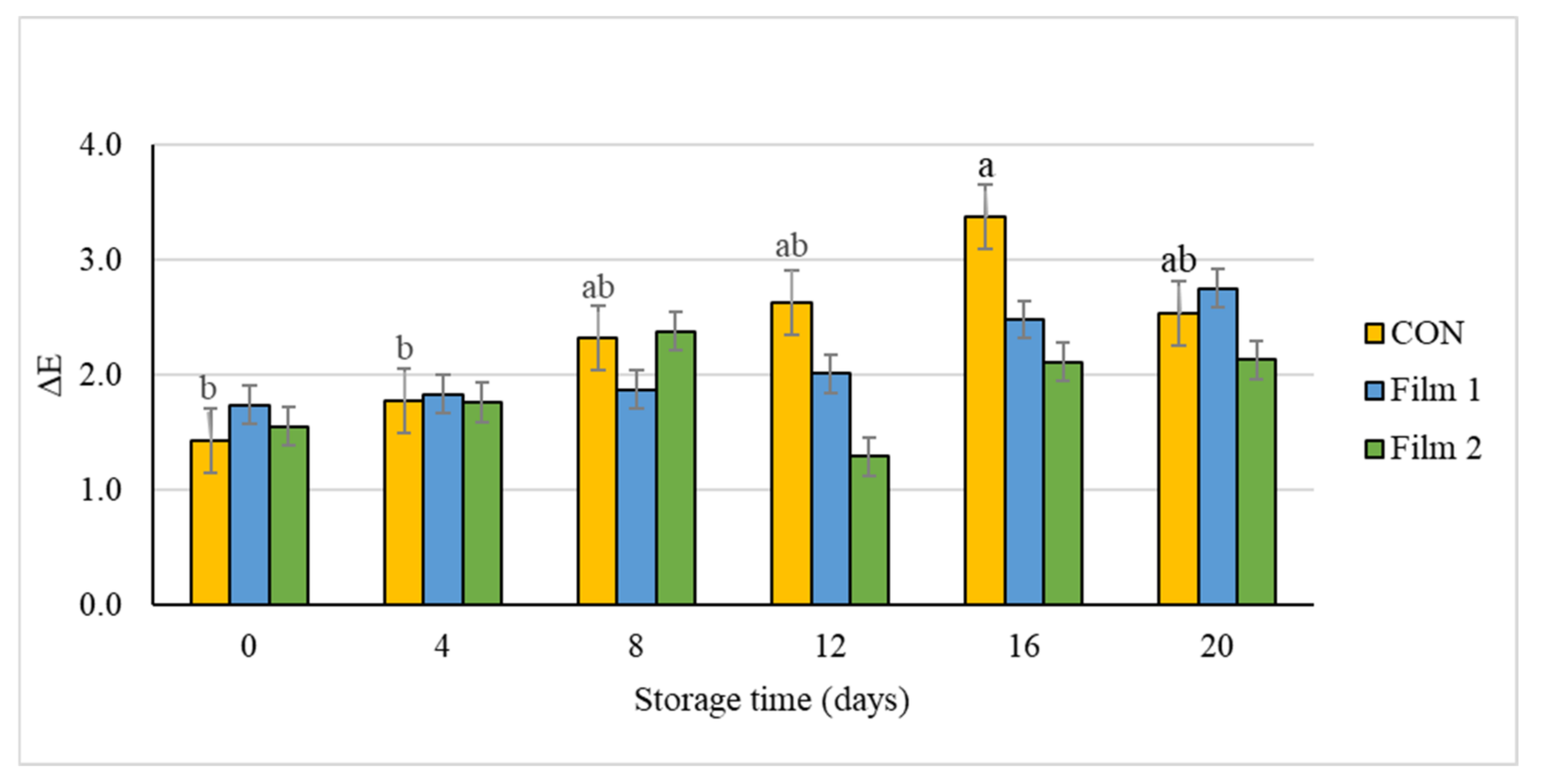
3.3.2. Instrumental Color
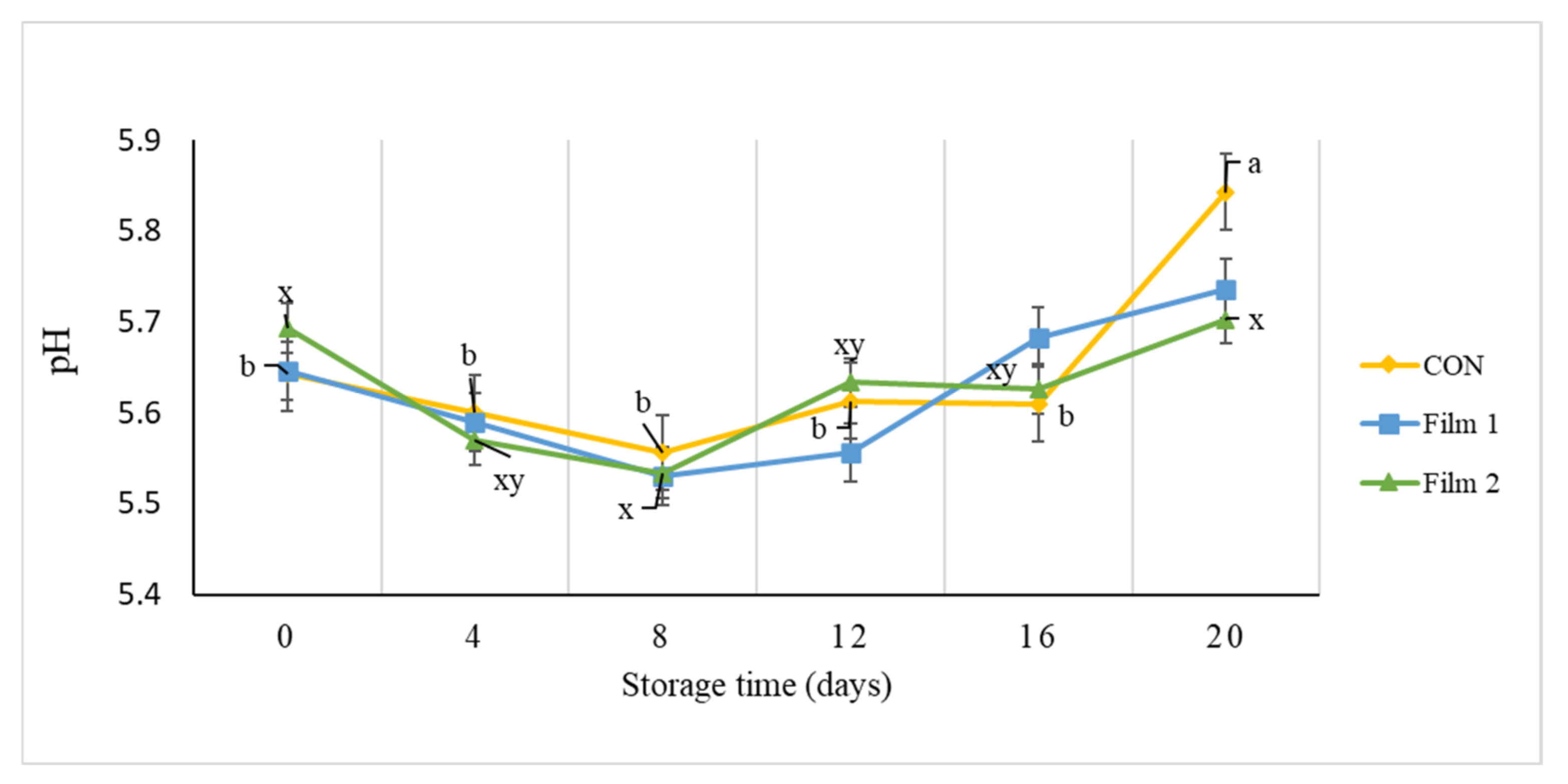
Changes in the color of meat during prolonged periods of storage can be indicative of lipid oxidation processes, protein oxidation processes, and microbiological contamination [54]. During the 20 days storage, there was a significant difference for instrumental lightness values among treatments on day 8, where the L* was greater (p < 0.05) in Film 2 samples as compared with Film 1 and CON samples. Instrumental redness (a*) value, which is an indicator of freshness for red meat, decreased during the 20 days storage period for all treatments. Samples trapped in Film 2 retained their initial redness for four days longer than the other treatments (day 0 compared to day 4), as reflected by the lower a* value for Film 2 samples on day 0. On day 12 of the storage period, pork samples wrapped in Film 1 and Film 2 had greater a* values compared with those wrapped in the CON film. This observation is indicative of the greater storage ability of samples with the coated films. For instrumental yellowness (b*) values, significant differences were observed on day 0 and day 12. On day 0, yellowness values were lower (p < 0.05) for Film 2 samples compared with Film 1 and CON samples. By contrast, on day 12, yellowness was greater for Film 1 samples compared with CON samples, while Film 2 samples were intermediate in value and not different compared with Film 1 and CON samples. The ΔE value is an indication of total color change over time and is often a meaningful instrumental color parameter to monitor during storage or display of meat products. The ΔE value of pork chops was affected during the storage time in CON samples in which ΔE value on day 16 was higher than on day 0 and day 4 (Figure 2). However, there were no significant changes in ΔE value for Film 1 and Film 2 samples throughout the storage period. This indicates improved color stability and thus preservation of color of Film 1 and Film 2 samples during the 20 days storage period. These results suggested that the antioxidant properties of chitosan and/or PE might have helped prevent the oxidation of the myoglobin protein and change in chemical state from myoglobin to metmyoglobin. Other studies have also confirmed that the addition of chitosan and PE either directly on samples or on edible films in meat matrices had a protective effect from oxidative deterioration [23,24,55].

3.3.3. pH

There was an increase (p < 0.05) in pH for CON samples on day 20 of the storage period compared to the other sampling days (Figure 3). No changes (p > 0.05) in pH were observed for the Film 1 samples during the storage period. The pH value of Film 2 samples on day 20 was higher than that on day 8, which may be indicative of the growth of lactic acid bacteria during the first 8 days of storage, due to the lag phase of PE release. These results may suggest that the chitosan and/or PE nonwoven coated LLDPE films could moderate the increase of pH for pork during the storage period. These findings lead to the speculation of two possible explanations. First, the increase in pH could be attributed to the accumulation of volatile basic nitrogen compounds from the action of microorganisms [56]. Second, endogenous meat proteases might have been involved in the change of pH during storage. It is possible for endogenous meat proteases to denature proteins during storage, causing an increase in the formation of free amino acids and peptides [57,58].

3.3.4. Lipid Oxidation

Lipid oxidation values of all samples during the 20 days storage period were below the detectable rancidity threshold levels of 0.50 mg MDA/kg of meat [59]. The bioactive LLDPE films did not significantly affect (p > 0.05) lipid oxidation of the pork samples. Conflicting results have been reported by researchers using chitosan and PE for shelf-life extension of meat products. For example, López-Caballero et al. [60] found that gelatin–chitosan coatings were not significant in preventing lipid oxidation of cod fillets. However, other studies demonstrated significant antioxidant activity when chitosan was used as a base polymer, both in pork loins [61] and in ready-to-cook meat products [62]. Additionally, significant antioxidant activity was demonstrated when PE was used in meat and fish products [23,32]. The negligible antioxidative effect of PE observed in the current study might be due to the low concentration of the bioactives present in the pork samples that conferred a minimal protective effect against lipid oxidation. It is noteworthy that the levels of chitosan and/or PE used by other researchers, in coatings or with direct addition into the food products, were considerably higher than the levels used in the present study [23,24,55,63].

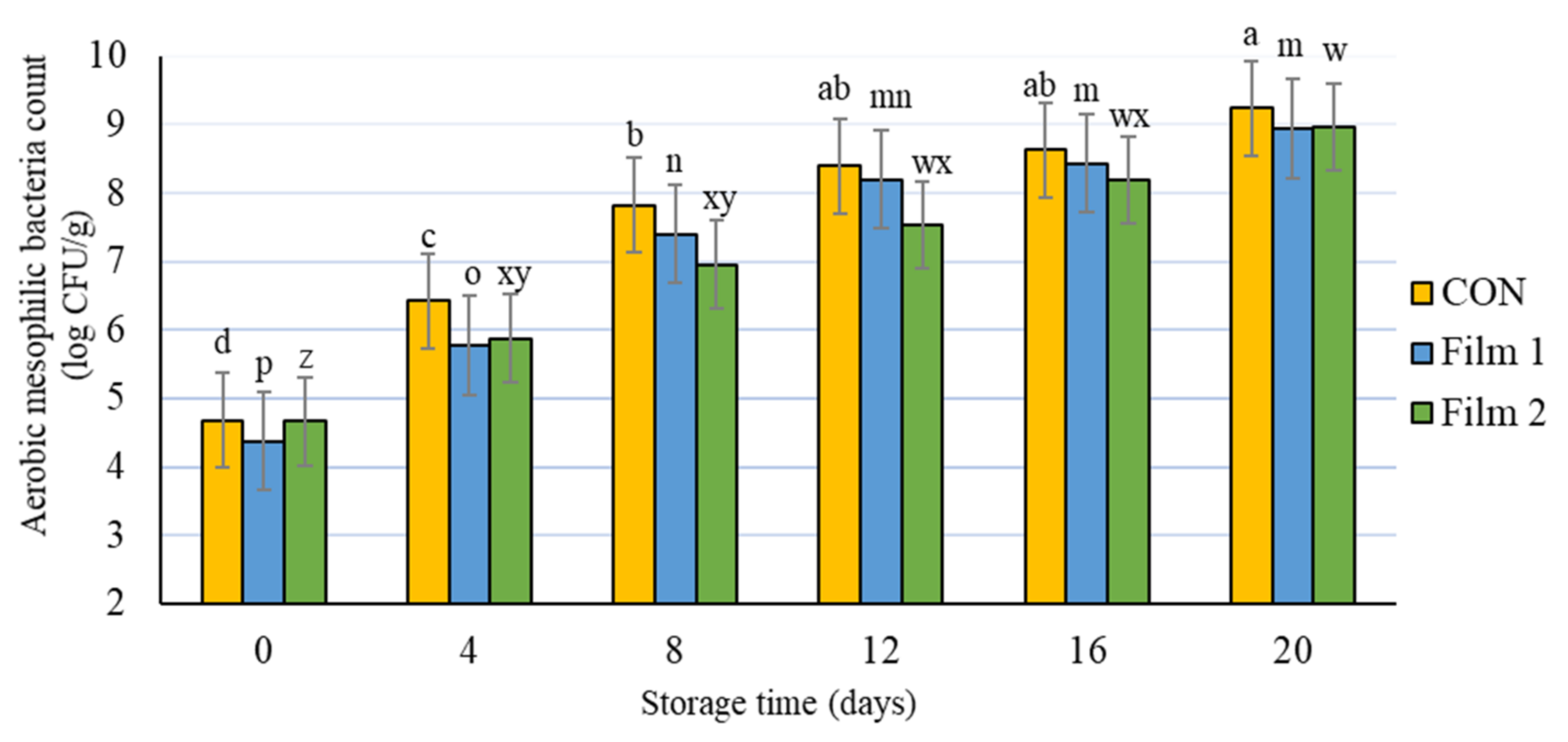
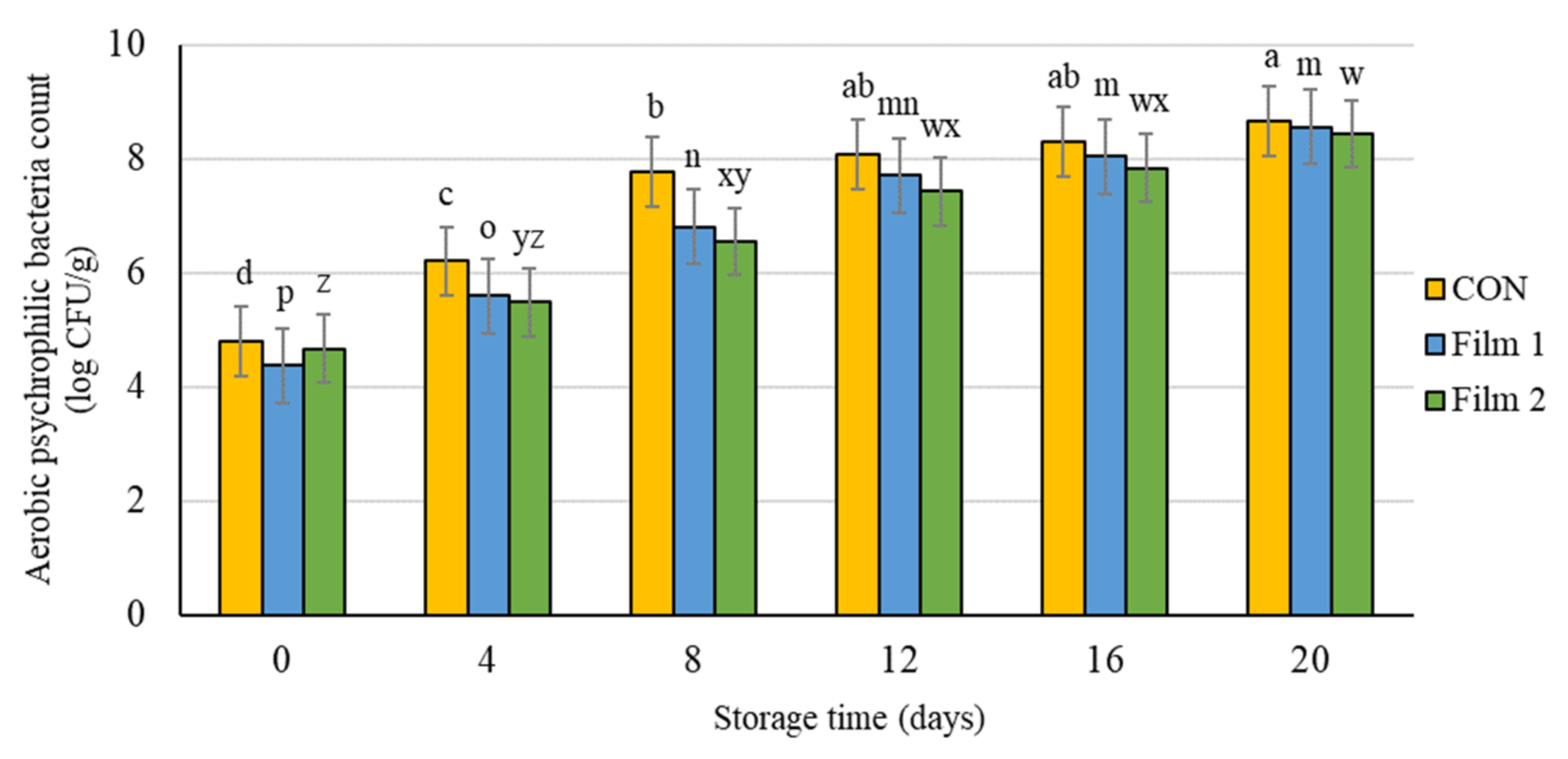
3.3.5. Microbial Analysis

There were no differences (p > 0.05) among treatments at any storage day for either aerobic mesophilic bacteria count or aerobic psychrophilic bacteria count. At the beginning of the experiment, the pork samples in this study had a bacterial count for total aerobic mesophilic bacteria of 4.6 log CFU/g (Figure 4) and a bacterial count for aerobic psychrophilic bacteria of 4.7 log CFU/ g (Figure 5). Bacterial counts increased as storage time increased for both groups of bacteria and for all treatments. Although the aerobic mesophilic and aerobic psychrophilic bacteria counts were not different among the treatments, samples treated with Film 2 had delayed bacteria growth compared with the Film 1 samples and the CON samples. Specifically, aerobic mesophilic bacteria and aerobic psychrophilic bacteria counts on day 4 were similar to day 0, and the bacteria counts on day 8 were similar to day 4 for this Film 2 samples. Moreover, these values (day 0 versus day 4 and day 4 versus day 8) were different for Film 1 samples and CON samples. This observation indicated that the presence of PE in the Film 2 samples could preserve the microbiology quality of pork chops for a longer period of storage time. In fact, the bacterial counts of Film 2 samples were lower or equal to log 107 CFU/g up to 12 d of storage. A value of log 107 CFU/g is the microbial growth threshold level of bacteria for some regulatory purposes, which, in turn, is often used as the maximum limit that guarantees the microbiological safety of meat products. This observation is consistent with other studies that reported an improved antimicrobial effect of combining chitosan and PE [36,64] and other antimicrobials [65,66]. The findings from this study indicate that the addition of PE with chitosan in a polycaprolactone nonwoven may be a promising combination for maintaining microbial stability of meat during prolonged periods of storage.

4. Conclusions

The results obtained from this study suggested that the application of LLDPE film coated with a polycaprolactone/chitosan nonwoven or LLDPE film coated with a polycaprolactone/chitosan nonwoven fortified with PE had the potential to extend the quality of fresh pork during prolonged periods of refrigerated storage. Specifically, the inclusion of coated films improved color stability and microbial stability of the pork samples as evidenced by delayed color deterioration and delayed onset of aerobic mesophilic and aerobic psychrophilic bacteria growth. However, the differences between the LLDPE film coated with polycaprolactone/chitosan nonwoven and the LLDPE film coated with a polycaprolactone/chitosan nonwoven fortified with Colombian propolis extract were generally minor, in terms of magnitude, and therefore both of the films tested in this study could be recommended to the meat industry as effective active packaging materials. Future research is needed for developing a systematic understanding of the mechanistic action of chitosan and PE when introduced into a meat system. Shelf-life testing of the products packaged using the composite LLDPE films, under typical distribution and storage conditions, will also be essential for commercial applications.

Author Contributions

Conceptualization, E.V.R., L.-T.L., H.S.M., and B.M.B.; methodology, E.V.R., L.-T.L., H.S.M., and B.M.B.; formal analysis, E.V.R., L.-T.L., H.S.M., and B.M.B.; investigation, E.V.R., L.-T.L., H.S.M., and B.M.B.; resources, L.-T.L. and B.M.B.; data curation, E.V.R., L.-T.L., and B.M.B.; writing—original draft preparation, E.V.R.; writing—review and editing, L.-T.L., H.S.M., and B.M.B.; supervision, L.-T.L., H.S.M., and B.M.B.; project administration, B.M.B.; funding acquisition, L.-T.L., H.S.M., and B.M.B. All authors have read and agreed to the published version of the manuscript.

Funding

Partial funding for this research was provided by the Natural Sciences and Engineering Research Council of Canada (NSERC) Discovery Grants Program (Grant 2018-04695).

Data Availability Statement

Data can be made available upon request.

Acknowledgments

The authors would like to acknowledge the Department of Food Science at the University of Guelph for administrative and technical support.

Conflicts of Interest

The authors declare no conflict of interest.

References

  1. Sun, X.D.; Holley, R.A. Antimicrobial and antioxidative strategies to reduce pathogens and extend the shelf life of fresh red meats. Compr. Rev. Food Sci. Food Saf. 2012, 11, 340–354. [Google Scholar] [CrossRef]
  2. Borch, E.; Kant-Muermans, M.L.; Blixt, Y. Bacterial spoilage of meat and cured meat products. Int. J. Food Microbiol. 1996, 33, 103–120. [Google Scholar] [CrossRef]
  3. Nychas, G.J.E.; Skandamis, P.N.; Tassou, C.C.; Koutsoumanis, K.P. Meat spoilage during distribution. Meat Sci. 2008, 78, 77–89. [Google Scholar] [CrossRef] [PubMed]
  4. Ahmed, I.; Lin, H.; Zou, L.; Brody, A.L.; Li, Z.; Qazi, I.M.; Pavase, T.; Lv, L. A comprehensive review on the application of active packaging technologies to muscle foods. Food Control 2017, 82, 163–178. [Google Scholar] [CrossRef]
  5. Dainelli, D.; Gontard, N.; Spyropoulos, D.; Zondervan-van den Beuken, E.; Tobback, P. Active and intelligent food packaging: Legal aspects and safety concerns. Trends Food Sci. Technol. 2008, 19, S103–S112. [Google Scholar] [CrossRef]
  6. Robertson, G.L. Food Packaging: Principles and Practice, 3rd ed.; CRC Press: Boca Raton, FL, USA, 2013; Available online: https://books.google.com.co/books?id=y27tL_7ZJFUC (accessed on 1 March 2021).
  7. Yam, K.L.; Takhistov, P.T.; Miltz, J. Intelligent Packaging: Concepts and Applications. J. Food Sci. 2005, 70, R1–R10. [Google Scholar] [CrossRef]
  8. Gue, M.; Wang, H.; Wang, Q.; Chen, M.; Li, L.; Li, X.; Jiang, S. Intelligent double-layer fiber mats with high colorimetric response sensitivity for food freshness monitoring and preservation. Food Hydrocoll. 2020, 101, 105468. [Google Scholar] [CrossRef]
  9. Li, L.; Wang, H.; Chen, M.; Jiang, S.; Cheng, J.; Li, X.; Zhang, M.; Jiang, S. Gelatin/zein fiber mats encapsulated with resveratrol: Kinetics, antibacterial activity and application for pork preservation. Food Hydrocoll. 2020, 101, 105577. [Google Scholar] [CrossRef]
  10. Roy, S.; Rhim, J.W. Preparation of antimicrobial and antioxidant gelatin/curcumin composite films for active food packaging application. Colloid Surf. B 2020, 188, 110761. [Google Scholar] [CrossRef]
  11. Feng, X.; Ng, V.K.; Mikš-Krajnik, M.; Yang, H. Effects of fish gelatin and tea polyphenol coating on the spoilage and degradation of myofibril in fish fillet during cold storage. Food Bioproc. Tech. 2017, 10, 89–102. [Google Scholar] [CrossRef]
  12. Jayasena, D.D.; Jo, C. Essential oils as potential antimicrobial agents in meat and meat products: A review. Trends Food Sci. Technol. 2013, 34, 96–108. [Google Scholar] [CrossRef]
  13. Yang, H.; Hewes, D.; Salaheen, S.; Federman, C.; Biswas, D. Effects of blackberry juice on growth inhibition of foodborne pathogens and growth promotion of Lactobacillus. Food Control 2014, 37, 15–20. [Google Scholar] [CrossRef]
  14. Woodruff, M.A.; Hutmacher, D.W. The return of a forgotten polymer—Polycaprolactone in the 21st century. Prog. Polym. Sci. 2010, 35, 1217–1256. [Google Scholar] [CrossRef] [Green Version]
  15. Cerkez, I.; Sezer, A.; Bhullar, S.K. Fabrication and characterization of electrospun poly(e-caprolactone) fibrous membrane with antibacterial functionality. R. Soc. Open Sci. 2017, 4, 160911. [Google Scholar] [CrossRef] [Green Version]
  16. Shao, S.; Li, L.; Yang, G.; Li, J.; Luo, C.; Gong, T.; Zhou, S. Controlled green tea polyphenols release from electrospun PCL/MWCNTs composite nanofibers. Inter. J. Pharm. 2011, 421, 310–320. [Google Scholar] [CrossRef]
  17. Xiao, J.; Shi, C.; Zheng, H.; Shi, Z.; Jiang, D.; Li, Y.; Huang, Q. Kafirin protein based electrospun fibers with tunable mechanical property, wettability, and release profile. J. Agric. Food Chem. 2016, 64, 3226–3233. [Google Scholar] [CrossRef]
  18. Tian, J.; Deng, H.; Huang, M.; Liu, R.; Yi, Y.; Dong, X. Electrospun nanofibers for food and food packaging technology. Micro Nano Technol. 2019, 455–516. [Google Scholar] [CrossRef]
  19. Bankova, V.; Bertelli, D.; Borba, R.; Conti, B.J.; da Silva Cunha, I.B.; Danert, C.; Eberlin, M.; I Falcão, S.; Isla, M.; Moreno, M.; et al. Standard methods for Apis mellifera propolis research. J. Apic. Res. 2016, 58, 1–49. [Google Scholar] [CrossRef] [Green Version]
  20. Falcão, S.I.; Vilas-Boas, M.; Estevinho, L.M.; Barros, C.; Domingues, M.R.M.; Cardoso, S.M. Phenolic characterization of Northeast Portuguese propolis: Usual and unusual compounds. Anal. Bioanal. Chem. 2010, 396, 887–897. [Google Scholar] [CrossRef] [Green Version]
  21. Silici, S.; Kutluca, S. Chemical composition and antibacterial activity of propolis collected by three different races of honeybees in the same region. J. Ethnopharmacol. 2005, 99, 69–73. [Google Scholar] [CrossRef]
  22. Barrera, E.; Gil, J.; Restrepo, A.; Mosquera, K.; Durango, D. A coating of chitosan and propolis extract for the postharvest treatment of papaya (Carica papaya L. cv. Hawaiiana). Rev. Fac. Nac. Agron. Medellín 2015, 68, 7667–7678. [Google Scholar] [CrossRef]
  23. Gutiérrez, C.; Suárez, H. Antimicrobial activity of propolis and its effect on the physicochemical and sensoral characteristics in sausages. Vitae 2014, 21, 90–96. [Google Scholar]
  24. Piedrahíta, D.G.; Fuenmayor, C.A.; Suárez, H. Effect of chitosan-propolis edible coatings on stability of refrigerated cachama (Piaractus brachypomus) vacuum-packed fish fillets. Packag. Technol. Sci. 2019, 32, 143–153. [Google Scholar] [CrossRef]
  25. Ahi, Z.B.; Renkler, N.Z.; Gul Seker, M.; Tuzlakoglu, K. Biodegradable polymer films with a natural antibacterial extract as novel periodontal barrier membranes. Int. J. Biomater. 2019, 7932470. [Google Scholar] [CrossRef] [PubMed] [Green Version]
  26. Asawahame, C.; Sutjarittangtham, K.; Eitssayeam, S.; Tragoolpua, Y.; Sirithunyalug, B.; Sirithunyalug, J. Antibacterial activity and inhibition of adherence of Streptococcus mutans by propolis electrospun fibers. AAPS PharmSciTech 2015, 16, 182–191. [Google Scholar] [CrossRef] [PubMed]
  27. Kim, J.I.; Pant, H.R.; Sim, H.J.; Lee, K.M.; Kim, C.S. Electrospun propolis/polyurethane composite nanofibers for biomedical applications. Mater. Sci. Eng. 2014, 44, 52–57. [Google Scholar] [CrossRef] [PubMed]
  28. Moradkhannejhad, L.; Abdouss, M.; Nikfarjam, N.; Mazinani, S.; Heydari, V. Electrospinning of zein/propolis nanofibers; antimicrobial properties and morphology investigation. J. Mater. Sci. Mater. Med. 2018, 29, 165. [Google Scholar] [CrossRef]
  29. Ohkawa, K.; Cha, D.; Kim, H.; Nishida, A.; Yamamoto, H. Electrospinning of chitosan. Marcomol. Rapid Commun. 2004, 25, 1600–1605. [Google Scholar] [CrossRef]
  30. Sutjarittangtham, K.; Sanpa, S.; Tunkasiri, T.; Chantawannakul, P.; Intatha, U.; Eitssayeam, S. Bactericidal effects of propolis/polylactic acid (PLA) nanofibres obtained via electrospinning. J. Apic. Res. 2014, 53, 109–115. [Google Scholar] [CrossRef]
  31. Tosi, E.A.; Ré, E.; Ortega, M.E.; Cazzoli, A.F. Food preservative based on propolis: Bacteriostatic activity of propolis polyphenols and flavonoids upon Escherichia coli. Food Chem. 2007, 104, 1025–1029. [Google Scholar] [CrossRef]
  32. Pobiega, P.; Igielska, M.; Włodarczyk, P.; Gniewosz, M. The use of pullulan coatings with propolis extract to extend the shelf life of blueberry (Vaccinium corymbosum) fruit. Int. J. Food Sci. Technol. 2021, 56, 1013–1020. [Google Scholar] [CrossRef]
  33. Pobiega, K.; Kraśniewska, K.; Gniewosz, M. Application of propolis in antimicrobial and antioxidative protection of food quality—A review. Trends Food Sci. Technol. 2019, 83, 53–62. [Google Scholar] [CrossRef]
  34. Bonou, J.; Ahouandjinou, H.F.; Baba-Moussa, F.; Adéoti, Z.; Dougnon, V.; Metongnon, I.; Gbenou, J.D.; Toukouro, F.; Baba-Moussa, L. Assessment of the antimicrobial activity of essential oils from some Beninese medicinal plants: Influence of different tweens. Issues Bio. Sci. Pharma. Res. 2016, 4, 43–49. [Google Scholar] [CrossRef]
  35. Singleton, V.L.; Orthofer, R.; Lamuela-Raventós, R.M. Analysis of total phenols and other oxidation substrates and antioxidants by means of folin-ciocalteu reagent. Meth. Enzymol. 1999, 299, 152–178. [Google Scholar] [CrossRef]
  36. Siripatrawan, U.; Vitchayakitti, W. Improving functional properties of chitosan films as active food packaging by incorporating with propolis. Food Hydrocoll. 2016, 61, 695–702. [Google Scholar] [CrossRef]
  37. Moon, J.K.; Shibamoto, T. Antioxidant assays for plant and food components. J. Agr. Food Chem. 2009, 57, 1655–1666. [Google Scholar] [CrossRef]
  38. Zaitoon, A.; Lim, L.T. Effect of poly(ethylene oxide) on the electrospinning behavior and characteristics of ethyl cellulose composite fibers. Materialia 2020, 10, 100649. [Google Scholar] [CrossRef]
  39. Vargas, H.; Bohrer, B.M. A preliminary investigation on the effects of a hot water shrink tunnel and chill tank following vacuum packaging on commercial pork quality and bacteria growth. Can. J. Anim. Sci. 2018, 98, 893–897. [Google Scholar] [CrossRef]
  40. Cottica, S.M.; Sawaya, A.C.; Eberlin, M.N.; Franco, S.L.; Zeoula, L.M.; Visentainer, J.V. Antioxidant activity and composition of propolis obtained by different methods of extraction. J. Braz. Chem. Soc. 2011, 22, 929–935. [Google Scholar] [CrossRef]
  41. Trusheva, B.; Trunkova, D.; Bankova, V. Different extraction methods of biologically active components from propolis: A preliminary study. Chem. Cent. J. 2007, 1, 1–4. [Google Scholar] [CrossRef] [Green Version]
  42. Anjum, S.I.; Ullah, A.; Khan, K.A.; Attaullah, M.; Khan, H.; Ali, H.; Bashir, M.; Tahir, M.; Ansari, M.; Ghramh, H.; et al. Composition and functional properties of propolis (bee glue): A review. Saudi J. Biol. Sci. 2018, 26, 1695–1703. [Google Scholar] [CrossRef] [PubMed]
  43. Harfouch, R. Antibacterial activity of Syrian propolis extract against several strains of bacteria in vitro. World J. Pharm. Pharm. Sci. 2017, 6, 42–46. [Google Scholar] [CrossRef]
  44. Martínez, J.; Garcia, C.; Durango, D.; Gil, J. Caracterización de propóleos provenientes del municipio de Caldas obtenido por dos métodos de recolección. Rev. MVZ Cordoba 2012, 17, 2861–2869. [Google Scholar] [CrossRef] [Green Version]
  45. Machado, B.S.; Pulcino, T.N.; Silva, A.L.; Melo, D.T.; Silva, R.G.; Mendonça, I.G. Propolis as an alternative in prevention and control of dental cavity. J. Apither. 2017, 1, 47–50. [Google Scholar] [CrossRef] [Green Version]
  46. Parolia, A.; Thomas, M.; Mala, K.; Mohan, M. Propolis and its potential uses in oral health. Int. J. Med. Med. Sci. 2010, 2, 210–215. [Google Scholar]
  47. Bankova, V. Chemical diversity of propolis and the problem of standardization. J. Ethnopharmacol. 2015, 100, 114–117. [Google Scholar] [CrossRef]
  48. Torel, J.; Cillard, J.; Cillard, P. Antioxidant activity of flavonoids and reactivity with peroxy radical. Phytochemistry 1986, 25, 383–385. [Google Scholar] [CrossRef]
  49. Bors, W.; Heller, W.; Michel, C.; Saran, M. Flavonoids as antioxidants: Determination of radical-scavenging efficiencies. Meth. Enzymol. 1990, 186, 343–355. [Google Scholar] [CrossRef]
  50. Palomino, G.L.R.; García, P.C.M.; Gil, G.J.H.; Rojano, B.A.; Durango, R.D.L. Determinación del contenido de fenoles y evaluación de la actividad antioxidante de propóleos recolectados en el departamento de Antioquia (Colombia). Vitae 2009, 16, 388–395. [Google Scholar]
  51. Salamanca, G.; Correa, I.L.; Principal, J. Perfil de flavonoides e índices de oxidación de algunos propóleos colombianos. Zootec. Trop. 2007, 25, 95–102. [Google Scholar]
  52. Yano, H.; Sugiyama, J.; Nakagaito, A.N.; Nogi, M.; Matsuura, T.; Hikita, M.; Handa, K. Optically transparent composites reinforced with networks of bacterial nanofibers. Adv. Mater. 2005, 17, 153–155. [Google Scholar] [CrossRef]
  53. Sánchez, Á.P. Preparación y caracterización de membranas poliméricas electrohiladas de policaprolactona y quitosano para la liberación controlada de clorhidrato de tiamina. Cienc. Desarro. 2016, 7, 133–152. [Google Scholar] [CrossRef] [Green Version]
  54. Mancini, R.A.; Hunt, M.C. Current research in meat color. Meat Sci. 2005, 71, 100–121. [Google Scholar] [CrossRef]
  55. No, H.K.; Meyers, S.P.; Prinyawiwatkul, W.; Xu, Z. Applications of chitosan for improvement of quality and shelf life of foods: A review. J. Food Sci. 2007, 72, R87–R100. [Google Scholar] [CrossRef] [PubMed]
  56. Miao, J.; Peng, W.; Liu, G.; Chen, Y.; Chen, F.; Cao, Y. Biopreservative effect of the natural antimicrobial substance from Lactobacillus paracasei subsp. tolerans FX-6 on fresh pork during chilled storage. Food Control 2015, 56, 53–56. [Google Scholar] [CrossRef]
  57. Fan, W.; Sun, J.; Chen, Y.; Qiu, J.; Zhang, Y.; Chi, Y. Effects of chitosan coating on quality and shelf life of silver carp during frozen storage. Food Chem. 2009, 115, 66–70. [Google Scholar] [CrossRef]
  58. Lorenzo, J.M.; Batlle, R.; Gómez, M. Extension of the shelf-life of foal meat with two antioxidant active packaging systems. LWT Food Sci. Technol. 2014, 59, 181–188. [Google Scholar] [CrossRef]
  59. Sheard, P.R.; Enser, M.; Wood, J.D.; Nute, G.R.; Gill, B.P.; Richardson, R.I. Shelf life and quality of pork and pork products with raised n-3 PUFA. Meat Sci. 2000, 55, 213–221. [Google Scholar] [CrossRef]
  60. López-Caballero, M.E.; Gómez-Guillén, M.C.; Pérez-Mateos, M.; Montero, P. A chitosan–gelatin blend as a coating for fish patties. Food Hydrocoll. 2005, 19, 303–311. [Google Scholar] [CrossRef] [Green Version]
  61. Cao, Y.; Warner, R.D.; Fang, Z. Effect of chitosan/nisin/gallic acid coating on preservation of pork loin in high oxygen modified atmosphere packaging. Food Control 2019, 101, 9–16. [Google Scholar] [CrossRef]
  62. Kanatt, S.R.; Rao, M.S.; Chawla, S.P.; Sharma, A. Effects of chitosan coating on shelf-life of ready-to-cook meat products during chilled storage. LWT Food Sci. Technol. 2013, 53, 321–326. [Google Scholar] [CrossRef]
  63. Dos Reis, A.S.; Diedrich, C.; de Moura, C.; Pereira, D.; de Almeida, J.F.; da Silva, L.D.; Plata-Ovideo, M.S.V.; Tavarez, R.A.W.; Carpes, S.T. Physico-chemical characteristics of microencapsulated propolis co-product extract and its effect on storage stability of burger meat during storage at −15 °C. LWT Food Sci. Technol. 2017, 76, 306–313. [Google Scholar] [CrossRef]
  64. Torlak, E.; Sert, D. Antibacterial effectiveness of chitosan-propolis coated polypropylene films against foodborne pathogens. Int. J. Biol. Macromol. 2013, 60, 52–55. [Google Scholar] [CrossRef] [PubMed]
  65. Orsi, R.D.O.; Sforcin, J.M.; Cunha Funari, S.R.; Fernandes, A.; Bankova, V. Synergistic effect of propolis and antibiotics on the Salmonella typhi. Braz. J. Microbiol. 2006, 37, 108–112. [Google Scholar] [CrossRef] [Green Version]
  66. Orsi, R.O.; Fernandes, A.; Bankova, V.; Sforcin, J.M. The effects of Brazilian and Bulgarian propolis in vitro against Salmonella Typhi and their synergism with antibiotics acting on the ribosome. Nat. Prod. Res. 2012, 26, 430–437. [Google Scholar] [CrossRef]
Figure 1. (A) (I) Spin dope solutions for electrospinning; (II) active nonwovens coated commercial linear low-density polyethylene (LLDPE); (III) treatments tested: CON (O.1)—LLDPE commercial packaging without a functional nonwoven component; Film 1 (O.2)—a commercial LLDPE film coated with a polycaprolactone/chitosan nonwoven; Film 2 (O.3)—a commercial LLDPE film coated with a polycaprolactone/chitosan nonwoven fortified with Colombian PE; (IV) example of coating during use; and (V) example of coating following use. (B). Scanning electron micrographs of (I) Film 1; (II) Film 2.
Foods 10 01110 g001
Figure 2. Total color difference (ΔE) of pork loin chops wrapped in active packaging for 20 days at 4 °C storage. Treatments included the following: CON—a commercial linear low-density polyethylene (LLDPE) film without a functional nonwoven component; Film 1—a commercial LLDPE film coated with a polycaprolactone/chitosan nonwoven; Film 2—commercial LLDPE film coated with a polycaprolactone/chitosan nonwoven fortified with Colombian propolis extract. ab Different letters indicate significant differences (p < 0.05) for the main effect of storage day for each respective treatment when a significant difference was detected. Note: no significant differences (p > 0.05) were detected for treatment × day, no significant differences (p > 0.05) were detected for storage day for Film 1 and Film 2 samples, and no significant differences (p > 0.05) were detected for the main effect of treatment within each day.
Foods 10 01110 g002
Figure 3. pH of pork loin chops wrapped in active packaging for 20 days at 4 °C storage. Treatments included the following: CON—a commercial linear low-density polyethylene (LLDPE) film without a functional nonwoven component; Film 1—a commercial LLDPE film coated with a polycaprolactone/chitosan nonwoven; Film 2—commercial LLDPE film coated with a polycaprolactone/chitosan nonwoven fortified with Colombian propolis extract. ab, xy Different letters indicate significant differences (p < 0.05) for the main effect of storage day for each respective treatment when a significant difference was detected. Note: no significant differences (p > 0.05) were detected for treatment × day, no significant differences (p > 0.05) were detected for storage day for Film 1 samples, and no significant differences (p > 0.05) were detected for the main effect of treatment within each day.
Foods 10 01110 g003
Figure 4. Aerobic mesophilic bacteria count of pork loin chops wrapped in active packaging for 20 days at 4 °C storage. Treatments included the following: CON—a commercial linear low-density polyethylene (LLDPE) film without a functional nonwoven component; Film 1—a commercial LLDPE film coated with a polycaprolactone/chitosan nonwoven; Film 2—commercial LLDPE film coated with a polycaprolactone/chitosan nonwoven fortified with Colombian propolis extract. abcd, mnop, wxyz Different letters indicate significant differences (p < 0.05) for the main effect of storage day for each respective treatment when a significant difference was detected. Note: no significant differences (p > 0.05) were detected for treatment × day, and no significant differences (p > 0.05) were detected for the main effect of treatment within each day.
Foods 10 01110 g004
Figure 5. Aerobic psychrophilic bacteria count of pork loin chops wrapped in active packaging for 20 days at 4 °C storage. Treatments included the following: CON—a commercial linear low-density polyethylene (LLDPE) film without a functional nonwoven component; Film 1—a commercial LLDPE film coated with a polycaprolactone/chitosan nonwoven; Film 2—commercial LLDPE film coated with a polycaprolactone/chitosan nonwoven fortified with Colombian propolis extract. abcd, mnop, wxyz Different letters indicate significant differences (p < 0.05) for the main effect of storage day for each respective treatment when a significant difference was detected. Note: no significant differences (p > 0.05) were detected for treatment × day, and no significant differences (p > 0.05) were detected for the main effect of treatment within each day.
Foods 10 01110 g005
Table 1. Antimicrobial and antioxidant determination of Colombian propolis extract (PE) sourced from Santander, Colombia.
Minimum Inhibitory Concentration (MIC) of PE from Santander, Colombia (mg/mL)Total Phenol Content ± SD (mg GAE/g of PE)Antioxidant Activity
ABTS TEAC ± SD (µmol Trolox/g PE)
Staphylococcus
aureus
Escherichia
coli
Salmonella
enteritidis
0.15201082.6 ± 21186 ± 52
Table 2. Purge loss, instrumental color, and TBARS of pork loin chops wrapped in active packaging for 20 days at 4 °C storage 1.
Day 0Day 4Day 8Day 12Day 16Day 20
Purge loss, %
CON0.87 ± 0.52 b3.18 ± 0.98 a3.62 ± 1.21 a4.23 ± 0.81 a4.33 ± 1.54 a4.37 ± 0.45 a
Film 11.20 ± 0.62 b2.55 ± 0.48 b4.05 ± 1.01 a4.06 ± 0.26 a4.31 ± 0.60 a5.10 ± 1.22 a
Film 21.57 ± 0.44 d2.37 ± 0.32 d4.34 ± 0.11 c4.54 ± 0.54 bc5.30 ± 0.41 ab5.59 ± 0.77 a
Minolta L* (lightness) color, units
CON47.35 ± 1.93 b47.90 ± 1.85 ab49.43 ± 2.23 ab, y50.33 ± 2.75 a50.36 ± 2.02 a47.90 ± 0.85 ab
Film 146.69 ± 2.31 b48.25 ± 2.05 ab49.16 ± 1.19 ab, y49.67 ± 1.59 a49.21 ± 1.13 ab49.00 ± 2.55 ab
Film 245.40 ± 2.1649.38 ± 2.9752.20 ± 0.92 x48.35 ± 0.4948.28 ± 1.7749.45 ± 1.95
Minolta a* (redness) color, units
CON7.01 ± 1.46 a4.68 ± 1.12 b4.90 ± 1.35 b2.73 ± 0.90 c, y3.62 ± 0.56 bc4.00 ± 0.76 b
Film 17.53 ± 1.77 a4.89 ± 0.31 bc4.82 ± 1.33 bc5.37 ± 0.93 b, x3.90 ± 0.71 c4.09 ± 0.93 bc
Film 25.94 ± 0.43 a5.31 ± 1.03 ab3.89 ± 0.74 bc5.59 ± 0.13 ab, x4.19 ± 0.80 c4.02 ± 0.84 c
Minolta b* (yellowness) color, units
CON4.34 ± 2.62 xy3.24 ± 0.923.27 ± 0.202.81 ± 0.24 y4.24 ± 2.043.44 ± 1.29
Film 15.07 ± 2.37 a, x3.48 ± 0.90 ab3.78 ± 1.50 ab4.28 ± 1.88 ab, x2.83 ± 0.59 b4.61 ± 1.53 a
Film 22.61 ± 0.97 y4.29 ± 2.283.83 ± 1.243.29 ± 0.36 xy3.50 ± 1.393.46 ± 1.39
Thiobarbituric acid reactive substances (TBARS), mg MDA/kg meat
CON0.023 ± 0.001 ab0.024 ± 0.002 a0.023 ± 0.001 b,y0.025 ± 0.001 a0.024 ± 0.001 a0.024 ± 0.001 ab
Film 10.023 ± 0.002 c0.025 ± 0.002 abc0.026 ± 0.001 ab,x0.026 ± 0.003 a0.023 ± 0.001 bc0.025 ± 0.001 abc
Film 20.023 ± 0.0020.024 ± 0.0010.024 ± 0.001 xy0.026 ± 0.0010.024 ± 0.0010.024 ± 0.001
abc For each parameter, values in the same row (day effect within each treatment) with different superscripts are different (p < 0.05). xyz For each parameter, values in the same column (treatment effect within each day) with different superscripts are different (p < 0.05). 1 Treatments included the following: CON—a commercial linear low-density polyethylene (LLDPE) film without a functional nonwoven component; Film 1—a commercial LLDPE film coated with a polycaprolactone/chitosan nonwoven; Film 2—commercial LLDPE film coated with a polycaprolactone/chitosan nonwoven fortified with Colombian propolis extract (PE).
Publisher’s Note: MDPI stays neutral with regard to jurisdictional claims in published maps and institutional affiliations.

