Article

In Silico Evidence of the Multifunctional Features of Lactiplantibacillus pentosus LPG1, a Natural Fermenting Agent Isolated from Table Olive Biofilms

by
Elio López-García
1,
Antonio Benítez-Cabello
1,*,
Javier Ramiro-García
1,
Victor Ladero
2 and
Francisco Noé Arroyo-López
1
1
Food Biotechnology Department, Instituto de la Grasa (CSIC), Carretera Utrera Km 1, Campus Universitario Pablo de Olavide, 41013 Seville, Spain
2
Technology and Biotechnology Department, Instituto de Productos Lácteos de Asturias (IPLA-CSIC), Paseo Río Linares s/n, 33300 Villaviciosa, Spain
*
Author to whom correspondence should be addressed.
Submission received: 16 January 2023 / Revised: 16 February 2023 / Accepted: 17 February 2023 / Published: 22 February 2023
(This article belongs to the Special Issue New Insight in Microbial Diversity and Genomic in Foods)

Abstract

:
In recent years, there has been a growing interest in obtaining probiotic bacteria from plant origins. This is the case of Lactiplantibacillus pentosus LPG1, a lactic acid bacterial strain isolated from table olive biofilms with proven multifunctional features. In this work, we have sequenced and closed the complete genome of L. pentosus LPG1 using both Illumina and PacBio technologies. Our intention is to carry out a comprehensive bioinformatics analysis and whole-genome annotation for a further complete evaluation of the safety and functionality of this microorganism. The chromosomic genome had a size of 3,619,252 bp, with a GC (Guanine-Citosine) content of 46.34%. L. pentosus LPG1 also had two plasmids, designated as pl1LPG1 and pl2LPG1, with lengths of 72,578 and 8713 bp (base pair), respectively. Genome annotation revealed that the sequenced genome consisted of 3345 coding genes and 89 non-coding sequences (73 tRNA and 16 rRNA genes). Taxonomy was confirmed by Average Nucleotide Identity analysis, which grouped L. pentosus LPG1 with other sequenced L. pentosus genomes. Moreover, the pan-genome analysis showed that L. pentosus LPG1 was closely related to the L. pentosus strains IG8, IG9, IG11, and IG12, all of which were isolated from table olive biofilms. Resistome analysis reported the absence of antibiotic resistance genes, whilst PathogenFinder tool classified the strain as a non-human pathogen. Finally, in silico analysis of L. pentosus LPG1 showed that many of its previously reported technological and probiotic phenotypes corresponded with the presence of functional genes. In light of these results, we can conclude that L. pentosus LPG1 is a safe microorganism and a potential human probiotic with a plant origin and application as a starter culture for vegetable fermentations.

1. Introduction

Lactobacillales, commonly called lactic acid bacteria (LAB), are an order of Gram-positive and acid-tolerant bacteria that produce lactic acid as the major metabolic end product obtained from carbohydrate fermentation. Their large genetic versatility allows them to colonize a wide range of ecological niches, ranging from the mammalian gut microbiota to a large number of fermented foods [1]. This is the case of table olives, one of the most important fermented vegetables in Mediterranean countries, with a worldwide production that exceeds 3 million tonnes/year [2]. Lactiplantibacillus pentosus and Lactiplantibacillus plantarum (formerly known as Lactobacillus pentosus and Lactobacillus plantarum), together with diverse species of yeasts, are among the main microorganisms and LAB species responsible for table olive fermentation, determining the flavor, quality, and safety of the final product [3,4,5].
Traditionally, the market for probiotic foods was dominated by fermented dairy products. However, in recent years, the demand for vegetal-based probiotic products has grown due to a shift in consumer preference toward healthier alternatives. In addition, vegetarian activism and lactose intolerance have driven the search for new dairy-free and vegan products. Therefore, the emergence of new vegetal-based probiotics should be encouraged to satisfy societal demand, contributing to the rapidly growing global market of approximately 50 billion USD [6].
In recent years, table olives have emerged as a solid alternative to dairy products as a carrier of beneficial microorganisms to consumers. Certain LAB strains have the ability to form biofilms on the olive epidermis, obtaining fermented olives with more than 10 million UFC/g [7]. L. pentosus LPG1 (hereinafter LPG1) is a fermentation agent obtained from table olive biofilms with proven probiotic and technological features based on previous in vitro and in vivo studies [8,9]. Thereby, LPG1 has shown anti-inflammatory, esterase, and phytase activities, a reduction in cholesterol levels, lactic acid production, and the inhibition of food-borne pathogens. It has also shown the ability to adhere to Caco-2 cells, as well as the absence of antibiotic resistance and hemolytic activity, among other features. However, the precise genomic mechanisms involving its functions are still unknown. Advances in genome sequencing techniques together with cost reduction have allowed an advancement of the knowledge, not only of the probiotic potential of the strains, but also of the technological potential. In recent years, several genomes of L. pentosus isolated from table olives have been sequenced [10,11,12,13,14], as well as from other vegetal-derived matrices [15].
Our aim in the present study is to expand our knowledge of the functional and safety features of the LPG1 strain. For this purpose, we have sequenced, annotated, and closed its full genome, including chromosome and plasmids, correlating the technological and probiotic features with the presence of functional genes after in silico analysis. Our work was performed under the recommendations of EFSA, which requires full genome sequencing and annotation of novel strains that are intended for biotechnological applications [16].

2. Materials and Methods

2.1. Bacterial Strain, Culture Conditions, and DNA Isolation

The LPG1 strain was previously isolated from Spanish-style table olive biofilms and identified by molecular methods [8]. LPG1 was grown in Man Rogosa and Sharpe agar (Oxoid, Basingstoke, UK) at 37 °C for 48 h prior to DNA isolation. Then, genomic and plasmid DNA was extracted and purified using the protocol described by Martín-Platero et al. [17]. The integrity of the extracted DNA was confirmed by visualization in agarose gel (0.9%), while its concentration was determined by a Qubit 4 fluorometer, obtaining a DNA concentration of 319 ng/µL.

2.2. Genome Sequencing, Assembly, and Annotation

Whole-genome sequencing of LPG1 was carried out using two platforms, Illumina HiSeq (Eurofins, Luxemburg) and PacBio (FISABIO, Valencia, Spain). The raw reads obtained from both methodologies were curated by Trim Galore and the fastp tool, respectively. Then, the Illumina and PacBio reads were checked for quality control by FastQC and the MultiQC tool, respectively.
A hybrid assembly was performed using Unicycler a recent assembler for bacterial genomes from a combination of short and long reads [18]. Finally, the genome annotation of the LPG1 strain was done using the Prokaryotic Genome Annotation System (Prokka) version 1.14.6 [19]. A circular genomic map was obtained through GView.js software [20]. Genomic data were deposited in the European Nucleotide Archive under Bioproject number PRJEB51357.

2.3. Phylogenetic and Pan-Genome Analysis

To verify the taxonomic identity of the LPG1 strain, an analysis of the Average Nucleotide Identity (ANI) was performed that included different strains of L. plantarum, L. paraplantarum, L. pentosus, and other genera from the Lactobacillaceae family (Table S1, Supplementary Materials). ANI analysis was performed using the JSpecies Web Server after plasmid removal [21]. Clustering and heatmap were performed by using HemI version 2.0 [22]. Moreover, a total of 62 fully assembled genomes of L. pentosus strains were retrieved from the NCBI GenBank (https://0-www-ncbi-nlm-nih-gov.brum.beds.ac.uk/genbank/) (accessed on 10 July 2022) for pan-genome analysis (Table S2, Supplementary Materials). All genomes were re-annotated with the same tool (Prokka) to avoid biases of the annotation process in the comparisons due to different annotation methods. The output general feature format (GFF) was used to perform a pan-genome analysis to identify core, accessory, and unique genes using Roary by setting a threshold of 95% BLASTp [23]. Thus, four different classes of genes belonging to the core (99% < strain < 100%), soft-core (95% < strain < 99%), shell (15% < strain < 95%), and cloud (0% < strain < 15%) groups were obtained.

2.4. Food Safety Assessment

The genome safety of LPG1 was assessed using several bioinformatics tools. First, the Comprehensive Antibiotic Resistance Database (CARD) and Resistance Gene Identifier (RGI) tool were used to identify antibiotic resistance genes [24]. Second, acquired antimicrobial resistance (AMR) genes were screened by ResFinder software version 4.1, with a threshold of 80% identity and 60% for minimum length [25,26,27]. In addition, PathogenFinder 1.1 and VirulenceFinder 2.0 were employed to predict the bacteria pathogenicity towards human hosts and the identification of acquired virulence genes, respectively [28,29,30].

2.5. Technological and Probiotic Genomic Assessment

Functional classification of genes into COGs (Cluster of Orthologous Groups) was performed by EggNOGmapper (version 2.0), a tool for functional annotation based on precomputed orthology assignments from the EggNOG database (version 5.0) [31]. Furthermore, KofamKOALA (version 2.2) was employed for the Kyoto Encyclopedia of Genes and Genomes Orthology (KEGG) assignment and mapping of the metabolic pathways [32]. Moreover, for the automated annotation of Carbohydrate-active enzymes (CAZymes), the dbCAN2 metaserver was used, which integrates three search tools, an HMMER search against the dbCAN HMM database, a DIAMOND search against the CAZy database, and an HMMER search against dbCAN-sub, a database of carbohydrate-active enzyme subfamilies for substrate annotation [33,34]. The annotation of CAZymes was considered valid when it was consistent across at least two tools.
The genes clusters coding bacteriocins and ribosomal-synthesized and post-translationally modified peptides (RiPPs) were predicted by Bagel Database through Bagel4 and the antiSMASH version 6.1.1 web server tool [35,36].
The prediction of probiotic and technological genes in LPG1 was carried out by searching the National Center for Biotechnology Information (NCBI). The whole LPG1 genome was subjected to BLAST from the amino acid sequences of genes of interest using BLASTp, with identity percentage, E-value, and query percentage coverage threshold values of 70%, 1E-20 and 70%, respectively. Subsequently, the gene resulting from the BLAST, and not annotated by the Prokka and eggNOG annotation method, was exposed to a new BLAST search in UniProt, as many entries in this database are manually reviewed by experts. Table S3 (Supplementary Materials) shows the list of genes with technological and probiotic potential studied in the present study.

2.6. Identification of Prophage, Genomic Island, and Other Insertion Sequences

The prophage regions were predicted using Phage Search Tool Enhanced Release (PHASTER) [37,38]. Thus, the length, localization, GC content, and gene annotation for each prophage were predicted. The predicted intact prophages were compared to the Virus-Host DB database, and a proteomics tree of the viral genome sequence was generated through the VIPtree tool, which was based on the genome-wide sequence similarities calculated by tBLASTx [39,40]. Moreover, the genomic islands (GI) were searched against several databases using IslandViewer 4 [41]. In addition, for the search of integrons, we used the software Integronfinder [42]. Finally, the ISFinder database and ISsaga tool were used for the annotation of transposase and mobile elements [43,44].

2.7. Analysis of CRISPR−Cas Sequences

Coding sequences for Clustered Regularly Interspaced Short Palindromic Repeats (CRISPR) and CRISPR-associated genes (Cas) were scanned using CRISPRCasFinder v.1.1.2-I2BC [45,46]. Then, CRISPR arrays were confirmed according to the CRISPRdb database.

3. Results and Discussion

3.1. Genome Features

The genome of LAB contains mobile elements and repeat regions, which makes it difficult to close the full genome using exclusively short-read sequences obtained from the Illumina platform. For this reason, the LPG1 genome was sequenced and closed in this study by the hybrid assembly of the sequences obtained from both Illumina (short reads) and PacBio (long reads) technologies.
The LPG1 complete genome consisted of 3,700,533 bp with a G+C content of 46.23%. This strain had a unique and circular chromosome of 3,619,252 bp with a G+C content of 46.34%, and two sequenced plasmids, designated as pl1LPG1 and pl2LPG1, with lengths of 72,578 and 8713 bp, respectively. The sequence blast results revealed that the plasmids pl1LPG1 and pl2LPG1 showed the highest similarity to the plasmid number 6 of L. pentosus KW2 and plasmid number 10 of L. plantarum M19, respectively. Further genome annotation showed that the sequenced genome consisted of 3,345 coding genes and 89 non-coding sequences (73 tRNA and 16 rRNA genes). Table S2 (Supplementary Materials) shows the length and GC content for a total of 62 L. pentosus genomes obtained from the NCBI database. The genome sizes ranged from 3,426,320 bp (KAC1 strain) to 4,036,510 (KW1 strain), while the GC content ranged from 44.9% (IG1 strain) to 46.34% (LPG1 strain). Therefore, LPG1 is currently the L. pentosus strain with the highest GC content.
The genomic size and GC content of strains can be indicative of their lifestyle and preferred environmental niches. Thereby, the LPG1 genome size was higher than that of the other genera from the Lactobacillaceae family such as Lactobacillus helveticus (2 Mb), Lactobacillus acidophilus (2 Mb), or Lacticaseibacillus casei (3 Mb). This fact is due to the ecological flexibility of L. pentosus and the diversity of the ecological niches this organism can colonize. Throughout their evolution, Lactobacillaceae species reduced their genome size to adapt to specific ecological niches such as mammalian gut microbiota. However, free-living species such as L. pentosus and L. plantarum have larger genomes due to horizontal gene transfer through plasmids, transposons, or prophages [47,48,49].
An overview of the LPG1 chromosome and its two plasmids is shown in Figure 1. It is noteworthy that two clusters encoding bacteriocins, as well as two clusters of exopolysaccharide production, were found in the genome. Likewise, a total of four prophages and six GI were predicted to be introduced into the chromosome. Finally, two CRISPR-Cas systems were found, as well as four CRISPR arrays.
Functional classification into COG categories performed by EggNOGmapper provided an overview of the genes that LPG1 contains. Protein-coding sequences were functionally divided into 19 COG categories; others were assigned to unknown functions or did not belong to COG. The category with the highest number of coding genes was transcription, which may be due to the genomic plasticity of the strain. Carbohydrate transport and metabolism was the second most abundant category in terms of genes coding for this function, which reflects the great capacity of LPG1 to capture sugars and transform them, either by catabolism or by anabolism to produce more complex carbohydrates. At the opposite extreme were the low numbers of genes coding for cell motility. Figure 2 shows the number of assigned genes by COG category.

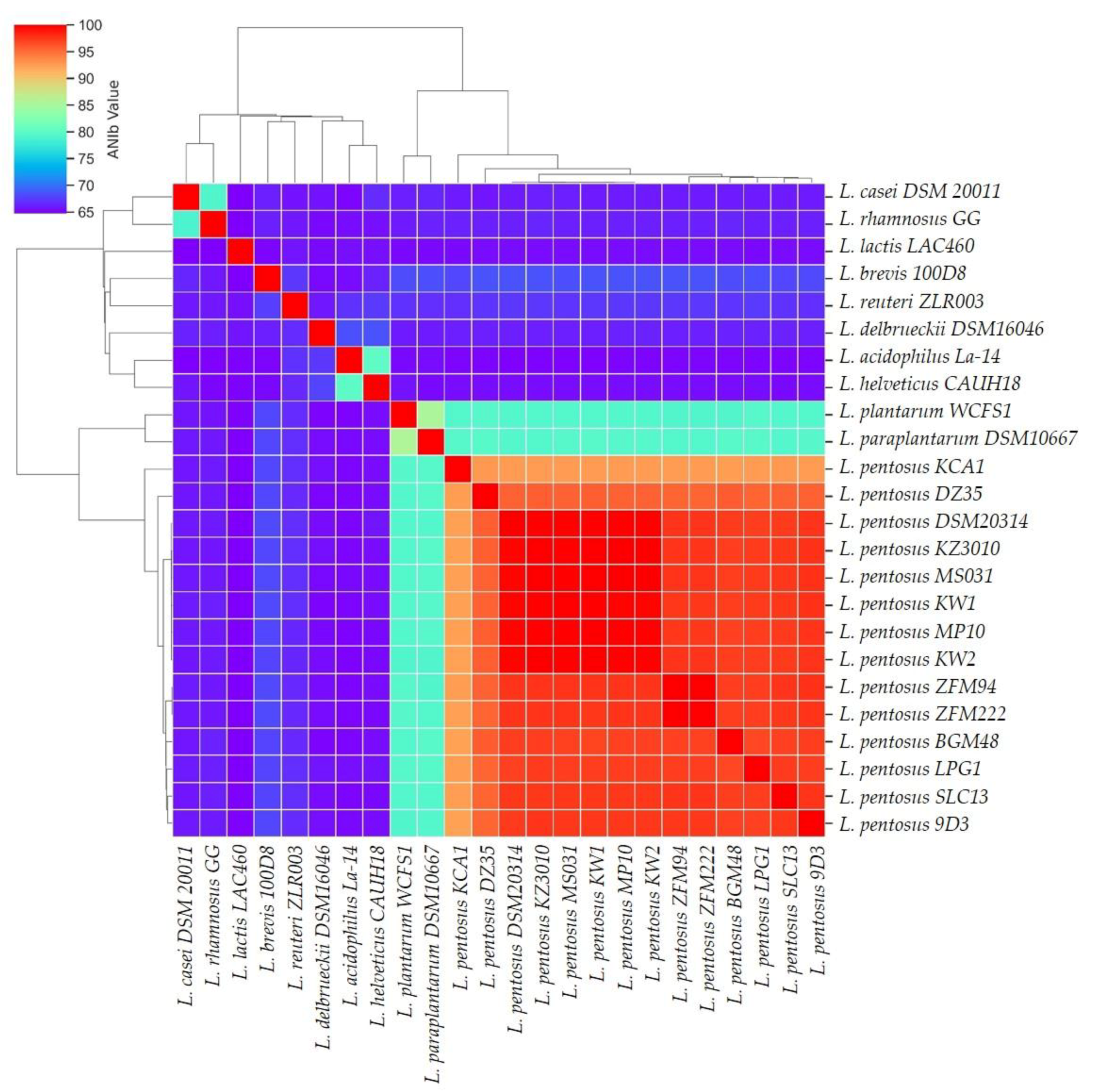
3.2. Phylogenetic and Comparative Analyses

The complete genome assembly of LPG1 was used to verify its taxonomy and phylogenetic relationship through ANI analysis. Studies recently showed ANI analysis as a robust measure of the genetic and evolutionary distance, as ANI analysis is based on a large number of conserved genes [50]. Moreover, phylogenetic analysis based exclusively on the 16S ribosomal gene does not distinguish between L. pentosus, L. paraplantarum or L. plantarum species [51]. For this reason, in the ANI analysis, we also included different strains of L. pentosus, L. plantarum, L. paraplantarum, as well as other genera from the Lactobacillaceae family (Table S1). ANI analysis revealed that LPG1 was grouped with the rest of the L. pentosus strains with >94% of the ANI value, above the threshold value to consider two organisms as the same species [52]. Moreover, L. plantarum WCFS1 was grouped together with L. paraplantarum DSM10667 strain in another sub-cluster, which was very different from the rest of the Lactobacillaceae genomes included in the present analysis (Figure 3).
The pan-genome of L. pentosus was analyzed, including our LPG1 strain as well as 62 other L. pentosus strains available in the NCBI database (https://0-www-ncbi-nlm-nih-gov.brum.beds.ac.uk/genbank/) (accessed on 10 July 2022) (Table S2, Supplementary Materials). For this purpose, we used the Roary program. The pan-genome composition consisted of 1407 core, 639 softcore, 2251 shell, and 5894 cloud genes (Table S4, Supplementary Materials). A total of 10,191 genes made up the full pan-genome; therefore, as genomes were added, the number of genes increased. The occurrence of a small number of core genes could be due to the small amount of L. pentosus genomes included in the analysis and to the fact that the threshold of presence in the analyzed sequences must be 99% for consideration as a core gene, which implies that if a gene is missing in any strain, it was not considered a core gene. Therefore, when the isolated strains originate from diverse environments such as fermented vegetables, female vaginas, fermented drink, soil, or fermented food, it is common for some genes to be lost. Figure 4 shows the hierarchical clustering and the heat map of the gene matrix (presence/absence) obtained after the pan-genome analysis of L. pentosus. As can be easily deduced from this figure, the L. pentosus strains more closely related to the LPG1 strain were IG8, IG9, IG11, and IG12, all of which were isolated from table olive biofilms.
The approach used for pan-genome analysis was also used to search probiotic marker genes (PMGs) within the shell and cloud genes present in our LPG1 strain. Genes involved in the tolerance of bile salt, adhesion, gut persistence, response to stress, and carbohydrate metabolism were found in the LPG1 strain in both the shell and cloud genes categories, which are also the genes shared with the rest of the L. pentosus strains (Table 1). Finally, from the total of 62 L. pentosus strains assayed, 15 strains with two complete exopolysaccharide (EPS) clusters and 48 strains with one complete EPS cluster were obtained. The genome comparison indicated the presence of only 78 wzy genes, which encoded the polysaccharide polymerase protein, an essential protein for EPS biosynthesis, although other crucial genes were found [53,54]. However, previous studies showed incomplete EPS clusters would likely be compensated by the genes located in other clusters [55]. Many other studies have reported the involvement of EPS in functions as diverse as biofilm formation, adhesion to the intestinal cells, inhibition of pathogen adhesion to gut cells, as well as resistance to bile salt and toxic compounds such as metal ions, and resistance under adverse conditions such as desiccation, a high salt concentration, and varying pH [56,57,58,59].

3.3. Food Safety Assessment

Lactobacilli have been commonly recognized as generally regarded as safe (GRAS) organisms due to their high safety profile. Additionally, L. pentosus has the qualified presumption of safety (QPS) status from the European Food Safety Agency (EFSA). However, previous studies have shown that certain Lactobacillus strains have antimicrobial resistance genes (AMR) that are involved in antibiotic resistance mechanisms, e.g., against chloramphenicol, tetracycline, beta-lactam, or aminoglycoside [69,70,71]. Therefore, the resistome of LPG1 was evaluated by the RGI tool (CARD) matching only complete genes. The CARD prediction matched strict category 2 gen, vanT and vanY in the VanG cluster, which encode alanine racemase and D-alanyl-D-alanine carboxypeptidase protein, respectively. Both genes were present in a vancomycin resistance cluster. On the one hand, vanT converts L-serine to D-serine for peptidoglycan synthesis, and DD-carboxypeptidase catalyzes the release of D-Ala from the diacetyl-L-Lys-D-Ala-D-Ala tripeptide [72]. Of the remaining genes in the cluster, only one was predicted by the RGI tool as a loose category, the vanS gene in the vanG cluster. Therefore, the vanG operon was predicted as incomplete. However, previous in vitro tests showed that LPG1 was a vancomycin-resistant strain [8]. This may be due to the presence of the D-alanine-D-alanine ligase protein encoded by the ddlA gene, as the vanG gene encodes to form the same protein, creating the dipeptide D-Ala-D-Ala, which after several transformations, develops a high affinity for vancomycin, binding it to the membrane and preventing the penetration of the antibiotic into the cytoplasm [73]. It is noteworthy that vancomycin resistance is considered intrinsic in Lactobacilli and not transmissible and is therefore not considered a safety concern by EFSA. As can be seen in Table S3 (pan-genome analysis) all of the L. pentosus strains carry both genes encoding alanine racemase and D-alanyl-D-alanine. Therefore, resistance to vancomycin is expected. On the other hand, no other AMR genes were obtained by the ResFinder tool for LPG1. These data agree with previous results obtained by Benítez-Cabello et al. [8], who described this strain as very sensitive to a wide range of antibiotics. Furthermore, no gene was predicted to be virulent based on a search in VirulenceFinder. Finally, the PathogenFinder tool classified the strain as a non-human pathogen, with a probability of being a human pathogen of only 0.075%.

3.4. Multifunctional Assessment

L. pentosus and L. plantarum are species typically found in a wide range of vegetable fermentations. This ability to colonize different niches is due to the presence, in both species, of an arsenal of enzymes that degrade carbohydrates released during fermentation [47,74].
Regarding technological features, in silico analysis of the LPG1 strain showed a complete Embden–Meyenhorf pathway. Thereby, LPG1 is able to ferment six-carbon carbohydrates such as glucose and fructose, both present in table olive fermentation brine. The enzymatic arsenal predicted in the annotation of the LPG1 strain allows it to capture and hydrolyze sucrose, a compound of glucose and fructose linked through an O-glucosidic bond that is present in large quantities in table olive brine. Moreover, LPG1 contains the mannitol degradative operon. This sugar is also common in table olive fermentations. However, in the absence of six-carbon carbohydrates, LPG1 can ferment five-carbon sugars through the pentose phosphate pathway. Furthermore, the LPG1 strain was predicted to contain other enzymatic tools involved in the degradation of more complex carbohydrates such as galactose, glycogen, starch, cellulose, or xylan (Table S3).
Moreover, CAZymes analysis revealed that this enzymatic arsenal was classified into five gene classes of CAZymes. The LPG1 genome contains 58 genes encoding the Glycosides Hydrolase family (GHs), 41 genes encoding Glucosyltransferase (GT), 3 Carbohydrates Esterases (CE), and 8 genes encoding Carbohydrate-Binding Modules (CBM). Similar results were described for other L. pentosus strains [65,75], which contrasts with the lower amount of CAZymes reported in other genera from the Lactobacillaceae family such as L. casei, L. rhamnosus, L. acidophilus, or L. helveticus [76]. This difference probably lies in the ability of L. pentosus to adapt to different niches and the use of diverse carbon sources as nutrients [4]. Therefore, species such as L. rhamnosus, L. casei, or L. acidophilus are more commonly found in host environments. In addition, to contribute to the fermentation process, the LPG1 strain contains three copies of the gene encoding for D-Lactate dehydrogenase and three copies of the genes encoding for L-Lactate dehydrogenase, supporting the results obtained in previous studies where the LPG1 strain was shown to be a major producer of lactic acid [8]. In these previous studies, this strain also showed high esterase activity, involved in aroma production and degradation of the bitter compound oleuropein. The subsequent in silico analysis confirmed the esterase activity, as the LPG1 strain is predicted to contain genes encoding esterase, carboxylesterase, gallate decarboxylase, p-coumaric acid decarboxylase, and the transcriptional regulator PadR, all of which are involved in the degradation of phenolic compounds [68]. On the contrary, no gene encoding β-glucosidase was predicted, also supporting the previous results that showed the phenotype was not positive for this activity [8].
Many microorganisms produce a diverse range of products with antibiotic, antifungal, or immunosuppressant activity such as bacteriocins and Ribosomally synthesized and Post translationally modified Peptides (RiPPs), encoded by different gene clusters [77]. The occurrence of bacteriocins in fermented food is very important because this can help bacteria to inhibit the growth or eliminate foodborne pathogens and spoilage bacteria [78]. After comprehensive bioinformatics analysis, two types of bacteriocins in the LPG1 genome were predicted; however, no RiPPs were predicted. A Pediocin gene cluster (PA-1) was identified between position 3.422.048 and 3.442.255 bp. Although bacteriocins commonly show more activity against Gram-positive bacteria, Escherichia coli and Salmonella Typhimurium have been shown to be sensitive to pediocin. Several studies have shown the relevant activity of Pediocin against Listeria monocytogenes [79]. In addition, the plantaricin EF gene cluster was predicted and encoded between position 420.305 and 435.375 bp. Plantaricin EF consists of two-peptide bacteriocins, and both genes were found side by side in the same operon. Both pediocin and plantaricin EF were found in other genomes of the Lactobacillus genus [80,81]. Previous studies showed the ability of LPG1 to inhibit E. coli and L. monocytogenes under in vitro conditions, improving the inhibitory activity shown by the recognized probiotics strains L. casei Shirota and L. rhamnosus GG [8]. All of these data give solid reasons for the commercial use of this microorganism as a starter culture in table olives (https://www.oleica.es/productos-y-servicios/oleicastarter/, accessed on 10 July 2022) and possibly other fermented vegetables.
Regarding the probiotic potential, this activity is determined by several factors such as the capacity to survive digestion and persistence in the human gut, attachment in the intestinal cells, ability to carry out metabolic processes in a highly competitive environment, and finally to produce a beneficial effect on the host [60]. In silico analysis of LPG1 predicted a large number of genes with the ability to perform all of these functions (Table S3). Firstly, the LPG1 strain was predicted to harbor three genes encoding choloylglicine hydrolase, involved in resistance to bile salt. Although no choloylglicine hydrolase gene was annotated as the bile salt hydrolase gene (bsh), previous studies showed the ability of the LPG1 strain to decrease cholesterol levels [8]. The ability of microorganisms to deconjugate bile via the production of bile salt hydrolase has been widely associated with their cholesterol-lowering potential in the prevention of hypercholesterolemia [61]. Additionally, other genes with important roles regarding resistance to bile salt were predicted. In this way, two chaperones genes encoding clpC and clpE, four genes encoding methionine sulfoxide reductase (msrA_1, msrA_2, mrsA_3, and msrB), two genes encoding histidine protein kinase (HKP) along with response regulator (RR), and two efflux systems related to the multidrug resistance transporter family encoding by emrB_1 and lmrA were predicted [82,83].
A large number of genes involved in the response to acid stress and ultimately involved in persistence in the gut, such as the DnaK, DnaJ, GroEL, and GroES chaperones, as well as the GrpE protein, were found in the genome of the LPG1 strain. Previous assays related an upregulated response to the acid environment and bile exposure to genes coding for these proteins [84,85]. Furthermore, a total of 16 complete PTS sugar transporter systems were annotated, reflecting an adaptative and flexible behavior in carbohydrate metabolism. More specifically, cellobiose PTS has been confirmed to play a key role in the competitive ability of Lactobacillus in the gut [86].
Regarding the ability of the LPG1 strain to adhere to intestinal cells, a variety of related genes have been predicted. Thus, five genes encoding a mucus-binding protein (MucBP), two genes encoding a collagen binding-protein and fibronectin-binding domain-containing protein, and two genes encoding glyceraldehyde-3-phosphate dehydrogenase were predicted, all of them involved mainly in the colonization of the gut [62,63,64,87]. In addition, others genes involved in intestinal cell adhesion such as the strA, luxS, dltA, or tuf genes were annotated in the LPG1 genome [88,89,90,91]. In contrast, the gene encoding S-layer protein, an important protein involved in the adhesion to intestinal cells, was not predicted [62]. EPS production also plays an important role in the adhesion to the surface, as well as conferring a certain degree of protection to the bacteria themselves [92]. Moreover, bacterial EPS has been associated with the pathogen’s growth inhibition [93] and modulation of the immune system [94,95]. In this sense, up to two complete EPS clusters were predicted in the LPG1 genome.
Alternatively, a certain degree of synergism of LPG1 with the gut microbiota could be expected due to a large number of enzymes producers of lactic acid that were predicted, especially butyrate-producing colon bacteria that transform lactic acid into butyric acid, which has been shown to be an excellent beneficial anti-inflammatory in the gut epithelium [96,97]. Previous studies showed the ability of this strain to modulate the immune response in human and murine cell lines and a colitis murine model, highlighting its anti-inflammatory activity [9].
Finally, some species of Lactobacillus are known to be producers of vitamin B9, also known as folate, an essential nutrient component of the human diet, that is involved in many metabolic pathways [98]. The efficiency of DNA replication, repair, and methylation is affected by folate; therefore, high amounts of folate are required by fast-proliferating cells such as leucocytes, erythrocytes, and enterocytes [99]. The LPG1 genome was predicted to harbor an active folate cluster including genes such as dfrA, fpgS_1, fpgS_2, folP, folE, folk, and folB.

3.5. Identification of Mobile Genetic Elements

Bacteriophages are viruses that infect and replicate in bacterial cells, being ubiquitous and the most abundant biological agent in the environment. Similar to most viruses, bacteriophages are species-specific or even strain-specific. When a bacteriophage infects a bacterial cell, two different strategies of replication can be developed, a lysogenic or a lytic cycle. Usually, during a lysogenic cycle, the phage genome is integrated into the bacterial chromosome, called a prophage. The prophage contains the necessary information to induce a lytic cycle, a viral reproduction method that involves the destruction of infected cells and that is usually induced in response to stress conditions [100,101]. The presence of prophages in bacterial strains with probiotic potential isolated from the fermentation process is common [102,103,104]. Furthermore, prophages are a source of new genes added to the genome, in some cases providing new features in the bacterial genomes [105,106]. These features could include antibiotic resistance genes or virulence factors [107,108]. The LPG1 strain was predicted to contain four intact prophages according to PHASTER. A similar number of prophages have been found in other L. pentosus genomes [14,75,108]. The length of the prophages ranged from 39.4 Kb to 62.5 kB, and each prophage encoded about 50 proteins. Integrase was present in all prophages. Among genes predicted in the prophages sequence, no genes involved in the drug resistance mechanisms or virulence factors were detected. A comparison of the database and the proteomic tree was performed for each latent phage (Figure 5). The prophages analyzed were related to the Siphoviridae family, the main family responsible for infecting the Lactiplantibacillus genus along with the Myoviridae family [109].
The IslandViewer 4 tool was used to predict GI matched with whole prophages predicted through PHASTER. GI refer to discrete DNA segments that establish horizontally transferred genes in a population and frequently encode functions such as pathogenesis, symbiosis, and adaptation [110,111]. Six putative GI in the LPG1 genome were predicted by at least one of the following methods: IslandPick, SIGI-HMM, and IslandPath-DIMOB [112,113,114]. Some of the most interesting genes found in GI of the LPG1 strain were involved in polysaccharide biosynthesis, teichoic acid synthesis, metal ion transporters, and carbohydrate metabolism, such as beta-galactosidase and extracellular alpha-L-arabinofuranosidase. These findings showed similarities with other Lactobacillus where GI included genes coding for sugar metabolism and transport and exopolysaccharide biosynthesis [115,116]. Furthermore, a search for integrons was carried out using IntegronFinder, and no results were obtained for the presence of integrons. Perhaps the most important mobile genetic elements are the integrons, which play a key role in the dissemination of antibiotic-resistance genes [117].
The GI coding for L-arabinofuranosidase and beta-galactosidase, both found within the shell genes in the pan-genome analysis, were used to find the most likely bacterium that transferred these genes to LPG1. Thus, BLAST-p was performed for both genes, prior to transformation into proteins. Later, the alignment was held with the sequences of most similar genomes found in the BLAST-p results. The phylogenetic tree showed that Levilactobacillus was probably the genus responsible for the transfer of the GI to LPG1 (Figure 6). Interestingly, species of the Levilactobacillus genus have been also identified in Sicilian and Algerian table olive processing [118,119], with both species sharing the same habitats. In addition, L. pentosus IG8, IG9, IG10, and IG11, all of them from table olive processing [13], contain these genes in common with LPG1. Therefore, it is likely that the LPG1 strain shares a common ancestor with all of them. These results agree with the hierarchical clustering obtained from the pan-genome analysis.
Finally, other mobile genetic elements such as the insertion sequence (IS) could play a role in gene disruption or activation due to promoter interaction to contribute to the genome plasticity [120]. Functional gene loss occurs in genomes with a large number of IS elements. Furthermore, IS elements with transposase genes flanked by two inverted repeats have the skill to move through transposition, causing DNA rearrangement [121,122]. Seventeen transposases were found in the chromosome of LPG1, and 13 were located in pl1LPg1, the most abundant plasmid in the LPG1 strain. A similar number was found in other Lactobacillus strains such as L. pentosus DSM20314 or L. reuteri PNW1 [67]. However, the number was lower than that of other L. pentosus with putative probiotic potential such as L. pentosus MP-10 or L. pentosus CF2-10N [14,123]. This could mean that the LPG1 strain has higher transcription stability than other genomes with a large amount of IS. The transposase included 10 different families, highlighting the ISL3, IS5, and IS30 families with multiple copies.

3.6. Identification of CRISPR−Cas Sequences

CRISPR-Cas systems are an adaptive immune system present in prokaryotic organisms, both bacteria, and Archaea, conferring resistance to exogenous genetic material, whether from pathogens, other bacteria, phages, or any element that could lead to a risk to the organism at the expression levels [124,125]. Previous studies have shown the occurrence of the prophages was lower in strains with the CRISPR-Cas system than in strains without this immune system, suggesting that CRISPR-Cas systems play a key role in preventing phage infection and consequently avoiding prophage integration into the genome [100]. In the LPG1 genome, two CRISPR-Cas systems were identified, CRISPR-Cas type I-E and type II-A. CRISPR-Cas operon type I-E and type II-A were formed by eight Cas genes (cas1, cas2, cas3, cse1, cse2, cas7, cas5, and cas6) and four Cas genes (cas9, cas1, cas2, and csn2), respectively. These results were very similar to those found in other L. pentosus strains such as KCA1, MP-10, IG4, IG7, IG8, IG9, IG11, or IG12 [12,13,65,66]. Up to six CRISPR arrays were predicted by the CRISPRcasFinder tool but, only four were confirmed by CRISPRdb. A CRISPR array is composed of repeated elements and spacers, where spacer sequences are fragments of foreign DNA originating from plasmids, phages, or other exogenous genetic material that are incorporated into the host [125]. Therefore, the occurrence of the CRISPR-Cas system could prevent the LPG1 strain from acquiring antimicrobial resistance genes or virulence or pathogenic factors through the horizontal transfer of genes.

4. Conclusions

Comprehensive bioinformatic analysis of the LPG1 genome has revealed the presence of a large number of genes related to multifunctional features, which could be allied with many of its technological and probiotic phenotypes obtained in previous studies. A safety assessment of the LPG1 genome also showed the absence of virulence and antibiotic resistance genes, as well as the presence of gene systems that could prevent their acquisition (CRISPR-Cas system). Conclusively, LPG1 is a safe microorganism with great potential for use as a human probiotic from plant origin or as a starter culture for vegetable fermentations.

Supplementary Materials

The following supporting information can be downloaded at https://0-www-mdpi-com.brum.beds.ac.uk/article/10.3390/foods12050938/s1, Table S1: List of Lactobacillaceae strains included in the present study for ANI (Average Nucleotide Identity) analysis; Table S2: List of L. pentosus strains used in the present study for pangenome analysis; Table S3: List of genes found in L. pentosus LPG1 related to its probiotic and technological potentials; Table S4: Results of the pan-genome analysis, presence/absence of genes in L. pentosus strains included in the analysis.

Author Contributions

Conceptualization, F.N.A.-L.; methodology, E.L.-G., A.B.-C., J.R.-G.; software, E.L.-G., A.B.-C., J.R.-G., V.L.; data curation, E.L.-G.; writing—original draft preparation, E.L.-G., A.B.-C., F.N.A.-L.; writing—review and editing, E.L.-G., F.N.A.-L., A.B.-C., V.L., J.R.-G.; supervision, F.N.A.-L., V.L.; project administration, F.N.A.-L.; funding acquisition, F.N.A.-L. All authors have read and agreed to the published version of the manuscript.

Funding

This research was funded through TOBE project (RTI2018-100883-B-I00, MCIU/AEI/FEDER, UE).

Data Availability Statement

ENA repository access number PRJEB51357.

Acknowledgments

ELG thanks the Spanish Ministry of Science and Innovation for his FPI contract (PRE2019-087812). ABC thanks Junta de Andalucia for his contract PAIDI2020-00162. JRG has received funding from the European Union’s Horizon 2020 research and innovation program under grant agreement Nº 101033411. We sincerely thank to Belén Caballero from the Microbiology Unit of Instituto Grasa (CSIC) for her invaluable help during the DNA extraction.

Conflicts of Interest

The authors declare no conflict of interest.

References

  1. Inglin, R.C.; Meile, L.; Stevens, M.J.A. Clustering of Pan- and Core-genome of Lactobacillus provides novel evolutionary insights for differentiation. BMC Genom. 2018, 19, 284. [Google Scholar] [CrossRef] [PubMed]
  2. IOC Economic Affairs & Promotion Unit—International Olive Council. Available online: https://www.internationaloliveoil.org/what-we-do/economic-affairs-promotion-unit/#figures%0Ahttps://www.internationaloliveoil.org/what-we- (accessed on 10 July 2022).
  3. Garrido-Fernandez, A.; Fernández Díez, M.J.; Adams, R.M. Table Olives: Production and Processing; Chapman & Hall: London, UK, 1997; pp. 67–109. [Google Scholar]
  4. Hurtado, A.; Reguant, C.; Bordons, A.; Rozès, N. Lactic acid bacteria from fermented table olives. Food Microbiol. 2012, 31, 1–8. [Google Scholar] [CrossRef]
  5. Arroyo-López, F.N.; Romero-Gil, V.; Bautista-Gallego, J.; Rodríguez-Gómez, F.; Jiménez-Díaz, R.; García-García, P.; Querol, A.; Garrido-Fernández, A. Potential benefits of the application of yeast starters in table olive processing. Front. Microbiol. 2012, 3, 161. [Google Scholar] [CrossRef] [PubMed] [Green Version]
  6. Grand View Research. Available online: https://www.grandviewresearch.com/industry-analysis/probiotics-market (accessed on 3 November 2022).
  7. Arroyo-López, F.N.; Bautista-Gallego, J.; Domínguez-Manzano, J.; Romero-Gil, V.; Rodríguez-Gómez, F.; García-García, P.; Garrido-Fernández, A.; Jiménez-Díaz, R. Formation of lactic acid bacteria-yeasts communities on the olive Surface during Spanish-style Manzanilla fermentations. Food Microbiol. 2012, 32, 295–301. [Google Scholar] [CrossRef] [PubMed]
  8. Benítez-Cabello, A.; Calero-Delgado, B.; Rodríguez-Gómez, F.; Garrido-Fernández, A.; Jiménez-Díaz, R.; Arroyo-López, F.N. Biodiversity and multifunctional features of lactic acid bacteria isolated from table olive biofilms. Front. Microbiol. 2019, 10, 836. [Google Scholar] [CrossRef] [PubMed] [Green Version]
  9. Benítez-Cabello, A.; Torres-Maravilla, E.; Bermúdez-Humarán, L.; Langella, P.; Martín, R.; Jiménez-Díaz, R.; Arroyo-López, F.N. Probiotic Properties of Lactobacillus Strains Isolated from Table Olive Biofilms. Probiotics Antimicrob. Proteins 2020, 12, 1071–1082. [Google Scholar] [CrossRef]
  10. Maldonado-Barragán, A.; Caballero-Guerrero, B.; Lucena-Padrós, H.; Ruiz-Barba, J.L. Genome sequence of Lactobacillus pentosus IG1, a strain isolated from Spanish-style green olive fermentations. J. Bacteriol. 2011, 193, 5605. [Google Scholar] [CrossRef] [Green Version]
  11. Abriouel, H.; Benomar, N.; Pulido, R.P.; Cañamero, M.M.; Gálvez, A. Annotated genome sequence of Lactobacillus pentosus MP-10, which has probiotic potential, from naturally fermented Aloreña green table olives. J. Bacteriol. 2011, 193, 4559–4560. [Google Scholar] [CrossRef] [Green Version]
  12. Calero-Delgado, B.; Martín-Platero, A.M.; Pérez-Pulido, A.J.; Benítez-Cabello, A.; Casimiro-Soriguer, C.S.; Martínez-Bueno, M.; Arroyo-López, F.N.; Rodríguez-Gómez, F.; Bautista-Gallego, J.; Garrido-Fernández, A.; et al. Draft genome sequences of six Lactobacillus pentosus strains isolated from brines of traditionally fermented Spanish-style green table olives. Genome Announc. 2018, 6, e00379-18. [Google Scholar] [CrossRef] [Green Version]
  13. Calero-Delgado, B.; Pérez-Pulido, A.J.; Benítez-Cabello, A.; Martín-Platero, A.M.; Casimiro-Soriguer, C.S.; Martínez-Bueno, M.; Arroyo-López, F.N.; Díaz, R.J. Multiple Genome Sequences of Lactobacillus pentosus Strains Isolated from Biofilms on the Skin of Fermented Green Table Olives. Microbiol. Resour. Announc. 2019, 8, e01546-18. [Google Scholar] [CrossRef] [Green Version]
  14. Abriouel, H.; Manestsberger, J.; Caballero-Cómez, N.; Benomar, N. In silico genomic analysis of the potential probiotic Lactiplantibacillus pentosus CF2-10N reveals promising beneficial effects with health promoting properties. Front. Microbiol. 2022, 13, 4201. [Google Scholar] [CrossRef] [PubMed]
  15. Page, C.A.; Pérez-Díaz, I.M. Whole-Genome Sequencing and Annotation of Selected Lactobacillales Isolated from Commercial Cucumber Fermentation. Microbiol. Resour. Announc. 2021, 10, e00625-21. [Google Scholar] [CrossRef] [PubMed]
  16. EFSA. Guidance on the characterisation of microorganisms used as feed additives or 30 as production organisms. EFSA J. 2018, 16, 5206. [Google Scholar] [CrossRef]
  17. Martín-Platero, A.M.; Valdivia, E.; Maqueda, M.; Martínez-Bueno, M. Fast, convenient, and economical method for isolating genomic DNA from lactic acid bacteria using a modification of the protein “salting-out” procedure. Anal. Biochem. 2007, 366, 102–104. [Google Scholar] [CrossRef]
  18. Son, S.; Lee, R.; Park, S.-M.; Lee, S.H.; Lee, H.-K.; Kim, Y.; Shin, D. Complete genome sequencing and comparative genomic analysis of Lactobacillus acidophilus C5 as a potential canine probiotics. J. Anim. Sci. Technol. 2021, 63, 1411–1422. [Google Scholar] [CrossRef]
  19. Seemann, T. Prokka: Rapid prokaryotic genome annotation. Bioinformatics 2014, 30, 2068–2069. [Google Scholar] [CrossRef] [Green Version]
  20. Stothard, P.; Wishart, D.S. Circular genome visualization and exploration using CGView. Bioinformatics 2005, 21, 537–539. [Google Scholar] [CrossRef] [Green Version]
  21. Richter, M.; Rosselló-Móra, R.; Oliver Glöckner, F.; Peplies, J. JSpeciesWS: A web server for prokaryotic species circumscription based on pairwise genome comparison. Bioinformatics 2016, 32, 929–931. [Google Scholar] [CrossRef]
  22. Ning, W.; Wei, Y.; Gao, L.; Han, C.; Gou, Y.; Fu, S.; Liu, D.; Zhang, C.; Huang, X.; Wu, S.; et al. HemI 2.0: An online service for heatmap illustration. Nucleic Acids Res. 2022, 50, W405–W411. [Google Scholar] [CrossRef]
  23. Page, A.J.; Cummins, C.A.; Hunt, M.; Wong, V.K.; Reuter, S.; Holden, M.T.G.; Fookes, M.; Falush, D.; Keane, J.A.; Parkhill, J. Roary: Rapid large-scale prokaryote pan genome analysis. Bioinformatics 2015, 31, 3691–3693. [Google Scholar] [CrossRef] [Green Version]
  24. Alcock, B.P.; Raphenya, A.R.; Lau, T.T.; Tsang, K.K.; Bouchard, M.; Edalatmand, A.; Huynh, W.; Nguyen, A.L.V.; Cheng, A.A.; Liu, S.; et al. CARD 2020: Antibiotic resistome surveillance with the comprehensive antibiotic resistance database. Nucleic Acids Res. 2020, 48, D517–D525. [Google Scholar] [CrossRef] [PubMed]
  25. Bortolaia, V.; Kaas, R.S.; Ruppe, E.; Roberts, M.C.; Schwarz, S.; Cattoir, V.; Philippon, A.; Allesoe, R.L.; Rita Rebelo, A.; Ferrer Florensa, A.; et al. Mette Pinholt 16, Muna F. Anjum 18, Nicholas A. Duggett 18. J. Antimicrob. Chemother. 2020, 16, 3491–3500. [Google Scholar] [CrossRef] [PubMed]
  26. Camacho, C.; Coulouris, G.; Avagyan, V.; Ma, N.; Papadopoulos, J.; Bealer, K.; Madden, T.L. BLAST+: Architecture and applications. BMC Bioinform. 2009, 10, 421. [Google Scholar] [CrossRef] [PubMed] [Green Version]
  27. Zankari, E.; Allesøe, R.; Joensen, K.G.; Cavaco, L.M.; Lund, O.; Aarestrup, F.M. PointFinder: A novel web tool for WGS-based detection of antimicrobial resistance associated with chromosomal point mutations in bacterial pathogens. J. Antimicrob. Chemother. 2017, 72, 2764–2768. [Google Scholar] [CrossRef] [PubMed] [Green Version]
  28. Cosentino, S.; Voldby Larsen, M.; Møller Aarestrup, F.; Lund, O. PathogenFinder—Distinguishing Friend from Foe Using Bacterial Whole Genome Sequence Data. PLoS ONE 2013, 8, 77302. [Google Scholar] [CrossRef]
  29. Joensen, K.G.; Scheutz, F.; Lund, O.; Hasman, H.; Kaas, R.S.; Nielsen, E.M.; Aarestrup, F.M. Real-time whole-genome sequencing for routine typing, surveillance, and outbreak detection of verotoxigenic Escherichia coli. J. Clin. Microbiol. 2014, 52, 1501–1510. [Google Scholar] [CrossRef] [PubMed] [Green Version]
  30. Tetzschner, A.M.M.; Johnson, J.R.; Johnston, B.D.; Lund, O.; Scheutz, F. In Silico genotyping of Escherichia coli isolates for extraintestinal virulence genes by use of whole-genome sequencing data. J. Clin. Microbiol. 2020, 58, e01269-20. [Google Scholar] [CrossRef]
  31. Huerta-Cepas, J.; Szklarczyk, D.; Heller, D.; Hernández-Plaza, A.; Forslund, S.K.; Cook, H.; Mende, D.R.; Letunic, I.; Rattei, T.; Jensen, L.J.; et al. EggNOG 5.0: A hierarchical, functionally and phylogenetically annotated orthology resource based on 5090 organisms and 2502 viruses. Nucleic Acids Res. 2019, 47, D309–D314. [Google Scholar] [CrossRef] [Green Version]
  32. Aramaki, T.; Blanc-Mathieu, R.; Endo, H.; Ohkubo, K.; Kanehisa, M.; Goto, S.; Ogata, H. KofamKOALA: KEGG Ortholog assignment based on profile HMM and adaptive score threshold. Bioinformatics 2020, 36, 2251–2252. [Google Scholar] [CrossRef] [Green Version]
  33. Zhang, H.; Yohe, T.; Huang, L.; Entwistle, S.; Wu, P.; Yang, Z.; Busk, P.K.; Xu, Y.; Yin, Y. DbCAN2: A meta server for automated carbohydrate-active enzyme annotation. Nucleic Acids Res. 2018, 46, W95–W101. [Google Scholar] [CrossRef] [PubMed] [Green Version]
  34. Yin, Y.; Mao, X.; Yang, J.; Chen, X.; Mao, F.; Xu, Y. DbCAN: A web resource for automated carbohydrate-active enzyme annotation. Nucleic Acids Res. 2012, 40, W445–W451. [Google Scholar] [CrossRef] [PubMed]
  35. Van Heel, A.J.; De Jong, A.; Song, C.; Viel, J.H.; Kok, J.; Kuipers, O.P. BAGEL4: A user-friendly web server to thoroughly mine RiPPs and bacteriocins. Nucleic Acids Res. 2018, 46, W278–W281. [Google Scholar] [CrossRef] [PubMed]
  36. Blin, K.; Shaw, S.; Kloosterman, A.M.; Charlop-Powers, Z.; Van Wezel, G.P.; Medema, M.H.; Weber, T. AntiSMASH 6.0: Improving cluster detection and comparison capabilities. Nucleic Acids Res. 2021, 49, W29–W35. [Google Scholar] [CrossRef] [PubMed]
  37. Arndt, D.; Grant, J.R.; Marcu, A.; Sajed, T.; Pon, A.; Liang, Y.; Wishart, D.S. PHASTER: A better, faster version of the PHAST phage search tool. Nucleic Acids Res. 2016, 44, W16–W21. [Google Scholar] [CrossRef] [Green Version]
  38. Zhou, Y.; Liang, Y.; Lynch, K.H.; Dennis, J.J.; Wishart, D.S. PHAST: A Fast Phage Search Tool. Nucleic Acids Res. 2011, 39, W347–W352. [Google Scholar] [CrossRef] [PubMed]
  39. Mihara, T.; Nishimura, Y.; Shimizu, Y.; Nishiyama, H.; Yoshikawa, G.; Uehara, H.; Hingamp, P.; Goto, S.; Ogata, H. Linking virus genomes with host taxonomy. Viruses 2016, 8, 66. [Google Scholar] [CrossRef] [Green Version]
  40. Nishimura, Y.; Yoshida, T.; Kuronishi, M.; Uehara, H.; Ogata, H.; Goto, S. ViPTree: The viral proteomic tree server. Bioinformatics 2017, 33, 2379–2380. [Google Scholar] [CrossRef]
  41. Bertelli, C.; Laird, M.R.; Williams, K.P.; Lau, B.Y.; Hoad, G.; Winsor, G.L.; Brinkman, F.S.L. IslandViewer 4: Expanded prediction of genomic islands for larger-scale datasets. Nucleic Acids Res. 2017, 45, W30–W35. [Google Scholar] [CrossRef]
  42. Néron, B.; Littner, E.; Haudiquet, M.; Perrin, A.; Cury, J.; Rocha, E.P.C. IntegronFinder 2.0: Identification and Analysis of Integrons across Bacteria, with a Focus on Antibiotic Resistance in Klebsiella. Microorganisms 2022, 10, 700. [Google Scholar] [CrossRef]
  43. Varani, A.M.; Siguier, P.; Gourbeyre, E.; Charneau, V.; Chandler, M. ISsaga is an ensemble of web-based methods for high throughput identification and semi-automatic annotation of insertion sequences in prokaryotic genomes. Genome Biol. 2011, 12, R30. [Google Scholar] [CrossRef] [Green Version]
  44. Siguier, P.; Perochon, J.; Lestrade, L.; Mahillon, J.; Chandler, M. ISfinder: The reference centre for bacterial insertion sequences. Nucleic Acids Res. 2006, 34, D32–D36. [Google Scholar] [CrossRef] [Green Version]
  45. Grissa, I.; Vergnaud, G.; Pourcel, C. CRISPRFinder: A web tool to identify clustered regularly interspaced short palindromic repeats. Nucleic Acids Res. 2007, 35, W52–W57. [Google Scholar] [CrossRef] [Green Version]
  46. Abby, S.S.; Néron, B.; Ménager, H.; Touchon, M.; Rocha, E.P.C. MacSyFinder: A program to mine genomes for molecular systems with an application to CRISPR-Cas systems. PLoS ONE 2014, 9, e110726. [Google Scholar] [CrossRef] [PubMed] [Green Version]
  47. Siezen, R.J.; Tzeneva, V.A.; Castioni, A.; Wels, M.; Phan, H.T.K.; Rademaker, J.L.W.; Starrenburg, M.J.C.; Kleerebezem, M.; van Hylckama Vlieg, J.E.T. Phenotypic and genomic diversity of Lactobacillus plantarum strains isolated from various environmental niches. Environ. Microbiol. 2010, 12, 758–773. [Google Scholar] [CrossRef] [PubMed]
  48. O’Sullivan, O.; O’Callaghan, J.; Sangrador-Vegas, A.; McAuliffe, O.; Slattery, L.; Kaleta, P.; Callanan, M.; Fitzgerald, G.F.; Ross, R.P.; Beresford, T. Comparative genomics of lactic acid bacteria reveals a niche-specific gene set. BMC Microbiol. 2009, 9, 50. [Google Scholar] [CrossRef] [PubMed] [Green Version]
  49. Holzapfel, W.H.; Haberer, P.; Geisen, R.; Björkroth, J.; Schillinger, U. Taxonomy and important features of probiotic microorganisms in food and nutrition. Am. J. Clin. Nutr. 2001, 73, 365s–373s. [Google Scholar] [CrossRef] [PubMed] [Green Version]
  50. Jain, C.; Rodriguez-R, L.M.; Phillippy, A.M.; Konstantinidis, K.T.; Aluru, S. High throughput ANI analysis of 90K prokaryotic genomes reveals clear species boundaries. Nat. Commun. 2018, 9, 5114. [Google Scholar] [CrossRef] [Green Version]
  51. Torriani, S.; Clementi, F.; Vancanneyt, M.; Hoste, B.; Dellaglio, F.; Kersters, K. Differentiation of Lactobacillus plantarum, L. pentosus and L. paraplantarum species by RAPD-PCR and AFLP. Syst. Appl. Microbiol. 2001, 24, 554–560. [Google Scholar] [CrossRef]
  52. Arahal, D.R. Whole-genome analyses: Average nucleotide identity. In Methods in Microbiology; Academic Press: Cambridge, MA, USA, 2014; Volume 41, pp. 103–122. [Google Scholar]
  53. Schmid, J.; Sieber, V.; Rehm, B. Bacterial exopolysaccharides: Biosynthesis pathways and engineering strategies. Front. Microbiol. 2015, 6, 496. [Google Scholar] [CrossRef] [Green Version]
  54. Zeidan, A.A.; Poulsen, V.K.; Janzen, T.; Buldo, P.; Derkx, P.M.F.; Øregaard, G.; Neves, A.R. Polysaccharide production by lactic acid bacteria: From genes to industrial applications. FEMS Microbiol. Rev. 2017, 41, S168–S200. [Google Scholar] [CrossRef] [Green Version]
  55. Remus, D.M.; van Kranenburg, R.; van Swam, I.I.; Taverne, N.; Bongers, R.S.; Wels, M.; Wells, J.M.; Bron, P.A.; Kleerebezem, M. Impact of 4 Lactobacillus plantarum capsular polysaccharide clusters on surface glycan composition and host cell signaling. Microb. Cell Fact. 2012, 11, 1–10. [Google Scholar] [CrossRef] [PubMed] [Green Version]
  56. Wang, K.; Li, W.; Rui, X.; Chen, X.; Jiang, M.; Dong, M. Characterization of a novel exopolysaccharide with antitumor activity from Lactobacillus plantarum 70810. Int. J. Biol. Macromol. 2014, 63, 133–139. [Google Scholar] [CrossRef] [PubMed]
  57. Li, S.; Huang, R.; Shah, N.P.; Tao, X.; Xiong, Y.; Wei, H. Antioxidant and antibacterial activities of exopolysaccharides from Bifidobacterium bifidum WBIN03 and Lactobacillus plantarum R315. J. Dairy Sci. 2014, 97, 7334–7343. [Google Scholar] [CrossRef] [PubMed] [Green Version]
  58. Dertli, E.; Mayer, M.J.; Narbad, A. Impact of the exopolysaccharide layer on biofilms, adhesion and resistance to stress in Lactobacillus johnsonii FI9785. BMC Microbiol. 2015, 15, 8. [Google Scholar] [CrossRef] [Green Version]
  59. Whitehead, K.; Versalovic, J.; Roos, S.; Britton, R.A. Genomic and genetic characterization of the bile stress response of probiotic Lactobacillus reuteri ATCC 55730. Appl. Environ. Microbiol. 2008, 74, 1812–1819. [Google Scholar] [CrossRef] [Green Version]
  60. Lebeer, S.; Vanderleyden, J.; De Keersmaecker, S.C.J. Genes and Molecules of Lactobacilli Supporting Probiotic Action. Microbiol. Mol. Biol. Rev. 2008, 72, 728–764. [Google Scholar] [CrossRef] [Green Version]
  61. Choi, S.B.; Lew, L.C.; Yeo, S.K.; Nair Parvathy, S.; Liong, M.T. Probiotics and the BSH-related cholesterol lowering mechanism: A Jekyll and Hyde scenario. Crit. Rev. Biotechnol. 2015, 35, 392–401. [Google Scholar] [CrossRef]
  62. Buck, B.L.; Altermann, E.; Svingerud, T.; Klaenhammer, T.R. Functional analysis of putative adhesion factors in Lactobacillus acidophilus NCFM. Appl. Environ. Microbiol. 2005, 71, 8344–8351. [Google Scholar] [CrossRef] [Green Version]
  63. Heinemann, C.; Hylckama Vlieg, J.E.T.; Janssen, D.B.; Busscher, H.J.; Mei, H.C.; Reid, G. Purification and characterization of a surface-binding protein from Lactobacillus fermentum RC-14 that inhibits adhesion of Enterococcus faecalis 1131. FEMS Microbiol. Lett. 2000, 190, 177–180. [Google Scholar] [CrossRef]
  64. Kinoshita, H.; Uchida, H.; Kawai, Y.; Kawasaki, T.; Wakahara, N.; Matsuo, H.; Watanabe, M.; Kitazawa, H.; Ohnuma, S.; Miura, K.; et al. Cell surface Lactobacillus plantarum LA 318 glyceraldehyde-3-phosphate dehydrogenase (GAPDH) adheres to human colonic mucin. J. Appl. Microbiol. 2008, 104, 1667–1674. [Google Scholar] [CrossRef]
  65. Abriouel, H.; Pérez Montoro, B.; Casimiro-Soriguer, C.S.; Pérez Pulido, A.J.; Knapp, C.W.; Caballero Gómez, N.; Castillo-Gutiérrez, S.; Estudillo-Martínez, M.D.; Gálvez, A.; Benomar, N. Insight into potential probiotic markers predicted in Lactobacillus pentosus MP-10 genome sequence. Front. Microbiol. 2017, 8, 891. [Google Scholar] [CrossRef] [PubMed] [Green Version]
  66. Anukam, K.C.; Macklaim, J.M.; Gloor, G.B.; Reid, G.; Boekhorst, J.; Renckens, B.; van Hijum, S.A.F.T.; Siezen, R.J. Genome Sequence of Lactobacillus pentosus KCA1: Vaginal Isolate from a Healthy Premenopausal Woman. PLoS ONE 2013, 8, e59239. [Google Scholar] [CrossRef] [PubMed] [Green Version]
  67. Alayande, K.A.; Aiyegoro, O.A.; Nengwekhulu, T.M.; Katata-Seru, L.; Ateba, C.N. Integrated genome-based probiotic relevance and safety evaluation of Lactobacillus reuteri PNW1. PLoS ONE 2020, 15, e235873. [Google Scholar] [CrossRef] [PubMed]
  68. Zotta, T.; Giavalisco, M.; Parente, E.; Picariello, G.; Siano, F.; Ricciardi, A. Selection of Lactiplantibacillus Strains for the Production of Fermented Table Olives. Microorganisms 2022, 10, 625. [Google Scholar] [CrossRef]
  69. Hummel, A.S.; Hertel, C.; Holzapfel, W.H.; Franz, C.M.A.P. Antibiotic resistances of starter and probiotic strains of lactic acid bacteria. Appl. Environ. Microbiol. 2007, 73, 730–739. [Google Scholar] [CrossRef] [Green Version]
  70. Sharma, P.; Tomar, S.K.; Goswami, P.; Sangwan, V.; Singh, R. Antibiotic resistance among commercially available probiotics. Food Res. Int. 2014, 57, 176–195. [Google Scholar] [CrossRef]
  71. Gueimonde, M.; Sánchez, B.; de los Reyes-Gavilán, C.G.; Margolles, A. Antibiotic resistance in probiotic bacteria. Front. Microbiol. 2013, 4, 202. [Google Scholar] [CrossRef] [Green Version]
  72. Rhazi, N.; Delmarcelle, M.; Sauvage, E.; Jacquemotte, F.; Devriendt, K.; Tallon, V.; Ghosez, L.; Frère, J.-M. Specificity and reversibility of the transpeptidation reaction catalyzed by the Streptomyces R61 D-Ala-D-Ala peptidase. Protein Sci. 2005, 14, 2922–2928. [Google Scholar] [CrossRef] [Green Version]
  73. Arias, C.A.; Weisner, J.; Blackburn, J.M.; Reynolds, P.E. Serine and alanine racemase activities of VanT: A protein necessary for vancomycin resistance in Enterococcus gallinarum BM4174. Microbiology 2000, 146, 1727–1734. [Google Scholar] [CrossRef] [Green Version]
  74. Vaccalluzzo, A.; Pino, A.; Russo, N.; De Angelis, M.; Caggia, C.; Randazzo, C.L. FoodOmics as a new frontier to reveal microbial community and metabolic processes occurring on table olives fermentation. Food Microbiol. 2020, 92, 103606. [Google Scholar] [CrossRef]
  75. Stergiou, O.S.; Tegopoulos, K.; Kiousi, D.E.; Tsifintaris, M.; Papageorgiou, A.C.; Tassou, C.C.; Chorianopoulos, N.; Kolovos, P.; Galanis, A. Whole-Genome Sequencing, Phylogenetic and Genomic Analysis of Lactiplantibacillus pentosus L33, a Potential Probiotic Strain Isolated from Fermented Sausages. Front. Microbiol. 2021, 12, 3295. [Google Scholar] [CrossRef] [PubMed]
  76. CAZy—Bacteria. Available online: http://www.cazy.org/b96.html (accessed on 30 September 2022).
  77. Arnison, P.G.; Bibb, M.J.; Bierbaum, G.; Bowers, A.A.; Bugni, T.S.; Bulaj, G.; Camarero, J.A.; Campopiano, D.J.; Challis, G.L.; Clardy, J.; et al. Ribosomally synthesized and post-translationally modified peptide natural products: Overview and recommendations for a universal nomenclature. Nat. Prod. Rep. 2013, 30, 108–160. [Google Scholar] [CrossRef] [PubMed]
  78. Hurtado, A.; Ben Othman, N.; Chammem, N.; Hamdi, M.; Ferrer, S.; Reguant, C.; Bordons, A.; Rozès, N. Characterization of Lactobacillus isolates from fermented olives and their bacteriocin gene profiles. Food Microbiol. 2011, 28, 1514–1518. [Google Scholar] [CrossRef] [PubMed]
  79. Barbosa, A.A.T.; Mantovani, H.C.; Jain, S. Bacteriocins from lactic acid bacteria and their potential in the preservation of fruit products. Crit. Rev. Biotechnol. 2017, 37, 852–864. [Google Scholar] [CrossRef] [PubMed]
  80. Rodríguez, J.M.; Martínez, M.I.; Kok, J. Pediocin PA-1, a wide-spectrum bacteriocin from lactic acid bacteria. Crit. Rev. Food Sci. Nutr. 2002, 42, 91–121. [Google Scholar] [CrossRef] [PubMed] [Green Version]
  81. Anderssen, E.L.; Diep, D.B.; Nes, I.F.; Eijsink, V.G.H.; Nissen-Meyer, J. Antagonistic activity of Lactobacillus plantarum C11: Two new two- peptide bacteriocins, plantaricins EF and JK, and the induction factor plantaricin A. Appl. Environ. Microbiol. 1998, 64, 2269–2272. [Google Scholar] [CrossRef] [PubMed] [Green Version]
  82. Ekblad, B.; Kyriakou, P.K.; Oppegård, C.; Nissen-Meyer, J.; Kaznessis, Y.N.; Kristiansen, P.E. Structure-Function Analysis of the Two-Peptide Bacteriocin Plantaricin EF. Biochemistry 2016, 55, 5106–5116. [Google Scholar] [CrossRef] [PubMed]
  83. Azcarate-Peril, M.A.; McAuliffe, O.; Altermann, E.; Lick, S.; Russell, W.M.; Klaenhammer, T.R. Microarray analysis of a two-component regulatory system involved in acid resistance and proteolytic activity in Lactobacillus acidophilus. Appl. Environ. Microbiol. 2005, 71, 5794–5804. [Google Scholar] [CrossRef] [Green Version]
  84. Pfeiler, E.A.; Azcarate-Peril, M.A.; Klaenhammer, T.R. Characterization of a novel bile-inducible operon encoding a two-component regulatory system in Lactobacillus acidophilus. J. Bacteriol. 2007, 189, 4624–4634. [Google Scholar] [CrossRef] [Green Version]
  85. Lorca, G.L.; Font De Valdez, G.; Ljungh, Å. Characterization of the protein-synthesis dependent adaptive acid tolerance response in Lactobacillus acidophilus. J. Mol. Microbiol. Biotechnol. 2002, 4, 525–532. [Google Scholar]
  86. Silva, J.; Carvalho, A.S.; Ferreira, R.; Vitorino, R.; Amado, F.; Domingues, P.; Teixeira, P.; Gibbs, P.A. Effect of the pH of growth on the survival of Lactobacillus delbrueckii subsp. bulgaricus to stress conditions during spray-drying. J. Appl. Microbiol. 2005, 98, 775–782. [Google Scholar] [CrossRef] [PubMed]
  87. Bron, P.A.; Molenaar, D.; De Vos, W.M.; Kleerebezem, M. DNA micro-array-based identification of bile-responsive genes in Lactobacillus plantarum. J. Appl. Microbiol. 2006, 100, 728–738. [Google Scholar] [CrossRef]
  88. Hynönen, U.; Westerlund-Wikström, B.; Palva, A.; Korhonen, T.K. Identification by flagellum display of an epithelial cell- and fibronectin-binding function in the S1pA surface protein of Lactobacillus brevis. J. Bacteriol. 2002, 184, 3360–3367. [Google Scholar] [CrossRef] [PubMed] [Green Version]
  89. Tannock, G.W.; Ghazally, S.; Walter, J.; Loach, D.; Brooks, H.; Cook, G.; Surette, M.; Simmers, C.; Bremer, P.; Dal Bello, F.; et al. Ecological behavior of Lactobacillus reuteri 100-23 is affected by mutation of the luxS gene. Appl. Environ. Microbiol. 2005, 71, 8419–8425. [Google Scholar] [CrossRef] [PubMed] [Green Version]
  90. Vélez, M.P.; De Keersmaecker, S.C.J.; Vanderleyden, J. Adherence factors of Lactobacillus in the human gastrointestinal tract. FEMS Microbiol. Lett. 2007, 276, 140–148. [Google Scholar] [CrossRef] [Green Version]
  91. Walter, J.; Loach, D.M.; Alqumber, M.; Rockel, C.; Hermann, C.; Pfitzenmaier, M.; Tannock, G.W. D-Alanyl ester depletion of teichoic acids in Lactobacillus reuteri 100-23 results in impaired colonization of the mouse gastrointestinal tract. Environ. Microbiol. 2007, 9, 1750–1760. [Google Scholar] [CrossRef]
  92. Caggianiello, G.; Kleerebezem, M.; Spano, G. Exopolysaccharides produced by lactic acid bacteria: From health-promoting benefits to stress tolerance mechanisms. Appl. Microbiol. Biotechnol. 2016, 100, 3877–3886. [Google Scholar] [CrossRef]
  93. Silva, L.A.; Lopes Neto, J.H.P.; Cardarelli, H.R. Exopolysaccharides produced by Lactobacillus plantarum: Technological properties, biological activity, and potential application in the food industry. Ann. Microbiol. 2019, 69, 321–328. [Google Scholar] [CrossRef]
  94. Chabot, S.; Yu, H.L.; De Léséleuc, L.; Cloutier, D.; Van Calsteren, M.R.; Lessard, M.; Roy, D.; Lacroix, M.; Oth, D. Exopolysaccharides from Lactobacillus rhamnosus RW-9595M stimulate TNF, IL-6 and IL-12 in human and mouse cultured immunocompetent cells, and IFN-γ in mouse splenocytes. Lait 2001, 81, 683–697. [Google Scholar] [CrossRef] [Green Version]
  95. Vinderola, G.; Perdigón, G.; Duarte, J.; Farnworth, E.; Matar, C. Effects of the oral administration of the exopolysaccharide produced by Lactobacillus kefiranofaciens on the gut mucosal immunity. Cytokine 2006, 36, 254–260. [Google Scholar] [CrossRef]
  96. Duncan, S.H.; Louis, P.; Flint, H.J. Lactate-utilizing bacteria, isolated from human feces, that produce butyrate as a major fermentation product. Appl. Environ. Microbiol. 2004, 70, 5810–5817. [Google Scholar] [CrossRef] [PubMed] [Green Version]
  97. Scheppach, W. Effects of short chain fatty acids on gut morphology and function. Gut 1994, 35, S35. [Google Scholar] [CrossRef] [PubMed] [Green Version]
  98. de Crécy-Lagard, V.; El Yacoubi, B.; de la Garza, R.D.; Noiriel, A.; Hanson, A.D. Comparative genomics of bacterial and plant folate synthesis and salvage: Predictions and validations. BMC Genom. 2007, 8, 245. [Google Scholar] [CrossRef] [PubMed] [Green Version]
  99. Fuchs, C.S.; Willett, W.C.; Colditz, G.A.; Hunter, D.J.; Stampfer, M.J.; Speizer, F.E.; Giovannucci, E.L. The influence of folate and multivitamin use on the familial risk of colon cancer in women. Cancer Epidemiol. Biomark. Prev. 2002, 11, 227–234. [Google Scholar]
  100. Moineau, S. Bacteriophage. In Brenner’s Encyclopedia of Genetics, 2nd ed.; Academic Press: Cambridge, MA, USA, 2013; pp. 280–283. ISBN 9780080961569. [Google Scholar]
  101. Pei, Z.; Sadiq, F.A.; Han, X.; Zhao, J.; Zhang, H.; Ross, R.P.; Lu, W.; Chen, W. Comprehensive Scanning of Prophages in Lactobacillus: Distribution, Diversity, Antibiotic Resistance Genes, and Linkages with CRISPR-Cas Systems. mSystems 2021, 6, e01211-20. [Google Scholar] [CrossRef]
  102. Ventura, M.; Canchaya, C.; Kleerebezem, M.; De Vos, W.M.; Siezen, R.J.; Brüssow, H. The prophage sequences of Lactobacillus plantarum strain WCFS1. Virology 2003, 316, 245–255. [Google Scholar] [CrossRef] [Green Version]
  103. Lugli, G.A.; Milani, C.; Turroni, F.; Tremblay, D.; Ferrario, C.; Mancabelli, L.; Duranti, S.; Ward, D.V.; Ossiprandi, M.C.; Moineau, S.; et al. Prophages of the genus Bifidobacterium as modulating agents of the infant gut microbiota. Environ. Microbiol. 2016, 18, 2196–2213. [Google Scholar] [CrossRef]
  104. Eraclio, G.; Fortina, M.G.; Labrie, S.J.; Tremblay, D.M.; Moineau, S. Characterization of prophages of Lactococcus garvieae. Sci. Rep. 2017, 7, 1856. [Google Scholar] [CrossRef] [Green Version]
  105. Brüssow, H.; Canchaya, C.; Hardt, W.-D. Phages and the Evolution of Bacterial Pathogens: From Genomic Rearrangements to Lysogenic Conversion. Microbiol. Mol. Biol. Rev. 2004, 68, 560–602. [Google Scholar] [CrossRef] [Green Version]
  106. Brüssow, H. Impact of Phages on Evolution of Bacterial Pathogenicity. Bact. Pathog. 2014, 267–300. [Google Scholar] [CrossRef]
  107. Quirós, P.; Colomer-Lluch, M.; Martínez-Castillo, A.; Miró, E.; Argente, M.; Jofre, J.; Navarro, F.; Muniesa, M. Antibiotic resistance genes in the bacteriophage DNA fraction of human fecal samples. Antimicrob. Agents Chemother. 2014, 58, 606–609. [Google Scholar] [CrossRef] [PubMed] [Green Version]
  108. Yin, Z.; Zhang, S.; Wei, Y.; Wang, M.; Ma, S.; Yang, S.; Wang, J.; Yuan, C.; Jiang, L.; Du, Y. Horizontal Gene Transfer Clarifies Taxonomic Confusion and Promotes the Genetic Diversity and Pathogenicity of Plesiomonas shigelloides. mSystems 2020, 5, e00448-20. [Google Scholar] [CrossRef] [PubMed]
  109. Brüssow, H. Phages of dairy bacteria. Annu. Rev. Microbiol. 2001, 55, 283–303. [Google Scholar] [CrossRef] [PubMed]
  110. Sulaiman, S.; Yusoff, N.S.; Mun, N.S.; Makmur, H.; Firdaus-Raih, M. Inference of horizontal gene transfer: Gaining insights into evolution via lateral acquisition of genetic material. Encycl. Bioinform. Comput. Biol. ABC Bioinform. 2018, 1–3, 953–964. [Google Scholar] [CrossRef]
  111. Ho Sui, S.J.; Fedynak, A.; Hsiao, W.W.L.; Langille, M.G.I.; Brinkman, F.S.L. The association of virulence factors with genomic Islands. PLoS ONE 2009, 4, e8094. [Google Scholar] [CrossRef]
  112. Hsiao, W.; Wan, I.; Jones, S.J.; Brinkman, F.S.L. IslandPath: Aiding detection of genomic islands in prokaryotes. Bioinformatics 2003, 19, 418–420. [Google Scholar] [CrossRef] [Green Version]
  113. Waack, S.; Keller, O.; Asper, R.; Brodag, T.; Damm, C.; Fricke, W.F.; Surovcik, K.; Meinicke, P.; Merkl, R. Score-based prediction of genomic islands in prokaryotic genomes using hidden Markov models. BMC Bioinform. 2006, 7, 142. [Google Scholar] [CrossRef] [Green Version]
  114. Langille, M.G.I.; Hsiao, W.W.L.; Brinkman, F.S.L. Evaluation of genomic island predictors using a comparative genomics approach. BMC Bioinform. 2008, 9, 329. [Google Scholar] [CrossRef] [Green Version]
  115. Kankainen, M.; Paulin, L.; Tynkkynen, S.; Von Ossowski, I.; Reunanen, J.; Partanen, P.; Satokari, R.; Vesterlund, S.; Hendrickx, A.P.A.; Lebeer, S.; et al. Comparative genomic analysis of Lactobacillus rhamnosus GG reveals pili containing a human-mucus binding protein. Proc. Natl. Acad. Sci. USA 2009, 106, 17193–17198. [Google Scholar] [CrossRef] [Green Version]
  116. Jeon, S.; Jung, J.; Kim, K.; Yoo, D.A.; Lee, C.; Kang, J.; Cho, K.; Kang, D.K.; Kwak, W.; Yoon, S.H.; et al. Comparative genome analysis of Lactobacillus plantarum GB-LP3 provides candidates of survival-related genetic factors. Infect. Genet. Evol. 2017, 53, 218–226. [Google Scholar] [CrossRef]
  117. Deng, Y.; Bao, X.; Ji, L.; Chen, L.; Liu, J.; Miao, J.; Chen, D.; Bian, H.; Li, Y.; Yu, G. Resistance integrons: Class 1, 2 and 3 integrons. Ann. Clin. Microbiol. Antimicrob. 2015, 14, 1–11. [Google Scholar] [CrossRef] [PubMed] [Green Version]
  118. Portilha-Cunha, M.F.; Macedo, A.C.; Malcata, F.X. A review on adventitious lactic acid bacteria from table olives. Foods 2020, 9, 948. [Google Scholar] [CrossRef] [PubMed]
  119. Botta, C.; Cocolin, L. Microbial dynamics and biodiversity in table olive fermentation: Culture-dependent and -independent approaches. Front. Microbiol. 2012, 3, 245. [Google Scholar] [CrossRef] [Green Version]
  120. Patrick, T.; Ehrlich, S.D.; Chopin, M.C. Characterization of IS1201, an insertion sequence isolated from Lactobacillus helveticus. Gene 1994, 145, 75–79. [Google Scholar] [CrossRef]
  121. De Las Rivas, B.; Marcobal, Á.E.; Gómez, A.; Muñoz, R. Characterization of ISLpl4, a functional insertion sequence in Lactobacillus plantarum. Gene 2005, 363, 202–210. [Google Scholar] [CrossRef] [Green Version]
  122. Sybesma, W.; Molenaar, D.; van IJcken, W.; Venema, K.; Korta, R. Genome instability in Lactobacillus rhamnosus GG. Appl. Environ. Microbiol. 2013, 79, 2233–2239. [Google Scholar] [CrossRef] [Green Version]
  123. Abriouel, H.; Montoro, B.P.; Casado Muñoz, M.D.C.; Knapp, C.W.; Gálvez, A.; Benomar, N. In silico genomic insights into aspects of food safety and defense mechanisms of a potentially probiotic Lactobacillus pentosus MP-10 isolated from brines of naturally fermented Aloreña green table olives. PLoS ONE 2017, 12, e176801. [Google Scholar] [CrossRef] [Green Version]
  124. Clark, D.P.; Pazdernik, N.J.; McGehee, M.R. Genome Defense. Mol. Biol. 2019, 622–653. [Google Scholar] [CrossRef]
  125. Roberts, A.; Barrangou, R. Applications of CRISPR-Cas systems in lactic acid bacteria. FEMS Microbiol. Rev. 2020, 44, 523–537. [Google Scholar] [CrossRef]
Figure 1. Chromosome and plasmid visualization of the LPG1 genome. The outermost and the second ring illustrate the CDS and important markers (labels) in the forward strand and reverse strand respectively; the third circle (black) presents the GC content, the following ring denotes the GC skew index, and the last innermost ring displays the genome size.
Foods 12 00938 g001
Figure 2. Functional classification into COG (clusters of orthologous groups) and the number of genes by categories of predicted CDSs of the LPG1 genome performed by EggNOGmapper. The genes with unknown functions and not associated with COG are not shown.
Foods 12 00938 g002
Figure 3. Taxonomy hierarchical clustering and heat map of ANI (average nucleotide identity) analysis included different strains of the L. pentosus, L. plantarum, and L. paraplantarum species, as well as other genera from the Lactobacillaceae family.
Foods 12 00938 g003
Figure 4. Hierarchical clustering and heat map of the gene matrix for the pan-genome analysis using the 63 L. pentosus strains. Blue/presence, gray/absence of the gene.
Foods 12 00938 g004
Figure 5. Circular proteomic tree of the viral genome sequences. The four hypothetical prophages are marked with a red star in the graph with all related viral genomes. The outermost ring presents the host group of bacteriophages; the inner ring symbolizes the virus family of phages.
Foods 12 00938 g005
Figure 6. Phylogenetic analysis performed by the neighbor-joining method with 1000 replications in the bootstrap test. (A) Tree based on the L-arabinofuranosidase gene. (B) Tree based on the beta-galactosidase gene.
Foods 12 00938 g006
Table 1. Shell and cloud genes found in the LPG1 genome related to its probiotic and technological potential.
Function InvolvedGeneLocusAnnotationPresence in Pangenome (%)Reference
Acid stress resistance and tolerance bile saltSecB_1LOPJBOPB_01128Protein-export chaperone SecB40[60]
secB_2LOPJBOPB_01741Protein-export chaperone SecB79[60]
oppA_2LOPJBOPB_00084Peptide ABC transporter substrate-binding protein87[60]
pva2LOPJBOPB_03149Choloylglycine hydrolase 68[61]
lmrALOPJBOPB_02793Multidrug resistance ABC transporter ATP-binding and permease protein40[59]
emrB_1LOPJBOPB_00043Multidrug efflux MFS transporter24[59]
speFLOPJBOPB_00263Inducible ornithine decarboxylase16[60]
AdhesionLOPJBOPB_02921LOPJBOPB_02921MucBP domain-containing protein59[62,63]
LNFPBAJA_00018LNFPBAJA_00018MucBP domain-containing protein37[62,63]
LOPJBOPB_01179LOPJBOPB_01179LPXTG cell wall anchor domain-containing protein24[60]
gap_1LOPJBOPB_00266Glyceraldehyde-3-phosphate dehydrogenase11[64]
Carbohydrate metabolismglgPLOPJBOPB_00022Glycogen phosphorylase81[65,66]
malSLOPJBOPB_00166Alpha-1,4-glucan:maltose-1-phosphate maltosyltransferase57[65]
mal_1LOPJBOPB_00161Oligo-1,6-glucosidase/alpha-amylase57[65,67]
iolILOPJBOPB_03268Inosose isomerase/epimerase37[65]
trePLOPJBOPB_00084Alpha-trehalose phosphorylase25[60]
ycjTLOPJBOPB_03255Kojibiose phosphorylase19[65]
abf2LOPJBOPB_03286Intracellular exo-alpha-L-arabinofuranosidase8[60]
bgaALOPJBOPB_03283beta-galactosidase8[65,66]
Response to stressdps_2NFJKPBJO_00003DNA protection during starvation protein10[60,67]
Degradation phenolic compoundLOPJBOPB_02786LOPJBOPB_02786Tannase59[68]
LOPJBOPB_00821LOPJBOPB_00821Carboxylesterase81[68]
LOPJBOPB_02878LOPJBOPB_02878PadR family transcriptional regulator56[68]
Disclaimer/Publisher’s Note: The statements, opinions and data contained in all publications are solely those of the individual author(s) and contributor(s) and not of MDPI and/or the editor(s). MDPI and/or the editor(s) disclaim responsibility for any injury to people or property resulting from any ideas, methods, instructions or products referred to in the content.

