Exploring Antioxidant and Enzymes (A-Amylase and B-Glucosidase) Inhibitory Activity of Morinda lucida and Momordica charantia Leaves from Benin

 , , , , , , , and
Abstract
:
1. Introduction
2. Materials and Methods
2.1. Chemicals
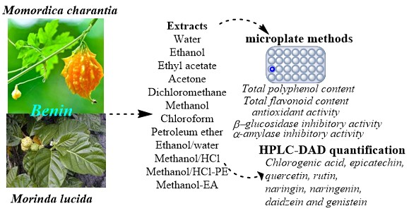
2.2. Plant Material
2.3. Preparation of Plants Extracts
2.4. Microplate Determination of Total Polyphenol Content
2.5. Microplate Determination of Total Flavonoid Content
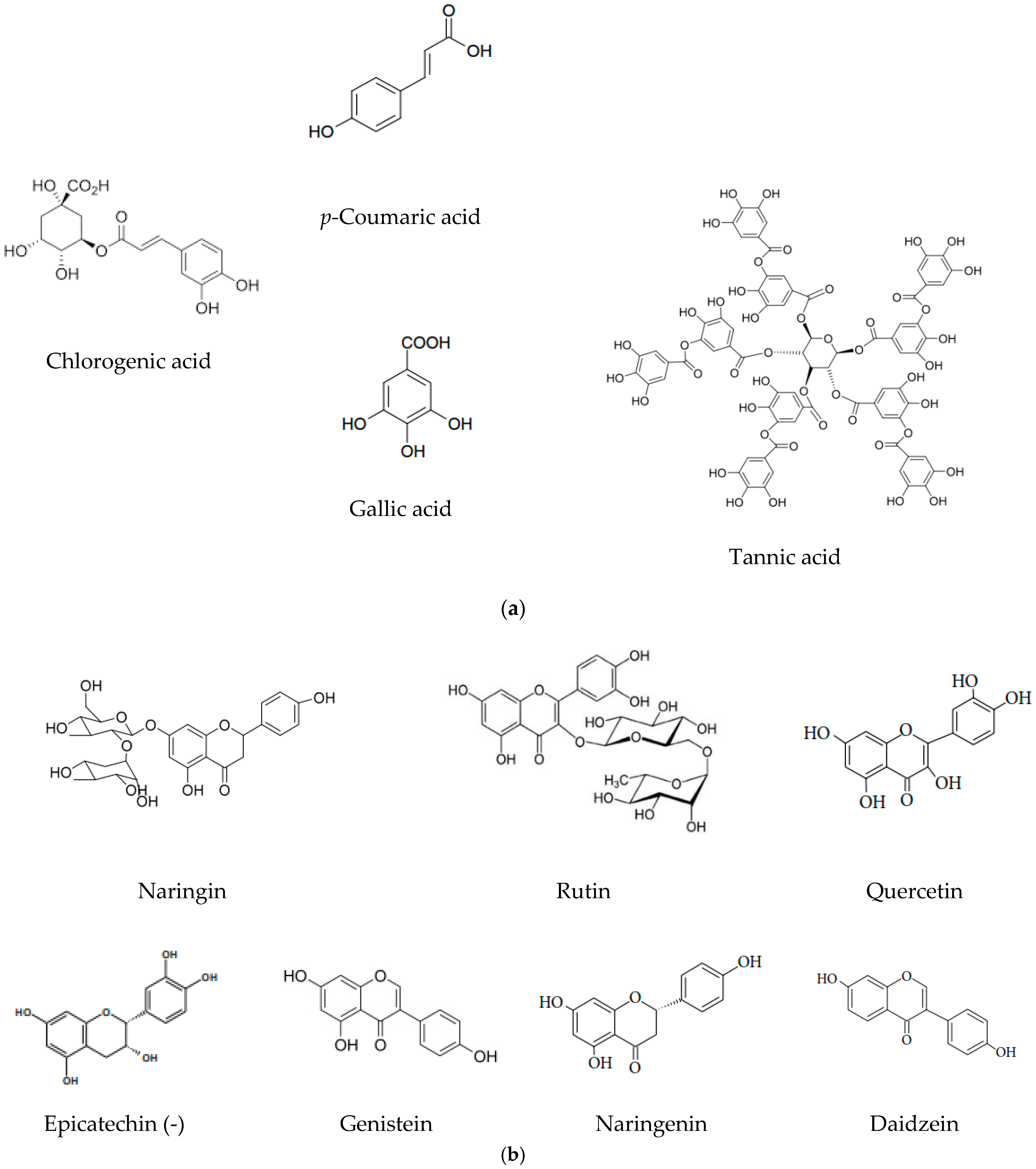
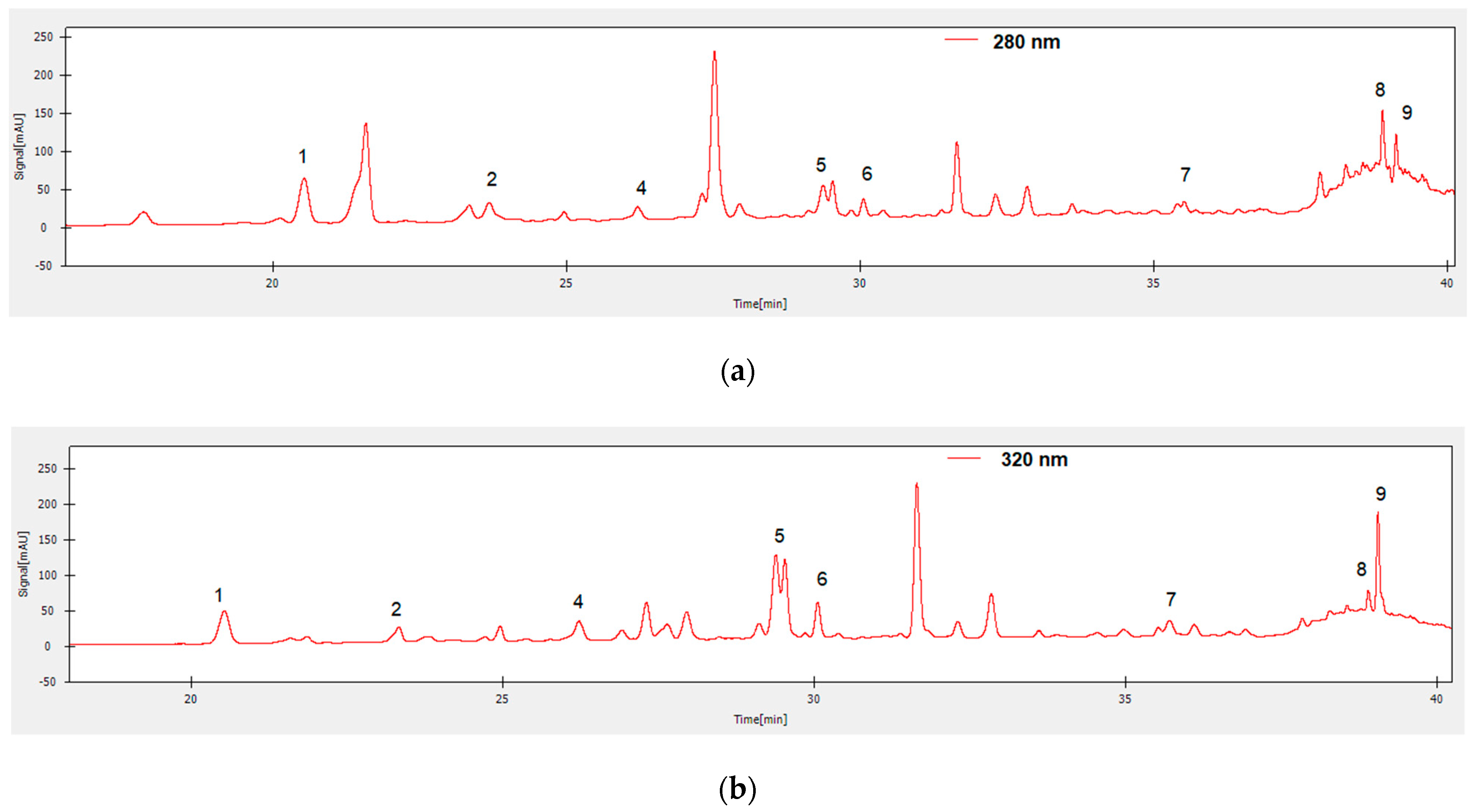
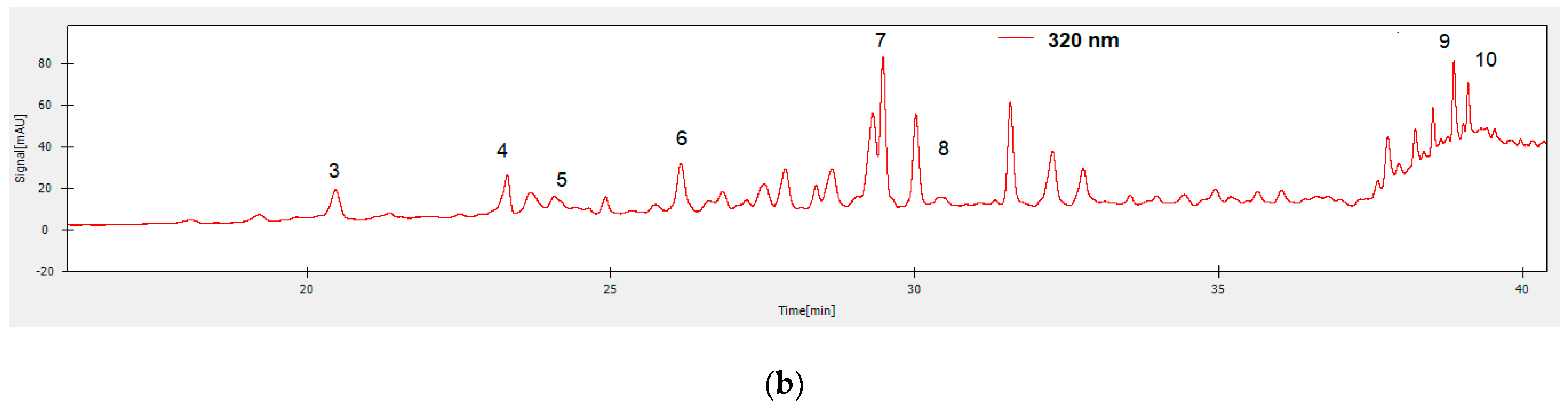
2.6. HPLC-DAD Quantification of Bioactive Polyphenols from M. lucida and M. charantia Extracts
2.7. Microplate Determination of Antioxidant Activity
2.7.1. DPPH radical-scavenging activity
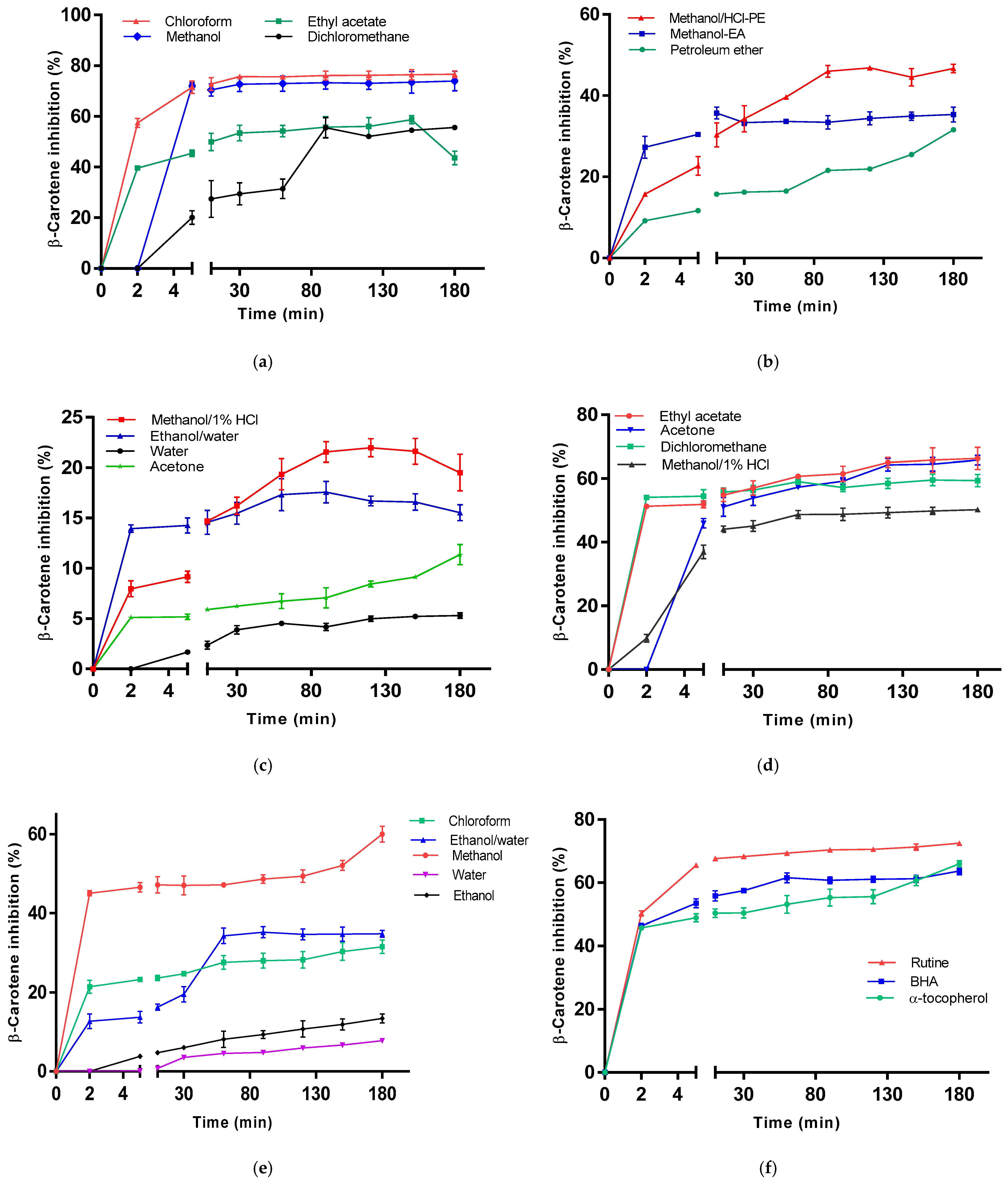
2.7.2. β-Carotene Bleaching Method
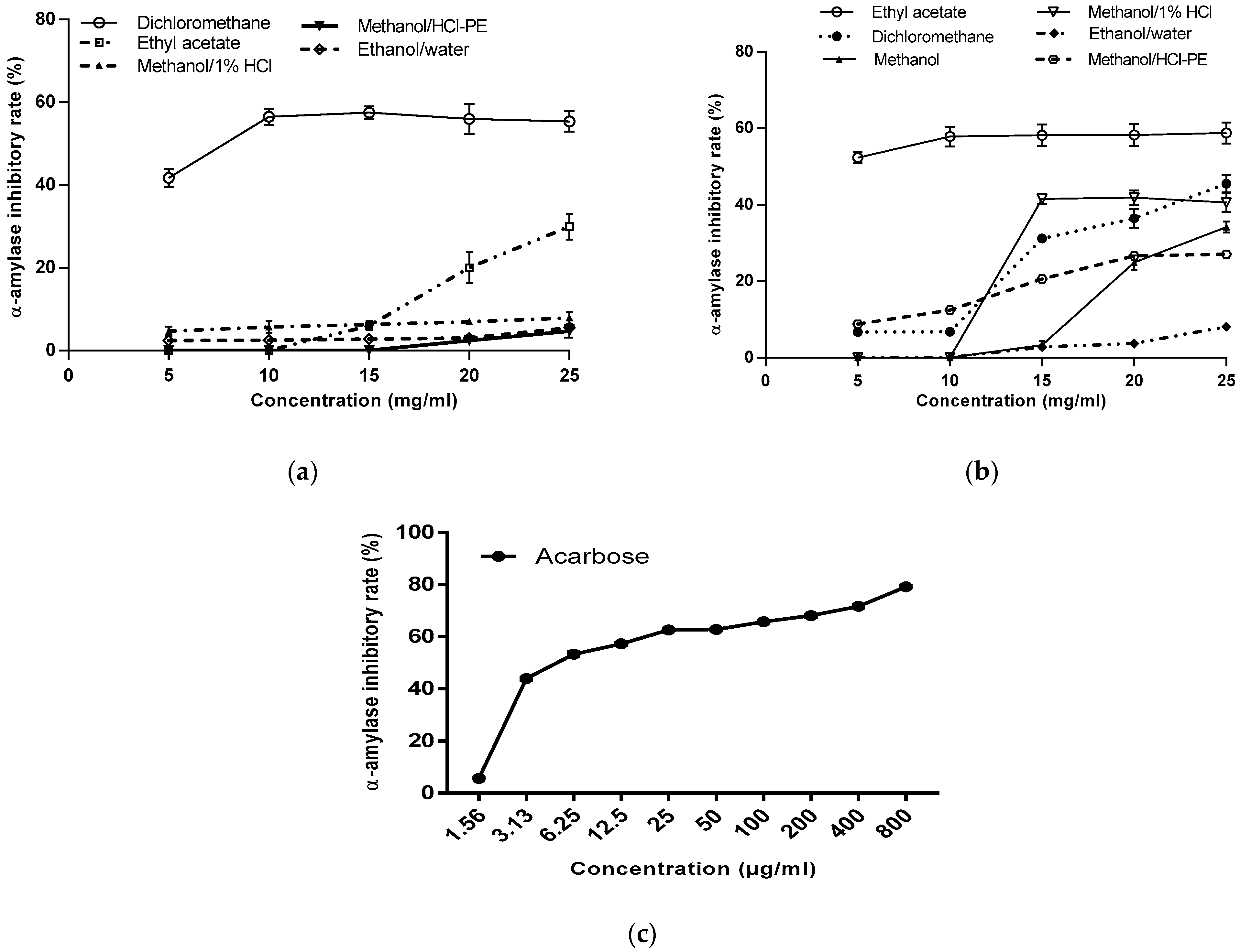
2.8. In vitro α-Amylase Inhibitory Activity Assay
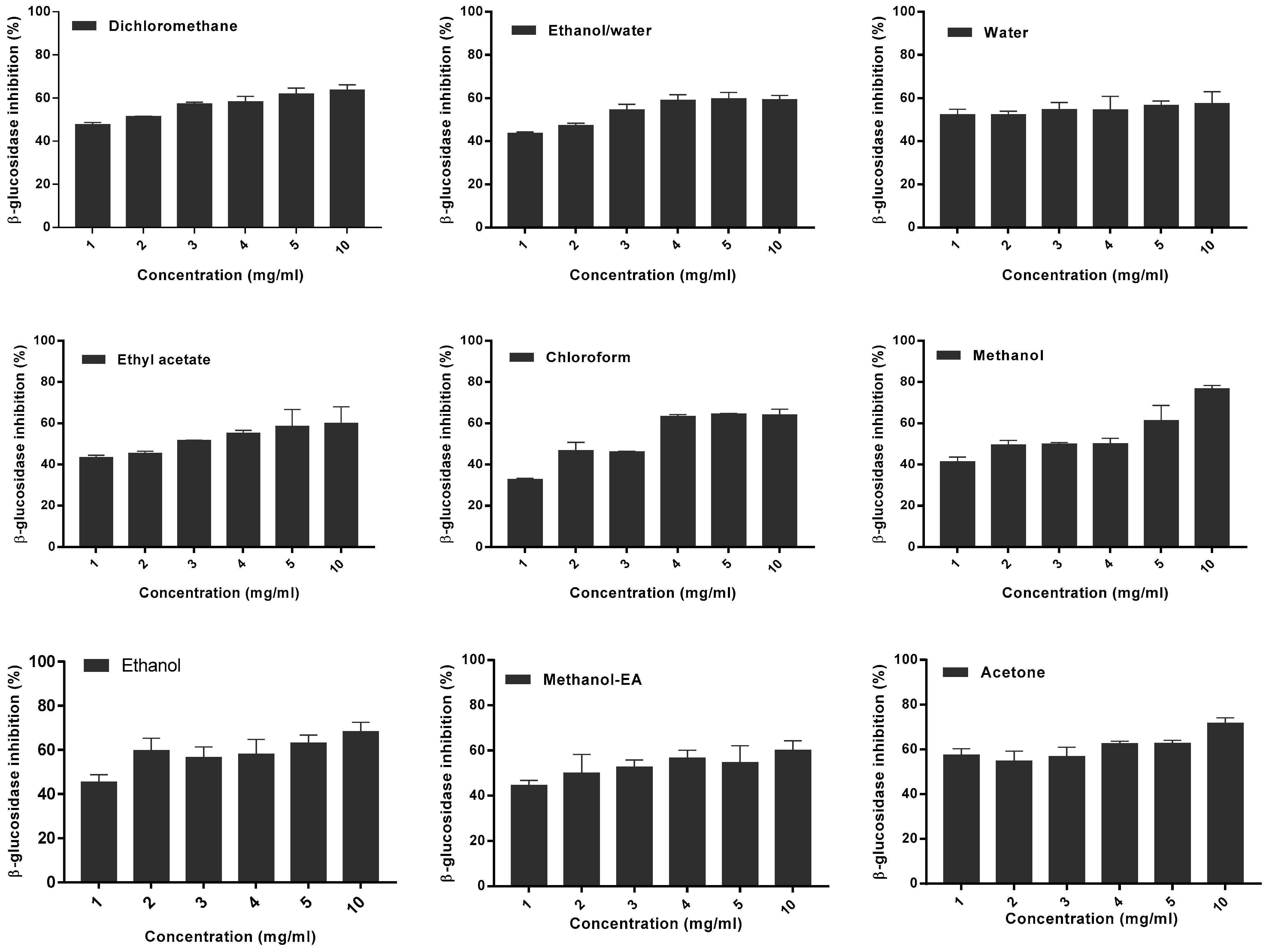
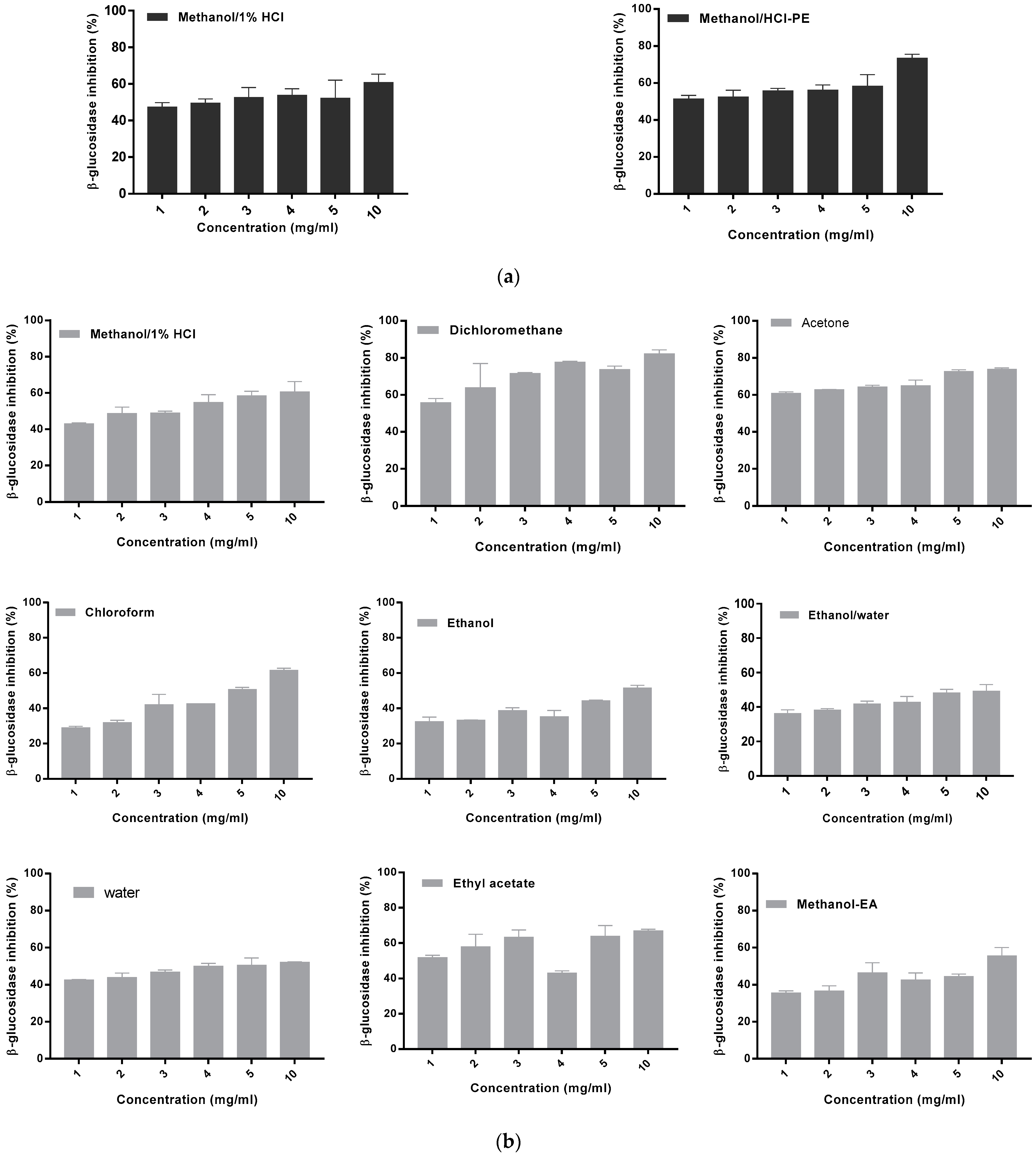
2.9. Microplate β-Glucosidase Inhibition Assay
2.10. Statistical Analysis
3. Results
3.1. Total Polyphenolic and Flavonoid Contents
3.2. HPLC-DAD Quantification of Bioactive Polyphenols
3.3. In Vitro Antioxidant Activities
3.4. Inhibition of α-amylase Activity
3.5. In Vitro β-Glucosidase Inhibitory Activity
4. Discussion
5. Conclusions
Author Contributions
Funding
Acknowledgments
Conflicts of Interest
References
- Ladoh-Yemeda, C.F.; Vandi, D.; Dibong, S.D.; Mpondo Mpondo, E.; Wansi, J.D.; Betti, J.L.; Choula, F.; Ndongo, D.; Tomedi-Eyango, M. Étude ethnobotanique des plantes médicinales commercialisées dans les marchés de la ville de Douala, Cameroun. J. Appl. Biosci. 2016, 99, 9452–9468. [Google Scholar] [CrossRef] [Green Version]
- King, H.; Aubert, R.E.; Herman, W.H. Global burden of diabetes, 1995–2025, prevalence, numerical estimates, and projections. Diabetes Care 1998, 21, 1414–1431. [Google Scholar] [CrossRef] [PubMed]
- Kameswararao, B.; Kesavulu, M.M.; Apparao, C. Evaluation of antidiabetic effect of Momordica cymbalaria fruit in alloxan-diabetic rats. Fitoterapia 2003, 74, 7–13. [Google Scholar] [CrossRef]
- Balan, K.; Ratha, P.; Prakash, G.; Viswanathamurthi, P.; Adisakwattana, S.; Palvannan, T. Evaluation of in vitro α-amylase and α-glucosidase inhibitory potential of N2O2 schiff base Zn complex. Arab. J. Chem. 2017, 10, 732–738. [Google Scholar] [CrossRef] [Green Version]
- De Melo, E.B.; Gomes, A.S.; Carvalho, I. α- and β-Glucosidase inhibitors: Chemical structure and biological activity. Tetrahedron Lett. 2006, 62, 10277–10302. [Google Scholar] [CrossRef]
- Parizadeh, H.; Garampalli, R.H. Evaluation of some lichen extracts for β-Glucosidase inhibitory as a possible source of herbal anti-diabetic drugs. Am. J. Biochem. 2016, 6, 46–50. [Google Scholar]
- Asayama, K.; Uchida, N.; Nakane, T.; Hayasnibe, H.; Dobashi, K.; Amemiya, S.; Kato, K.; Nakazawa, S. Antioxidants in the serum of children with insulin dependent diabetes mellitus. Free Radic. BioI. Med. 1993, 15, 597–602. [Google Scholar] [CrossRef]
- Mathew, S.; Abraham, T.E. In vitro antioxidant activity and scavenging effects of Cinnamomum verum leaf extract assayed by different methodologies. Food. Chem. Toxicol. 2006, 44, 198–206. [Google Scholar] [CrossRef]
- Kubola, J.; Siriamornpun, S. Phenolic contents and antioxidant activities of bitter gourd (Momordica charantia L.) leaf, stem and fruit fraction extracts in vitro. Food Chem. 2008, 110, 881–890. [Google Scholar] [CrossRef]
- Basch, E.; Gabardi, S.; Ulbricht, C. Bitter melon (Momordica charantia): A review of efficacy and safety. Am. J. Health Syst. Pharm. 2003, 60, 356–359. [Google Scholar] [CrossRef] [Green Version]
- Braca, A.; Siciliano, T.; D’Arrigo, M.; Germanò, M.P. Chemical composition and antimicrobial activity of Momordica charantia seed essential oil. Fitoterapia 2008, 79, 123–125. [Google Scholar] [CrossRef] [PubMed]
- Ahmed, I.; Lakhani, M.S.; Gillett, M.; John, A.; Raza, H. Hypotriglyceridemic and hypocholesterolemic effects of anti-diabetic Momordica charantia (karela) fruit extract in streptozotocin-induced rats. Diabetes Res. Clin. Pract. 2001, 51, 155–161. [Google Scholar] [CrossRef]
- Raman, A.; Lau, C. Anti-diabetic properties and phytochemistry of Momordica charantia L. (Cucurbitaceae). Phytomedicine 1996, 2, 349–362. [Google Scholar] [CrossRef]
- Grover, J.K.; Yadav, S.P. Pharmacological actions and potential uses of Momordica charantia: A review. J. Ethnopharmacol. 2004, 93, 123–132. [Google Scholar] [CrossRef]
- Matsuda, H.; Li, Y.; Murajami, T.; Matsumura, N.; Yamahara, J.; Yoshikawa, M. Antidiabetic principles of natural medicines, Part III. Structure-related inhibitory activity and action mode of oleanolic acid glycosides on hypoglycemic activity. Chem. Pharm. Bull. 1998, 46, 1399–1403. [Google Scholar] [CrossRef] [Green Version]
- Nag, B.; Medicherla, S.; Sharma, S.D. Orally Active Fraction of Momordica charantia, Active Peptides Thereof, and Their Use in the Treatment of Diabetes. U.S. Patent 6127338, 3 October 2000. [Google Scholar]
- Makinde, J.M.; Obih, P.O. Screening of Morinda lucida leaf extract for antimalarial action on Plasmodium berghei in mice. Afr. J. Med. Med. Sci. 1985, 17, 59–63. [Google Scholar]
- Karou, S.D.; Tchacondo, T.; Ilboudo, D.P.; Simpore, J. Saharan Rubiaceae: A review of their traditional uses, phytochemistry and biological activities. Pak. J. Biol. Sci. 2011, 14, 149–169. [Google Scholar] [CrossRef]
- Joppa, K.M.; Vovor, A.; Eklu-Gadegbeku, K.; Agbonon, A.; Aklikokou, K.; Gbeassor, M. Effect of Morinda Lucida Benth. (Rubiaceae) and Newbouldia leavis P. Beauv. (Bignoniaceae) on sickling of red blood cells. Med. Trop. 2008, 68, 251–256. [Google Scholar]
- Kazeem, M.I.; Adamson, J.O.; Ogunwande, I.A. Modes of inhibition of α-amylase and α-glucosidase by aqueous extract of Morinda lucida Benth. leaf. BioMed. Res. Int. 2013, 2013, 1–6. [Google Scholar] [CrossRef] [Green Version]
- Dicko, M.H.; Hilhorst, R.; Gruppen, H.; Traore, A.S.; Laane, C.; van Berkel, W.J.H.; Voragen, A.G.J. Comparison of content in phenolic compounds, polyphenol oxidase, and peroxidase in grains of fifty sorghum varieties from Burkina Faso. J. Agric. Food Chem. 2002, 50, 3780–3788. [Google Scholar] [CrossRef]
- Dah-Nouvlessounon, D.; Adjanohoun-Sagbadja, H.; Diarrasouba, N.; Sina, H.; Noumavo, P.A.; Baba-Moussa, F.; Adjanohoun, A.; Gbenou, J.D.; Baba-Moussa, L. Antimicrobial, antioxidant, cytotoxic Activities and phytochemical assessment of Cola acuminata used in Benin. Int. J. Pharm. Pharm. Sci. 2015, 7, 102–109. [Google Scholar]
- Cudalbeanu, M.; Ghinea, I.O.; Furdui, B.; Dah-Nouvlessounon, D.; Raclea, R.; Costache, T.; Cucolea, I.E.; Urlan, F.; Dinica, R.M. Exploring new antioxidant and mineral compounds from Nymphaea alba wild-Grown in Danube Delta Biosphere. Molecules 2018, 23, 1247. [Google Scholar] [CrossRef] [PubMed] [Green Version]
- Schmeda-Hirschmann, G.; Rodriguez, J.A.; Theoduloz, C.; Astudillo, S.L.; Feresin, G.E.; Tapia, A. Free-radical scavengers and antioxidants from Peumus boldus Mol. (“Boldo”). Free Rad. Res. 2003, 37, 447–452. [Google Scholar] [CrossRef] [PubMed]
- Scherer, R.; Godoy, H.T. Antioxidant activity index (AAI) by the 2,2-diphenyl-1-picrylhydrazyl method. Food Chem. 2009, 112, 654–658. [Google Scholar] [CrossRef]
- Lage, M.Á.P.; García, M.A.M.; Álvarez, J.A.V.; Anders, Y.; Curran, T.P. A new microplate procedure for simultaneous assessment of lipophilic and hydrophilic antioxidants and pro-oxidants, using crocin and β-carotene bleaching methods in a single combined assay: Tea extracts as a case study. Food Res. Int. 2013, 53, 836–846. [Google Scholar] [CrossRef] [Green Version]
- Telagari, M.; Hullatti, K. In-vitro α-amylase and α-glucosidase inhibitory activity of Adiantum caudatum Linn. and Celosia argentea Linn. extracts and fractions. Indian J Pharmacol. 2015, 47, 42–59. [Google Scholar]
- Sánchez-Medina, A.; García-Sosa, K.; May-Pat, F.; Peña-Rodríguez, L.M. Evaluation of biological activity of crude extracts from plants used in Yucatecan traditional medicine part, I. antioxidant, antimicrobial and β-glucosidase inhibition activities. Phytomedicine 2001, 8, 144–151. [Google Scholar] [CrossRef]
- Landoulsi, A. Etude chimiotaxonomique et activités biologiques des métabolites secondaires des plantes du genre Eryngium. Ph.D. Thesis, University of Droit et de la Santé -Lille II, Talence Cedex, France, 20 December 2016. [Google Scholar]
- Romdhane, M. Extraction solide-liquide sous ultrasons: Mise en oeuvre d’un capteur de puissance locale ultrasonore. Ph.D. Thesis, University of Toulouse, INPT, Tolouse, France, 12 Septembre 1993. [Google Scholar]
- Bourgou, S.; Serairi Beji, R.; Medini, F.; Ksouri, R. Effet du solvant et de la méthode d’extraction sur la teneur en composés phénoliques et les potentialités antioxydantes d’Euphorbia helioscopia. J. New Sci. 2016, 28, 1649–1655. [Google Scholar]
- Nagarani, G.; Abirami, A.; Siddhuraju, P. A comparative study on antioxidant potentials, inhibitory activities against key enzymes related to metabolic syndrome, and anti-inflammatory activity of leaf extract from different Momordica species. Food Sci. Hum. Well. 2014, 3, 36–46. [Google Scholar] [CrossRef] [Green Version]
- Budrat, P.; Shotipruk, A. Enhanced recovery of phenolic compounds from bitter melon (Momordica charantia) by subcritical water extraction. Sep. Purif. Technol. 2009, 66, 125–129. [Google Scholar] [CrossRef]
- Heim, K.E.; Tagliaferro, A.R.; Bobilya, D.J. Flavonoid antioxidants: Chemistry, metabolism and structure-activity relationships. J. Nutr. Biochem. 2002, 13, 572–584. [Google Scholar] [CrossRef]
- Rice-Evans, C.A.; Miller, N.J.; Paganga, G. Structure-antioxidant activity relationships of flavonoids and phenolic acids. Free Radic. Biol. Med. 1996, 20, 9339–9356. [Google Scholar] [CrossRef]
- Singh, A.K.; Raj, V.; Keshari, A.K.; Rai, A.; Kumar, P.; Rawat, A.; Maity, B.; Kumar, D.; Prakash, A.; De, A.; et al. Isolated mangiferin and naringenin exert antidiabetic effect via PPARγ/GLUT4 dual agonistic action with strong metabolic regulation. Chem. Biol. Interact. 2018, 280, 33–44. [Google Scholar] [CrossRef] [PubMed]
- Ahmed, O.M.; Hassan, M.A.; Abdel-Twab, S.M.; Abdel Azeem, M.N. Navel orange peel hydroethanolic extract, naringin and naringenin have anti-diabetic potentials in type 2 diabetic rats. Biomed. Pharmacother. 2017, 94, 197–205. [Google Scholar] [CrossRef]
- Sirovina, D.; Oršolić, N.; Gregorović, G.; Končić, M.Z. Naringenin ameliorates pathological changes in liver and kidney of diabetic mice: A preliminary study. Arch. Ind. Hyg. Toxicol. 2016, 67, 19–24. [Google Scholar]
- Ruijters, E.J.B.; Weseler, A.R.; Kicken, C.; Haenen, G.R.M.M.; Bast, A. The flavanol (-)-epicatechin and its metabolites protect against oxidative stress in primary endothelial cells via a direct antioxidant effect. Eur. J. Pharmacol. 2013, 715, 147–153. [Google Scholar] [CrossRef]
- Ranilla, L.G.; Kwon, Y.I.; Apostolidis, E.; Shetty, K. Phenolic compounds, antioxidant activity and in vitro inhibitory potential against key enzymes relevant for hyperglycemia and hypertension of commonly used medicinal plants, herbs and spices in Latin America. Bioresour. Technol. 2010, 101, 4676–4689. [Google Scholar] [CrossRef]
- El-Kaissi, S.; Sherbeeni, S. Pharmacological management of type 2 diabetes mellitus: An update. Curr. Diabetes Rev. 2011, 7, 392–405. [Google Scholar] [CrossRef]
- Baron, A.D. Post prandial hypoglycemia and α-glucosidase inhibitors. Diabetes Res. Clin. Pract. 1998, 40, S51–S55. [Google Scholar] [CrossRef]
- Lebovitz, H.E. Effect of the posprandial state on nontraditional risk factors. Am. J. Cardiol. 2001, 80, 20–25. [Google Scholar] [CrossRef]
- Weinman, S.; Méhul, P. Macanismes de régulation de l’activité enzymatique. Partie II: Catalyse biologique: Structure et mécanisme d’action des enzymes. In Toute la Biochimie, 2nd ed.; Sciences Sup: Dunod, Paris, 2004; pp. 138–152. [Google Scholar]
- Scheen, A.J.; Letiexhe, M.R.; Geronooz, I.; Paquot-Jandrain, N.B. l’hyperglycémie post-prandiale. Approches thérapeutiques médicamenteuses. Rev. Med. Liege 2002, 57, 196–201. [Google Scholar] [PubMed]
- Tundis, R.; Loizzo, M.R.; Menichini, F. Natural products as alpha-amylase and alpha-glucosidase inhibitors and their hypoglycaemic potential in the treatment of diabetes: An update. Mini. Rev. Med. Chem. 2010, 10, 315–331. [Google Scholar] [CrossRef] [PubMed]
- Kandra, L.; Gyeman, G.; Zajaez, A.; Batta, G. Inhibitory effects of tamnius of human salivary α-amylase. Biochem. Biophys. Res Commun. 2004, 319, 1265–1271. [Google Scholar] [CrossRef] [PubMed]
- Hunyadi, A.; Martins, A.; Hsieh, T.-J.; Seres, A.; Zupko, I. Chlorogenic acid and rutin play a major role in the in vivo anti-diabetic activity of Morus alba leaf extract on type II diabetic rats. PLoS ONE 2012, 7, e50619. [Google Scholar] [CrossRef] [PubMed] [Green Version]
- Hernández-Aquino, E.; Zarco, N.; Casas-Grajales, S.; Ramos-Tovar, E.; Flores-Beltrán, R.E.; Arauz, J.; Shibayama, M.; Favari, L.; Tsutsumi, V.; Segovia, J.; et al. Naringenin prevents experimental liver fibrosis by blocking TGFβ-Smad3 and JNK-Smad3 pathways. World J. Gastroenterol. 2017, 23, 4354–4368. [Google Scholar] [CrossRef] [PubMed]
- Shay, J.; Elbaz, H.A.; Lee, I.; Zielske, S.P.; Malek, M.H.; Hüttemann, M. Molecular mechanisms and therapeutic effects of (-)epicatechin and other polyphenols in cancer, inflammation, diabetes and neurodegeneration. Oxidative Med. Cell. Longev. 2015, 2015, 1–13. [Google Scholar] [CrossRef] [Green Version]
- Guo, T.L.; Germolec, D.R.; Zheng, J.F.; Auttachoat, W.; Smith, M.J.; White, J.; Elmore, S.A. Genistein protects female nonobese diabetic mice from developing type 1 diabetes when fed a soy- and alfalfa-free diet. Toxicol. Pathol. 2015, 43, 435–448. [Google Scholar] [CrossRef] [Green Version]
- Ahmad, F.; Khalid, P.; Khan, M.M.; Rastogi, A.K.; Kidwai, J.R. Insulin like activity in (-)epicatechin. Acta Diabetol. Lat. 1989, 26, 291–300. [Google Scholar] [CrossRef]
- Sheehan, E.W.; Zemaitis, M.A.; Slatkin, D.J.; Schiff, P.L.J. A constituent of Pterocarpus marsupium, (-)-epicatechin, as a potential antidiabetic agent. J. Nat. Prod. 1983, 46, 232–234. [Google Scholar] [CrossRef]
- Shim, Y.J.; Doo, H.K.; Ahn, S.Y.; Kim, Y.S.; Seong, J.K.; Park, I.S.; Min, B.H. Inhibitory effect of aqueous extract from the gall of Rhus chinensis on alpha-glucosidase activity and postprandial blood glucose. J. Ethanopharmacol. 2003, 85, 283–287. [Google Scholar] [CrossRef]
- Chau, C.F.; Huang, Y.L.; Lee, M.H. In vitro hypoglycemic effects of different insoluble fiber-rich fractions prepared from the peel of Citrus sinensis L. cv. Liucheng. J. Agric. Food Chem. 2003, 51, 6623–6626. [Google Scholar] [CrossRef] [PubMed]
- Kwon, G.J.; Choi, D.S.; Wang, M.H. Biological activities of hot water extracts from Euonymus alatus leaf. Korean J. Food. Sci. Technol. 2007, 39, 569–574. [Google Scholar]
- Lee, Y.A.; Cho, E.J.; Tanaka, T.; Yokozawa, T. Inhibitory activities of proanthocyanidins from persimmon against oxidative stress and digestive enzymes related to diabetes. J. Nutr. Sci. Vitaminol. 2007, 53, 287–292. [Google Scholar] [CrossRef] [PubMed] [Green Version]
- Antoun, M.D.; Ríos, Y.R.; Mendoza, N.T.; Proctor, G. Glucosidase inihibiton assay as prescreen for natural products. P. R. Health Sci. J. 1994, 13, 13–15. [Google Scholar] [PubMed]
- Vlietinck, A.J.; De bruyne, T.; Apers, S.; Pieters, L.A. Plant-derived leading compounds for chemotherapy of human inmunodeficiency virus (HIV) infection. Planta Med. 1998, 64, 97–109. [Google Scholar] [CrossRef] [PubMed] [Green Version]
- Akpan, E.J.; Umoh, I.B. Inhibitory activity of seed extract from Picralima nitida, (Staph) on -D-glucosidase. Biochemistry 2004, 16, 72–78. [Google Scholar] [CrossRef]
- Daniel, P.; Supe, U.; Roymon, M.G. A review on phytochemical analysis of Momordica charantia. Int. J. Adv. Pharm. Biol. Chem. 2014, 31, 214–220. [Google Scholar]
- Ebiloma, G.U.; Omale, J.; Aminu, R.O. Suppressive, curative and prophylactic potentials of Morinda lucida (Benth) against erythrocytic stage of mice infective chloroquine sensitive plasmodium berghei NK-65. Br. J. Appl. Sci. Technol. 2011, 1, 131–140, 201. [Google Scholar]
- Montefiori, D.C.; Robinson, W.E.; Mitchell, W.M. Role of protein N-glycosilation in pathogenesis of human inmunodeficiency virus type 1. Proc. Natl. Acad. Sci. USA 1988, 85, 9248–9252. [Google Scholar] [CrossRef] [Green Version]
- Wong, C.; Provencher, L.; Porco, J.A.; Jung, S.; Wang, Y.; Chen, L.; Wang, R.; Steensma, D.H. Synthesis and evaluation of homoazasugar as glycosidase inhibitors. J. Org. Chem. 1995, 60, 1492–1501. [Google Scholar] [CrossRef]
- Gruters, R.A.; Neefjes, J.J.; Tersmette, M.; de Goede, R.E.Y.; Tulp, A.; Huisman, H.G.; Miedema, F.; Ploegh, H.L. Interference with HIV-induced syncitium formation and viral infectivity by inhibitors of trimming glucosidase. Nature 1987, 330, 74–77. [Google Scholar] [CrossRef] [PubMed]
- Karpas, A.; Fleet, G.W.J.; Dwek, R.A.; Petursson, S.; Namgoong, S.K.; Ramsden, N.G.; Jacob, G.S.; Rademacher, T.W. Aminosugar derivatives as potential anti-human immunodeficiency virus agents. Proc. Natl. Acad. Sci. USA 1988, 85, 9229–9233. [Google Scholar] [CrossRef] [PubMed] [Green Version]
- Hernández-Aquino, E.; Muriel, P. Beneficial effects of naringenin in liver diseases: Molecular mechanisms. World J. Gastroenterol. 2018, 24, 1679–1707. [Google Scholar] [CrossRef] [PubMed]
- Shi, X.; Zhang, Y.; Zheng, J.; Pan, J. Reactive oxygen species in cancer stem cells. Antioxid. Redox. Signal. 2012, 16, 1215–1228. [Google Scholar] [CrossRef] [Green Version]
- Blois, M.S. Antioxidant determination by the use of a stable free radical. Nature 1958, 181, 1199–1200. [Google Scholar] [CrossRef]
- Leelaprakash, G.; Caroline-Rose, J.; Gowtham, B.M. In vitro antimicrobial and antioxidant activity of Momordica charantia leaves. Pharmacophore 2011, 2, 244–252. [Google Scholar]
- Temitope, O.O.; Olajubu, F.A.; Olorunnipa, T.A.; Thonda, O.A. Elemental composition, evaluation of anti-nutrients, and antioxidant potentials of Morinda lucida. Eur. J. Med. Plants. 2016, 12, 1–10. [Google Scholar] [CrossRef]
- Zheng, Y.-Z.; Deng, G.; Liang, Q.; Chen, D.-F.; Guo, R.; Lai, R.-C. Antioxidant activity of quercetin and Its glucosides from propolis: A theoretical study. Sci. Rep. 2017, 7, 7543. [Google Scholar] [CrossRef] [Green Version]
- Abarikwu, S.O.; Olufemi, P.D.; Lawrence, C.J.; Wekere, F.C.; Ochulor, A.C.; Barikuma, A.M. Rutin, an antioxidant flavonoid, induces glutathione and glutathione peroxidase activities to protect against ethanol effects in cadmium-induced oxidative stress in the testis of adult rats. Andrologia 2017, 49, e12696. [Google Scholar] [CrossRef]
- Ungar, Y.; Osundahunsi, O.F.; Shimoni, E. Thermal stability of genistein and daidzein and its effect on their antioxidant activity. J. Agric. Food Chem. 2003, 51, 4394–4399. [Google Scholar] [CrossRef]
- Jayaprakasha, G.K.; Singh, R.P.; Sakariah, K.K. Antioxidant activity of grape seed (Vitis vinifera) extracts on peroxidation models in vitro. Food. Chem. 2001, 73, 285–290. [Google Scholar] [CrossRef]
- Frankel, E.N.; Meyer, A.S. The problems of using one-dimensional methods to evaluate multifunctional food and biological antioxidants. J. Sci. Food. Agric. 2000, 80, 1925–1940. [Google Scholar] [CrossRef]
- Liyana-Pathirana, C.M.; Shahidi, F. Antioxydant propreties of commercial soft and hard winter wheats (Triticum aestivium L.) and their milling fractions. J. Sci. Food. Agric. 2006, 86, 477–485. [Google Scholar] [CrossRef]
- Ferreria, A.; Proenca, C.; Serralheiro, M.L.M.; Araujo, M.E.M. The in vitro screening for acetylcholinesterase inhibition and antioxidant activity of medicinal plant from Portugal. J. Ethnopharmacol. 2006, 108, 31–37. [Google Scholar] [CrossRef] [PubMed]


| Extracts | - | M. charantia | - | M. lucida | ||
|---|---|---|---|---|---|---|
| Extract Yield (%) | Total Polyphenols (µg GAE/mg) | Flavonoids (µg QE/mg) | Extract Yield (%) | Total Polyphenols (µg GAE/mg) | Flavonoids (µg QE/mg) | |
| Water | 12.30 ± 0.20 | 113.22 ± 11.46 | 65.84 ± 12.07 | 19.01 ± 0.10 | 1501.11±76.67 | 147.32±1.85 |
| Ethanol | 23.23 ± 0.45 | 1853.44 ± 180.99 | 41.69 ± 2.78 | 9.63 ± 0.15 | 2689.11 ± 314.76 | 487.41 ± 17.08 |
| Ethyl acetate | 7.13 ± 0.20 | 1440.55 ± 21.76 | 96.33 ± 2.74 | 4.50 ± 0.55 | 1173.33 ± 58.16 | 38.49 ± 3.98 |
| Acetone | 9.20 ± 0.30 | 320.92 ± 8.05 | 182.20 ± 51.78 | 6.60 ± 1.15 | 791.09 ± 24.43 | 131.33 ± 08.17 |
| Dichloromethane | 9.50 ± 0.20 | 1287.77 ± 26.21 | 38.45 ± 6.24 | 7.40 ± 0.85 | 220.33 ± 88.12 | 115.86 ± 56.70 |
| Methanol | 17.80 ± 0.26 | 989.55 ± 28.16 | 123.09 ± 9.63 | 9.83 ± 1.10 | 3048.33 ± 63.63 | 156.71 ± 18.02 |
| Chloroform | 5.10 ± 0.36 | 209.59 ± 31.03 | 150.78 ± 9.89 | 2.50 ± 0.70 | 175.78 ± 22.07 | 22.35 ± 14.7 |
| Petroleum Ether | 1.03 ± 0.05 | 185.78 ± 32.05 | 08.96 ± 3.92 | 1.43 ± 0.60 | 1353.77 ± 173.05 | 212.15 ± 57.89 |
| Ethanol/water | 22.13 ± 0.15 | 842.11 ± 52.07 | 302.28 ± 46.34 | 17.50 ± 1.17 | 2184.44 ± 103.21 | 272.37 ± 66.37 |
| Methanol/1%HCl | 18.50 ± 0.10 | 6833.88 ± 89.23 | 692.39 ± 1.89 | 11.23 ± 0.92 | 906.66 ± 51.47 | 336.61 ± 15.78 |
| Methanol/HCl-PE | 16.30 ± 0.26 | 705.16 ± 91.21 | 201.15 ± 1.69 | 9.60 ± 0.26 | 2236.11 ± 26.34 | 191.45 ± 43.82 |
| Methanol-EA | 8.80 ± 0.26 | 700.33 ± 77.66 | 440.43 ± 25.14 | 6.73 ± 0.75 | 2286.00 ± 143.84 | 441.65 ± d93.37 |
| Extracts Content (mg/kg) | ||||||||||||
|---|---|---|---|---|---|---|---|---|---|---|---|---|
| Peak | Compound | TR ** (min) | TR * (min) | λ max (nm) | H2O | H2O-EtOH 30:70 | MeOH | MeOH/1% HCl | EtOH | EAC | MeOH-EA | MeOH/ HCl-PE |
| 1 | Chlorogenic acid | 20.86 | 20.88 | 280, 300 | - | - | 6.72 ± 0.47 | 11.72 ± 0.52 | - | - | 5.31 ± 0.43 | 8.67 ± 0.52 |
| 2 | Epicatechin (-) | 23.56 | 23.74 | 230, 280 | - | 107.47 ± 0.72 | - | 85.44 ± 0.98 | 71.53 ± 0.83 | - | 50.59 ± 1.01 | 74.11 ± 0.14 |
| 3 | p-Coumaric acid | 24.10 | 24.22 | 300, 320 | 37.68 ± 0.59 | 52.25 ± 0.63 | - | - | - | - | - | - |
| 4 | Daidzein | 26.44 | 26.62 | 300, 320 | 15.31 ± 1.02 | - | 4.49 ± 0.26 | 19.10 ± 0.01 | - | - | 7.40 ± 0.70 | 10.69 ± 0.46 |
| 5 | Rutin | 29.70 | 29.68 | 230, 300 | 82.02 ± 1.11 | 74.23 ± 0.03 | 29.26 ± 0.09 | 54.53 ± 0.04 | 23.82 ± 0.07 | 16.14 ± 0.22 | 29.19 ± 0.07 | 28.85 ± 1.16 |
| 6 | Naringin | 31.51 | 31.50 | 320, 370 | 730.42 ± 38.22 | 286.31 ± 3.07 | 45.67 ± 0.02 | 425.04 ± 1.63 | 91.37 ± 0.06 | 3.04 ± 0.03 | 264.31 ± 1.03 | 161.17 ± 1.12 |
| 7 | Quercetin | 37.80 | 37.97 | 280, 300 | - | 13.75 ± 1.04 | 10.50 ± 2.09 | 12.76 ± 0.04 | - | 5.20 ± 1.05 | 4.94 ± 0.08 | - |
| 8 | Naringenin | 38.96 | 38.97 | 280, 300 | 50.80 ± 0.10 | 39.59 ± 1.03 | 24.39 ± 0.02 | 27.73 ± 0.05 | 23.29 ± 1.74 | 19.82 ± 0.06 | - | 21.24 ± 1.05 |
| 9 | Genistein | 39.11 | 39.19 | 320, 370 | 119.68 ± 0.07 | 57.07 ± 0.22 | 16.40 ± 0.06 | 42.80 ± 0.56 | 15.58 ± 0.13 | 38.32 ± 0.67 | 10.36 ± 0.03 | 16.93 ± 1.53 |
| Extracts Content (mg/kg) | ||||||||||||
|---|---|---|---|---|---|---|---|---|---|---|---|---|
| Peak | Compound | TR ** (min) | TR * (min) | λ max (nm) | H2O | H2O-EtOH 30:70 | MeOH | MeOH/1% HCl | EtOH | EAC | MeOH -EA | MeOH/ HCl-PE |
| 1 | Tannic acid | 2.30 | 2.32 | 280 | 18.74 ± 0.04 | - | - | - | - | - | - | - |
| 2 | Gallic acid | 3.51 | 3.31 | 250 | 30.21 ± 0.23 | - | - | - | - | - | - | - |
| 3 | Chlorogenic acid | 20.86 | 20.93 | 280, 300 | 7.24 ± 1.02 | - | - | 11.35 ± 0.20 | - | - | - | 0.97 ± 0.01 |
| 4 | Epicatechin (-) | 23.56 | 23.68 | 230, 280 | - | - | - | 143.34 ± 0.90 | - | 55.67 ± 0.44 | - | 12.49 ± 0.09 |
| 5 | p-Coumaric acid | 24.10 | 24.10 | 300, 320 | - | 65.88 ± 0.05 | - | 55.12 ± 0.57 | - | - | - | 4.86 ± 0.02 |
| 6 | Daidzein | 26.44 | 26.48 | 280, 320 | - | 9.37 ± 0.04 | 10.16 ± 0.15 | 20.82 ± 0.01 | 10.06 ± 0.52 | - | 3.56 ± 0.02 | 1.83 ± 0.05 |
| 7 | Rutin | 29.70 | 29.71 | 320, 370 | 21.88 ± 0.38 | 38.61 ± 0.06 | 41.56 ± 0.47 | 45.75 ± 0.46 | 40.87 ± 1.05 | - | 20.54 ± 0.10 | 3.83 ± 0.04 |
| 8 | Naringin | 31.51 | 31.72 | 320 | - | - | - | 1.07 ± 0.02 | - | - | - | - |
| 9 | Naringenin | 38.96 | 38.96 | 280, 300 | 29.85 ± 0.53 | - | 38.89 ± 0.36 | 45.44 ± 0.09 | - | - | 19.17 ± 0.38 | 3.96 ± 0.02 |
| 10 | Genistein | 39.11 | 39.20 | 300, 320 | 36.02 ± 0.15 | - | - | 54.77 ± 0.04 | - | - | - | 1.17 ± 0.02 |
| Extracts Type | M. charantia | M. lucida | ||
|---|---|---|---|---|
| IC50 (mg/mL) | AAI | IC50 (mg/mL) | AAI | |
| Water | >10 | nd | 3.35 ± 1.20 | 0.01 ± 0.11 |
| Ethanol | 1.24 ± 0.07 | 0.04 ± 0.01 | 1.53 ± 0.37 | 0.03 ± 0.00 |
| Ethyl acetate | 1.25 ± 0.21 | 0.04 ± 0.02 | 7.02 ± 09 | 0.01 ± 0.00 |
| Acetone | 1.03 ± 0.11 | 0.05 ± 0.02 | 0.91 ± 0.02 | 0.05 ± 0.00 |
| Dichloromethane | >10 | nd | >10 | nd |
| Methanol | 1.30 ± 0.12 | 0.03 ± 0.01 | 0.51 ± 0.01 | 0.10 ± 0.00 |
| Chloroform | 6.95 ± 0.21 | 0.01 ± 0.00 | >10 | nd |
| Petroleum Ether | >25 | nd | >25 | nd |
| Ethanol/water | 2.36 ± 0.08 | 0.02 ± 0.00 | 1.00 ± 0.00 | 0.05 ± 0.00 |
| Methanol/1%HCl | 1.14 ± 0.02 | 0.04 ± 0.00 | 6.05 ± 0.13 | 0.01 ± 0.07 |
| Methanol/HCl-PE | 3.60 ± 0.26 | 0.01 ± 0.00 | - | - |
| Methanol-EA | 1.33 ± 0.11 | 0.03 ± 0.01 | - | - |
| Reference compound | IC50 (µg/mL) | AAI | - | - |
| Ascorbic acid | 0.38 ± 0.02 | 130.57 ± 5.14 | - | - |
| Gallic acid | 0.69 ± 0.01 | 71.78 ± 1.17 | - | - |
© 2020 by the authors. Licensee MDPI, Basel, Switzerland. This article is an open access article distributed under the terms and conditions of the Creative Commons Attribution (CC BY) license (http://creativecommons.org/licenses/by/4.0/).
Share and Cite
Chokki, M.; Cudălbeanu, M.; Zongo, C.; Dah-Nouvlessounon, D.; Ghinea, I.O.; Furdui, B.; Raclea, R.; Savadogo, A.; Baba-Moussa, L.; Avamescu, S.M.; et al. Exploring Antioxidant and Enzymes (A-Amylase and B-Glucosidase) Inhibitory Activity of Morinda lucida and Momordica charantia Leaves from Benin. Foods 2020, 9, 434. https://0-doi-org.brum.beds.ac.uk/10.3390/foods9040434
Chokki M, Cudălbeanu M, Zongo C, Dah-Nouvlessounon D, Ghinea IO, Furdui B, Raclea R, Savadogo A, Baba-Moussa L, Avamescu SM, et al. Exploring Antioxidant and Enzymes (A-Amylase and B-Glucosidase) Inhibitory Activity of Morinda lucida and Momordica charantia Leaves from Benin. Foods. 2020; 9(4):434. https://0-doi-org.brum.beds.ac.uk/10.3390/foods9040434
Chicago/Turabian StyleChokki, Michaelle, Mihaela Cudălbeanu, Cheikna Zongo, Durand Dah-Nouvlessounon, Ioana Otilia Ghinea, Bianca Furdui, Robert Raclea, Aly Savadogo, Lamine Baba-Moussa, Sorin Marius Avamescu, and et al. 2020. "Exploring Antioxidant and Enzymes (A-Amylase and B-Glucosidase) Inhibitory Activity of Morinda lucida and Momordica charantia Leaves from Benin" Foods 9, no. 4: 434. https://0-doi-org.brum.beds.ac.uk/10.3390/foods9040434