
Preparation and Evaluation of Chitosan/PVA Based Hydrogel Films Loaded with Honey for Wound Healing Application

 , , and
Abstract
:1. Introduction
2. Results and Discussion
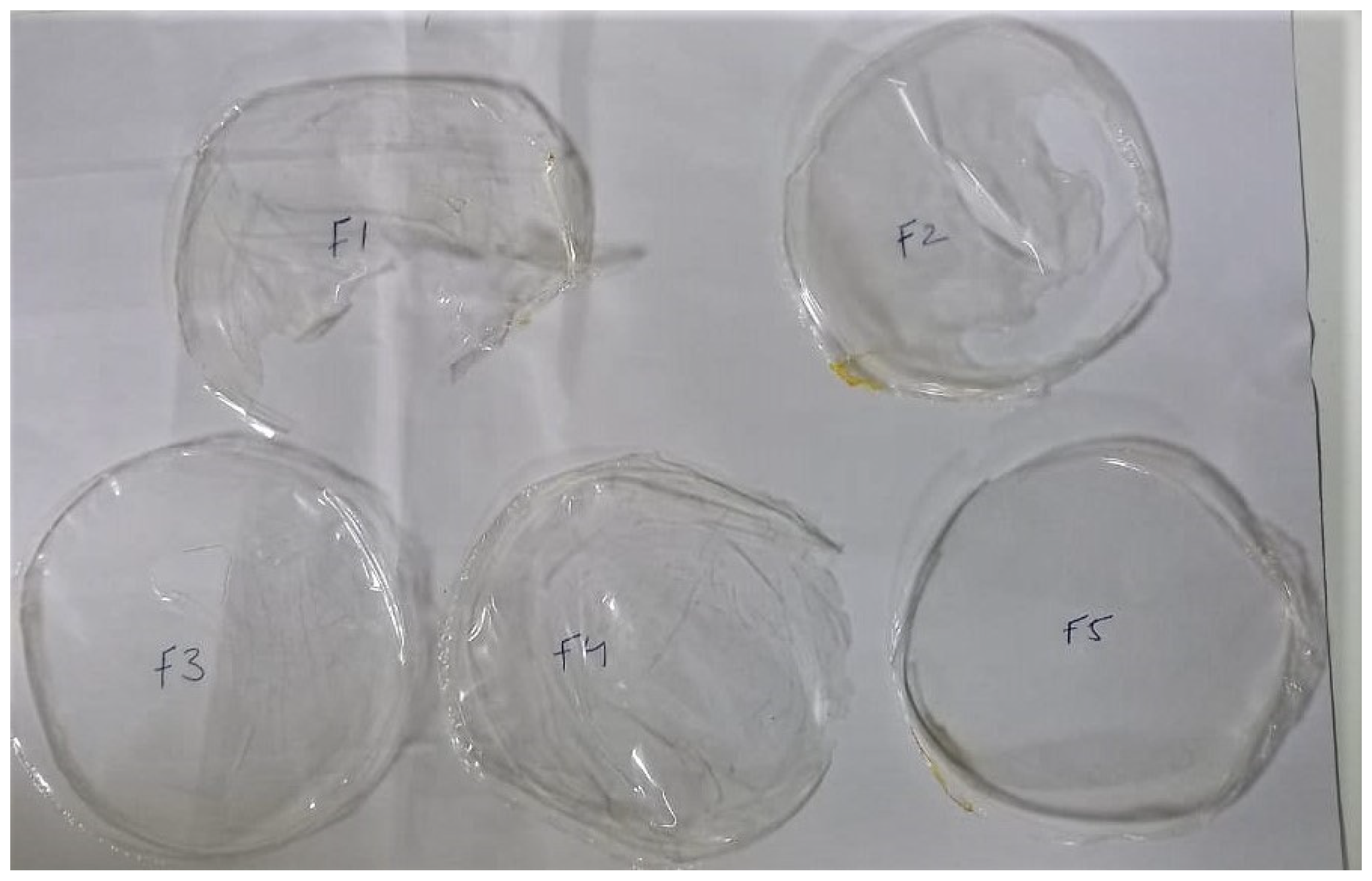
2.1. Thickness, Weight Variation and Folding Endurance
2.2. Moisture Content and Moisture Uptake
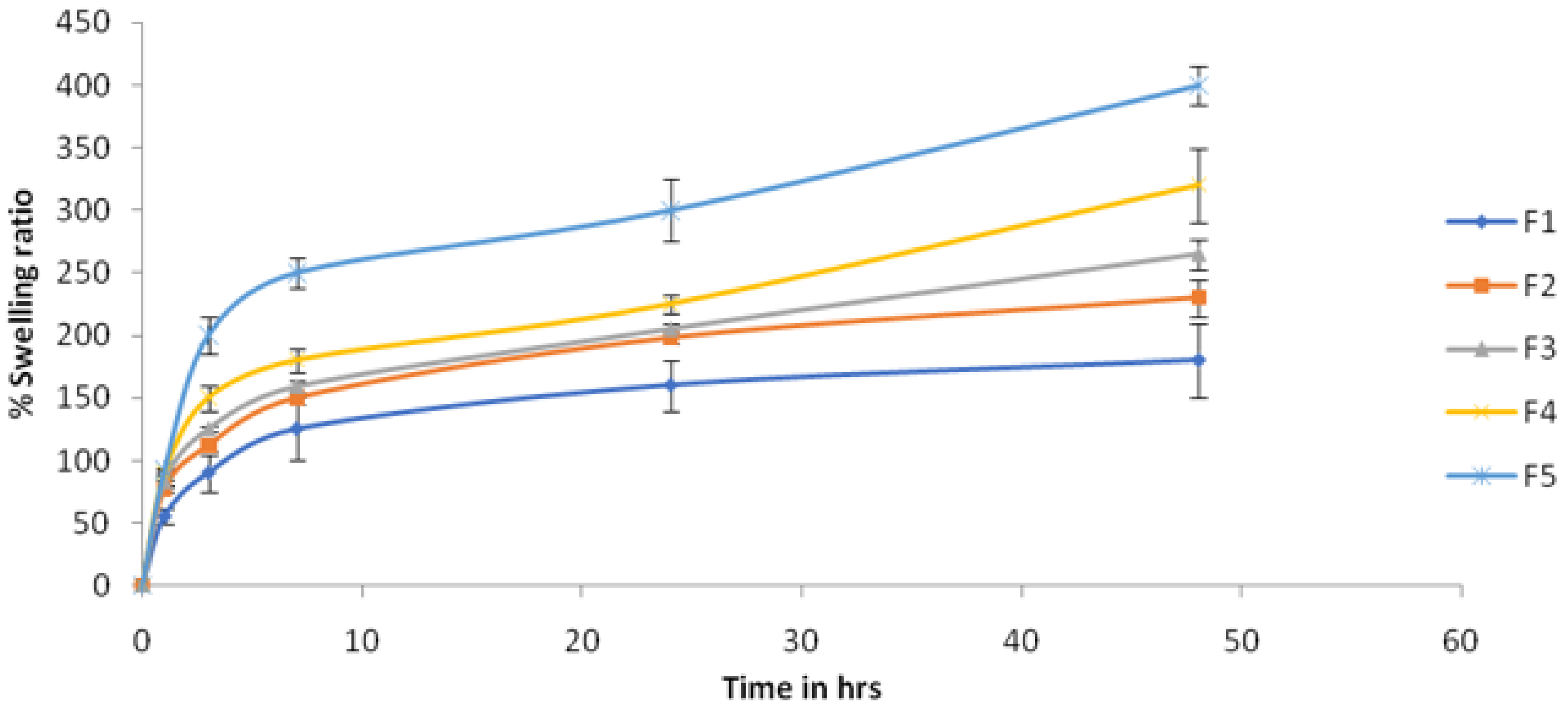
2.3. Swelling Ratio
2.4. WVTR
2.5. Tensile Strength and Elongation at Break
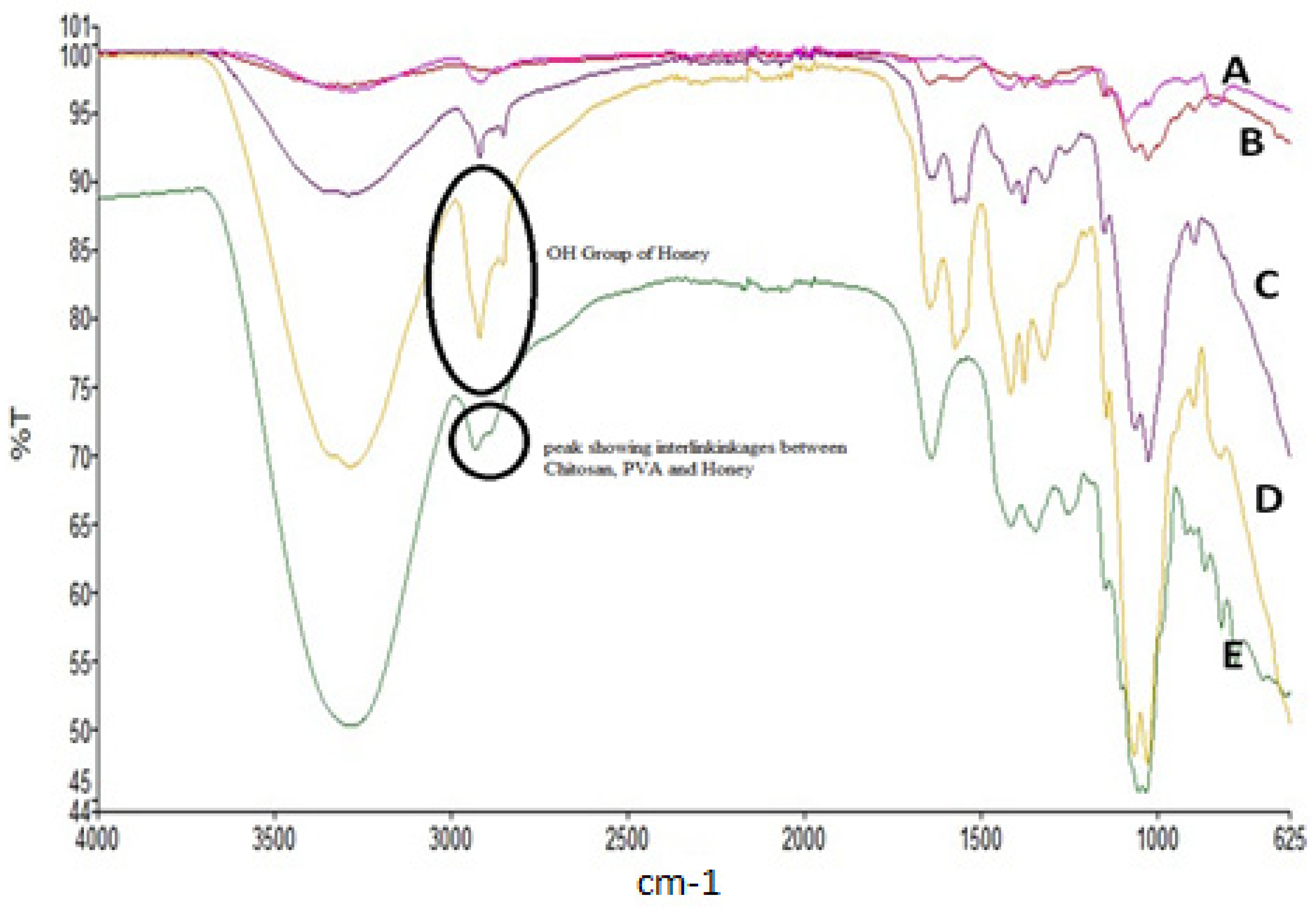
2.6. FTIR
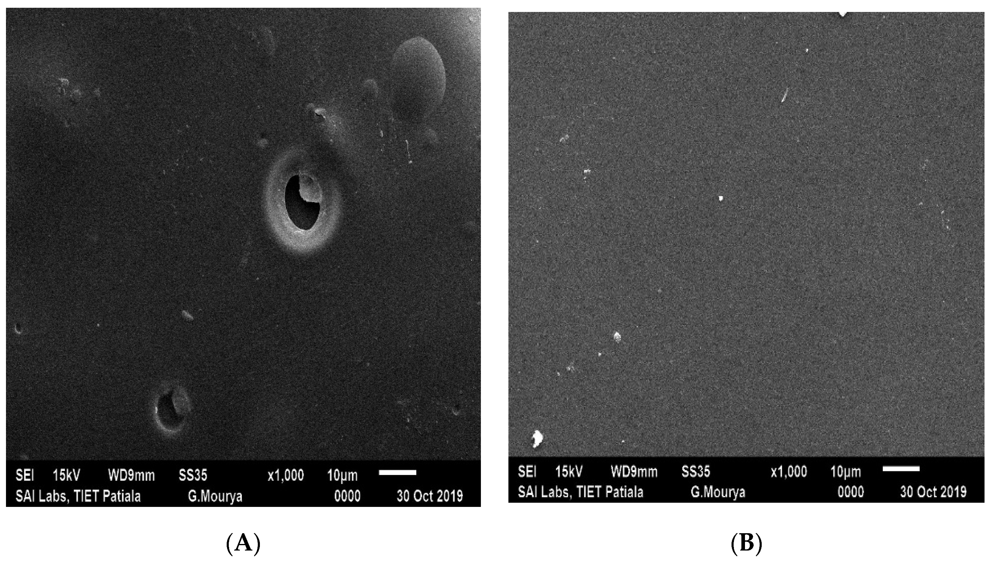
2.7. SEM
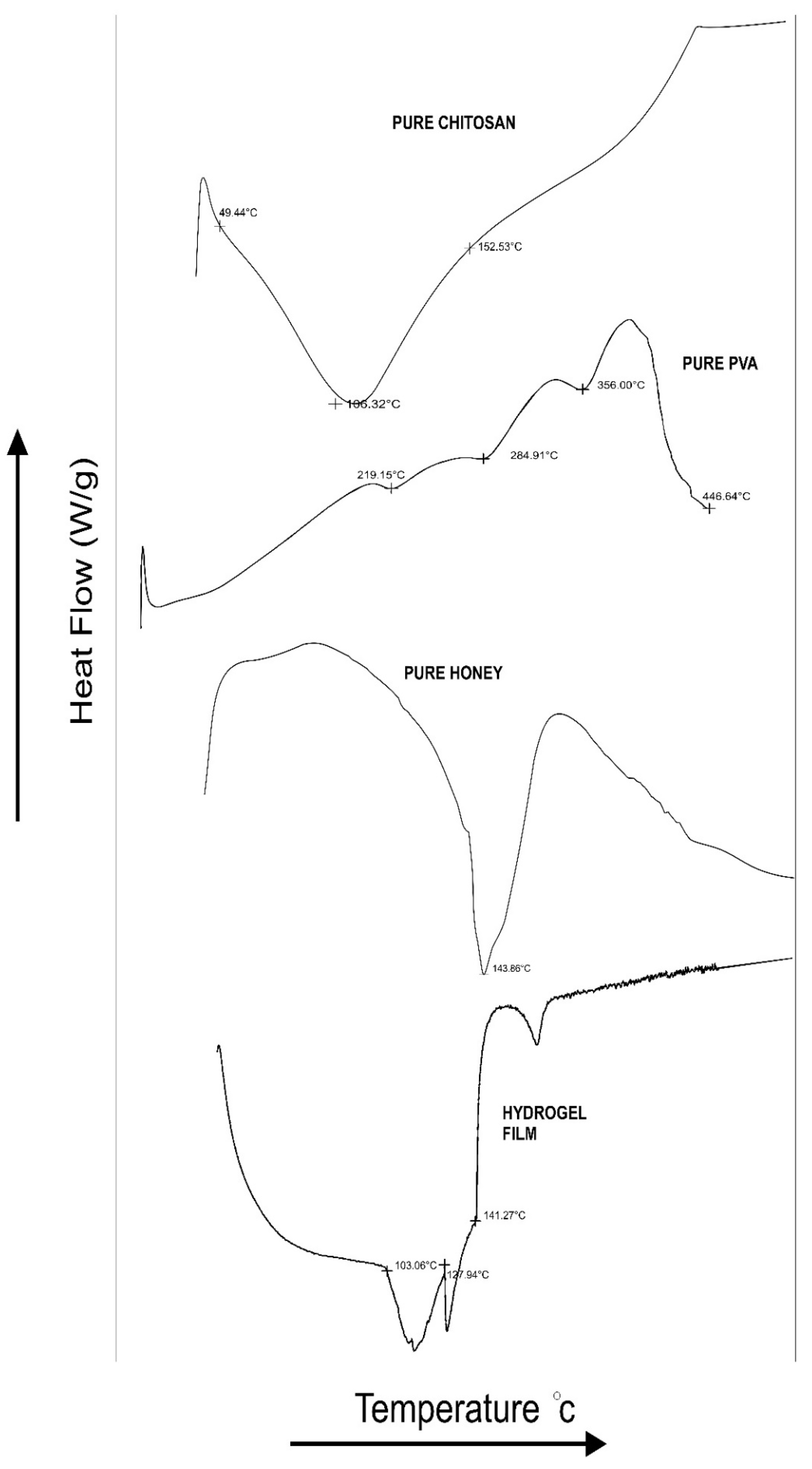
2.8. DSC
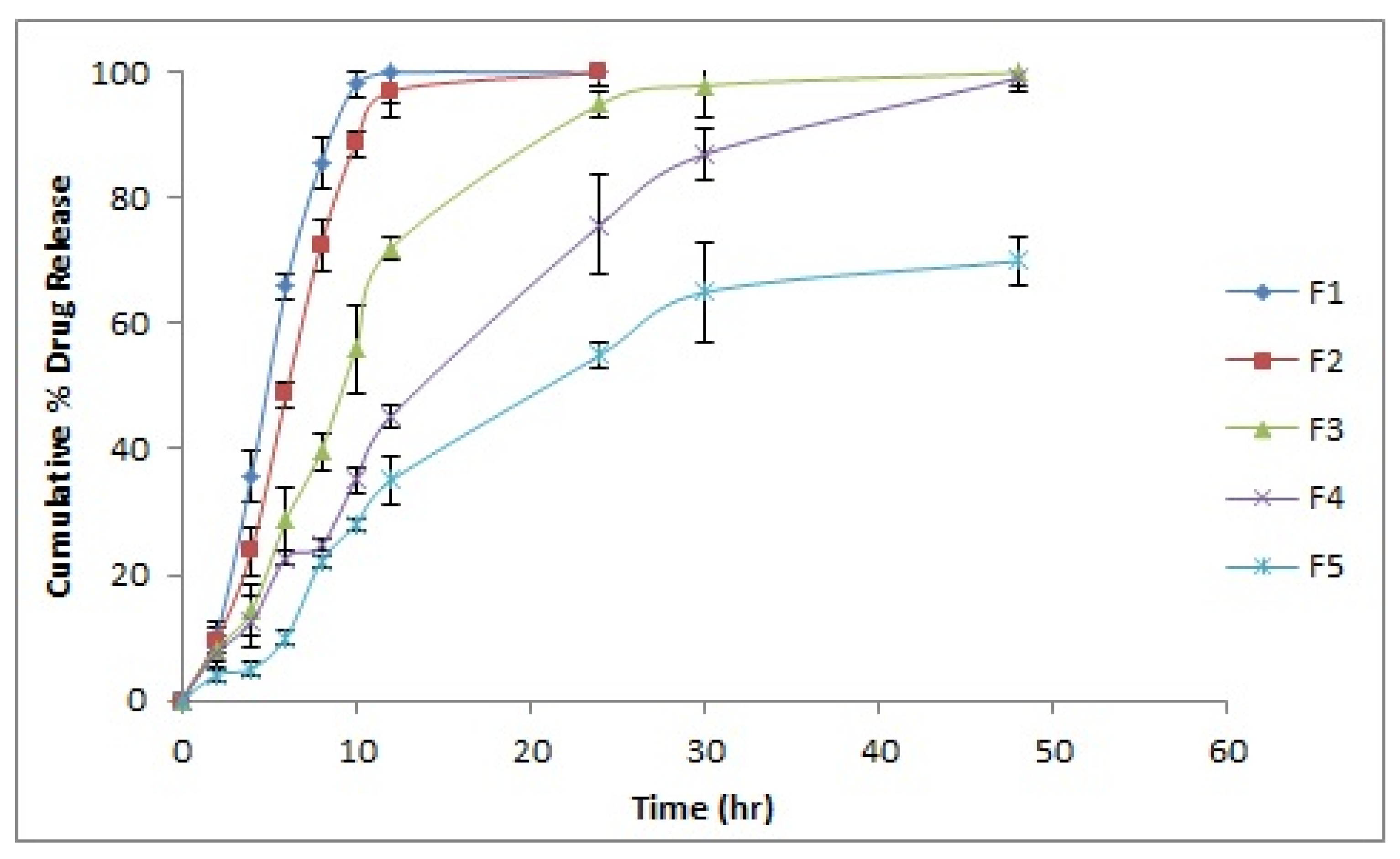
2.9. In Vitro Drug Release
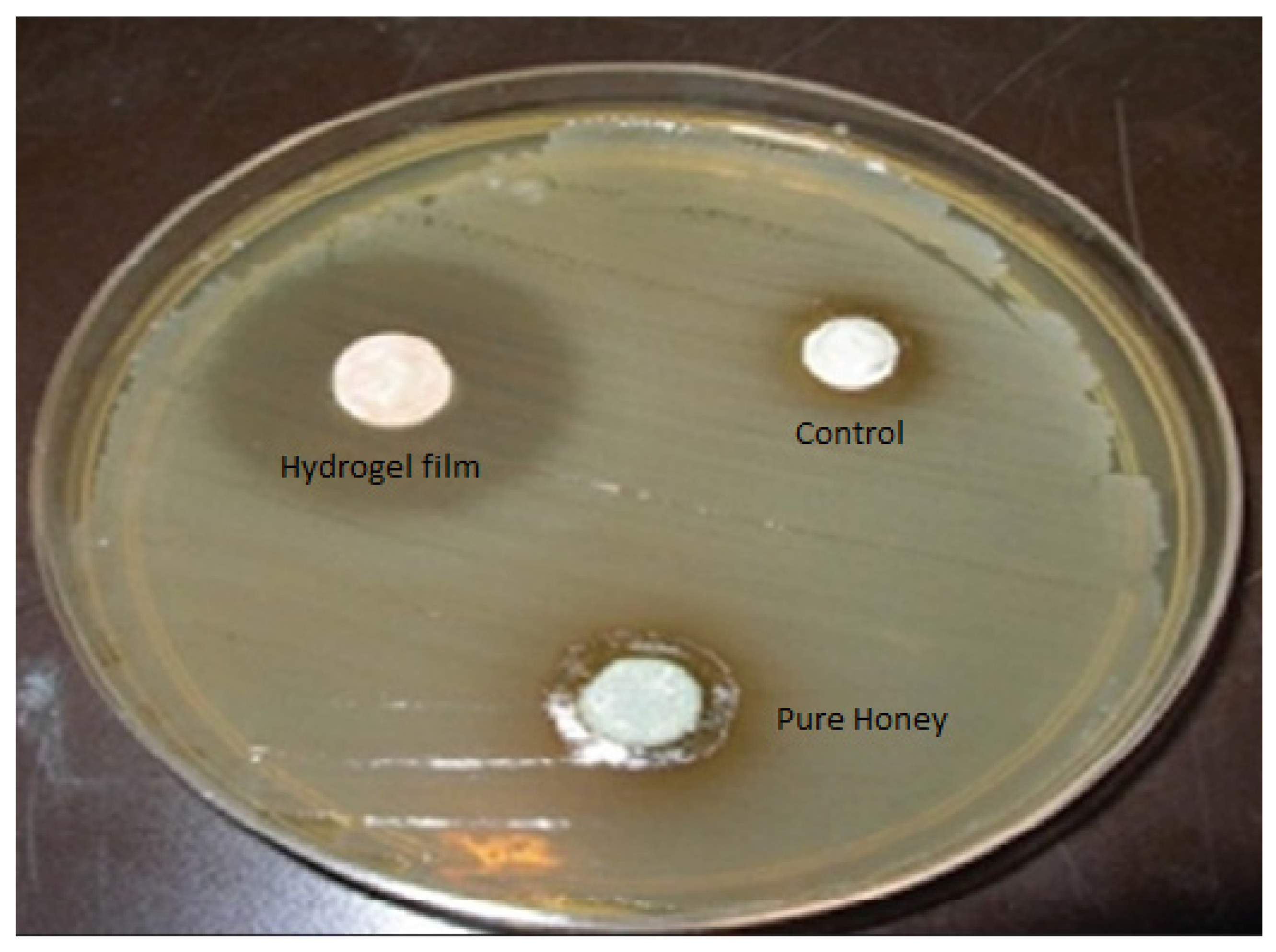
2.10. Antimicrobial Study
2.11. Stability Study
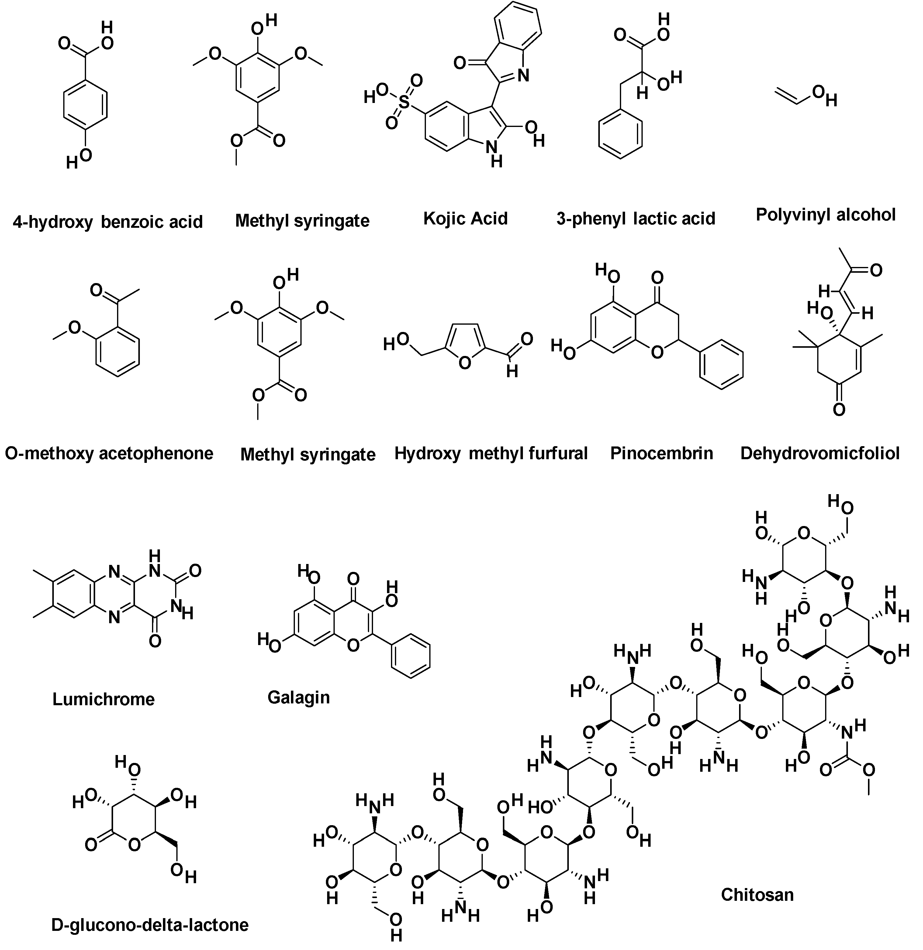
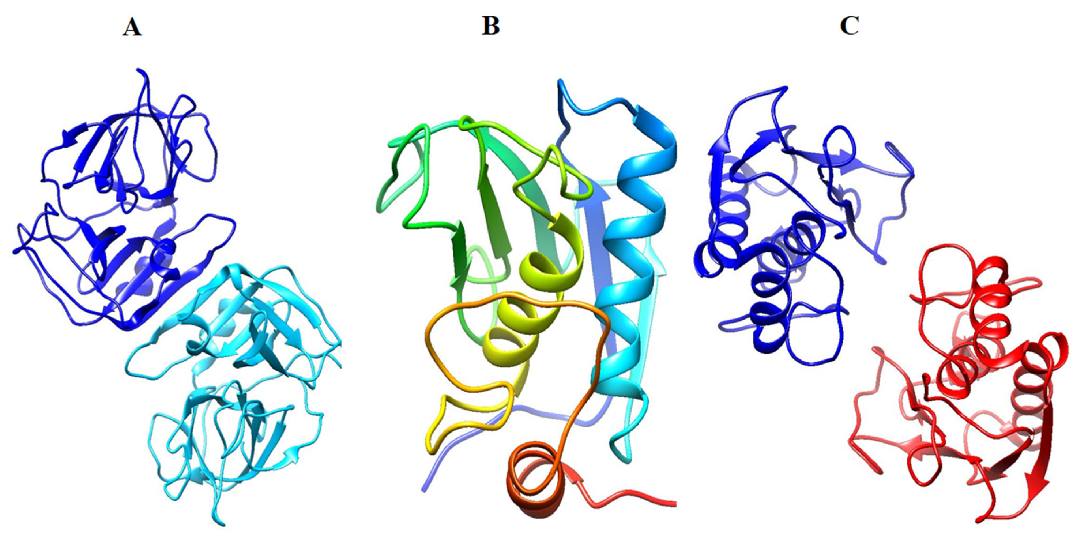
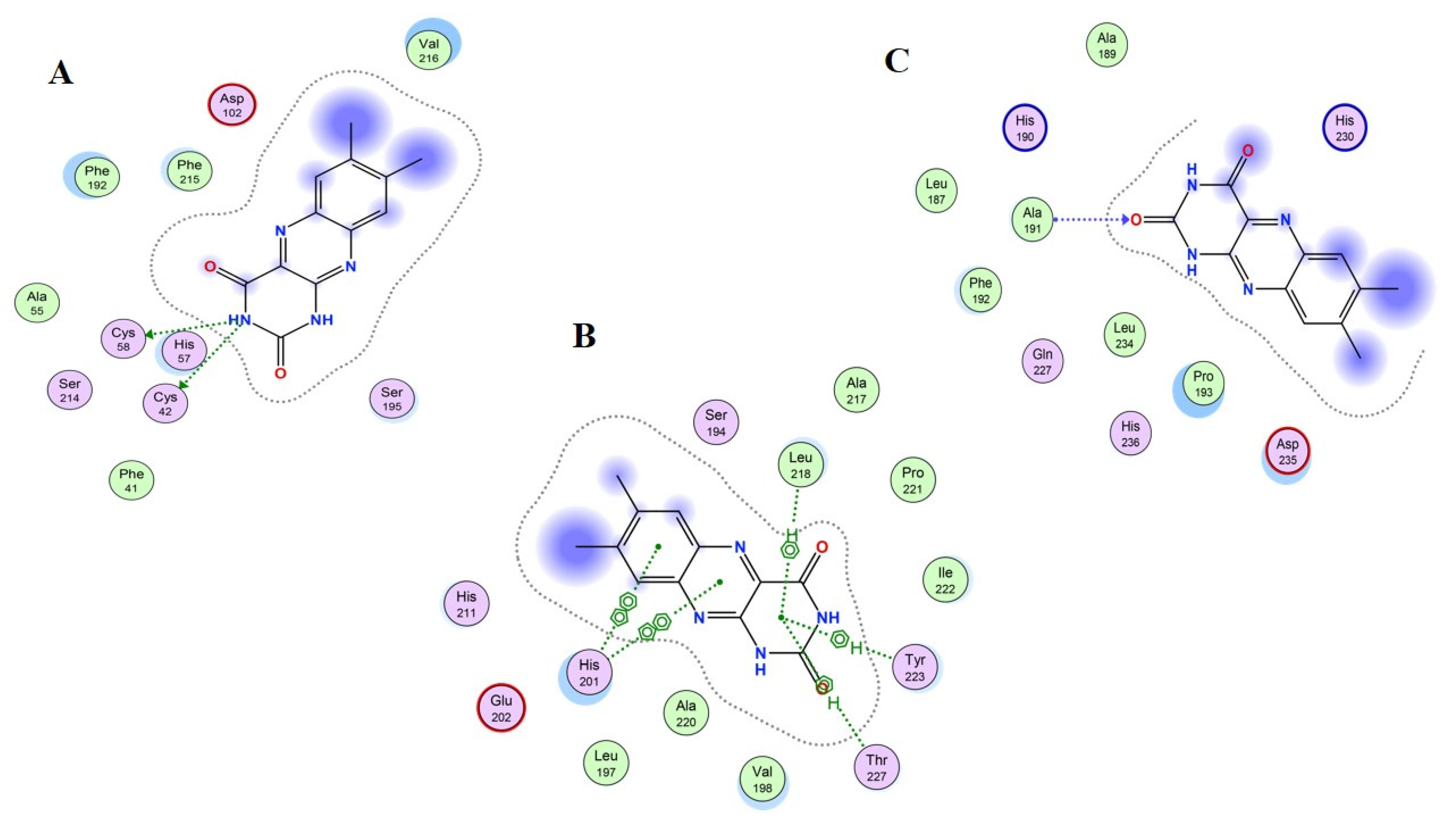
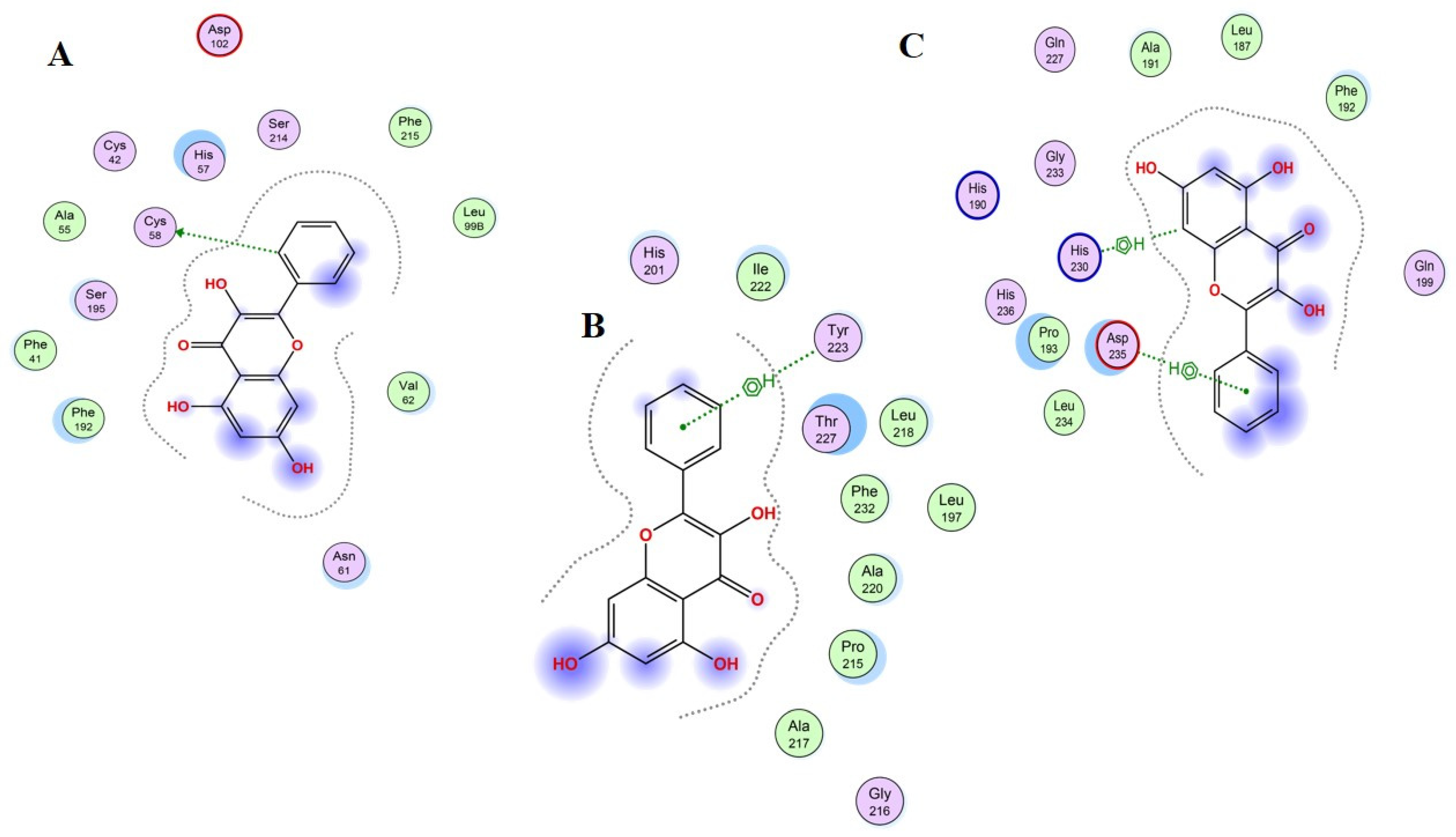
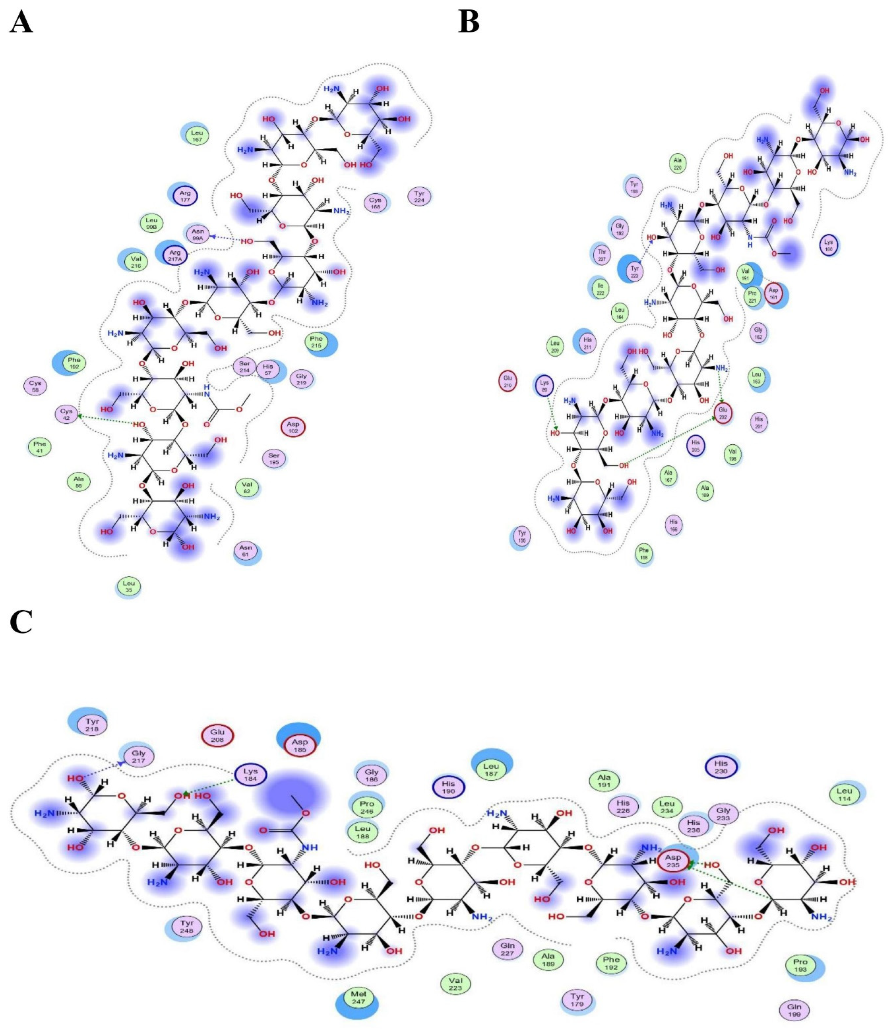
2.12. Molecular Docking Investigations
3. Conclusions
4. Material and Methods
4.1. Materials
4.2. Preparation of Hydrogel Films
4.3. Thickness and Weight Variation
4.4. Folding Endurance
4.5. Moisture Content
4.6. Moisture Uptake
4.7. Swelling Ratio
4.8. Water Vapor Transmission Rate (WVTR)
4.9. Mechanical Properties
4.10. Fourier-Transform Infrared Spectroscopy (FTIR)
4.11. SEM
4.12. DSC
4.13. In Vitro Drug Release
4.14. Antimicrobial Study
4.15. Stability Study
4.16. Construction of Chemical Database for In Silico Screening
4.17. Protein Target Selection
4.18. Protein–Ligand Docking and Interactions Analysis
4.19. Statistical Analysis
Author Contributions
Funding
Institutional Review Board Statement
Informed Consent Statement
Data Availability Statement
Acknowledgments
Conflicts of Interest
References
- Okabayashi, R.; Nakamura, M.; Okabayashi, T.; Tanaka, Y.; Nagai, A.; Yamashita, K. Efficacy of polarized hydroxyapatite and silk fibroin composite dressing gel on epidermal recovery from full-thickness skin wounds. J. Biomed. Mater. Res. Part B Appl. Biomater. 2009, 90 B, 641–646. [Google Scholar] [CrossRef]
- Chopra, H.; Kumar, S.; Singh, I. Strategies and Therapies for Wound Healing: A Review. Curr. Drug Targets 2021, 23, 87–98. [Google Scholar] [CrossRef] [PubMed]
- Chopra, H.; Singh, I.; Kumar, S.; Bhattacharya, T.; Habibur Rahman, M.; Akter, R.; Kabir, M.T. Comprehensive Review on Hydrogels. Curr. Drug Deliv. 2021, 18. [Google Scholar] [CrossRef] [PubMed]
- Chopra, H.; Kumar, S.; Singh, I. Bioinks for 3D printing of artificial extracellular matrices. In Advanced 3D-Printed Systems and Nanosystems for Drug Delivery and Tissue Engineering; Elsevier: Amsterdam, The Netherlands, 2020; pp. 1–37. [Google Scholar]
- Chopra, H.; Kumar, S.; Singh, I. Bioadhesive Hydrogels and Their Applications. In Bioadhesives in Drug Delivery; Scrivener Publishing LLC: Beverly, MA, USA, 2020; pp. 147–170. [Google Scholar] [CrossRef]
- Momin, M.; Kurhade, S.; Khanekar, P.; Mhatre, S. Novel biodegradable hydrogel sponge containing curcumin and honey for wound healing. J. Wound Care 2016, 25, 364–372. [Google Scholar] [CrossRef] [PubMed]
- El-Kased, R.F.; Amer, R.I.; Attia, D.; Elmazar, M.M. Honey-based hydrogel: In vitro and comparative in vivo evaluation for burn wound healing. Sci. Rep. 2017, 7, 9692. [Google Scholar] [CrossRef] [PubMed] [Green Version]
- Sasikala, L.; Rathinamoorthy, R.; Dhurai, B. Optimization of process conditions for chitosan-manuka honey film as wound contact layer for wound dressings. Wound Med. 2018, 23, 11–21. [Google Scholar] [CrossRef]
- Bagher, Z.; Ehterami, A.; Safdel, M.H.; Khastar, H.; Semiari, H.; Asefnejad, A.; Davachi, S.M.; Mirzaii, M.; Salehi, M. Wound healing with alginate/chitosan hydrogel containing hesperidin in rat model. J. Drug Deliv. Sci. Technol. 2020, 55, 101379. [Google Scholar] [CrossRef]
- Ahmad, S.; Minhas, M.U.; Ahmad, M.; Sohail, M.; Abdullah, O.; Badshah, S.F. Preparation and Evaluation of Skin Wound Healing Chitosan-Based Hydrogel Membranes. AAPS PharmSciTech 2018, 19, 3199–3209. [Google Scholar] [CrossRef]
- Mndlovu, H.; Du Toit, L.C.; Kumar, P.; Choonara, Y.E.; Marimuthu, T.; Kondiah, P.P.D.; Pillay, V. Bioplatform fabrication approaches affecting chitosan-based interpolymer complex properties and performance as wound dressings. Molecules 2020, 25, 222. [Google Scholar] [CrossRef] [Green Version]
- Shefa, A.A.; Sultana, T.; Park, M.K.; Lee, S.Y.; Gwon, J.G.; Lee, B.T. Curcumin incorporation into an oxidized cellulose nanofiber-polyvinyl alcohol hydrogel system promotes wound healing. Mater. Des. 2020, 186, 108313. [Google Scholar] [CrossRef]
- Yaghoobi, R.; Kazerouni, A.; Kazerouni, O. Evidence for clinical use of honey in wound healing as an anti-bacterial, anti-inflammatory anti-oxidant and anti-viral agent: A review. Jundishapur J. Nat. Pharm. Prod. 2013, 8, 100–104. [Google Scholar] [CrossRef] [PubMed] [Green Version]
- Moore, O.A.; Smith, L.A.; Campbell, F.; Seers, K.; McQuay, H.J.; Moore, R.A. Systematic review of the use of honey as a wound dressing. BMC Complement. Altern. Med. 2001, 1, 2. [Google Scholar] [CrossRef] [PubMed] [Green Version]
- Shamloo, A.; Aghababaie, Z.; Afjoul, H.; Jami, M.; Bidgoli, M.R.; Vossoughi, M.; Ramazani, A.; Kamyabhesari, K. Fabrication and evaluation of chitosan/gelatin/PVA hydrogel incorporating honey for wound healing applications: An in vitro, in vivo study. Int. J. Pharm. 2021, 592, 120068. [Google Scholar] [CrossRef] [PubMed]
- Sangnim, T.; Limmatvapirat, S.; Nunthanid, J.; Sriamornsak, P.; Sittikijyothin, W.; Wannachaiyasit, S.; Huanbutta, K. Design and characterization of clindamycin-loaded nanofiber patches composed of polyvinyl alcohol and tamarind seed gum and fabricated by electrohydrodynamic atomization. Asian J. Pharm. Sci. 2018, 13, 450–458. [Google Scholar] [CrossRef]
- Cazón, P.; Velázquez, G.; Vázquez, M. Characterization of bacterial cellulose films combined with chitosan and polyvinyl alcohol: Evaluation of mechanical and barrier properties. Carbohydr. Polym. 2019, 216, 72–85. [Google Scholar] [CrossRef]
- Abdeen, Z. Swelling and reswelling characteristics of cross-linked poly(vinyl alcohol)/chitosan hydrogel film. J. Dispers. Sci. Technol. 2011, 32, 1337–1344. [Google Scholar] [CrossRef]
- Casey, L.S.; Wilson, L.D. Investigation of chitosan-PVA composite films and their adsorption properties. J. Geosci. Environ. Prot. 2015, 3, 55214. [Google Scholar] [CrossRef]
- Kanatt, S.R.; Rao, M.S.; Chawla, S.P.; Sharma, A. Active chitosan-polyvinyl alcohol films with natural extracts. Food Hydrocoll. 2012, 29, 290–297. [Google Scholar] [CrossRef]
- Pelissari, F.M.; Grossmann, M.V.E.; Yamashita, F.; Pined, E.A.G. Antimicrobial, mechanical, and barrier properties of cassava starch-chitosan films incorporated with oregano essential oil. J. Agric. Food Chem. 2009, 57, 7499–7504. [Google Scholar] [CrossRef]
- Li, B.; Peng, J.; Yie, X.; Xie, B. Enhancing physical properties and antimicrobial activity of konjac glucomannan edible films by incorporating chitosan and nisin. J. Food Sci. 2006, 71, C174–C178. [Google Scholar] [CrossRef]
- Wang, Q.; Du, Y.M.; Fan, L.H. Properties of chitosan/poly(vinyl alcohol) films for drug-controlled release. J. Appl. Polym. Sci. 2005, 96, 808–813. [Google Scholar] [CrossRef]
- Liang, S.; Liu, L.; Huang, Q.; Yam, K.L. Preparation of single or double-network chitosan/poly(vinyl alcohol) gel films through selectively cross-linking method. Carbohydr. Polym. 2009, 77, 718–724. [Google Scholar] [CrossRef]
- Tripathi, S.; Mehrotra, G.K.; Dutta, P.K. Physicochemical and bioactivity of cross-linked chitosan-PVA film for food packaging applications. Int. J. Biol. Macromol. 2009, 45, 372–376. [Google Scholar] [CrossRef]
- Kouchak, M.; Ameri, A.; Naseri, B.; Kargar Boldaji, S. Chitosan and polyvinyl alcohol composite films containing nitrofurazone: Preparation and evaluation. Iran. J. Basic Med. Sci. 2014, 17, 14–20. [Google Scholar] [CrossRef]
- Wang, T.; Zhu, X.K.; Xue, X.T.; Wu, D.Y. Hydrogel sheets of chitosan, honey and gelatin as burn wound dressings. Carbohydr. Polym. 2012, 88, 75–83. [Google Scholar] [CrossRef]
- Park, J.S.; An, S.J.; Jeong, S.I.; Gwon, H.J.; Lim, Y.M.; Nho, Y.C. Chestnut honey impregnated carboxymethyl cellulose hydrogel for diabetic ulcer healing. Polymers 2017, 9, 248. [Google Scholar] [CrossRef] [Green Version]
- Koosha, M.; Hamedi, S. Intelligent Chitosan/PVA nanocomposite films containing black carrot anthocyanin and bentonite nanoclays with improved mechanical, thermal and antibacterial properties. Prog. Org. Coat. 2019, 127, 338–347. [Google Scholar] [CrossRef]
- Muxika, A.; Etxabide, A.; Uranga, J.; Guerrero, P.; de la Caba, K. Chitosan as a bioactive polymer: Processing, properties and applications. Int. J. Biol. Macromol. 2017, 105, 1358–1368. [Google Scholar] [CrossRef]
- Saikaly, S.K.; Khachemoune, A. Honey and Wound Healing: An Update. Am. J. Clin. Dermatol. 2017, 18, 237–251. [Google Scholar] [CrossRef]
- Narayanaswamy, R.; Kok Wai, L.; Ismail, I.S. In silico analysis of selected honey constituents as human neutrophil elastase (HNE) and matrix metalloproteinases (MMP 2 and 9) inhibitors. Int. J. Food Prop. 2015, 18, 2155–2164. [Google Scholar] [CrossRef]
- Caley, M.P.; Martins, V.L.C.; O’Toole, E.A. Metalloproteinases and wound healing. Adv. Wound Care 2015, 4, 225–234. [Google Scholar] [CrossRef] [PubMed] [Green Version]
- Novo, E.; Parola, M. The role of redox mechanisms in hepatic chronic wound healing and fibrogenesis. Fibrogenesis Tissue Repair 2012, 5, S4. [Google Scholar] [CrossRef] [PubMed] [Green Version]
- Bibi, S.; Sakata, K. An Integrated Computational Approach for Plant-Based Protein Tyrosine Phosphatase Non-Receptor Type 1 Inhibitors. Curr. Comput. Aided. Drug Des. 2017, 13, 319–335. [Google Scholar] [CrossRef] [PubMed] [Green Version]
- Kaur, R.; Sharma, A.; Puri, V.; Singh, I. Preparation and characterization of biocomposite films of carrageenan/locust bean gum/montmorrillonite for transdermal delivery of curcumin. BioImpacts 2019, 9, 37–43. [Google Scholar] [CrossRef] [PubMed] [Green Version]
- Standard Test Method for Determining Water Vapor Transmission Rates through Nonwoven and Plastic Barriers. Available online: https://www.astm.org/d6701-21.html (accessed on 26 January 2022).
- Thakur, G.; Singh, A.; Singh, I. Formulation and evaluation of transdermal composite films of chitosan-montmorillonite for the delivery of curcumin. Int. J. Pharm. Investig. 2016, 6, 23–31. [Google Scholar] [CrossRef] [Green Version]
- Kim, S.; Thiessen, P.A.; Bolton, E.E.; Chen, J.; Fu, G.; Gindulyte, A.; Han, L.; He, J.; He, S.; Shoemaker, B.A.; et al. PubChem substance and compound databases. Nucleic Acids Res. 2016, 44, D1202–D1213. [Google Scholar] [CrossRef]
- Macdonald, S.J.F.; Dowle, M.D.; Harrison, L.A.; Clarke, G.D.E.; Inglis, G.G.A.; Johnson, M.R.; Shah, P.; Smith, R.A.; Amour, A.; Fleetwood, G.; et al. Discovery of further pyrrolidine trans-lactams as inhibitors of human neutrophil elastase (HNE) with potential as development candidates and the crystal structure of HNE complexed with an inhibitor (GW475151). J. Med. Chem. 2002, 45, 3878–3890. [Google Scholar] [CrossRef] [PubMed]
- Dhanaraj, V.; Williams, M.G.; Ye, Q.Z.; Molina, F.; Johnson, L.L.; Ortwine, D.F.; Pavlovsky, A.; Rubin, J.R.; Skeean, R.W.; White, A.D.; et al. X-ray Structure of Gelatinase A Catalytic Domain Complexed with a Hydroxamate Inhibitor. Croat. Chem. Acta 1999, 72, 575–591. [Google Scholar] [CrossRef]
- Antoni, C.; Vera, L.; Devel, L.; Catalani, M.P.; Czarny, B.; Cassar-Lajeunesse, E.; Nuti, E.; Rossello, A.; Dive, V.; Stura, E.A. Crystallization of bi-functional ligand protein complexes. J. Struct. Biol. 2013, 182, 246–254. [Google Scholar] [CrossRef] [PubMed]
- Bibi, S.; Sakata, K. Current Status of Computer-Aided Drug Design for Type 2 Diabetes. Curr. Comput. Aided-Drug Des. 2016, 12, 167–177. [Google Scholar] [CrossRef]
- Vilar, S.; Cozza, G.; Moro, S. Medicinal Chemistry and the Molecular Operating Environment (MOE): Application of QSAR and Molecular Docking to Drug Discovery. Curr. Top. Med. Chem. 2008, 8, 1555–1572. [Google Scholar] [CrossRef] [PubMed]
- Yousafi, Q.; Batool, J.; Khan, M.S.; Perveen, T.; Sajid, M.W.; Hussain, A.; Mehmood, A.; Saleem, S. In Silico Evaluation of Food Derived Bioactive Peptides as Inhibitors of Angiotensin Converting Enzyme (ACE). Int. J. Pept. Res. Ther. 2021, 27, 341–349. [Google Scholar] [CrossRef]
- Xiong, G.; Shen, C.; Yang, Z.; Jiang, D.; Liu, S.; Lu, A.; Chen, X.; Hou, T.; Cao, D. Featurization strategies for protein–ligand interactions and their applications in scoring function development. Wiley Interdiscip. Rev. Comput. Mol. Sci. 2021, e1567, in press. [Google Scholar] [CrossRef]


| Formulation Code | Thickness (mm) | Weight Variation (g) | Folding Endurance | Moisture Content (%) | Moisture Uptake (%) |
|---|---|---|---|---|---|
| F1 | 0.052 ± 0.003 | 0.462 ± 0.09 | 350 ± 15 | 18.10 ± 1.05 | 11.35 ± 0.07 |
| F2 | 0.046 ± 0.006 | 0.429 ± 0.06 | 405 ± 9 | 12.52 ± 1.14 | 11.95 ± 1.01 |
| F3 | 0.055 ± 0.004 | 0.480 ± 0.04 | 430 ± 11 | 17.38 ± 2.56 | 12.25 ± 0.08 |
| F4 | 0.041 ± 0.006 | 0.425 ± 0.02 | 433 ± 10 | 21.57 ± 1.93 | 13.65 ± 0.09 |
| F5 | 0.048 ± 0.007 | 0.447 ± 0.08 | 445 ± 7 | 24.22 ± 2.37 | 14.96 ± 0.06 |
| Formulation Code | Tensile Strength (N) | Elongation at Break (mm) | WVTR (g/m2/day) |
|---|---|---|---|
| F1 | 4.74 ± 0.83 | 30.58 ± 3.64 | 2698.65 ± 76.29 |
| F2 | 10.52 ± 1.45 | 31.10 ± 4.56 | 2458.87 ± 71.40 |
| F3 | 23.77 ± 3.85 | 31.62 ± 5.25 | 2150.66 ± 80.19 |
| F4 | 25.15 ± 2.66 | 31.98 ± 3.09 | 1911.53 ± 55.41 |
| F5 | 38.36 ± 5.39 | 33.51 ± 2.47 | 1650.50 ± 35.86 |
| Formulation Code | Zero-Order | First-Order | Higuchi | Korsmeyer–Peppas | Hixson–Crowell | ||||||
|---|---|---|---|---|---|---|---|---|---|---|---|
| r2 | k0 | r2 | k1 | r2 | kH | r2 | kKP | n | r2 | kHC | |
| F1 | 0.981 | 10.67 | 0.842 | −0.161 | 0.978 | 3.318 | 0.975 | 0.637 | 0.428 | 0.94 | −0.342 |
| F2 | 0.981 | 8.898 | 0.873 | −0.122 | 0.98 | 3.032 | 0.987 | 0.582 | 0.359 | 0.945 | −0.273 |
| F3 | 0.926 | 4.441 | 0.912 | −0.087 | 0.966 | 2.201 | 0.961 | 0.581 | 0.403 | 0.973 | −0.16 |
| F4 | 0.923 | 2.212 | 0.945 | −0.041 | 0.961 | 1.507 | 0.976 | 0.638 | 0.688 | 0.997 | −0.078 |
| F5 | 0.891 | 2.087 | 0.993 | −0.028 | 0.934 | 1.475 | 0.945 | 0.659 | 0.604 | 0.98 | −0.064 |
| Batch | Time Interval (Months) | Test Parameters | |||
|---|---|---|---|---|---|
| Folding Endurance | Moisture Content (%) | Tensile Strength (N/mm2) | WVTR (g/m2/day) | ||
| F1 | 0 | 350 ± 15 | 18.10 ± 1.05 | 4.74 ± 0.83 | 2698.65 ± 76.29 |
| 1 | 348 ± 14 | 18.10 ± 1.01 | 4.70 ± 0.59 | 2695.54 ± 77.58 | |
| 2 | 349 ± 17 | 18.10 ± 1.09 | 4.71 ± 0.78 | 2696.60 ± 75.41 | |
| 3 | 350 ± 17 | 18.10 ± 1.12 | 4.74 ± 0.65 | 2697.54 ± 74.57 | |
| F2 | 0 | 405 ± 9 | 12.52 ± 1.14 | 10.52 ± 1.45 | 2458.87 ± 71.40 |
| 1 | 403 ± 6 | 12.45 ± 1.18 | 10.51 ± 1.49 | 2459.65 ± 70.45 | |
| 2 | 404 ± 7 | 12.50 ± 1.11 | 10.48 ± 1.50 | 2454.98 ± 71.50 | |
| 3 | 401 ± 8 | 12.54 ± 1.16 | 10.49 ± 1.40 | 2452.45 ± 70.45 | |
| F3 | 0 | 430 ± 11 | 17.38 ± 2.56 | 23.77 ± 3.85 | 2150.66 ± 80.19 |
| 1 | 429 ± 10 | 17.36 ± 2.41 | 23.72 ± 3.80 | 2149.65 ± 79.74 | |
| 2 | 428 ± 9 | 17.32 ± 2.52 | 23.74 ± 3.79 | 2151.46 ± 79.85 | |
| 3 | 430 ± 10 | 17.38 ± 2.50 | 23.71 ± 3.75 | 2148.85 ± 79.90 | |
| F4 | 0 | 433 ± 10 | 21.57 ± 1.93 | 25.15 ± 2.66 | 1911.53 ± 55.41 |
| 1 | 432 ± 9 | 21.51 ± 1.93 | 25.13 ± 2.61 | 1912.33 ± 55.41 | |
| 2 | 430 ± 9 | 21.48 ± 1.90 | 25.15 ± 2.60 | 1911.65 ± 55.41 | |
| 3 | 431 ± 8 | 21.49 ± 1.89 | 25.14 ± 2.65 | 1913.54 ± 55.41 | |
| F5 | 0 | 445 ± 7 | 24.22 ± 2.37 | 38.36 ± 5.39 | 1650.50 ± 35.86 |
| 1 | 444 ± 7 | 24.20 ± 2.35 | 38.34 ± 5.37 | 1648.49 ± 34.54 | |
| 2 | 444 ± 6 | 24.15 ± 2.31 | 38.31 ± 5.30 | 1649.65 ± 33.12 | |
| 3 | 443 ± 7 | 24.18 ± 2.38 | 38.34 ± 5.35 | 1651.45 ± 35.65 | |
| Sr. No | Compound Names | PubChem CID | Protein PDB ID: 1H1B | Protein PBD ID: 1QIB | Protein PBD ID: 4H1Q | |||
|---|---|---|---|---|---|---|---|---|
| Score | RMSD | Score | RMSD | Score | RMSD | |||
| 1 | 4-hydroxybenzoic acid | 135 | −4.1647 | 1.5935 | −4.9175 | 1.5940 | −4.9309 | 1.4805 |
| 2 | Methyl syringate | 880 | −3.6409 | 2.1469 | −3.7874 | 1.1233 | −3.8436 | 1.1154 |
| 3 | Kojic Acid | 3708 | −5.7348 | 1.1504 | −7.1493 | 1.6731 | −7.3917 | 0.6537 |
| 4 | 3-phenyl lactic acid | 3848 | −4.6707 | 0.8809 | −5.5804 | 1.4010 | −5.7754 | 1.6327 |
| 5 | Polyvinyl alcohol | 11199 | −3.1533 | 3.0526 | −3.1736 | 0.9737 | −3.1732 | 3.4360 |
| 6 | O-methoxyacetophenone | 68481 | −4.7006 | 0.9142 | −5.1265 | 0.8240 | −5.4133 | 1.1089 |
| 7 | Methyl syringate | 70164 | −5.1639 | 1.1198 | −5.7789 | 1.5015 | −6.3107 | 0.7194 |
| 8 | Hydroxymethylfurfural | 237332 | −4.382 | 1.6009 | −4.7615 | 1.2097 | −4.8671 | 2.7057 |
| 9 | Pinocembrin | 238782 | −5.0966 | 2.2995 | −6.3780 | 1.1495 | −6.7934 | 1.7762 |
| 10 | Dehydrovomifoliol | 688492 | −5.9192 | 2.5804 | −6.0208 | 0.7519 | −5.7665 | 2.1204 |
| 11 | Lumichrome | 5326566 | −6.0911 | 1.1315 | −7.3801 | 1.0649 | −6.9001 | 0.8905 |
| 12 | Galagin | 5281616 | −6.0133 | 0.8527 | −7.3211 | 1.3412 | −6.8556 | 1.8762 |
| 13 | Chitosan | 71853 | −11.8369 | 4.7772 | −11.6352 | 4.0294 | −12.8897 | 3.7408 |
| 14 | D-glucono delta-lactone | 7043900 | −4.7835 | 0.6276 | −5.2760 | 0.9685 | −5.4439 | 0.9102 |
| Compound | Dock Score | Interacting Residues in the Binding Pocket | ||||
|---|---|---|---|---|---|---|
| Ligand | Receptor | Interaction | Distance | E (kcal/mol) | ||
| Human Neutrophil Elastase (HNE) (PDB ID: 1H1B) | ||||||
| Lumichrome | −6.0911 | O6 O6 | SG:CYS42(A) SG:CYS58(A) | H-donor H-donor | 3.42 3.42 | −1.7 −1.9 |
| Galagin | −6.0133 | C16 | SG:CYS58(A) | H-donor | 3.82 | −0.5 |
| Chitosan | −11.8369 | O21 O28 | SG:CYS42(A) O:ASN99(A) | H-donor H-donor | 3.93 2.89 | −0.5 −1.1 |
| Matrix metalloproteinase-3 (MMP-3) (PDB ID: 1QIB) | ||||||
| Lumichrome | −7.3801 | 6-ring 6-ring 6-ring 6-ring 6-ring 6-ring | CD1:LEU218(A) CA:TYR223(A) CD1:TYR223(A) N:THR227(A) 5-ring:HIS201(A) 5-ring:HIS201(A) | Pi–H Pi–H Pi–H Pi–H Pi–Pi Pi–Pi | 4.45 4.03 4.30 4.22 3.75 3.57 | −0.6 −1.2 −0.6 −3.9 −0.0 −0.0 |
| Galagin | −7.3211 | BR2 5-ring | OE2:Glu202(A) CA:LEU218(A) | H-donor Pi–H | 3.45 3.93 | −0.9 −1.6 |
| Chitosan | −11.6352 | O32 O43 O15 O22 | OE1:GLU202(A) OE2:GLU202(A) N:TYR223(A) NZ:LYS89(A) | H-donor H-donor H-acceptor H-acceptor | 2.79 2.84 2.87 316 | −3.7 −1.7 −1.9 −2.4 |
| Matrix metallopeptidase 9 (MMP-9) (PDB ID: 4H1Q) | ||||||
| Lumichrome | −6.9001 | O2 | N: ALA191(A) | H-acceptor | 3.38 | −0.5 |
| Galagin | −6.8556 | C13 6-ring | 5-ring: HIS230(A) CB:ASP235(A) | H–Pi Pi–H | 3.61 3.75 | −1.2 −1.1 |
| Chitosan | −12.8897 | O32 O35 C90 O37 | OD2:ASP235(A) O:GLY217(A) OD2:ASP235(A) NZ:LYS184(A) | H-donor H-donor H-donor H-acceptor | 3.04 3.08 3.05 3.43 | −2.3 −0.6 −1.4 −1.4 |
| Formulation Code | Chitosan Solution | PVA Solution (5% w/v) (mL) | Honey (g) | |
|---|---|---|---|---|
| Concentration of Chitosan (% w/v) | Amount of Chitosan (mL) | |||
| F1 | 0.25 | 80 | 20 | 1 |
| F2 | 0.50 | 80 | 20 | 1 |
| F3 | 0.75 | 80 | 20 | 1 |
| F4 | 1.0 | 80 | 20 | 1 |
| F5 | 2.0 | 80 | 20 | 1 |
Publisher’s Note: MDPI stays neutral with regard to jurisdictional claims in published maps and institutional affiliations. |
© 2022 by the authors. Licensee MDPI, Basel, Switzerland. This article is an open access article distributed under the terms and conditions of the Creative Commons Attribution (CC BY) license (https://creativecommons.org/licenses/by/4.0/).
Share and Cite
Chopra, H.; Bibi, S.; Kumar, S.; Khan, M.S.; Kumar, P.; Singh, I. Preparation and Evaluation of Chitosan/PVA Based Hydrogel Films Loaded with Honey for Wound Healing Application. Gels 2022, 8, 111. https://0-doi-org.brum.beds.ac.uk/10.3390/gels8020111
Chopra H, Bibi S, Kumar S, Khan MS, Kumar P, Singh I. Preparation and Evaluation of Chitosan/PVA Based Hydrogel Films Loaded with Honey for Wound Healing Application. Gels. 2022; 8(2):111. https://0-doi-org.brum.beds.ac.uk/10.3390/gels8020111
Chicago/Turabian StyleChopra, Hitesh, Shabana Bibi, Sandeep Kumar, Muhammad Saad Khan, Pradeep Kumar, and Inderbir Singh. 2022. "Preparation and Evaluation of Chitosan/PVA Based Hydrogel Films Loaded with Honey for Wound Healing Application" Gels 8, no. 2: 111. https://0-doi-org.brum.beds.ac.uk/10.3390/gels8020111