Article

Dynamic Transcriptome Analysis Reveals Uncharacterized Complex Regulatory Pathway Underlying Genotype-Recalcitrant Somatic Embryogenesis Transdifferentiation in Cotton

State Key Laboratory of Crop Biology, College of Agronomy, Shandong Agricultural University, Tai’an 271018, China
*
Author to whom correspondence should be addressed.
Submission received: 10 March 2020 / Revised: 21 April 2020 / Accepted: 5 May 2020 / Published: 7 May 2020
(This article belongs to the Special Issue Evolutionary Genetics of Plant Reproduction)

Abstract

:
As a notable illustration of totipotency and plant regeneration, somatic embryogenesis (SE) is the developmental reprogramming of somatic cells toward the embryogenesis pathway, the key step for genetic engineering. Investigations examining the totipotency process are of great fundamental and practical importance in crop biotechnology. However, high-frequency regeneration of cotton via SE has been limited due to genotype-dependent response. The molecular basis deciphering SE genotype recalcitrance remains largely unexplored in cotton. In the current study, to comprehensively investigate the dynamic transcriptional profiling and gene regulatory patterns involved in SE process, a genome-wide RNA sequencing analysis was performed in two cotton genotypes with distinct embryogenic abilities, the highly embryogenic genotype Yuzao 1 (YZ) and the recalcitrant genotype Lumian 1 (LM). Three typical developmental staged cultures of early SE—hypocotyls (HY), nonembryogenic calli (NEC) and primary embryogenic calli (PEC)—were selected to establish the transcriptional profiles. Our data revealed that a total of 62,562 transcripts were present amongst different developmental stages in the two genotypes. Of these, 18,394 and 26,514 differentially expressed genes (DEGs) were identified during callus dedifferentiation (NEC-VS-HY) and embryogenic transdifferentiation (PEC-VS-NEC), respectively in the recalcitrant genotype, 21,842 and 22,343 DEGs in the highly embryogenic genotype. Furthermore, DEGs were clustered into six expression patterns during cotton SE process in the two genotypes. Moreover, functional enrichment analysis revealed that DEGs were significantly enriched in fatty acid, tryptophan and pyruvate metabolism in the highly embryogenic genotype and in DNA conformation change otherwise in the recalcitrant genotype. In addition, critical SE-associated expressed transcription factors, as well as alternative splicing events, were notably and preferentially activated during embryogenic transdifferentiation in the highly embryogenic genotype compared with the recalcitrant genotype. Taken together, by systematically comparing two genotypes with distinct embryogenic abilities, the findings in our study revealed a comprehensive overview of the dynamic gene regulatory patterns and uncharacterized complex regulatory pathways during cotton SE genotype-dependent response. Our work provides insights into the molecular basis and important gene resources for understanding the underlying genotype recalcitrance during SE process and plant regeneration, thereby holding great promise for accelerating the application of biotechnology to cotton for improving its breeding efficiency.

1. Introduction

Somatic embryogenesis (SE) is a powerful tool for plant genetic improvement when used in combination with traditional agricultural techniques, and it is also an important technique to understand the different processes that occur during the development of plant embryogenesis [1]. Moreover, the regeneration of whole plants from differentiated cells cultured in vitro is a clear demonstration of the plasticity of plant cells [2]. In response to specific environmental signals, cells acquire competence to switch fate, which is accomplished by undergoing dedifferentiation and transdifferentiation processes followed by the implementation of a new developmental pathway [3]. The dedifferentiation process involves a terminally differentiated cell from its own lineage to a relatively low differentiated stage. This process allows cells to proliferate before redifferentiation. The process of transdifferentiation takes dedifferentiation a step further, in which cells can switch their lineages, allowing them to differentiate into another cell type [4,5].
Somatic embryogenesis plays an important role in cell fusion, genetic engineering, and somaclone variation. Many major crop varieties are known to be capable of somatic embryogenesis and the subsequent plant regeneration. Soybeans and cotton have proven to be the most difficult to regenerate [6,7]. To establish high-efficient embryogenic cultures that are stable and homogeneous, it is necessary to obtain information about the mechanisms involved in dedifferentiation and transdifferentiation, which are key developmental processes of the SE [8]. Moreover, the underlying mechanisms of somatic embryogenesis are significant for revealing important scientific theoretical problems such as cell development, differentiation, and morphogenesis [1,9,10,11]. Scientists have conducted extensive research on the mechanism of somatic embryogenesis, and the molecular events involved in the transition of a somatic cell to an embryogenic-competent cell have been investigated. A large number of genes are specifically expressed during somatic embryogenesis, and transdifferentiation is the result of the regulation of gene sequential expression [12,13,14,15,16,17,18,19,20].
SE is a complex process of molecular regulation in which somatic cells acquire totipotency to transform into embryogenic cells. Candidate specifically SE-related genes were identified. Results showed that auxin response factor (ARF) [21], leafy cotyledon (LEC) [22], Wuschel (WUS) [23], somatic embryogenesis receptor kinase (SERK) [24], and baby boom (BBM) [22] participated and played a decisive role in SE. In addition, the salicylic acid (SA) and jasmonic acid (JA) signaling pathways were also predicted to regulate SE [25]. During cotton SE, the high-mobility group box 3 (GhHmgb3) gene interference was reported to enhance embryogenic callus differentiation [26]. The specific AGP protein (GhPLA1) secreted from embryogenic callus was identified to enhance SE [27]. The casein kinase gene (GhCKI) negatively regulated SE process via a complex regulatory network [28]. Genes related to stress, such as SERK1, abscisic aldehyde synthesis enzyme 2 (ABA2), abscisic acid insensitive 3 (ABI3), jasmonate ZIM-domain 1 (JAZ1), late embryogenesis abundant protein 1 (LEA1), and transcription factors were also involved in callus induction [29]. Transcriptome sequencing was carried out during callus dedifferentiation and transdifferentiation. Auxin and stress response molecules were found to be involved and played an important regulatory role in cotton SE [30,31].
Although the ability to regenerate whole plants from cells, tissues, or organs cultured in vitro has long been known, the question of how a somatic cell can differentiate into a whole plant has been considered to be one of the most important questions facing science over the next quarter-century [32,33]. SE is difficult to obtain in some species due to genetic variabilities [3,34,35]. Moreover, in some crops, the SE efficiency has been compromised by the genotype recalcitrance, which in some cases, is a major problem to be resolved. How different plant genotypes can result in a variety of phenotypic outcomes under the same in vitro culture conditions has not been thoroughly elucidated to date.
Upland cotton is one of the most important economic crops, and the earliest commercialized transgenic crop worldwide [19]. Genetic engineering and tissue culture approaches have been widely applied in cotton biotechnology breeding. Cotton transgenic technology relies heavily on somatic embryogenesis [13,36]. However, genotypes have a decisive influence on somatic embryogenesis in cotton. The genotype of the material is the first factor to affect the somatic embryogenesis and plant regeneration of cotton, followed by the type of phytohormone and its concentration [36,37]. More than 100 cotton varieties have been used for tissue culture research. Furthermore, approximately half of these materials have embryogenic ability, while the remaining half are unable or extremely unlikely to induce somatic embryos [36,38,39]. Most cultivars have been recalcitrant, and few reports of high-frequency regeneration in cotton via SE have come forth, due to a genotype-dependent response [36,40,41,42]. Two cotton varieties with different SE capabilities were analyzed by high throughout RNA sequencing at three callus dedifferentiation induction stages during SE [16]. Transcriptomic analysis revealed that protein kinase and oxidoreductase activity were highly enriched during the same and different treatment stages between two cotton varieties. The SERK gene showed different expression patterns between the two varieties. Moreover, several stress-related transcription factors and the complex auxin and ethylene signaling pathway might contribute to differentiation in SE [16]. However, the dynamic transcriptional regulation characteristics during embryogenic transdifferentiation, which is the key SE developmental process, have not been investigated.
Cotton SE is the concerted process involving multiple cellular pathways controlled by a complicated gene regulatory network. High-frequency regeneration of cotton via SE has been limited due to the genotype-dependent response. The regulatory networks and molecular basis underlying SE genotype recalcitrance remains largely unexplored in cotton. In this study, a genome-wide transcriptome sequencing (RNA-seq) was performed to comprehensively investigate the dynamic transcriptomic profiles and gene regulatory patterns involved in the process of cotton somatic embryogenesis in two genotypes with distinct embryogenic abilities. By comprehensively analyzing the transcriptomic data, we were able to identify a set of differentially expressed genes at molecular levels, and to draw picture of events underpinning the SE development. Our work provides an important molecular basis and gene resources for elucidating the SE genotype recalcitrance during cell totipotency and further expanding crop genetic engineering.

2. Materials and Methods

2.1. Plant Materials and Culture Conditions

Upland cotton (Gossypium hirsutum cultivars cv. LM-1 and Gossypium hirsutum cultivars cv. YZ-1) seeds were surface sterilized with 0.1% HgCl2 (w/v) for 8 min and rinsed 3-4 times with distilled water. Seeds were then germinated on Murashige and Skoog (MS) medium supplemented with 3% (w/v) sucrose and 0.25% (w/v) phytagel. Hypocotyls (HYs) were cultured on MS plus B5 vitamin (MSB) medium containing 0.45 μmol·L−1 2,4-dichlorophenoxyacetic acid (2,4-D) and 0.46 μmol·L−1 kinetin (KT). Nonembryogenic calli (NEC) were maintained on MSB medium without any hormone to induce primary embryogenic calli (PEC), as described by Guo et al. [43]. The embryogenic nature of cotton cell strains was determined by our previous research [44]. HYs (dedifferentiation induction 0 day), NEC (dedifferentiation induction ~45 days) and PEC (transdifferentiation initiation) samples were collected and frozen immediately in liquid nitrogen with three biological replicates for the following transcriptomic profiling.

2.2. Library Construction for RNA Sequencing

The RNA extraction method was operated according to the instructions of the Aidlab® Plant RNA Extraction Kit (RN38), from HY, NEC and PEC samples in the two genotypes. RNA purity was first checked using the kaiaoK5500® Spectrophotometer (Kaiao, Beijing, China). RNA integrity and concentration were assessed using the RNA Nano 6000 Assay Kit of the Bioanalyzer 2100 system (Agilent Technologies, CA, USA). A total amount of 2 μg RNA per sample was used as input material for the RNA sample preparations. Sequencing libraries were generated using the NEBNext® UltraTM RNA Library Prep Kit for Illumina® (E7530L, NEB, USA) following the manufacturer’s recommendations, and index codes were added to attribute sequences to each sample. Briefly, mRNA was purified from total RNA using poly-T oligo-attached magnetic beads. Fragmentation was carried out using divalent cations under elevated temperature in NEBNext First Strand Synthesis Reaction Buffer. First strand cDNA was synthesized using random hexamer primer and RNase H. Second strand cDNA synthesis was subsequently performed using buffer, dNTPs, DNA polymerase I and RNase H. The library fragments were purified with QIAquick PCR kits and eluted with EB buffer, and then terminal repair, A-tailing and adapter added were implemented. The whole library was completed by PCR amplification and then sequenced on an Illumina platform (provided by Annoroad Gene Technology Co., Ltd., Beijing, China) with 150 bp paired-end reads.

2.3. RNA-Seq Data and Gene Regulatory Pattern Analysis

Due to the limited amount of available materials of embryogenic transdifferentiation and unqualified sequencing distribution uniformity for a few replicates were observed among the samples. Considering unified comparison analysis in parallel for all samples in this study and repeatability of the RNA sequencing results, two ideal consistent replicates from each sample were used for subsequent transcriptome analysis. The reference genome file was downloaded from the CottonGen database (https://www.cottongen.org/species/Gossypium_hirsutum/nbi-AD1_genome_v1.1). Bowtie2 v2.2.3 was used for building the genome index, and clean transcriptome data were then aligned to the reference genome using TopHat v2.0.12. The reads count for each gene in each sample was counted by HTSeq v0.6.0, and RPKM (reads per kilobase million mapped reads) was then calculated to estimate the expression level of genes in each sample. DESeq2 v1.6.3 was used to estimate the expression level of each gene in per sample by linear regression and calculate the p-value with the Wald test. Finally, the p-value was corrected by Benjamini and Hochberg’s method. Genes with q ≤ 0.05 and |log2_ratio| ≥ 1 were identified as differentially expressed genes (DEGs). The normalized sequencing data from two biological replicates were used for DEGs identification. The GO (Gene Ontology, http://geneontology.org/) enrichment of DEGs was implemented by the hypergeometric test, in which p-value was calculated and adjusted as q-value. GO terms with q < 0.05 were considered to be significantly enriched. The KEGG (Kyoto Encyclopedia of Genes and Genomes, http://www.kegg.jp/) enrichment of DEGs was implemented by the hypergeometric test, in which q-value was adjusted by multiple comparisons as q-value. KEGG terms with q < 0.05 were considered to be significantly enriched. PlantTFDB (Plant Transcription Factor Database) was used for transcription factor identification. ASprofile v1.0.4 classified alternative splicing into 12 types and was used to present different AS events for each sample.

3. Results

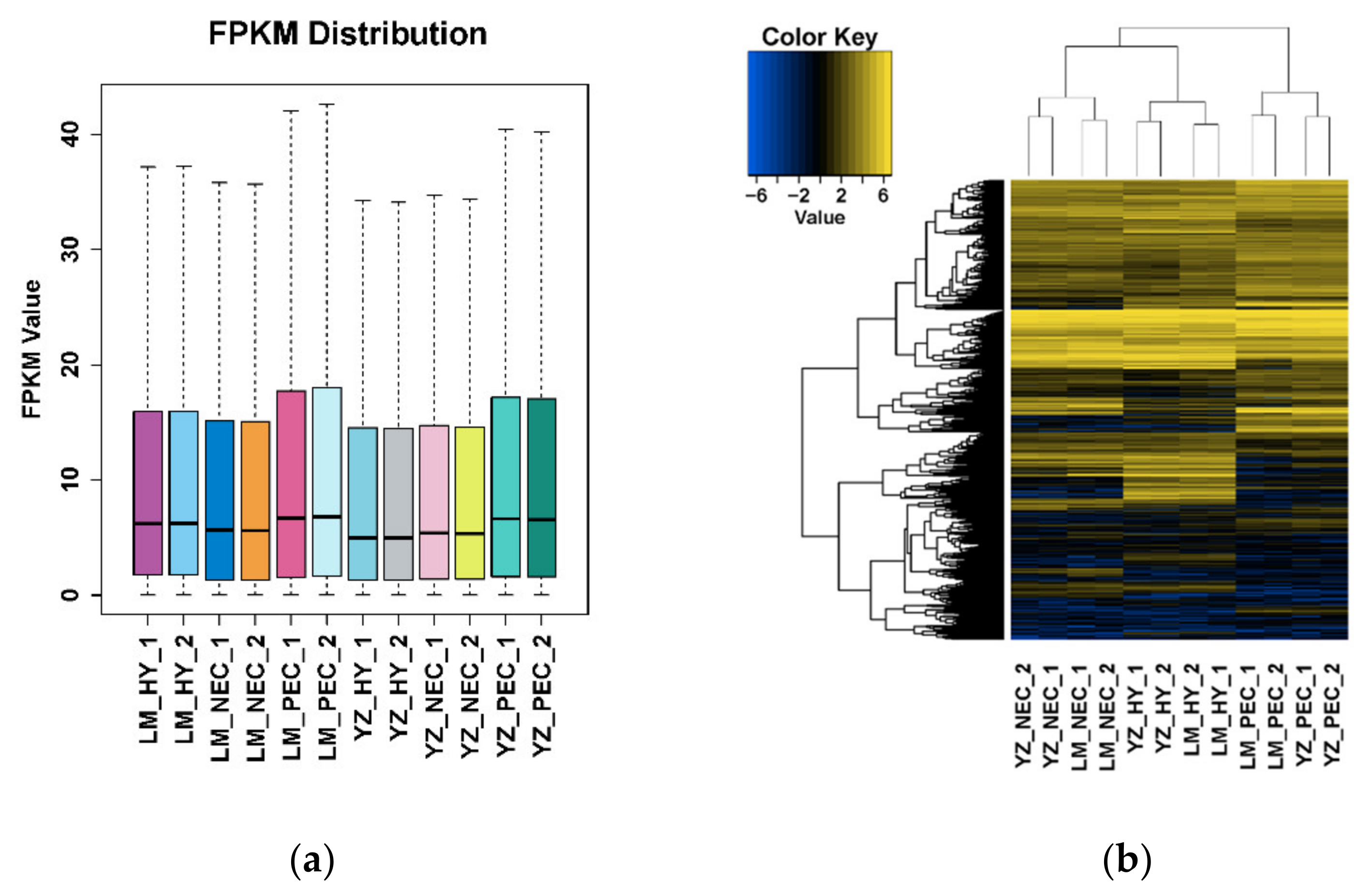
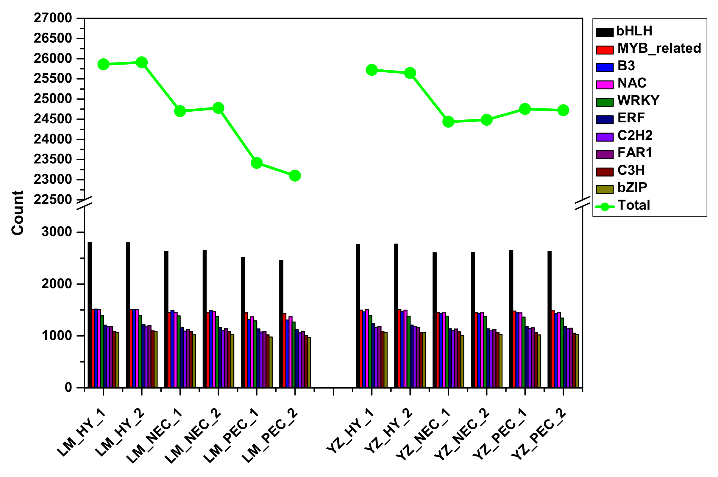
3.1. Landscape of RNA Transcriptome During Cotton SE in Two Genotypes

To comprehensively analyze the gene regulatory patterns involved in SE process, we performed a genome-wide analysis of transcripts using the high throughput RNA-seq technology in two cotton genotypes with distinct embryogenic abilities, Yuzao 1 (YZ) with a high embryogenic ability (embryogenic transdifferentiation rate >80%,) and Lumian 1 (LM) with a very low ability (embryogenic transdifferentiation rate <10%). Three typical developmental staged cultures of early SE—hypocotyls (HY), nonembryogenic calli (NEC) and primary embryogenic calli (PEC)—were selected to establish the transcriptional profiles.
The output results showed that more than 6 Gb RNA-seq clean data were filtered individually, with a high mapping rate (>94%) (Table S1). The quantitative expression levels for total 62,562 transcripts were calculated as FPKM (fragments per kilobase per million mapped fragments) based on the number of uniquely mapped reads that overlapped with exon regions [45] (Table S2). The FPKM distribution stability provided an important guarantee of the reliability of our transcriptome data (Figure 1a). Meanwhile, transcripts in the two genotypes were combined to perform a hierarchical clustering. We found that callus dedifferentiation and embryogenic transdifferentiation periods were clustered separately (Figure 1b), indicating that there were different gene expression patterns during the two developmental processes.

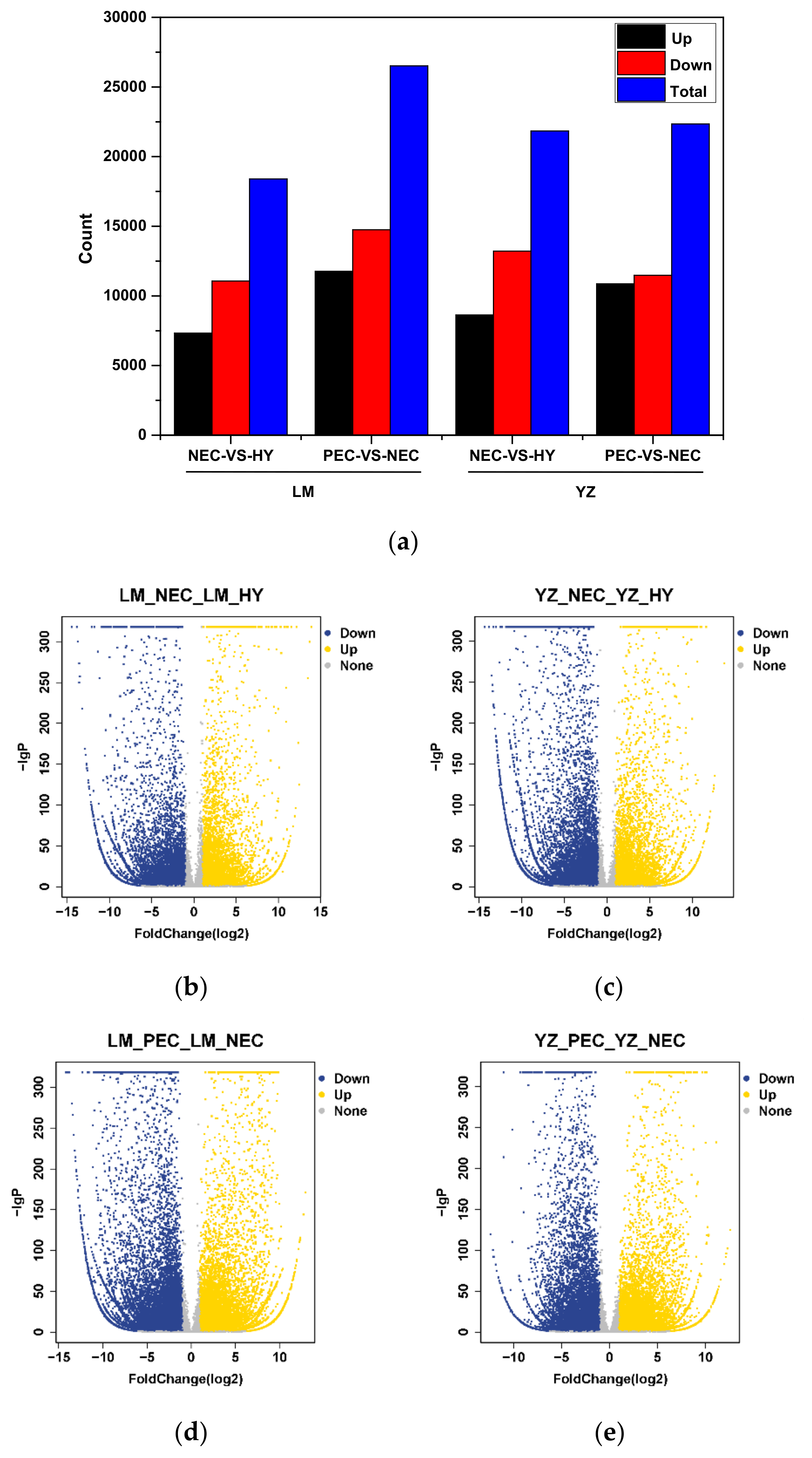
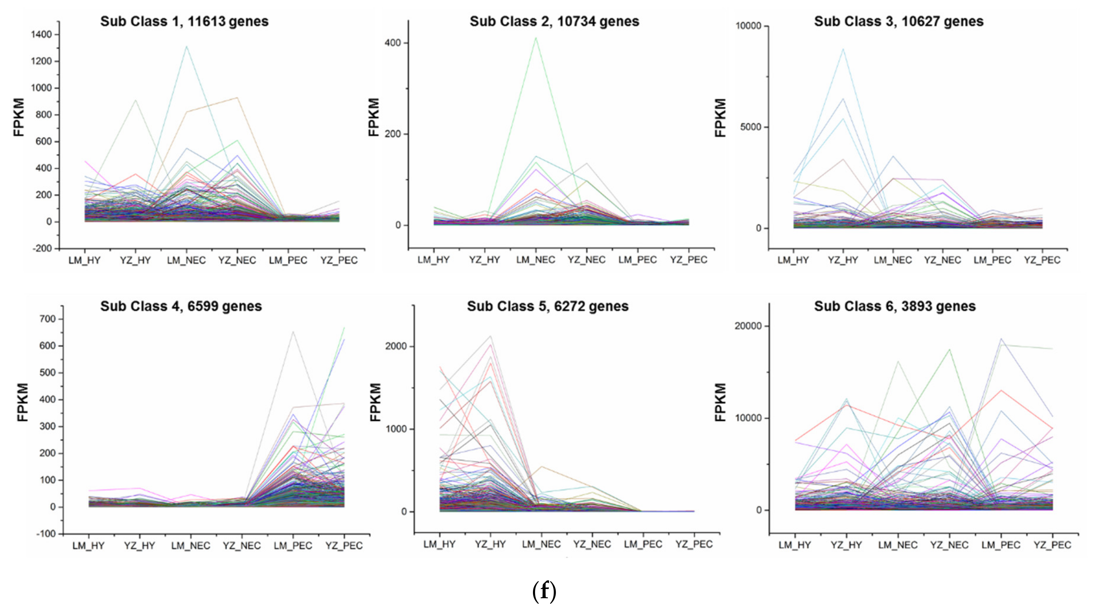
3.2. Differentially Expressed Gene Regulatory Patterns During Cotton SE

The false discovery rate (FDR) was used to determine the threshold of p-values in multiple tests, which corresponded to the differential gene expression test [46,47]. In this study, q < 0.05 and the absolute value of |Log2Ratio| ≥ 1 was used as a threshold to judge the significant differences in gene expression. A total of 18,394, 26,514, 21,842, and 22,343 differentially expressed genes (DEGs) were identified in LM_NEC-VS-LM_HY, LM_PEC-VS-LM_NEC, YZ_NEC-VS-YZ_HY and YZ_PEC-VS-YZ_NEC, respectively (Figure 2a; Table S3). The results showed that compared to LM, more DEGs were expressed during callus dedifferentiation (Figure 2b,c), whereas fewer DEGs were expressed during the embryogenic transdifferentiation process in YZ (Figure 2d,e).
In addition, by k-means clustering, the differential genes were divided into several similar expression patterns. Results suggested that DEGs were clustered into six groups during the cotton SE process in two genotypes (Figure 2f; Table S4). Of these groups, the genes in class 4 were more highly expressed in the PEC stage compared to HY and NEC and positively correlated with the embryogenic transdifferentiation process. In contrast, genes were severely repressed in PEC stage in class 5 and were active to callus dedifferentiation during early SE.

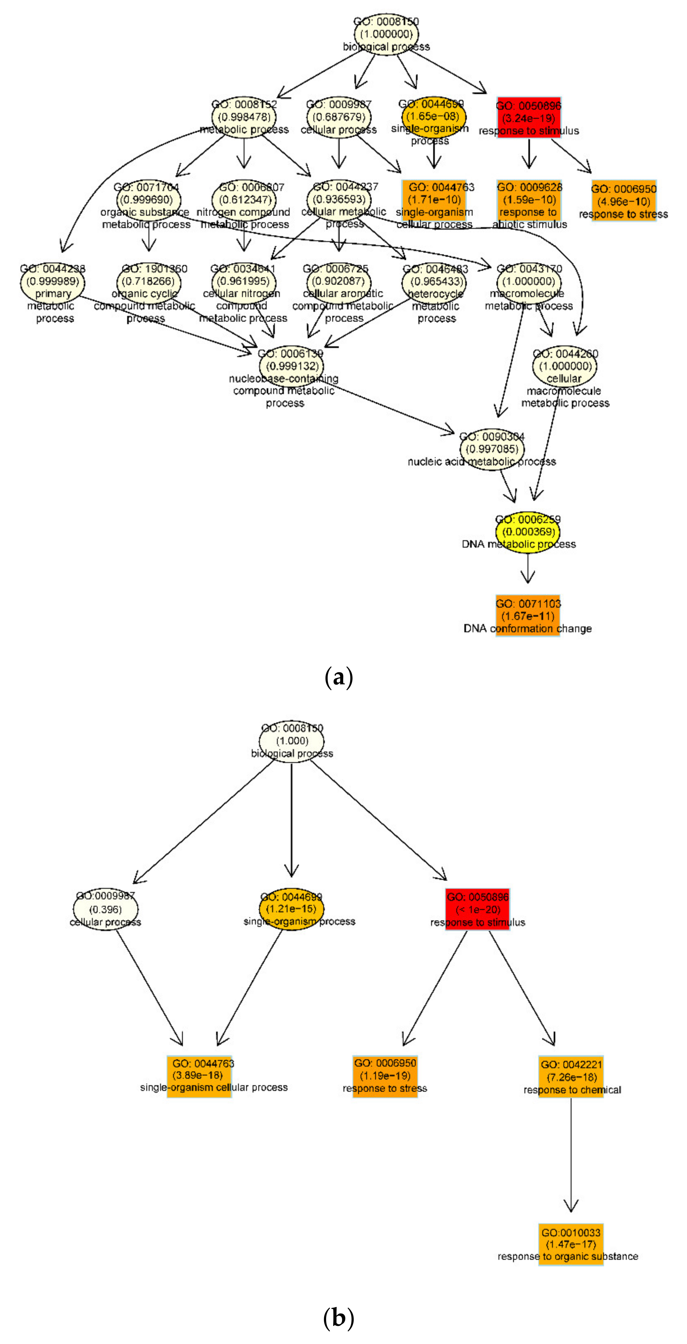
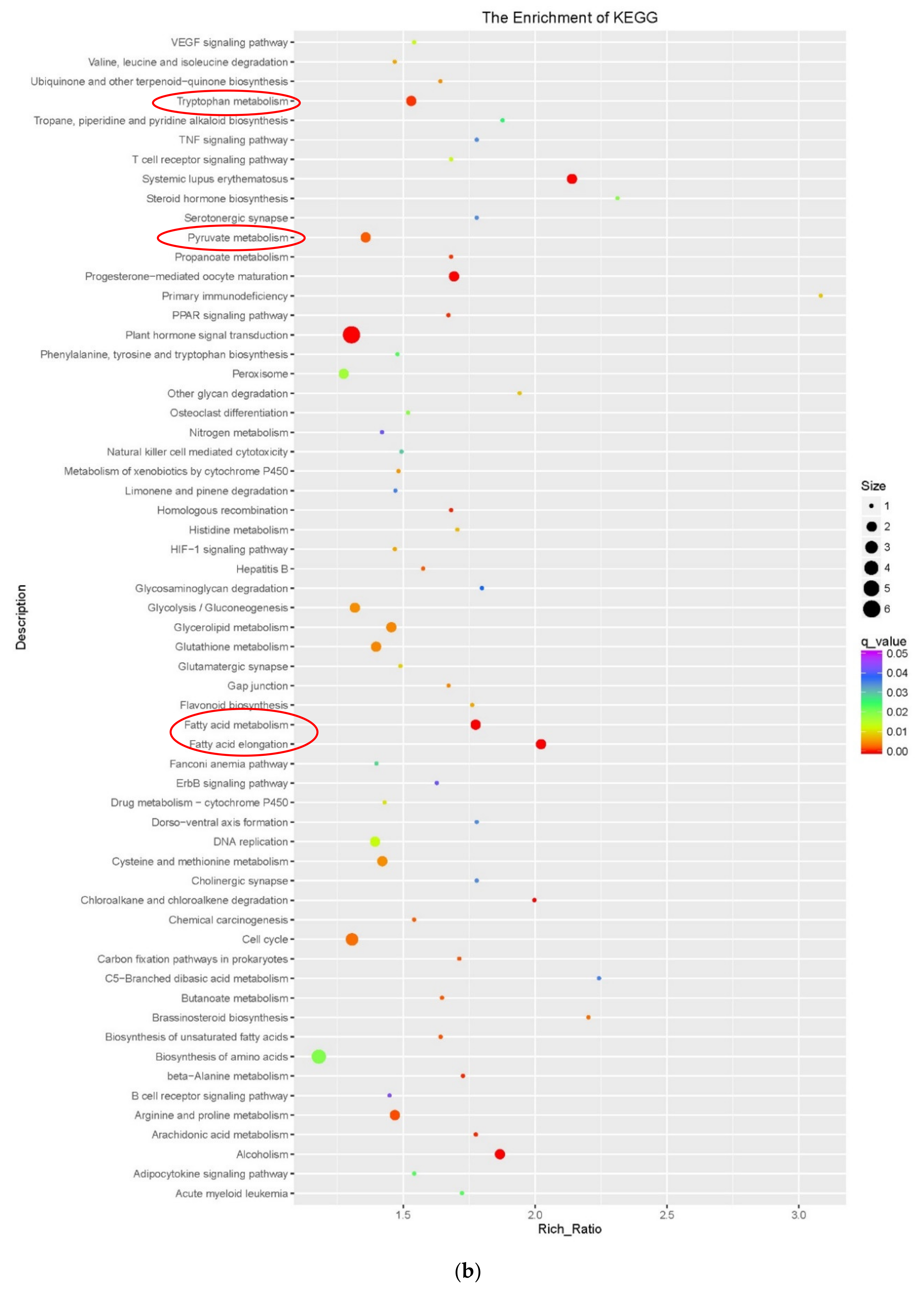
3.3. Functional Enrichment Analysis of DEGs During Embryogenic Transdifferentiation in Two Genotypes

3.3.1. DNA Conformation Change Involved in the Recalcitrant Genotype

To investigate the functioning of differentially expressed genes, Gene Ontology (GO) analysis investigated DEG enrichment of biological processes during embryogenic transdifferentiation in the two genotypes [48] (Figure 3; Table S5). Strong enrichment in different biological processes were observed (p < 0.05). In LM, the recalcitrant genotype, DEGs were highly clustered in ‘response to stimulus’, ‘single -organism cellular process’, ‘response to abiotic stimulus’, ‘response to stress’ and ‘DNA conformation change’ processes (Figure 3a). In YZ, the highly embryogenic genotype, ‘response to stimulus’, ‘single-organism cellular process’, ‘response to stress’, ‘response to chemical’ and ‘response to organic substance’ processes were significantly enriched (Figure 3b). Regarding the most significant difference in the two genotypes, a high cluster pointed to ‘DNA conformation change’ was significantly observed in the recalcitrant genotype. The results were consistent with the report that transcription variations were more modulated by DNA methylation in the recalcitrant genotype [49].
We identified the DEGs enriched in DNA conformation change process during embryogenic differentiation in the two genotypes (Table 1). Of these, several major factors that are associated with DNA-binding, DNA helicase, sister chromatid cohesion, nucleosome assembly, structural maintenance of chromosomes, transcription initiation, cold shock, signal activation, cell division, histone, DNA gyrase, structural maintenance of chromosomes, DNA annealing helicase and endonuclease, DNA replication licensing factor, DNA replication ATP-dependent helicase, increased DNA methylation, cis-trans isomerase and E3 ubiquitin-protein ligase, were significantly observed. Of these, GYRB (DNA gyrase subunit B, chloroplastic/mitochondrial), ZRANB3 (DNA annealing helicase and endonuclease), MCM3 (DNA replication licensing factor), HMGIY2 (HMG-Y-related protein A), and FKBP53 (Peptidyl-prolyl cis-trans isomerase) were specifically activated. IGHMBP2 (DNA-binding protein SMUBP-2), SRS2 (ATP-dependent DNA helicase SRS2-like protein), SYN1 (sister chromatid cohesion 1 protein 1), NAP1 (nucleosome assembly protein 1), TFG2 (transcription initiation factor IIF subunit beta), CSP1 (cold shock protein 1), PHRF1 (PHD and RING finger domain-containing protein 1), ASCC3 (activating signal cointegrator 1 complex subunit 3), and CDC45 (cell division control protein 45 homolog) were specifically downregulated during embryogenic differentiation process in LM, the recalcitrant genotype (Table 1).

3.3.2. Fatty Acid, Tryptophan and Pyruvate Metabolism Involved in the Highly Embryogenic Genotype

To further understand the function of SE-related genes, we analyzed the differences and dynamic changes of gene clustering by KEGG (Kyoto Encyclopedia of Genes and Genomes) enrichment analysis [50] (Figure A1; Table S6). Results showed that the terms ‘plant hormone signal transduction’, ‘biosynthesis of amino acids’, and ‘alcoholism’ pathways were significantly enriched in LM, the recalcitrant genotype (Figure A1a). While in YZ, the highly embryogenic genotype, DEGs were highly accumulated in ‘plant hormone signal transduction’, ‘tryptophan metabolism’, ‘pyruvate metabolism’, ‘fatty acid metabolism’, ‘fatty acid elongation’, ‘arginine and proline metabolism’ and ‘alcoholism’ pathways (Figure A1b). It is worth noting that fatty acid metabolism were significantly observed in the highly embryogenic genotype. The results in the highly embryogenic genotype were consistent with the report that a lipid transfer protein (SELTP) drives the totipotency of somatic cells in cotton [43].
The representative DEGs significantly enriched in fatty acid metabolism and fatty acid elongation processes during embryogenic differentiation in the two genotypes were listed in Table 2. Several major factors that are associated with enoyl-CoA hydratase, palmitoyl-protein thioesterase, 3-ketoacyl-CoA synthase, very-long-chain 3-oxoacyl-CoA reductase, very-long-chain (3R)-3-hydroxyacyl-CoA dehydratase, very-long-chain enoyl-CoA reductase, long chain acyl-CoA synthetase, acyl-coenzyme A oxidase, aldehyde dehydrogenase and alcohol dehydrogenase were significantly observed. Of these, PPT3 (palmitoyl-protein thioesterase 3), ECR (very-long-chain enoyl-CoA reductase) were specifically activated, and ADH6 (alcohol dehydrogenase 6) was specifically downregulated during embryogenic differentiation process in YZ, the highly embryogenic genotype (Table 2).
We also identified the DEGs significantly involved in tryptophan metabolism and pyruvate metabolism processes during embryogenic differentiation in the two genotypes (Table 2). The representative factors, including L-tryptophan-pyruvate aminotransferase, indole-3-pyruvate monooxygenase, indole-3-acetaldehyde oxidase, cytochrome P450, UDP-glycosyltransferase, aldehyde dehydrogenase, phosphoenolpyruvate carboxylase, phosphoenolpyruvate carboxykinase (ATP), pyruvate kinase, dihydrolipoamide dehydrogenase, dihydrolipoamide acetyltransferase, acetyl-CoA synthetase and acetyl-CoA acetyltransferase were investigated. Of these, UGT (UDP-glycosyltransferase), DLAT (dihydrolipoamide acetyltransferase) were specifically activated, and CYP (Cytochrome P450), AAT1 (Acetyl-CoA acetyltransferase) were specifically downregulated during embryogenic differentiation process in YZ, the highly embryogenic genotype (Table 2).

3.4. Stress-Related Transcription Factors Were Specifically Activated and Preferentially Expressed During Embryogenic Transdifferentiation in the Highly Embryogenic Genotype

Transcription factors (TFs) are a group of protein molecules that can specifically bind to a specific sequence of a gene, which can ensure that the target gene is expressed in a specific time and space. TFs during early SE were explored in the two genotypes. The results showed that bHLH, followed by MYB-related, B3, NAC and WRKY, was identified and highly expressed among the samples (Figure 4; Table S7). Moreover, during the embryogenic transdifferentiation process, the number of TFs in YZ was higher than that in LM.
Previous studies confirmed that these transcription factors were differentially expressed under stress conditions and acted as a link between stress and the developmental pathway [51]. bHLH participates in the process of adaptation to various stresses. MYB-related is involved in hormone and environmental responses. B3 plays an important role in responsing to various stresses. NAC is involved in defensive responses to biotic and abiotic stresses. WRKY participates in responsing to biotic and abiotic stresses and hormone signaling.
In the process of cotton somatic embryogenesis, a large number of stress-related transcription factors were specifically activated and preferentially expressed during embryogenic transdifferentiation in the highly embryogenic genotype. These critical SE-associated transcription factors might play a significant role in regulating the stress response during embryogenic transdifferentiation.

3.5. Active Alternative Splicing Significantly Involved in Embryogenic Transdifferentiation in the Highly Embryogenic Genotype

Various RNA splicing isomers can be produced by different splicing modes or different splicing sites. This process is alternative splicing (AS). AS exists widely in eukaryotes and is an important mechanism for regulating gene expression and protein diversity [52]. There are five types of AS events: alternative exon end (AE), intron retention (IR), skipped exon (SKIP), transcript start site (TSS) and transcript end site (TTS). The AS events during early SE were classified and quantitatively counted in the two genotypes (Figure 5). The results showed that TSS and TTS accounted for the highest proportion followed by IR and AE. During the embryogenic transdifferentiation process, compared with LM, the AS events continued to rise in YZ. Results suggested that the active AS events might play a positive role during embryogenic transdifferentiation in the highly embryogenic genotype.

4. Discussion

Plant in vitro propagation, such as somatic embryogenesis, has been possible because plant cells have the capacity to regenerate a whole organism from differentiated somatic cells [9,16,53]. Moreover, SE is an important tool for clonal propagation of important economical and agronomical species [54]. The high-frequency regeneration of cotton via SE has been limited due to genotype-dependent response. The molecular basis underlying SE genotype recalcitrance remains largely unexplored in cotton. During dedifferentiation induction of cotton SE, Cao et al. performed a high throughout RNA sequencing to analyze gene expression patterns in two cotton varieties with different SE capabilities [16]. Transcriptomic analysis revealed that protein kinase and oxidoreductase activities were highly enriched between two the cotton varieties. Several stress-related transcription factors and the auxin and ethylene signaling pathways might contribute to differentiation in SE. Moreover, the SERK gene showed different expression patterns between the two varieties [16]. However, the dynamic transcriptional regulation characteristics during embryogenic transdifferentiation have not been investigated so far. Compared with this report, in our current study, two cotton genotypes with distinct embryogenic abilities (YZ and LM), at three typical developmental staged cultures of early SE—hypocotyls, nonembryogenic calli and primary embryogenic calli—were selected to establish the transcriptional profiles. We found that DEGs identified in callus dedifferentiation and embryogenic transdifferentiation were clustered into six expression patterns during cotton SE process in the two genotypes. Moreover, functional enrichment analysis revealed that DEGs were significantly enriched in fatty acid, tryptophan and pyruvate metabolism in the highly embryogenic genotype and in DNA conformation change otherwise in the recalcitrant genotype. In addition, critical SE-associated expressed transcription factors, as well as alternative splicing events, were notably and preferentially activated during embryogenic transdifferentiation in the highly embryogenic genotype compared with the recalcitrant genotype.
The realization of plant high-frequency regeneration via SE depends largely on whether somatic cells can re-differentiate into embryogenic state. Our present work focused on the dynamic transcriptional regulation characteristics during embryogenic transdifferentiation, which is the key SE developmental process, with the expectation to provide a better understanding of the SE genotype recalcitrance during cell totipotency at molecular levels.

4.1. Effects of DNA Conformation Change During Embryogenic Transdifferentiation

In our current study, to investigate the function of differentially expressed genes, we performed GO enrichment of biological processes during embryogenic transdifferentiation in two genotypes (Figure 3; Table S5). Results suggested that a strong cluster related to ‘DNA conformation change’ was significantly observed in LM, the recalcitrant genotype (Figure 3a).
The effects of DNA conformation change in SE process have been demonstrated previously. In Scots pine (Pinus sylvestris L.) SE cultures, the cellular polyamine contents, which may provide protection by inducing conformational changes in DNA were analyzed throughout SE induction. Results revealed that manipulation of stress response pathways, such as changes in polyamine metabolism may provide a way to enhance somatic embryo production in recalcitrant lines [55,56]. In addition, molecular differences between cell lines previously characterized in terms of their embryogenic potential as embryogenic (E) and non-embryogenic (NE) were investigated during Pinus radiata SE process. Furthermore, the detection of vibrational markers of DNA conformation indicated that DNA samples obtained from E and NE lines presented different conformation, with the common B-DNA conformation in E samples, while Z-conformation in NE samples [57].
Moreover, Corredoira et al. reported that among the molecular mechanisms involved in regulation of SE, DNA methylation is an epigenetic modification of the chromatin that leads to a transcriptionally inactive conformation and gene silencing [58]. Cell reprogramming, totipotency and somatic embryogenesis initiation involve changes in the developmental genetic program of the cell, which affects global genome organization; in this sense, epigenetic modifications constitute key factors of genome flexibility and may be involved in these genome organization changes [58,59]. In this study, the IDM1 (Increased DNA methylation 1) gene, which was enriched in DNA conformation change process, was significantly upregulated during embryogenic transdifferentiation in LM, the recalcitrant genotype (Table 1). The findings were consistent with our previous report that global DNA hypomethylation was observed during embryogenic redifferentiation in YZ [49]. To differentiate nonembryogenic and embryogenic cells, the mechanism of DNA methylation could be utilized as a candidate marker. DNA hypomethylation might play a positive role in regulating cotton somatic embryogenesis. In Pinus nigra, low methylation levels were observed in embryogenic cell lines [60,61]. The Arabidopsis SE was also encouraged by global DNA hypomethylation during the induction process [23]. The role of DNA methylation and the embryogenic capacity in Chestnut (Castanea sativa M.) have been evaluated. It was found that embryogenic competence could be established under in vitro conditions with DNA demethylation [62]. The loss in DNA methylation level was observed during Coffea canephora embryogenesis. The decrease in DNA methylation levels was related to the onset of dedifferentiation and cellular proliferation [63]. The low level of DNA methylation promotes gene expression associated with cell dedifferentiation. The reduction in the DNA methylation level permitted the triggering of cell dedifferentiation by expressing related genes. Because the lowest level of DNA methylation was always found in the embryogenic cells, it was possible that DNA hypomethylation was involved in the signals that lead to the induction of SE. The decrease in DNA methylation levels could be an important step during vegetative-embryogenic transition, in which somatic cells acquired the competency to form embryonic cells. The above results reveal the significant role of DNA conformation change in embryogenic cultures proliferation.

4.2. Fatty Acid Metabolism Involved in Embryogenic Transdifferentiation

As protective chemicals in reproduction and seed preservation, fatty acids are the major structural components of membrane phospholipids and triacylglycerol storage oils, which play a significant role in the evolution and developmental processes in plants [15]. In the current study, the differences and dynamic changes of gene clustering by KEGG enrichment were analyzed to understand the function of SE-related genes in two cotton genotypes (Figure A1; Table S6). Results show that the term fatty acid metabolism was significantly observed in the highly embryogenic genotype (Figure A1b).
The significant role of fatty acid metabolism in embryogenic transdifferentiation process has been investigated. During cotton SE, genes related to fatty acid biosynthesis were upregulated in embryogenic callus compared to non-embryogenic callus. The results indicated that fatty acids accumulated during embryogenic callus development [15]. The proteomic analysis indicated that fatty acid biosynthesis and metabolism was one of the top pathways involved in cotton somatic embryogenesis, confirming the transcriptomic results on the important role of the fatty acid metabolism in cotton SE [19]. In addition, many fatty acid biosynthesis and metabolism related proteins, such as ‘cytochrome P450 86B1-like’ and ‘cytochrome P450 86A8-like’, were differentially accumulated during cotton SE. In the fatty acid biosynthesis pathway, these proteins were up regulated in PEC, implying the important contribution to the SE initiation in cotton [6]. The results above were consistent with our previous report that a lipid transfer protein (SELTP) drives the totipotency of somatic cells in cotton [43].
In tobacco cell cultures, the ectopic expression of LEAFY COTYLEDON (LEC) genes induced accumulation of fatty acid biosynthetic enzymes [64]. Most of genes acting in fatty acid and lipid biosynthesis were globally upregulated in LEC overexpression lines [65,66]. In the tree fern Cyathea delgadii, a comparative proteomic analysis was performed in stipe explants undergoing direct SE. The differentially regulated proteins during early SE were assigned to seven functional categories, including fatty acid metabolism [67]. The acetyl-CoA carboxylase (ACC) activity was crucial to SE process, which is a key enzyme in oil biosynthesis and catalyzes the first step in fatty acid biosynthesis [68]. A proteomic analysis also reported that energy production is increased during Quercus suber somatic embryogenesis probably to participate in the synthesis of primary metabolites such as fatty acids [69]. The amount of fatty acid may be used as a marker of embryogenic potential when promising cell lines of Siberian larch are screened in the stage of early embryogenesis [70]. The fatty acids, which affect cell function and growth patterns, appear to be a part of the thidiazuron action pattern and may play an important role in inducing regeneration [6]. In this study, some special genes were identified to be involved in fatty acid metabolism. Of these, KCR1 (Very-long-chain 3-oxoacyl-CoA reductase 1) and PAS2 (Very-long-chain-3-hydroxyacyl-CoA dehydratase 2) genes were significantly upregulated during embryogenic transdifferentiation in LM, ECR (Very-long-chain enoyl-CoA reductase) gene was significantly upregulated during embryogenic transdifferentiation in YZ (Table 2). Very long chain fatty acids are essential for many aspects of plant development, including cell proliferation. They were speculated to be required for polar auxin transport and tissue patterning during plant development [71]. Additionally, the ADH6 (Alcohol dehydrogenase 6) gene was significantly downregulated during embryogenic transdifferentiation in YZ (Table 2). The expression of the ADH gene increased markedly in barley zygotic embryo development [72]. It was concluded that ADH gene expression in zygotic embryos was regulated by an ABA/gibberellin interaction. These conclusions demonstrated that fatty acid might frequently be associated with embryogenic transdifferentiation during cotton SE.

4.3. Pyruvate and Tryptophan Metabolism Involved in Embryogenic Transdifferentiation

Pyruvate is a derivative of pyruvate acid and plays a role in glycolysis, or sugar metabolism. Furthermore, tryptophan is an important precursor for auxin biosynthesis in plants. The structure of tryptophan is similar to that of IAA, which is common in higher plants. In the current study, KEGG enrichment analyzed the differences and dynamic changes of gene clustering in two cotton genotypes (Figure A1; Table S6). Results indicated that the pyruvate and tryptophan metabolism were also significantly observed in the highly embryogenic genotype (Figure A1b).
The effects of pyruvate and tryptophan metabolism in SE process have been demonstrated. In cotton SE cultures, de novo transcriptome analysis investigated the genes expressed during SE and their expression dynamics using RNAs isolated from nonembryogenic callus and embryogenic callus [15]. Results showed that the differentially expressed genes related to pyruvate metabolites were identified between NEC and PEC [15]. Furthermore, iTRAQ approach was applied to study proteome changes and to identify differentially accumulated proteins between NEC and PEC. Through identification and annotation of differentially accumulated proteins, the key genes/proteins related to pyruvate metabolism involved in cotton SE were uncovered [19].
It has been shown that de novo auxin production via the tryptophan-dependent indole-3-pyruvate (IPA)-YUC auxin biosynthesis pathway was involved in cellular totipotency reprogramming during SE induction [73,74]. In sugarcane callus tissues, levels of different aliphatic compounds belonging to pyruvate metabolism changed significantly in response to different biochemical relationships between callus tissues and their nutrient media. Based on biochemical pathway analysis, it can be speculated that in PEC, alanine, valine, and leucine can act as possible precursors to carbohydrate synthesis through pyruvate metabolism [75]. Furthermore, several metabolites, including tryptophan present in embryogenic calluses [75]. In Maize cell lines, enrichment analysis of differential expressed genes revealed that pyruvate metabolism, biosynthesis of plant hormones, and fatty acid biosynthesis were identified to be three of the most affected pathways, indicating the conserved and important roles of pyruvate metabolism in the somatic embryogenesis process [76]. In the current study, some special genes were identified to be involved in tryptophan metabolism. Of these, TAA1 (L-tryptophan-pyruvate aminotransferase) gene was significantly upregulated during embryogenic transdifferentiation in LM. YUC4 (Probable indole-3-pyruvate monooxygenase) gene was significantly upregulated during embryogenic transdifferentiation in YZ (Table 2). During SE in Coffea canephora, there was an important increase in the expression of YUC and TAA1 genes [77]. The YUC gene, an auxin biosynthesis enzyme, was highly accumulated in EC which leads to increase the level of free IAA [78]. This finding is supported by the evidence that biosynthesis of auxin through YUCs is involved in embryogenesis and induction of somatic embryos. The YUC gene might play an essential role in auxin signaling process to enhance SE. Moreover, PPC (Phosphoenolpyruvate carboxylase) gene, which was enriched in pyruvate metabolism was upregulated during embryogenic transdifferentiation in YZ (Table 2). Phosphoenolpyruvate carboxylase was found in increased abundance in zygotic embryos [79]. PPC gene can yield oxaloacetate that can be converted to aspartate, malate or other intermediates of the TCA cycle. Thus, this metabolic pathway maintains the pool of carbon residues necessary for the biosynthesis of oil and storage proteins that take place in later stage of embryo development. It can be speculated that PPC gene is involved in somatic embryogenesis through the TCA cycle. Results above reveled the significant role of pyruvate and tryptophan metabolism in embryogenic transdifferentiation during SE process.

4.4. Stress-Response Transcription Factors Involved in Embryogenic Transdifferentiation

Stress is an important signal regulator for cell dedifferentiation, which can induce the expression of a large number of transcription factors, cell fate reorganization and acquisition of embryogenic capacity [80,81,82,83]. In response to stress, somatic embryogenesis originated from embryogenic capability acquisition, cell dedifferentiation and changes of gene expression patterns [84,85].
Plant cells with different types specifically responded to stress factors, and different stress treatments had different effects on somatic cells [82]. Developmental variabilities induced by stress explained to some extent that culture conditions can affect somatic embryogenesis in tissue cultures. In this study, several transcription factors (TF), such as bHLH, MYB-related, B3, NAC and WRKY were dynamically changed during different developmental stages of somatic embryogenesis in the two genotypes (Figure 4). Previous studies have confirmed that these transcription factors played an important role in responding to various stresses. They were differentially expressed under stress conditions and acted as a link between stress and the developmental pathway [50]. During cotton SE, genes related to stress response were identified and specifically involved in cotton somatic embryogenesis [29,30,86]. In addition, TF genes were also investigated during Arabidopsis thaliana SE induced by in vitro culture. Transcripts related with phytohormones and stress responses were found to be most abundant [87].
In the current study, some specific transcription factors were identified to be involved in embryogenic transdifferentiation, such as Wind, ESR, WUS, LEC genes. As an AP2/ERF transcription factor, Wind1 (Wound induced dedifferentiation 1) was proven to upregulate the expression of ESR1 (Enhancer of shoot regeneration 1) gene that encodes another AP2/ERF transcription factor, promoting Arabidopsis shoot regeneration [88]. Another important gene that marks embryonic cells was the transcription factor gene WUS, which was involved in the promotion and/or maintenance of totipotent embryogenic stem cells. Overexpression of LEC2 led to activation of YUC2 and YUC4 genes, encoding auxin biosynthetic enzymes, suggesting that LEC2-mediated somatic embryogenesis might be achieved by an endogenous upregulation of the auxin biosynthetic pathway [89].
In addition, the interaction between stress and auxin played an important regulatory role in chromatin recombination and embryogenic capacity acquisition. 2,4-D is not only a plant growth regulator, but also a stress inducer. About half of the stress-related transcription factors were involved in Arabidopsis thaliana SE process induced by 2,4-D [87]. The auxin and stress response were found to be involved in cotton somatic embryogenesis via transcriptome sequencing during callus dedifferentiation and transdifferentiation processes [30]. The stress response was activated and regulated auxin signaling during cotton somatic embryogenesis [86]. Moreover, abiotic stress factors can also affect auxin synthesis, transportation and stability in various ways during morphogenic response [80,83]. However, the mechanism of the interaction between stress and auxin for embryogenic capacity acquisition needs to be further investigated.

Supplementary Materials

Supplementary materials can be found at https://0-www-mdpi-com.brum.beds.ac.uk/2073-4425/11/5/519/s1. Table S1. RNA-Seq output data through filtering and mapping, Table S2. The expression levels (FPKM) of all transcripts during early SE, Table S3. Differentially expressed genes (DEGs) during early SE in the two genotypes, Table S4. K-means clustering of differentially expressed genes (DEGs) during early SE in the two genotypes, Table S5. Gene Ontology (GO) analysis of DEGs during embryonic redifferentiation process in the two genotypes, Table S6. KEGG enrichment analysis of DEGs during embryonic redifferentiation process in the two genotypes, Table S7. Transcription factors (TFs) during early SE in the two genotypes.

Author Contributions

Data curation, H.G. (Haixia Guo), L.Z., Y.F. (Yijie Fan), J.W., Z.T., Y.F. (Yupeng Fan) and F.Z.; Formal analysis, H.G. (Huihui Guo) and L.Z.; Investigation, H.G. (Huihui Guo), H.G. (Haixia Guo) and L.Z.; Methodology, Y.F. (Yijie Fan) and Z.T.; Resources, Y.Z.; Software, J.W.; Supervision, F.Z.; Validation, Y.Z. and Y.F. (Yupeng Fan); Writing—original draft, H.G. (Huihui Guo); Writing—review & editing, H.G. (Haixia Guo) and F.Z. All authors have read and agreed to the published version of the manuscript.

Funding

This work was funded by National Key Research and Development Program (2016YFD0100306), Taishan Scholar Talent Project from PRC (TSQN20161018), China Postdoctoral Science Foundation funded project (2019M662411) and the National Natural Science Foundation of China (31401428).

Conflicts of Interest

The authors declare no conflict of interest.

Appendix A

Figure A1. Functional KEGG enrichment analysis of differentially expressed genes during embryogenic transdifferentiation in the two genotypes. (a) KEGG enrichment of DEGs in LM_PEC compared to LM_NEC; (b) KEGG enrichment of DEGs in YZ_PEC compared to YZ_NEC. The KEGG pathway with a false-discovery rate adjusted p-value < 0.05 are shown. KEGG, Kyoto Encyclopedia of Genes and Genomes.
Genes 11 00519 g0a1aGenes 11 00519 g0a1b

References

  1. Loyola-Vargas, V.M.; De-la-Peña, C.; Galaz-Avalos, R.M.; Quiroz-Figueroa, F.R. Plant tissue culture. In Molecular Biomethods Handbook; Walker, J.M., Rapley, R., Eds.; Humana Press: Totowa, NJ, Canada, 2008; pp. 875–904. [Google Scholar]
  2. Yang, X.; Zhang, X. Regulation of somatic embryogenesis in higher plants. Crit. Rev. Plant Sci. 2010, 29, 36–57. [Google Scholar] [CrossRef]
  3. Miguel, C.; Marum, L. An epigenetic view of plant cells cultured in vitro: Somaclonal variation and beyond. J. Exp. Bot. 2011, 62, 3713–3725. [Google Scholar] [CrossRef] [PubMed] [Green Version]
  4. Jopling, C.; Boue, S.; Belmonte, J. Dedifferentiation, transdifferentiation and reprogramming: Three routes to regeneration. Nat. Rev. Mol. Cell Biol. 2011, 12, 79–89. [Google Scholar] [CrossRef] [PubMed]
  5. Fehér, A. Callus, Dedifferentiation, Totipotency, Somatic Embryogenesis: What These Terms Mean in the Era of Molecular Plant Biology? Front. Plant Sci. 2019, 10, 536–546. [Google Scholar] [CrossRef] [PubMed] [Green Version]
  6. Guo, H.; Guo, H.; Zhang, L.; Fan, Y.; Fan, Y.; Tang, Z.; Zeng, F. Dynamic TMT-based quantitative proteomics analysis of critical initiation process of totipotency during cotton somatic embryogenesis transdifferentiation. Int. J. Mol. Sci. 2019, 20, 1691–1724. [Google Scholar]
  7. Evans, D.A.; Sharp, W.R.; Flick, C.E. Growth and Behavior of Cell Cultures: Embryogenesis and Organogenesis. In Plant Tissue Cultures; Trevor, A.T., Ed.; Academic Press: New York, NY, USA, 1981; pp. 45–113. [Google Scholar]
  8. Nic-Can, G.I.; De-la-Peña, C. Epigenetic advances on somatic embryogenesis of agronomical and important crops. In Epigenetics in Plants of Agronomic Importance: Fundamentals and Applications; Alvarez, V., Ed.; Springer: Berlin, Germany, 2014; pp. 91–109. [Google Scholar]
  9. Ikeuchi, M.; Sugimoto, K.; Iwase, A. Plant callus: Mechanisms of induction and repression. Plant Cell 2013, 25, 3159–3173. [Google Scholar] [CrossRef] [Green Version]
  10. Zeng, F.; Zhang, X.; Cheng, L. A draft gene regulatory network for cellular totipotency reprogramming during plant somatic embryogenesis. Genomics 2007, 90, 620–628. [Google Scholar] [CrossRef] [Green Version]
  11. Sakhanokho, H.; Rajasekaran, K. Cotton regeneration in vitro. In Fiber Plants; Ramawat, K.G., Ahuja, M.R., Eds.; Springer: Berlin, Germany, 2016; pp. 87–110. [Google Scholar]
  12. Wu, J.; Zhang, X.; Nie, Y.; Jin, S.; Liang, S. Factors affecting somatic embryogenesis and plant regeneration from a range of recalcitrant genotypes of Chinese cottons (Gossypium hirsutum L.). In Vitro Cell. Dev. Plant 2004, 40, 371–375. [Google Scholar] [CrossRef]
  13. Zeng, F.; Zhang, X.; Zhu, L.; Tu, L.; Guo, X.; Nie, Y. Isolation and characterization of genes associated to cotton somatic embryogenesis by suppression subtractive hybridization and macroarray. Plant Mol. Biol. 2006, 60, 167–183. [Google Scholar] [CrossRef]
  14. Ge, X.; Zhang, C.; Wang, Q.; Yang, Z.; Wang, Y.; Zhang, X.; Wu, Z.; Hou, Y.; Wu, J.; Li, F. iTRAQ protein profile differential analysis between somatic globular and cotyledonary embryos reveals stress, hormone, and respiration involved in increasing plantlet regeneration of Gossypium hirsutum L. J. Proteome. Res. 2014, 14, 268–278. [Google Scholar] [CrossRef]
  15. Cheng, W.H.; Zhu, H.G.; Tian, W.G.; Zhu, S.H.; Xiong, X.P.; Sun, Y.Q.; Zhu, Q.H.; Sun, J. De novo transcriptome analysis reveals insights into dynamic homeostasis regulation of somatic embryogenesis in upland cotton (G. hirsutum L.). Plant Mol. Biol. 2016, 92, 279–292. [Google Scholar] [CrossRef] [Green Version]
  16. Cao, A.; Zheng, Y.; Yu, Y.; Wang, X.; Shao, D.; Sun, J.; Cui, B. Comparative Transcriptome Analysis of SE initial dedifferentiation in cotton of different SE capability. Sci. Rep. UK 2017, 7, 8583–8595. [Google Scholar] [CrossRef] [PubMed]
  17. Da Cunha Soares, T.; da Silva, C.R.C.; Chagas Carvalho, J.M.F.; Cavalcanti, J.J.V.; de Lima, L.M.; de Albuquerque, M.F.P.; Severino, L.S.; Dos Santos, R.C. Validating a probe from GhSERK1 gene for selection of cotton genotypes with somatic embryogenic capacity. J. Biotechnol. 2018, 270, 44–50. [Google Scholar] [CrossRef] [PubMed]
  18. Xiao, Y.; Chen, Y.; Ding, Y.; Wu, J.; Wang, P.; Yu, Y.; Wei, X.; Wang, Y.; Zhang, C.; Li, F.; et al. Effects of GhWUS from upland cotton (Gossypium hirsutum L.) on somatic embryogenesis and shoot regeneration. Plant Sci. 2018, 270, 157–165. [Google Scholar] [CrossRef] [PubMed]
  19. Zhu, H.G.; Cheng, W.H.; Tian, W.G.; Li, Y.J.; Liu, F.; Xue, F.; Zhu, Q.H.; Sun, Y.Q.; Sun, J. iTRAQ-based comparative proteomic analysis provides insights into somatic embryogenesis in Gossypium hirsutum L. Plant Mol. Biol. 2018, 96, 89–102. [Google Scholar] [CrossRef] [PubMed] [Green Version]
  20. Xu, J.; Yang, X.; Li, B.; Chen, L.; Min, L.; Zhang, X. GhL1L1 affects cell fate specification by regulating GhPIN1-mediated auxin distribution. Plant Biotechnol. J. 2019, 17, 63–74. [Google Scholar] [CrossRef] [Green Version]
  21. Su, Y.; Liu, Y.; Zhou, C.; Li, X.; Zhang, X. The microRNA167 controls somatic embryogenesis in Arabidopsis through regulating its target genes ARF6 and ARF8. Plant Cell Tiss. Org. Cult. 2016, 124, 405–417. [Google Scholar] [CrossRef]
  22. Rupps, A.; Raschke, J.; Rümmler, M.; Linke, B.; Zoglauer, K. Identification of putative homologs of Larix decidua to BABY BOOM (BBM), LEAFY COTYLEDON1 (LEC1), WUSCHEL-related HOMEOBOX2 (WOX2) and SOMATIC EMBRYOGENESIS RECEPTOR-LIKE KINASE (SERK) during somatic embryogenesis. Planta 2016, 243, 473–488. [Google Scholar] [CrossRef]
  23. Elhiti, M.; Tahir, M.; Gulden, R.; Khamiss, K.; Stasolla, C. Modulation of embryo-forming capacity in culture through the expression of Brassica genes involved in the regulation of the shoot apical meristem. J. Exp. Bot. 2010, 61, 4069–4085. [Google Scholar] [CrossRef] [Green Version]
  24. Hu, H.; Xiong, L.; Yang, Y. Rice SERK1 gene positively regulates somatic embryogenesis of cultured cell and host defense response against fungal infection. Planta 2005, 222, 107–117. [Google Scholar] [CrossRef]
  25. Kumar, V.; Van Staden, J. New insights into plant somatic embryogenesis: An epigenetic view. Acta Physiol. Plant 2017, 39, 194–211. [Google Scholar] [CrossRef]
  26. Hu, L.; Yang, X.; Yuan, D.; Zeng, F.; Zhang, X. GhHmgB3 deficiency deregulates proliferation and differentiation of cells during somatic embryogenesis in cotton. Plant Biotechnol. J. 2011, 9, 1038–1048. [Google Scholar] [CrossRef]
  27. Poon, S.; Heath, R.; Clarke, A. A chimeric arabinogalactan protein promotes somatic embryogenesis in cotton cell culture. Plant Physiol. 2012, 160, 684–695. [Google Scholar] [CrossRef] [PubMed] [Green Version]
  28. Min, L.; Hu, Q.; Li, Y.; Xu, J.; Ma, Y.; Zhu, L.; Yang, X.; Zhang, X. Leafy cotyledon1-casein kinase I-TCP15-phytochrome interacting factor 4 network regulates somatic embryogenesis by regulating auxin homeostasis. Plant Physiol. 2015, 169, 2805–2821. [Google Scholar] [PubMed] [Green Version]
  29. Jin, F.; Hu, L.; Yuan, D.; Xu, J.; Gao, W.; He, L.; Yang, X.; Zhang, X. Comparative transcriptome analysis between somatic embryos (SEs) and zygotic embryos in cotton: Evidence for stress response functions in SE development. Plant Biotechnol. J. 2014, 12, 161–173. [Google Scholar] [CrossRef] [PubMed]
  30. Yang, X.; Zhang, X.; Yuan, D.; Jin, F.; Zhang, Y.; Xu, J. Transcript profiling reveals complex auxin signaling pathway and transcription regulation involved in dedifferentiation and redifferentiation during somatic embryogenesis in cotton. BMC Plant Biol. 2012, 12, 110–128. [Google Scholar] [CrossRef] [Green Version]
  31. Xu, Z.; Zhang, C.; Zhang, X.; Liu, C.; Wu, Z.; Yang, Z.; Zhou, K.; Yang, X.; Li, F. Transcriptome profiling reveals auxin and cytokinin regulating somatic embryogenesis in different sister lines of cotton cultivar CCRI24. J. Integr. Plant Biol. 2013, 55, 631–642. [Google Scholar] [CrossRef]
  32. Kennedy, D.; Norman, C. What don’t we know? Science 2005, 309, 75. [Google Scholar] [CrossRef] [Green Version]
  33. Vogel, G. How does a single somatic cell become a whole plant? Science 2005, 309, 86. [Google Scholar] [CrossRef] [Green Version]
  34. Kaeppler, S.M.; Kaeppler, H.F.; Rhee, Y. Epigenetic aspects of somaclonal variation in plants. Plant Mol. Biol. 2000, 43, 179–188. [Google Scholar] [CrossRef]
  35. De-la-Peña, C.; Nic-Can, G.I.; Galaz-Ávalos, R.M.; Avilez-Montalvo, R.; Loyola-Vargas, V.M. The role of chromatin modifications in somatic embryogenesis in plants. Front. Plant Sci. 2015, 6, 635–649. [Google Scholar] [CrossRef] [PubMed] [Green Version]
  36. Fan, Y.; Yu, X.; Guo, H.; Wei, J.; Guo, H.; Zhang, L.; Zeng, F. Dynamic Transcriptome Analysis Reveals Uncharacterized Complex Regulatory Pathway Underlying Dose IBA-Induced Embryogenic Redifferentiation in Cotton. Int. J. Mol. Sci. 2020, 21, 426. [Google Scholar] [CrossRef] [PubMed] [Green Version]
  37. Xie, D.; Jin, S.; Guo, X.; Zhang, X. Somatic Embryogenesis and Plant Regeneration in Cotton Cultivars from Yellow and Yangtze River Planting Areas. Acta Agron. Sin. 2007, 33, 394–400. [Google Scholar]
  38. Feng, R.; Wang, Q.; Zhang, B.; Yu, X. Genotype analysis in cotton tissue culture and plant regeneration. Acta Agric. Boreali Occident. Sin. 1997, 6, 27–30. [Google Scholar]
  39. Zhang, C.; Yu, S.; Fan, S.; Zhang, J.; Li, F. Inheritance of somatic embryogenesis using leaf petioles as explants in upland cotton. Euphytica 2011, 181, 55–63. [Google Scholar] [CrossRef]
  40. Zhang, X.L.; Sun, J.Z.; Liu, J.L. Somatic embryogenesis and plant regeneration in upland cotton. Acta Genet. Sin. 1991, 18, 461–467. [Google Scholar]
  41. Mishra, R.; Wang, H.Y.; Yadav, N.R.; Wilkins, T.A. Development of a highly regenerable elite Acala cotton (Gossypium hirsutum cv. Maxxa): A step towards genotype-independent regeneration. Plant Cell Tiss. Org. 2003, 73, 21–35. [Google Scholar] [CrossRef]
  42. Ganesan, M.; Jayabalan, N. Evaluation of haemoglobin (erythrogen): For improved somatic embryogenesis and plant regeneration in cotton (Gossypium hirsutum L. cv. SVPR 2). Plant Cell Rep. 2004, 23, 181–187. [Google Scholar] [CrossRef]
  43. Guo, H.; Guo, H.; Zhang, L.; Fan, Y.; Fan, Y.; Zeng, F. SELTP assembled battery drives totipotency of somatic plant cell. Plant Biotechnol. J. 2019, 17, 1188–1190. [Google Scholar] [CrossRef] [Green Version]
  44. Guo, H.; Wu, J.; Chen, C.; Wang, H.; Zhao, Y.; Zhang, C.; Jia, Y.; Liu, F.; Ning, T.; Chu, Z.; et al. Identification and characterization of cell cultures with various embryogenic/regenerative potential in cotton based on morphological, cytochemical, and cytogenetical assessment. J. Integr. Agr. 2019, 18, 1–8. [Google Scholar] [CrossRef]
  45. Trapnell, C.; Williams, B.A.; Pertea, G.; Mortazavi, A.; Kwan, G.; van Baren, M.J.; Salzberg, S.L.; Wold, B.J.; Pachter, L. Transcript assembly and quantification by RNA-Seq reveals unannotated transcripts and isoform switching during cell differentiation. Nat. Biotechnol. 2010, 28, 511–515. [Google Scholar] [CrossRef] [PubMed] [Green Version]
  46. Lister, R.; Pelizzola, M.; Dowen, R.H.; Hawkins, R.D.; Hon, G.; Tonti-Filippini, J.; Nery, J.R.; Lee, L.; Ye, Z.; Ngo, Q.M.; et al. Human DNA methylomes at base resolution show widespread epigenomic differences. Nature 2009, 462, 315–322. [Google Scholar] [CrossRef] [PubMed] [Green Version]
  47. Li, X.; Zhu, J.; Hu, F.; Ge, S.; Ye, M.; Xiang, H.; Zhang, G.; Zheng, X.; Zhang, H.; Zhang, S.; et al. Single-base resolution maps of cultivated and wild rice methylomes and regulatory roles of DNA methylation in plant gene expression. BMC Genom. 2012, 13, 300–314. [Google Scholar] [CrossRef] [PubMed] [Green Version]
  48. Ye, J.; Fang, L.; Zheng, H.; Zhang, Y.; Chen, J.; Zhang, Z.; Wang, J. WEGO: A web tool for plotting GO annotations. Nucleic Acids Res. 2006, 34, 293–297. [Google Scholar] [CrossRef]
  49. Guo, H.; Fan, Y.; Guo, H.; Wu, J.; Yu, X.; Wei, J.; Lian, X.; Zhang, L.; Gou, Z.; Fan, Y.; et al. Somatic embryogenesis critical initiation stage-specific mCHH hypomethylation reveals epigenetic basis underlying embryogenic redifferentiation in cotton. Plant Biotechnol. J. 2020. [Google Scholar] [CrossRef] [Green Version]
  50. Kanehisa, M.; Goto, S. KEGG: Kyoto encyclopedia of genes and genomes. Nucleic Acids Res. 2000, 28, 27–30. [Google Scholar] [CrossRef]
  51. Iyerpascuzzi, A.; Jackson, T.; Cui, H.; Petricka1, J.; Busch, W.; Tsukagoshi, H.; Benfey, P. Cell identity regulators link development and stress responses in the Arabidopsis root. Dev. Cell 2011, 21, 770–782. [Google Scholar] [CrossRef] [Green Version]
  52. Florea, L.; Song, L.; Salzberg, S.L. Thousands of exon skipping events differentiate among splicing patterns in sixteen human tissues. F1000Research 2013, 2, 188–211. [Google Scholar] [CrossRef]
  53. Duarte-Aké, F.; De-la-Peña, C. Epigenetic advances in somatic embryogenesis in sequenced genome crops. In Somatic Embryogenesis: Fundamental Aspects and Applications; Loyola-Vargas, V.M., Ochoa-Alejo, N., Eds.; Springer: Berlin, Germany, 2016; pp. 81–102. [Google Scholar]
  54. Loyola-Vargas, V.M.; Ochoa-Alejo, N. An introduction to plant cell culture: The future ahead. In Plant Cell Culture Protocols; Loyola-Vargas, V.M., Ochoa-Alejo, N., Eds.; Humana Press: Totowa, Canada, 2012; pp. 1–8. [Google Scholar]
  55. Feuerstein, B.G.; Pattabiraman, N.; Marton, L.J. Molecular mechanics of the interactions of spermine with DNA: DNA bending as a result of ligand binding. Nucleic Acids Res. 1990, 18, 1271–1282. [Google Scholar] [CrossRef]
  56. Salo, H.M.; Sarjala, T.; Jokela, A.; Häggman, H.; Vuosku, J. Moderate stress responses and specific changes in polyamine metabolism characterize Scots pine somatic embryogenesis. Tree Physiol. 2016, 36, 392–402. [Google Scholar] [CrossRef] [Green Version]
  57. Bravo, S.; Bertín, A.; Turner, A.; Sepúlveda, F.; Jopia, P.; Parra, M.J.; Castillo, R.; Hasbún, R. Differences in DNA methylation, DNA structure and embryogenesis-related gene expression between embryogenic and non embryogenic lines of Pinus radiata D. don. Plant Cell Tiss. Org. 2017, 130, 521–529. [Google Scholar] [CrossRef]
  58. Corredoira, E.; Cano, V.; Bárány, I.; Solís, M.T.; Rodríguez, H.; Viéitez, M.; Ana, M.; Risueño, M.C.; Testillano, P.S. Initiation of leaf somatic embryogenesis involves high pectin esterification, auxin accumulation and DNA demethylation in Quercus alba. J. Plant Physiol. 2017, 213, 42–54. [Google Scholar] [CrossRef] [PubMed] [Green Version]
  59. Arnholdt-Schmitt, B. Stress-induced cell reprogramming. A role for global genome regulation? Plant Physiol. 2004, 136, 2579–2586. [Google Scholar] [CrossRef] [PubMed] [Green Version]
  60. Chakrabarty, D.; Yu, K.; Paek, K. Detection of DNA methylation changes during somatic embryogenesis of Siberian ginseng (Eleuterococcus senticosus). Plant Sci. 2003, 165, 61–68. [Google Scholar] [CrossRef]
  61. Noceda, C.; Salaj, T.; Pérez, M.; Viejo, M.; Cañal, J.; Salaj, J.; Rodriguez, R. DNA demethylation and decrease on free polyamines is associated with the embryogenic capacity of Pinus nigra Arn. cell culture. Trees 2009, 23, 1285–1293. [Google Scholar] [CrossRef]
  62. Viejo, M.; Rodríguez, R.; Valledor, L.; Pérez, M.; Cañal, M.; Hasbún, R. DNA methylation during sexual embryogenesis and implications on the induction of somatic embryogenesis in Castanea sativa Miller. Sex. Plant Reprod. 2010, 23, 315–323. [Google Scholar] [CrossRef]
  63. Nic-Can, G.; Lopez-Torres, A.; Barredo-Pool, F.; Wrobel, K.; Loyola-Vargas, V.; Rojas-Herrera, R.; De-la-Peña, C. New insights into somatic embryogenesis: LEAFY COTYLEDON1, BABY BOOM1 and WUSCHEL-RELATED HOMEOBOX4 are epigenetically regulated in Coffea canephora. PLoS ONE 2013, 8, e72160. [Google Scholar] [CrossRef] [Green Version]
  64. Guo, F.; Liu, C.; Xia, H.; Bi, Y.; Zhao, C.; Zhao, S.; Hou, L.; Li, F.; Wang, X. Induced expression of AtLEC1 and AtLEC2 differentially promotes somatic embryogenesis in transgenic tobacco plants. PLoS ONE 2013, 8, e71714. [Google Scholar] [CrossRef] [Green Version]
  65. Mu, J.; Tan, H.; Zheng, Q.; Fu, F.; Liang, Y.; Zhang, J.; Yang, X.; Wang, T.; Chong, K.; Wang, X.J.; et al. LEAFY COTYLEDON1 is a key regulator of fatty acid biosynthesis in Arabidopsis. Plant Physiol. 2008, 148, 1042–1054. [Google Scholar] [CrossRef] [Green Version]
  66. Mendoza, M.S.; Dubreucq, B.; Miquel, M.; Caboche, M.; Lepiniec, L. LEAFY COTYLEDON 2 activation is sufficient to trigger the accumulation of oil and seed specific mRNAs in Arabidopsis leaves. FEBS Lett. 2005, 579, 4666–4670. [Google Scholar] [CrossRef] [Green Version]
  67. Domżalska, L.; Kędracka-Krok, S.; Jankowska, U.; Grzyb, M.; Sobczak, M.; Rybczyński, R.J.J.; Mikuła, A. Proteomic analysis of stipe explants reveals differentially expressed proteins involved in early direct somatic embryogenesis of the tree fern Cyathea delgadii Sternb. Plant Sci. 2017, 258, 61–76. [Google Scholar] [CrossRef] [PubMed]
  68. O’Hara, P.; Slabas, A.R.; Fawcett, T. Fatty acid and lipid biosynthetic genes are expressed at constant molar ratios but different absolute levels during embryogenesis. Plant Physiol. 2002, 129, 310–320. [Google Scholar] [CrossRef] [PubMed] [Green Version]
  69. Gomez-Garay, A.; Lopez, J.A.; Camafeita, E.; Bueno, M.A.; Pintos, B. Proteomic perspective of Quercus suber somatic embryogenesis. J. Proteom. 2013, 93, 314–325. [Google Scholar] [CrossRef] [PubMed]
  70. Makarenko, S.P.; Shmakov, V.N.; Dudareva, L.V.; Stolbikova, A.V.; Semenova, N.V.; Tret’yakova, I.N.; Konstantinov, Y.M. Fatty acid composition of total lipids in embryogenic and nonembryogenic callus lines of larch. Russ. J. Plant Physiol. 2016, 63, 252–258. [Google Scholar] [CrossRef]
  71. Molino, D.; Markham, J.; Bellec, Y.; Gissot, L.; Palauqui, J.; Moreau, P.; Napier, J.; Faure, J. Very long chain fatty acids are required for cell polarity and organogenesis during plant development. Chem. Phys. Lipids 2010, 163, S17. [Google Scholar] [CrossRef]
  72. MacNicol, P.; Jacobsen, J. Regulation of alcohol dehydrogenase gene expression in barley aleurone by giberellin and abscisic acid. Physiol. Plant. 2001, 111, 533–539. [Google Scholar] [CrossRef]
  73. Altamura, M.M.; Della Rovere, F.; Fattorini, L.; D’Angeli, S.; Falasca, G. Recent advances on genetic and physiological bases of in vitro somatic embryo formation. In Vitro Embryogenesis in Higher Plants; Humana Press: New York, NY, USA, 2016; pp. 47–85. [Google Scholar]
  74. Wójcikowska, B.; Jaskóla, K.; Gąsiorek, P.; Meus, M.; Nowak, K.; Gaj, M.D. LEAFY COTYLEDON2 (LEC2) promotes embryogenic induction in somatic tissues of Arabidopsis, via YUCCA -mediated auxin biosynthesis. Planta 2013, 238, 425–440. [Google Scholar] [CrossRef] [Green Version]
  75. Mahmud, I.; Thapaliya, M.; Boroujerdi, A.; Chowdhury, K. NMR-based metabolomics study of the biochemical relationship between sugarcane callus tissues and their respective nutrient culture media. Anal. Bioanal. Chem. 2014, 406, 5997–6005. [Google Scholar] [CrossRef]
  76. Shen, Y.; Jiang, Z.; Yao, X.; Zhang, Z.; Lin, H.; Zhao, M.; Liu, H.; Peng, H.; Li, S.; Pan, G. Genome expression profile analysis of the immature maize embryo during dedifferentiation. PLoS ONE 2012, 7, e32237. [Google Scholar] [CrossRef] [Green Version]
  77. Ayil-Gutiérrez, B.; Galaz-Ávalos, R.; Peña-Cabrera, E.; Loyola-Vargas, V. Dynamics of the concentration of IAA and some of its conjugates during the induction of somatic embryogenesis in Coffea canephora. Plant Signal. Behav. 2013, 8, e26998. [Google Scholar] [CrossRef] [Green Version]
  78. Kumaravel, M.; Uma, S.; Backiyarani, S.; Saraswathi, M.; Vaganan, M.; Muthusamy, M.; Sajith, K. Differential proteome analysis during early somatic embryogenesis in Musa spp. AAA cv. Grand Naine. Plant Cell Rep. 2016, 36, 1–16. [Google Scholar] [CrossRef] [PubMed]
  79. Noaha, A.; Niemenaka, N.; Sunderhausb, S.; Haaseb, C.; Omokoloa, D.; Winkelmannc, T.; Braunb, H. Comparative proteomic analysis of early somatic and zygotic embryogenesis in Theobroma cacao L. J. Proteom. 2013, 78, 123–133. [Google Scholar] [CrossRef] [PubMed]
  80. Tognetti, V.; Mühlenbock, P.; Van Breusegem, B. Stress homeostasis-the redox and auxin perspective. Plant Cell Environ. 2012, 35, 321–333. [Google Scholar] [CrossRef] [PubMed]
  81. Grafi, G.; Barak, S. Stress induces cell dedifferentiation in plants. Biochim. Biophys. Acta 2015, 1849, 378–384. [Google Scholar] [CrossRef] [PubMed]
  82. Fehér, A. Somatic embryogenesis-stress induced remodeling of plant cell fate. Biochim. Biophys. Acta 2015, 1849, 385–402. [Google Scholar] [CrossRef] [PubMed]
  83. Guisez, Y. Stress-induced morphogenic responses: Growing out of trouble? Trends Plant Sci. 2007, 12, 98–105. [Google Scholar]
  84. Fehér, A.; Pasternak, T.; Dudits, D. Transition of somatic plant cells to an embryogenic state. Plant Cell Tiss. Org. 2003, 74, 201–228. [Google Scholar] [CrossRef]
  85. Namasivayam, P. Acquisition of embryogenic competence during somatic embryogenesis. Plant Cell Tiss. Org. 2007, 90, 1–8. [Google Scholar] [CrossRef]
  86. Zhou, T.; Yang, X.; Guo, K.; Deng, J.; Xu, J.; Gao, W.; Lindsey, K.; Zhang, X. ROS homeostasis regulates somatic embryogenesis via the regulation of auxin signaling in cotton. Mol. Cell. Proteom. 2016, 15, 2108–2124. [Google Scholar] [CrossRef] [Green Version]
  87. Gliwicka, M.; Nowak, K.; Balazadeh, S.; Mueller-Roeber, B.; Gaj, M. Extensive modulation of the transcription factor transcriptome during somatic embryogenesis in Arabidopsis thaliana. PLoS ONE 2013, 8, e69261. [Google Scholar] [CrossRef] [Green Version]
  88. Iwase, A.; Harashima, H.; Ikeuchi, M.; Rymen, B.; Ohnuma, M.; Komaki, S.; Morohashi, K.; Kurata, T.; Nakata, M.; Ohme-Takagi, M.; et al. WIND1 promotes shoot regeneration through transcriptional activation of ENHANCER OF SHOOT REGENERATION1 in Arabidopsis. Plant Cell 2017, 29, 54–69. [Google Scholar] [CrossRef] [PubMed] [Green Version]
  89. Elhiti, M.; Stasolla, C.; Wang, A. Molecular regulation of plant somatic embryogenesis. In Vitro Cell. Dev. Plant 2013, 49, 631–642. [Google Scholar] [CrossRef]
Figure 1. Overall data analysis for the transcriptome assays during cotton SE in two genotypes. (a) Fragments Per Kilobase Million (FPKM) distribution in different developmental stages during Somatic embryogenesis (SE) in Lumian 1 (LM) and Yuzao 1 (YZ); (b) Hierarchical clustering of transcripts in different developmental stages during early SE in LM and YZ.
Genes 11 00519 g001
Figure 2. Analysis of differentially expressed genes in different developmental stages during cotton SE. (a) Identified differentially expressed genes (DEGs) during callus dedifferentiation and embryogenic callus transdifferentiation periods; (b) Volcano map of DEGs during callus dedifferentiation in LM; (c) Volcano map of DEGs during callus dedifferentiation in YZ; (d) Volcano map of DEGs during embryogenic callus transdifferentiation in LM; (e) Volcano map of DEGs during embryogenic callus transdifferentiation in YZ; (f) K-means clustering of DEGs in different developmental stages during early SE in LM and YZ. Colored lines indicate changes in gene expression levels.
Genes 11 00519 g002aGenes 11 00519 g002b
Figure 3. Functional GO enrichment analysis of differentially expressed genes (DEGs) during embryogenic transdifferentiation in the two cotton genotypes. (a) GO enrichment of DEGs in LM_PEC-VS-LM_NEC; (b) GO enrichment of DEGs in YZ_PEC-VS-YZ_NEC. The biological processes with a false-discovery rate adjusted p-value < 0.05 are shown. GO, Gene Ontology. Each node represents a GO term, and the boxes represent the top five GO terms. The color depth represents the enrichment degree. The redder the color, the higher the enrichment degree, the more yellow the color, the lower the enrichment degree. The name of the GO term and the adjusted p-value of enrichment analysis are shown on each node.
Genes 11 00519 g003
Figure 4. Transcription factors involved in different developmental stages during cotton SE in the two genotypes.
Genes 11 00519 g004
Figure 5. Alternative splicing events in different developmental stages during cotton SE in the two genotypes. TSS, transcript start site. TTS, transcript end site. IR, intron retention. AE, alternative exon end. SKIP, skipped exon. XIR, IR-like. XAE, AE-like. XSKIP, SKIP-like. MIR, Multi-IR. XMIR, MIR-like. MSKIP, Multi-exon SKIP. XMSKIP, MSKIP-like.
Genes 11 00519 g005
Table 1. Significantly representative SE-related DEGs enriched in DNA conformation change process during embryogenic transdifferentiation.
Gene IDGene NameDescriptionLog2 (Fold Change)
LM_PEC/
LM_NEC
YZ_PEC/
YZ_NEC
Gh_A11G2024IGHMBP2DNA-binding protein SMUBP-2−1.86
Gh_D09G2407SRS2ATP-dependent DNA helicase SRS2-like protein−1.65
Gh_D11G1733RECQL3ATP-dependent DNA helicase Q-like 3−6.37−4.37
Gh_A05G2891SYN1Sister chromatid cohesion 1 protein 1−3.24
Gh_A07G0113NAP1Nucleosome assembly protein 1−1.01
Gh_A09G0152SMC4Structural maintenance of chromosomes protein 4−4.982.74
Gh_A08G2038CENPVCentromere protein V−2.63−1.10
Gh_A05G2192TFG2Transcription initiation factor IIF subunit beta−1.43
Gh_A05G3742CSP1Cold shock protein 1−6.26
Gh_A07G0517PHRF1PHD and RING finger domain-containing protein 1−1.57
Gh_A12G1650ASCC3Activating signal cointegrator 1 complex subunit 3−1.02
Gh_D07G2021CDC45Cell division control protein 45 homolog−2.74
Gh_A01G1890HIS4Histone H45.073.56
Gh_A02G1361HIS4Histone H44.552.42
Gh_A03G0342HIS4Histone H45.363.23
Gh_A08G2531HIS4Histone H43.611.72
Gh_A08G2548HIS4Histone H44.922.95
Gh_A10G0449HIS4Histone H44.172.23
Gh_A02G0490CAPH2Condensin-2 complex subunit H23.172.29
Gh_A02G1767GYRBDNA gyrase subunit B, chloroplastic/mitochondrial2.10-
Gh_A03G0513SMC2-1Structural maintenance of chromosomes protein 2-14.012.17
Gh_A03G0582NRP2NAP1-related protein 21.991.55
Gh_A06G1021ZRANB3DNA annealing helicase and endonuclease1.10-
Gh_A03G0737MCM2DNA replication licensing factor2.451.70
Gh_A09G0025MCM2DNA replication licensing factor4.833.86
Gh_A05G1256MCM3DNA replication licensing factor3.44-
Gh_A03G1940MCM6DNA replication licensing factor5.182.27
Gh_A04G1267HMGIY2HMG-Y-related protein A1.69-
Gh_A07G2178HMGIY2HMG-Y-related protein A2.601.51
Gh_A05G2910DNA2DNA replication ATP-dependent helicase/nuclease3.852.54
Gh_A07G0948IDM1Increased DNA methylation 16.014.99
Gh_A07G1821SNA41Cell division control protein 45 homolog6.212.52
Gh_A09G0431FKBP53Peptidyl-prolyl cis-trans isomerase1.87-
Gh_A09G1577ORTH2E3 ubiquitin-protein ligase ORTHRUS 22.372.67
Table 2. Significantly representative SE-related DEGs enriched in fatty acid, tryptophan and pyruvate metabolism during embryogenic transdifferentiation.
Gene IDGene NameDescriptionPathway AnnotationLog2 (Fold Change)
LM_PEC/
LM_NEC
YZ_PEC/
YZ_NEC
Gh_D02G0833ECHS1Probable enoyl-CoA hydratase, mitochondrialFatty acid elongation−1.65−1.29
Gh_A12G1958PPT3Palmitoyl-protein thioesterase 3Fatty acid elongation-2.69
Gh_A08G1244PPT1Palmitoyl-protein thioesterase 1Fatty acid elongation−7.51−2.61
Gh_A01G0045KCS93-ketoacyl-CoA synthase 9Fatty acid elongation4.634.02
Gh_A03G0701KCS43-ketoacyl-CoA synthase 4Fatty acid elongation−1.94−2.45
Gh_A03G1143KCR1Very-long-chain 3-oxoacyl-CoA reductase 1Fatty acid elongation5.434.23
Gh_A11G3137PAS2Very-long-chain (3R)-3-hydroxyacyl-CoA dehydratase 2Fatty acid elongation3.452.92
Gh_A05G3048ECRVery-long-chain enoyl-CoA reductaseFatty acid elongation-1.16
Gh_A10G1742GPSN2Very-long-chain enoyl-CoA reductaseFatty acid elongation−4.89−2.61
Gh_A01G1527LACS2Long chain acyl-CoA synthetase 2Fatty acid metabolism8.416.67
Gh_A01G0167LACS7Long chain acyl-CoA synthetase 7, peroxisomalFatty acid metabolism−1.90−1.50
Gh_A01G0141ACX3Acyl-coenzyme A oxidase 3, peroxisomalFatty acid metabolism−2.01−1.50
Gh_A02G1616ALDH3Aldehyde dehydrogenase family 3Fatty acid metabolism3.676.33
Gh_D08G2387ADH6Alcohol dehydrogenase 6Fatty acid metabolism-−1.37
Gh_A03G1194TAA1L-tryptophan—pyruvate aminotransferaseTryptophan metabolism5.763.69
Gh_A05G0035YUC4Probable indole-3-pyruvate monooxygenase YUCCA4Tryptophan metabolism4.857.68
Gh_A08G0657YUC10Probable indole-3-pyruvate monooxygenase YUCCA10Tryptophan metabolism−4.92−2.40
Gh_D09G0992AAO2Indole-3-acetaldehyde oxidase 2Tryptophan metabolism−2.03−2.98
Gh_A02G1509CYPCytochrome P450Tryptophan metabolism-−4.14
Gh_A05G2791UGTUDP-glycosyltransferaseTryptophan metabolism-4.15
Gh_D04G0292ALDH3I1Aldehyde dehydrogenase family 3 member I1Tryptophan metabolism1.252.10
Gh_A01G0390PPCPhosphoenolpyruvate carboxylasePyruvate metabolism3.862.07
Gh_D08G2350PCKAPhosphoenolpyruvate carboxykinase (ATP)Pyruvate metabolism1.641.20
Gh_A10G1783PYKPyruvate kinasePyruvate metabolism−1.91−1.40
Gh_A09G2065DLDDihydrolipoamide dehydrogenasePyruvate metabolism2.422.18
Gh_A05G2129DLATDihydrolipoamide acetyltransferasePyruvate metabolism-1.26
Gh_A05G3511ACSSAcetyl-CoA synthetasePyruvate metabolism−2.07−1.82
Gh_D11G3501AAT1Acetyl-CoA acetyltransferase, cytosolic 1Pyruvate metabolism-−3.92

