Review

Genetics of Peripartum Cardiomyopathy: Current Knowledge, Future Directions and Clinical Implications

by
Timothy F. Spracklen
1,2,
Graham Chakafana
1,2,
Peter J. Schwartz
1,3,
Maria-Christina Kotta
3,
Gasnat Shaboodien
1,2,
Ntobeko A. B. Ntusi
1,2,4 and
Karen Sliwa
1,2,*
1
Hatter Institute for Cardiovascular Research in Africa & CHI, Department of Medicine, Faculty of Health Sciences, University of Cape Town, Cape Town 7935, South Africa
2
Division of Cardiology, Department of Medicine, Groote Schuur Hospital, Faculty of Health Sciences, University of Cape Town, Cape Town 7925, South Africa
3
Center for Cardiac Arrhythmias of Genetic Origin and Laboratory of Cardiovascular Genetics, Istituto Auxologico Italiano, IRCCS, 20135 Milan, Italy
4
Cape Universities Body Imaging Centre, Faculty of Health Sciences, University of Cape Town, Cape Town 7925, South Africa
*
Author to whom correspondence should be addressed.
Submission received: 18 December 2020 / Revised: 8 January 2021 / Accepted: 9 January 2021 / Published: 15 January 2021
(This article belongs to the Special Issue Cardiovascular Genetics)

Abstract

:
Peripartum cardiomyopathy (PPCM) is a condition in which heart failure and systolic dysfunction occur late in pregnancy or within months following delivery. Over the last decade, genetic advances in heritable cardiomyopathy have provided new insights into the role of genetics in PPCM. In this review, we summarise current knowledge of the genetics of PPCM and potential avenues for further research, including the role of molecular chaperone mutations in PPCM. Evidence supporting a genetic basis for PPCM has emanated from observations of familial disease, overlap with familial dilated cardiomyopathy, and sequencing studies of PPCM cohorts. Approximately 20% of PPCM patients screened for cardiomyopathy genes have an identified pathogenic mutation, with TTN truncations most commonly implicated. As a stress-associated condition, PPCM may be modulated by molecular chaperones such as heat shock proteins (Hsps). Recent studies have led to the identification of Hsp mutations in a PPCM model, suggesting that variation in these stress-response genes may contribute to PPCM pathogenesis. Although some Hsp genes have been implicated in dilated cardiomyopathy, their roles in PPCM remain to be determined. Additional areas of future investigation may include the delineation of genotype-phenotype correlations and the screening of newly-identified cardiomyopathy genes for their roles in PPCM. Nevertheless, these findings suggest that the construction of a family history may be advised in the management of PPCM and that genetic testing should be considered. A better understanding of the genetics of PPCM holds the potential to improve treatment, prognosis, and family management.

1. Introduction

Peripartum cardiomyopathy (PPCM) is a rare form of cardiac muscle disease associated with pregnancy. PPCM is a potentially lethal condition which is defined as heart failure (HF) and left ventricular systolic dysfunction occurring late in pregnancy or within months following delivery and in the absence of other causes of HF [1]. Although left ventricular recovery is possible up to 12 months following diagnosis, adverse outcomes such as irreversible HF, arrhythmia and sudden cardiac death are frequent [2,3].
The reported incidence of PPCM varies greatly between population groups, from approximately 1:1000 pregnancies in South Africa [4] to 1:10,000 pregnancies in Denmark [5]. Higher incidence of PPCM in Nigeria, South Africa and African Americans reported historically suggests that individuals of African descent may be at greater risk of PPCM [4,6]. However, a multicentre registry spanning 43 countries indicated that PPCM is a global disease that is likely to be under-reported in many instances and can occur in women of any ethnic background [7]. Other documented PPCM risk factors include multiparity, increased maternal age, and family history of cardiovascular disease [1]. The disparity in PPCM prevalence across various populations suggests that apart from environmental factors, a strong genetic basis for the condition may exist.
Precisely how PPCM develops is unclear, although there are several proposed mechanisms (Figure 1). It has been observed that pregnancy induces dramatic haemodynamic changes, such as reduced afterload and an increase in cardiac output and blood volume [8]. These changes trigger homeostatic and structural remodelling of cardiovascular tissues which results in exacerbated cardiac stress. As such, the hormonal changes associated with parturition may trigger endothelial dysfunction and PPCM in susceptible women [9]. Over the last decade, much progress has been made towards understanding the genetics of familial forms of cardiomyopathy such as dilated cardiomyopathy (DCM). Given the role of family history and genetic factors in PPCM, these advances have afforded opportunities to better understand the genetics of PPCM. This review summarises current knowledge of the genetic contribution to PPCM and highlights potential avenues for future research.

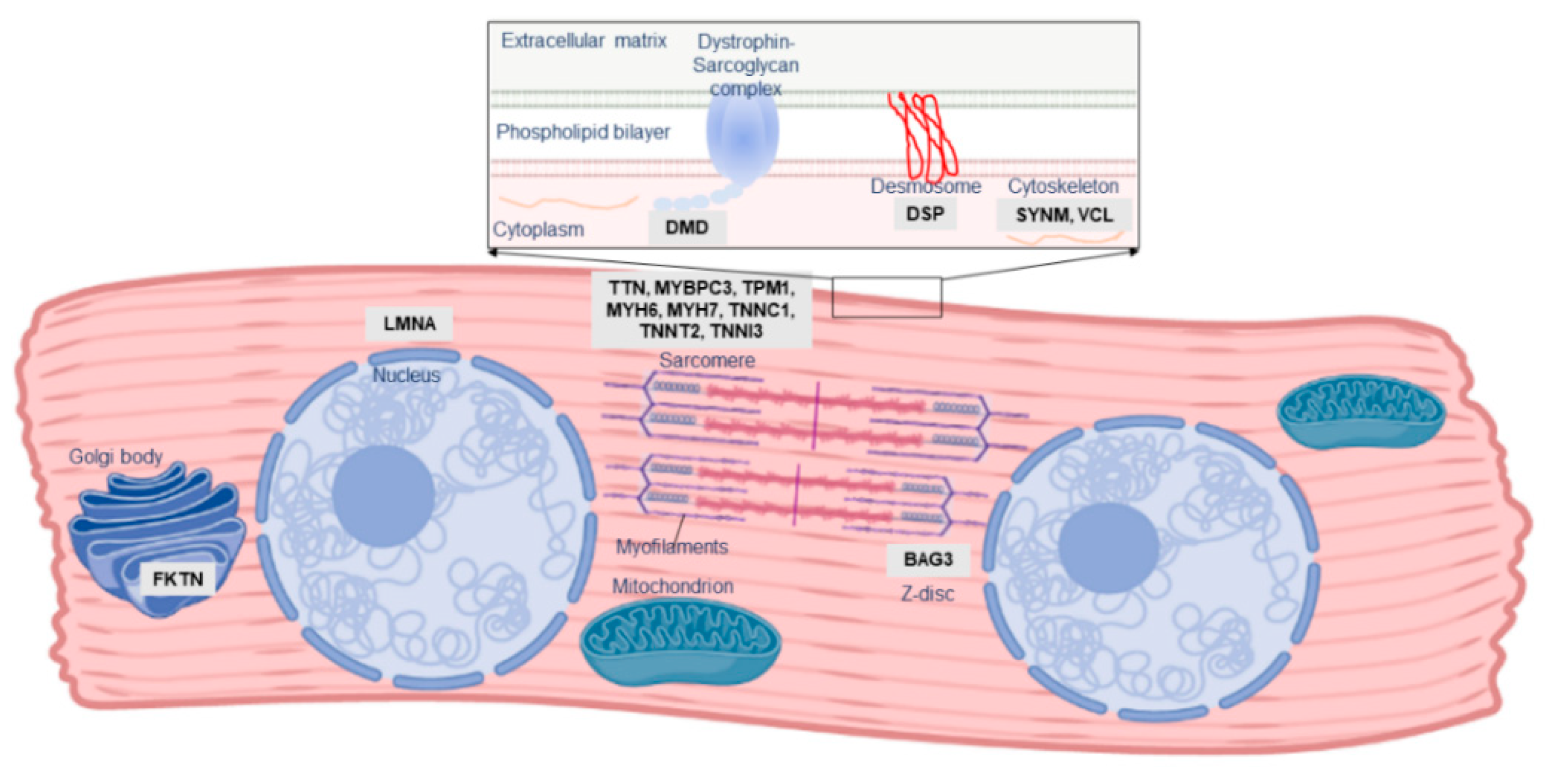
2. The Role of Familial Cardiomyopathy Genes in PPCM

One of the first indications of the role of genetic susceptibility in PPCM pathogenesis was familial clustering of PPCM with other forms of cardiomyopathy. Three instances of familial PPCM were reported in 1963 by Pierce et al. [10]; many similar familial occurrences of PPCM and DCM have been subsequently documented [11,12,13,14,15,16,17]. Although PPCM is a distinct clinical entity, these observations of overlap with familial DCM indicate that, in at least a subset of patients, PPCM may form part of the clinical and genetic spectrum of DCM. DCM itself is a vastly heterogeneous disease, genetically overlapping with hypertrophic cardiomyopathy (HCM), arrhythmogenic cardiomyopathy (ACM), and channelopathies: mutations in many of these genes have also been described in PPCM patients (Table 1; Figure 2).
In what appears to be the first documented genetic cause of PPCM, a 2001 case report described a female carrier of a Duchenne Muscular Dystrophy-causing DMD mutation who developed PPCM during the 36th week of pregnancy [18]. As an X-linked gene, most female carriers of DMD mutations are asymptomatic although some may develop mild disease or cardiomyopathy, and it was unclear if the PPCM, in this case, was related to her carrier status. However, similar case reports of PPCM amongst DMD and LAMP2 X-linked mutation carriers have been described [19,20,21]. LAMP2 mutations cause Danon disease, a form of cardiomyopathy and skeletal myopathy, through impairment of macroautophagy [22], while DMD mutations cause muscular dystrophy and/or DCM through the loss of the stabilising protein dystrophin, leaving myocytes vulnerable to oxidative stress and calcium overload [23]. In both cases, it is thought that skewed X-chromosome inactivation, in which only some cells express the DMD or LAMP2 mutation, underlies the comparatively milder cardiac phenotype observed in females [24]. It is possible, too, that this mosaic status in females may be exacerbated by stress conditions such as pregnancy, although no evidence of this has been reported as yet.
DMD and LAMP2 mutations are very rare causes of DCM; notably, most known DCM-causing mutations affect the sarcomere, causing cardiomyopathy through impaired force generation or reduced contractility in the heart. Early investigations reported several mutations in the sarcomeric genes MYBPC3, MYH6, MYH7, TNNC1 and TNNT2 in Dutch and American PPCM cohorts [13,17,25]. As such, similar studies across several other populations may provide critical information into the role of these mutations in PPCM.
As knowledge of DCM genetics has evolved over time, so too has knowledge of the genetic contribution to PPCM. Subsequent to these initial investigations, the giant sarcomeric protein-coding gene TTN emerged as a key gene in cardiomyopathy, accounting for up to 25% of familial DCM cases [26]. As the second-longest gene in humans, genetic variation in TTN is common [27], and it may be challenging to differentiate benign polymorphisms from true cardiomyopathy-causing mutations. However, frameshift insertions/deletions and nonsense mutations (truncating mutations) in TTN have been reported at an increased prevalence in DCM patients [26,27]. The mechanisms underlying TTN cardiomyopathy are thought to involve impairment of force generation or transmission in the myocardium or disrupted signalling [28]. BAG3 has also recently emerged as a major cardiomyopathy gene [29]. BAG3 encodes a multifunction co-chaperone protein involved in myocardial protein homeostasis, stabilisation of the myocardial Z disk, and modulation of cardiac contraction [30]. Mutations in BAG3 can cause cardiomyopathy through impairments in any of the protein’s diverse functions [31].
The emergence of high throughput next-generation sequencing (NGS) platforms enabled the rapid sequencing of patient genes and has highlighted the genetic overlap between different forms of heritable cardiomyopathy. Following these genetic advances, NGS studies of PPCM cohorts have revealed truncating mutations in TTN as the predominant genetic contributor to PPCM in American, Australian and European patients [32,33,34]. Although such TTN mutations are present in healthy individuals, they were found to be significantly more prevalent amongst PPCM patients [33]. In addition, the TTN mutations were mostly localised to the protein domains already associated with DCM (A-band), thereby implicating their possible role in PPCM [33]. In these NGS studies, mutations were reported in other cardiac disease genes, including disruptions of BAG3 [32,33,34]. Cumulatively, these cohort studies of PPCM have identified pathogenic mutations in 23% of cases (Figure 3). Studies of isolated PPCM cases have further implicated TTN in severe PPCM [35], as well as other disease genes such as KCNH2, RET and TXNRD2 [16,36,37], although the contribution of these genes to PPCM is unclear: the mutations in these cases may merely reflect the co-occurrence of genetic disorders with PPCM. Mutations in FKTN, RBM20, LMNA and DSP were also linked to PPCM through large cardiomyopathy family studies [38,39,40,41].
The diverse genetic aetiology of PPCM (Table 1) mirrors the genetic profile of DCM and strengthens the notion that PPCM may share genetic determinants with familial cardiomyopathy. In PPCM, as in DCM, a high proportion of TTN truncating variants is observed, and at present appears to be the greatest single genetic contributor to disease pathogenesis [32,33]. The mutation detection rate of genetic testing in DCM ranges from 15% to 40%, with familial history and larger gene panels contributing to higher mutation yields [42]. Although within this range, the relatively low yield (~20%) in PPCM may be attributed to the substantially fewer patients who have been screened so far, combined with fewer genes being sequenced in earlier studies.
When considering the worldwide distribution of PPCM-associated mutations (Figure 4), it is noteworthy that the majority have been identified in American and European populations, but even then, many of the genes are limited to single observations and case reports. Analysis of participants of African descent by Ware et al. [33] revealed mutation yields that were akin to those in individuals of European ancestry, indicating a similar genetic basis of PPCM in different population groups. This also suggests that mutation screening of African PPCM cohorts may be warranted. In DCM, the clinical utility of genetic testing can extend to family management and individual treatment and prognosis, through the characterisation of genotype-phenotype correlations. For example, LMNA mutations are associated with severe DCM with high penetrance and increased risk of sudden death, while TTN mutations have been associated with milder disease with reduced penetrance [43]. Mutations in these genes have been reported in PPCM, but their implications on phenotype and outcome are still to be determined. Further research is also needed to determine whether the co-occurrence of cardiomyopathy mutations in PPCM patients reflects causality or if, rather, it is the physiological strain of pregnancy that reveals cardiomyopathy in previously asymptomatic mutation carriers.

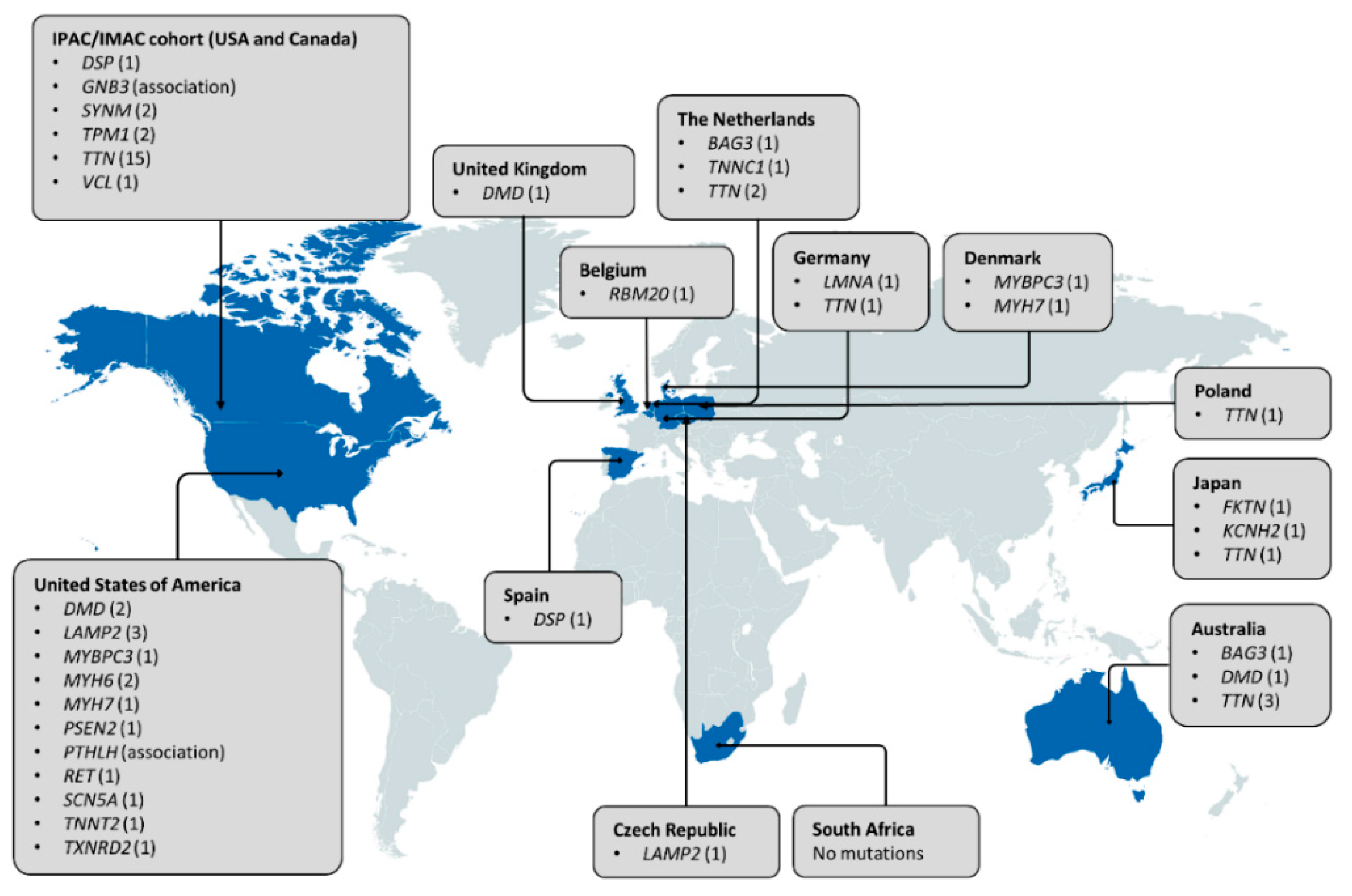
3. Other Genetic Determinants of PPCM

As a complex disorder, the role of genetics in PPCM may not be limited to rare, high-impact mutations as observed in familial cardiomyopathy. PPCM may also be influenced by more common variants with smaller effects on individual susceptibility or outcome: such variants are best identified through genetic association studies. In a 2011 genome-wide association study of 41 PPCM cases and 49 healthy controls, Horne et al. [44] investigated common genetic variation for associations with PPCM. Despite the small sample size, PTHLH rs258415 reached statistical significance, and its association with PPCM was replicated in a second cohort of 30 cases and 124 controls. PTHLH has documented cardiovascular roles including modulation of ventricular contraction and regulation of blood flow in the placenta and uterus. The authors hypothesised that the gene may be upregulated in pregnancy to prevent uterine contraction, and variation in PTHLH could compromise heart function and predispose individuals to PPCM [44].
In an outcomes-based genetic analysis, Sheppard et al. [45] revealed that the GNB3 c.825C>T polymorphism, specifically the homozygous TT allele, was significantly associated with poorer outcomes, with lower ejection fractions up to a year postpartum compared to individuals without the variant. The TT allele was also determined to be more prevalent in patients of African ethnicity. The gene may play a role in hypertension and cardiac remodelling, although its role in PPCM remains to be confirmed.

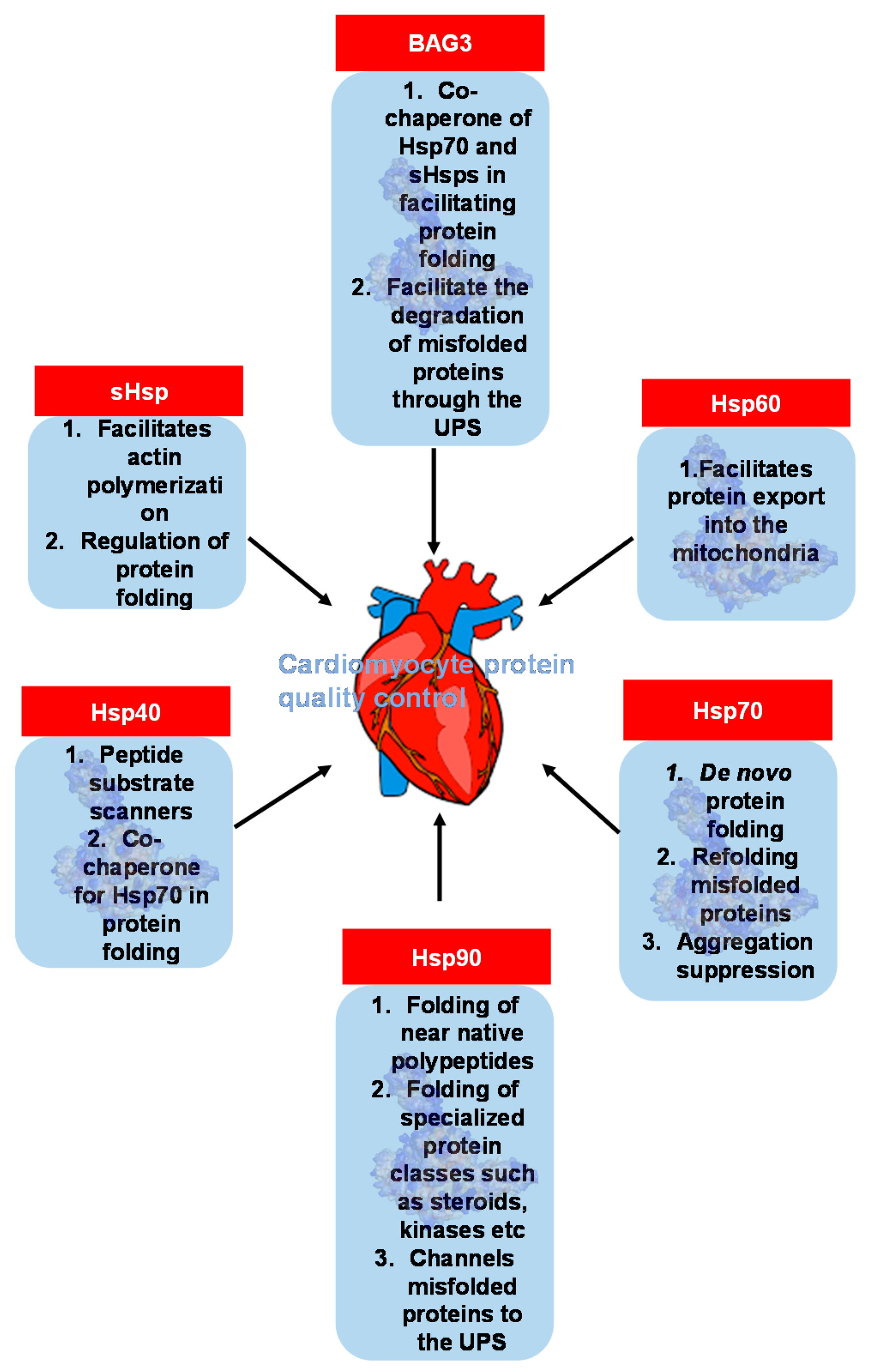
4. An Emerging Field: Heat Shock Protein and Molecular Chaperone Genes in PPCM

The aforementioned studies indicate a clear role of genetics in PPCM pathophysiology. Nevertheless, the 23% mutation yield from the screening of cardiomyopathy genes (Figure 3) suggests that many genetic determinants of PPCM are yet to be discovered. Indeed, many of the reported PPCM/DCM families had no identified mutation [13,32]. There is, therefore, a need to consider other genes for their contribution to PPCM. Given the role of cardiac stress in the development of the disease, molecular chaperones and heat shock proteins (Hsps) may prove of interest due to their stress-protective functions.
Lending support to this notion is the prior identification of BAG3 mutations in PPCM patients [32,34]. As a molecular chaperone, BAG3 is actively involved in a variety of cellular mechanisms during stress which includes protein folding, autophagy, and the ubiquitin-proteasome system [46]. BAG3 exerts some of these functions by acting as a co-chaperone for Hsp70 and the small Hsps (sHsps) HspB5, HspB6 and HspB8 [47]. The formation of the BAG3-Hsp70-sHsp complex is essential in facilitating denatured proteins to refold thus preventing protein aggregation in cell stress conditions. Because BAG3 is a key cardiomyopathy gene, with several reported DCM-causing mutations occurring across the gene, many more causative variants in the gene are likely still to be discovered amongst PPCM patients.
Hsps are a class of molecular chaperones that perform a myriad of housekeeping and stress-protective roles in cells to maintain proteostasis (Figure 5) [48]. Hsps broadly function to facilitate the correct folding and assembly of polypeptides, thus preventing the formation of aggregation-prone misfolded proteins that are toxic to cardiomyocytes [49]. Since the contractile and metabolic demands of the heart require robust protein quality control, Hsps may play crucial cardioprotective roles. Included in the Hsp family are small heat shock proteins (sHsps), which are ubiquitously expressed regulators of cellular protein folding [30]. The high expression of sHsps in the heart has been reported, where they are implicated in the maintenance of normal cardiac function and regulation of the cardiac stress response [50,51].
Genetic investigations have recently implicated Hsps in PPCM pathogenesis. Sequencing the HSPB6 gene in DCM patients led to the identification of the rare variant p.Ser10Phe [52]. In vitro, this mutation reduced thermal resilience of the chaperone, potentially compromising its cardioprotective activity [53]. In a murine model, the p.Ser10Phe mutation-induced progressive cardiac dysfunction and HF in males, while female mice had preserved cardiac function and survival [52,54]. However, the HSPB6 p.Ser10Phe mutation appeared to induce lethal PPCM, with a 100% fatality rate within 4 pregnancies and significant cardiac dilatation and dysfunction after 3 pregnancies [54]. This is the first description of a mutant mouse model of PPCM in which the mutation was originally discovered in humans, although STAT3 and Akt transgene mice have been used previously to study the pathophysiology of PPCM [9,55]. The clinical and demographic characteristics of the p.Ser10Phe mutation carriers in the original cohort were not described, and the potential role of Hsp genes in PPCM may warrant further investigation. Although this is the first study demonstrating Hsp mutations in PPCM, genetic variation in HSPB5, HSPB7 and HSPD1 has been reported in other cardiomyopathies.
The proteostatic functions of Hsps and molecular chaperones can be hypothesised to play a crucial role in the physiological adaptation of the heart to pregnancy. Mutations in these genes may alter protein quality control systems in the heart, thereby predisposing individuals to PPCM. However, at present no other Hsp gene mutations have been described in PPCM patients to our knowledge. As such, other molecular chaperone genes such as HSPB5 (CRYAB), HSPB7 and HSPD1 may be worth exploring for their potential as novel genetic determinants of PPCM.
HSPB7, sometimes referred to as the “cardiovascular Hsp” due to its high cardiac expression, is an sHsp that has attracted interest as a possible susceptibility locus for cardiomyopathy and HF [56,57]. Several genetic association studies have described protective effects of variation in HSPB7 against DCM [29,57,58] and systolic HF [57,59,60]. These studies have been limited to individuals of European descent, and the lack of association with DCM in African Americans and Chinese populations [56,61,62] indicates that the effects of HSPB7 variation may be population-specific, or are influenced by underlying population genetic differences. The causal relationship between HSPB7 variation and cardiomyopathy is unclear, as many of the variants occurred in non-coding regions of the gene, did not appear to be in linkage disequilibrium with overtly protein-altering variants, and targeted sequencing of the gene in DCM patients did not yield any pathogenic variation [29,57]. However, the multiple observations of HSPB7 polymorphisms in HF and cardiomyopathy suggests a common genetic basis for these related phenotypes. Several of these studies also reported BAG3 variation to associate with disease risk [29,58,60], with the most recent study also demonstrating direct interaction of BAG3 protein with HspB7 [58]. These findings support the notion that HSPB7 genetic variation may contribute to cardiomyopathy, even though the precise mechanisms are not yet known.
The CRYAB gene encodes αβ-crystallin or HspB5, a member of the sHsp family. In cardiomyocytes, HspB5 primarily acts to prevent the accumulation of potentially toxic protein aggregates [63], although the binding of HspB5 to TTN, actin and desmin is essential to ensure proper sarcomeric assembly and function. Mutations in CRYAB can trigger cardiac disease through the accumulation of protein aggregates [64]. Such a scenario has been reported in patients with cardiomyopathy, usually presenting in conjunction with skeletal myopathy and/or ocular disorders such as cataracts [64,65,66]. CRYAB mutations have been reported to cause isolated DCM and HCM in individuals with positive family histories for cardiomyopathy [67,68]. One of these mutations, CRYAB p.Arg157His, was demonstrated to impair interaction with cardiac-specific isoforms of TTN [67], suggesting that mutations affecting this uncharacterised domain of CRYAB may play a role in cardiomyopathy through impaired CRYAB-TTN interaction in cardiomyocytes. Given the major role of TTN truncations in PPCM, the effect of genetic variation in CRYAB may warrant further investigation.
The HSPD1 gene encodes for a constitutively expressed Hsp60 chaperonin protein. A recent study has linked HSPD1 mutations with cardiomyopathy when the mutation p.Thr320Ala was described in a Japanese family with DCM and arrhythmia [69]. Although the mechanisms whereby HSPD1 may contribute to cardiomyopathy are unclear, the authors demonstrated that a similar mutation in zebrafish resulted in a DCM phenotype with mitochondrial damage and increased levels of reactive oxygen species, as well as a reduced tolerance to exercise stress [69]. Cardiac-specific deletion of the gene in mice resulted in the development of lethal DCM and HF in males, although the effects in females were not described [70]. Other HSPD1 mutations may need to be reported in additional cardiomyopathy families before its role in disease pathogenesis is clarified.

5. Clinical Implications

5.1. Genetic Testing May Be Indicated in PPCM Patients with Family History

The identification of pathogenic cardiomyopathy-causing mutations in women with PPCM indicates that PPCM overlaps with heritable cardiomyopathy not just phenotypically, but genetically as well. In patients with genetic forms of cardiomyopathy such as DCM, ACM and HCM, current guidelines recommend the construction of a detailed family history (usually at least 3 generations), periodic clinical screening of first-degree relatives by echocardiography and electrocardiography, as well as genetic testing in the case of familial disease [71,72,73,74,75]. This approach allows at-risk family members to be identified and, when disease-causing mutations are found, cardiac follow-up can be guided by the presence of the mutation in family members. A similar strategy may be beneficial in PPCM, in which family history should be obtained from index cases and, when familial cardiovascular disease is present, genetic testing may be warranted.
Genetic techniques such as NGS allow massively parallel sequencing of vast tracts of DNA and can be used to accurately screen numerous cardiomyopathy genes in a single experiment. Such targeted sequencing panels are becoming routinely incorporated into the diagnosis of heritable cardiomyopathy in some countries [76]. However, it should be noted that there is uncertainty about whether inconclusive genetic results should be reported to patients [77]. Nevertheless, the increasing availability of NGS implies that it now may be considered more cost-effective to conduct genetic testing on asymptomatic family members than to phenotypically screen them [78,79]. In the context of familial PPCM, if a patient is found to carry a disease-causing mutation, this could allow clinical follow-up to be limited to relatives who also carry the mutation.
At this stage, it is still unclear whether individuals with mutations in any of the cardiomyopathy-causing genes require additional management during pregnancy and delivery. Limited reports have indicated that patients with DCM, HCM and ACM tend to tolerate pregnancy well, although risks of adverse cardiac events such as arrhythmias, HF, syncope and death are associated with advanced left ventricular dysfunction and prior cardiac events [80,81]. Although emergency caesarean section amongst DCM, HCM and ACM patients is rare [81], a global PPCM registry recently reported caesarean sections for up to 59% of its patients, for whom mutation carrier status was unknown [82].

5.2. Areas of Future Investigation

While the contribution of cardiomyopathy genes to PPCM has been demonstrated, relatively few mutations and genes have been reported so far. There is therefore still much scope for future research into the genetics of PPCM. It is likely that further screening will identify more mutations, potentially in genes that have not been described in PPCM to date, since upwards of 60 genes have been associated with DCM and other forms of cardiomyopathy [83]. Correlating genetic information with phenotypic presentation and disease outcome could have profound implications on patient management and should be explored further. This is particularly true in African populations, where the risk of PPCM is higher [4,84], the prognosis is poorer [85], and the knowledge of cardiomyopathy genetics is scarce [86]. PPCM has a broader aetiology than other forms of heritable cardiomyopathy, and further research is needed into the role of additional genes such as those involved in (auto)immunity, angiogenesis, metabolism, and oxidative stress.
Molecular chaperones and Hsp genes hold great promise as candidate genes for PPCM. In this review, several Hsp genes have been proposed as crucial genetic determinants of PPCM pathology. Recently, the mutation HSPB6 p.Ser10Phe was demonstrated to cause PPCM in mice, indicating a role of the chaperone in protein quality control systems and stress response [54]. BAG3 gene mutations are some of the most documented chaperone-associated mutations in cardiomyopathy. Indeed, this has led to the identification of 2 PPCM-related gene mutations in BAG3 [32,34]. However, mutations occurring in other Hsp family genes are yet to be fully documented. Since pregnancy can induce extra stress on the heart, it is therefore plausible that Hsps may play an integral cardioprotective role in pregnant women. As such, in-depth analysis into Hsp and molecular chaperone genes may lead to the identification of novel PPCM genetic determinants that will aid in subsequent case management and drug design.

6. Conclusions

Recent advances have emphasised an important role of genetics in the pathophysiology of PPCM. Several studies, reviewed in this paper, suggest that up to 20% of women with PPCM have an identifiable disease-causing mutation. While a substantial overlap with heritable forms of cardiomyopathy is observed, there is much scope for future research. Given the range of physiological stresses that the body is subjected to during pregnancy, the role of Hsps in PPCM provides great potential which warrants further investigation. A better understanding of the genetics of PPCM holds the potential to improve patient management, with possible implications on treatment, prognosis, and family management.

Author Contributions

T.F.S. and G.C. performed the literature review and drafted the manuscript. The concept was conceived by K.S., G.C. and T.F.S. Critical feedback was provided by P.J.S., M.-C.K., N.A.B.N. and K.S. Academic support was provided by K.S., G.S. and N.A.B.N. All authors have read and agreed to the published version of the manuscript.

Funding

The article processing charge was funded by the Hippocrate Fund.

Institutional Review Board Statement

Not applicable.

Informed Consent Statement

Not applicable.

Data Availability Statement

No new data were created or analysed in this study. Data sharing is not applicable to this article.

Acknowledgments

We would like to acknowledge institutional support in the form of an unconditional research grant from Servier: Intitut La Conference Hippocrate; post-graduate student support from the University of Cape Town (G.C., T.F.S.); G.C. also gratefully acknowledges support from the URC Faculty of Sciences Fellowship, University of Cape Town; and N.A.B.N. gratefully acknowledges support from the National Research Foundation and the South African Medical Research Council, as well as from the Harry Crossley Foundation and Lily & Ernst Hausmann Trust. Figure 2 was created using BioRender.com.

Conflicts of Interest

The authors declare no conflict of interest.

References

  1. Bauersachs, J.; Konig, T.; van der Meer, P.; Petrie, M.C.; Hilfiker-Kleiner, D.; Mbakwem, A. Pathophysiology, diagnosis and management of peripartum cardiomyopathy: A position statement from the Heart Failure Association of the European Society of Cardiology Study Group on peripartum cardiomyopathy. Eur. J. Heart Fail 2019, 21, 827–843. [Google Scholar] [CrossRef] [PubMed]
  2. Hoevelmann, J.; Hahnle, L.; Hahnle, J.; Sliwa, K.; Viljoen, C. Detection and management of arrhythmias in peripartum cardiomyopathy. Cardiovasc. Diagn. Ther. 2020, 10, 325–335. [Google Scholar] [CrossRef] [PubMed]
  3. McNamara, D.M.; Elkayam, U.; Alharethi, R.; Damp, J.; Hsich, E.; Ewald, G. Clinical outcomes for peripartum cardiomyopathy in North America: Results of the IPAC study (Investigations of Pregnancy-Associated Cardiomyopathy). J. Am. Coll. Cardiol. 2015, 66, 905–914. [Google Scholar] [CrossRef] [PubMed] [Green Version]
  4. Desai, D.; Moodley, J.; Naidoo, D. Peripartum cardiomyopathy: Experiences at King Edward VIII Hospital, Durban, South Africa and a review of the literature. Trop. Doct. 1995, 25, 118–123. [Google Scholar] [CrossRef] [PubMed]
  5. Ersbøll, A.S.; Johansen, M.; Damm, P.; Rasmussen, S.; Vejlstrup, N.G.; Gustafsson, F. Peripartum cardiomyopathy in Denmark: A retrospective, population-based study of incidence, management and outcome. Eur. J. Heart Fail 2017, 19, 1712–1720. [Google Scholar] [CrossRef] [Green Version]
  6. Kolte, D.; Khera, S.; Aronow, W.S.; Palaniswamy, C.; Mujib, M.; Ahn, C. Temporal trends in incidence and outcomes of peripartum cardiomyopathy in the United States: A nationwide population-based study. J. Am. Heart Assoc. 2014, 3, e001056. [Google Scholar] [CrossRef] [Green Version]
  7. Sliwa, K.; Mebazaa, A.; Hilfiker-Kleiner, D.; Petrie, M.C.; Maggioni, A.P.; Laroche, C. Clinical characteristics of patients from the worldwide registry on peripartum cardiomyopathy (PPCM): EURObservational Research Programme in conjunction with the Heart Failure Association of the European Society of Cardiology Study Group on PPCM. Eur. J. Heart Fail 2017, 19, 1131–1141. [Google Scholar] [CrossRef]
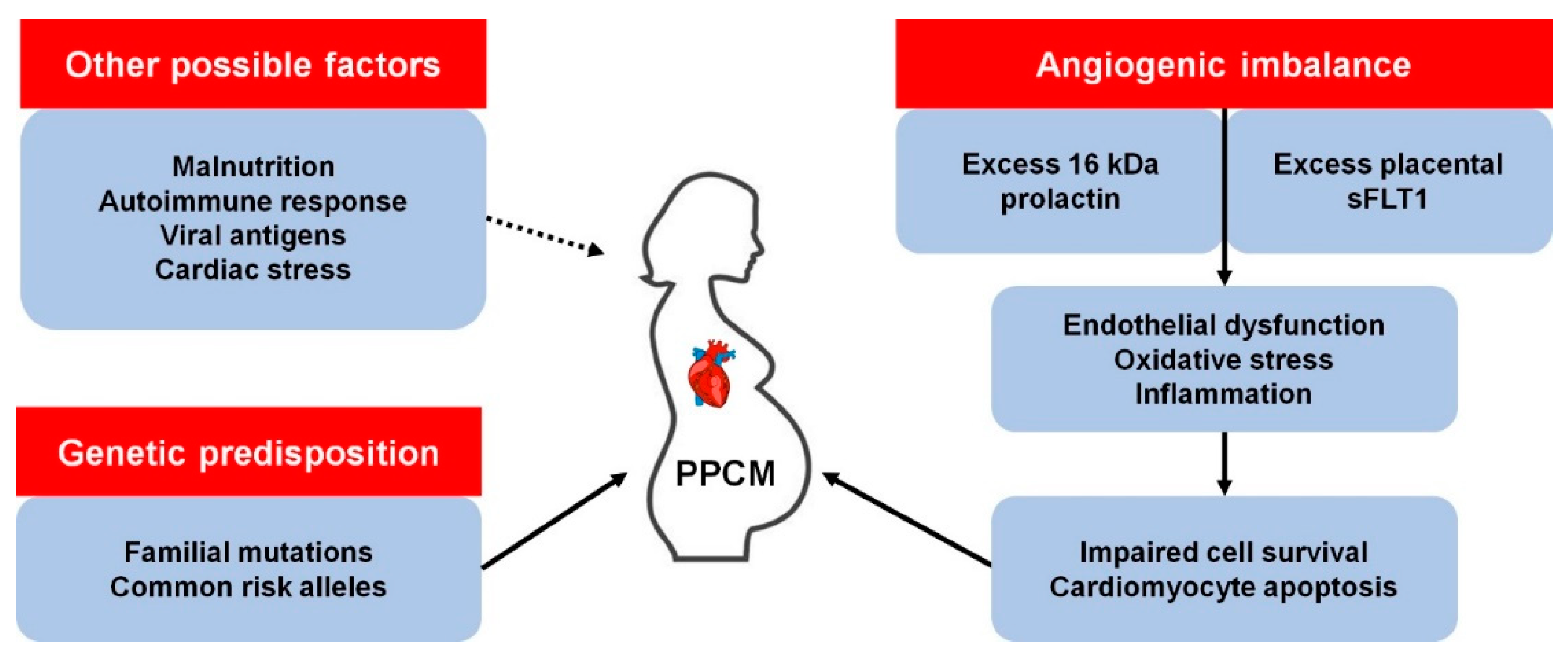
  8. Hall, M.E.; George, E.M.; Granger, J.P. The heart during pregnancy. Rev. Esp. Cardiol. 2011, 64, 1045–1050. [Google Scholar] [CrossRef] [Green Version]
  9. Hilfiker-Kleiner, D.; Kaminski, K.; Podewski, E.; Bonda, T.; Schaefer, A.; Sliwa, K. A cathepsin D-cleaved 16 kDa form of prolactin mediates postpartum cardiomyopathy. Cell 2007, 128, 589–600. [Google Scholar] [CrossRef] [Green Version]
  10. Pierce, J.A.; Price, B.O.; Joyce, J.W. Familial occurrence of postpartal heart failure. Arch Intern Med 1963, 111, 651–655. [Google Scholar] [CrossRef]
  11. Baruteau, A.-E.; Leurent, G.; Schleich, J.-M.; Gervais, R.; Daubert, J.-C.; Mabo, P. Can peripartum cardiomyopathy be familial? Int. J. Cardiol. 2009, 137, 181–185. [Google Scholar] [CrossRef] [PubMed]
  12. Haghikia, A.; Podewski, E.; Libhaber, E.; Labidi, S.; Fischer, D.; Roentgen, P. Phenotyping and outcome on contemporary management in a German cohort of patients with peripartum cardiomyopathy. Basic Res. Cardiol. 2013, 108, 366. [Google Scholar] [CrossRef] [PubMed] [Green Version]
  13. Morales, A.; Painter, T.; Li, R.; Siegfried, J.D.; Li, D.; Norton, N. Rare variant mutations in pregnancy-associated or peripartum cardiomyopathy. Circulation 2010, 121, 2176–2182. [Google Scholar] [CrossRef] [PubMed]
  14. Ntusi, N.A.; Wonkam, A.; Shaboodien, G.; Badri, M.; Mayosi, B.M. Frequency and clinical genetics of familial dilated cardiomyopathy in Cape Town: Implications for the evaluation of patients with unexplained cardiomyopathy. S. Afr. Med. J. 2011, 1010, 394–398. [Google Scholar]
  15. Pearl, W. Familial occurrence of peripartum cardiomyopathy. Am. Heart J. 1995, 129, 421–492. [Google Scholar] [CrossRef]
  16. Rajapreyar, I.; Sinkey, R.; Pamboukian, S.V.; Tita, A. Did a shared thioredoxin-reductase gene mutation lead to maternal peripartum cardiomyopathy and fatal dilated cardiomyopathy in her son? A case report. Case Rep. Womens Health 2020, 26, e00196. [Google Scholar] [CrossRef]
  17. van Spaendonck-Zwarts, K.Y.; van Tintelen, J.P.; van Veldhuisen, D.J.; van der Werf, R.; Jongbloed, J.D.; Paulus, W.J. Peripartum cardiomyopathy as a part of familial dilated cardiomyopathy. Circulation 2010, 121, 2169–2175. [Google Scholar] [CrossRef] [Green Version]
  18. Davies, J.E.; Winokur, T.S.; Aaron, M.F.; Benza, R.L.; Foley, B.A.; Holman, W.L. Cardiomyopathy in a carrier of Duchenne’s muscular dystrophy. J. Heart Lung Transpl. 2001, 20, 781–784. [Google Scholar] [CrossRef]
  19. Ahmed, A.; Spinty, S.; Murday, V.; Longman, C.; Khand, A. A de-novo deletion of dystrophin provoking severe ’peri-partum cardiomyopathy: The importance of genetic testing in peripartum cardiomyopathy to uncover female carriers. Int. J. Cardiol. 2016, 203, 1084–1085. [Google Scholar] [CrossRef]
  20. Cheng, V.E.; Prior, D.L. Peripartum cardiomyopathy in a previously asymptomatic carrier of Duchenne muscular dystrophy. Heart Lung Circ 2013, 22, 677–681. [Google Scholar] [CrossRef]
  21. Gurka, J.; Piherova, L.; Majer, F.; Chaloupka, A.; Zakova, D.; Pelak, O. Danon disease is an underdiagnosed cause of advanced heart failure in young female patients: A LAMP2 flow cytometric study. ESC Heart Fail 2020. [Google Scholar] [CrossRef]
  22. Rowland, T.J.; Sweet, M.E.; Mestroni, L.; Taylor, M.R. Danon disease-dysregulation of autophagy in a multisystem disorder with cardiomyopathy. J. Cell Sci. 2016, 129, 2135–2143. [Google Scholar] [CrossRef] [PubMed] [Green Version]
  23. Shirokova, N.; Niggli, E. Cardiac phenotype of Duchenne Muscular Dystrophy: Insights from cellular studies. J. Mol. Cell Cardiol. 2013, 58, 217–224. [Google Scholar] [CrossRef] [PubMed] [Green Version]
  24. Lim, K.R.Q.; Sheri, N.; Nguyen, Q.; Yokota, T. Cardiac Involvement in Dystrophin-Deficient Females: Current Understanding and Implications for the Treatment of Dystrophinopathies. Genes 2020, 11, 765. [Google Scholar] [CrossRef] [PubMed]
  25. Moller, D.V.; Andersen, P.S.; Hedley, P.; Ersboll, M.K.; Bundgaard, H.; Moolman-Smook, J. The role of sarcomere gene mutations in patients with idiopathic dilated cardiomyopathy. Eur. J. Hum. Genet. 2009, 17, 1241–1249. [Google Scholar] [CrossRef] [Green Version]
  26. Herman, D.S.; Lam, L.; Taylor, M.R.; Wang, L.; Teekakirikul, P.; Christodoulou, D. Truncations of titin causing dilated cardiomyopathy. N. Engl. J. Med. 2012, 366, 619–628. [Google Scholar] [CrossRef] [Green Version]
  27. Walsh, R.; Thomson, K.L.; Ware, J.S.; Funke, B.H.; Woodley, J.; McGuire, K.J. Reassessment of Mendelian gene pathogenicity using 7,855 cardiomyopathy cases and 60,706 reference samples. Genet. Med. 2017, 19, 192–203. [Google Scholar] [CrossRef] [Green Version]
  28. Tharp, C.A.; Haywood, M.E.; Sbaizero, O.; Taylor, M.R.G.; Mestroni, L. The giant protein Titin’s role in cardiomyopathy: Genetic, transcriptional, and post-translational modifications of TTN and their contribution to cardiac disease. Front. Physiol. 2019, 10, 1436. [Google Scholar] [CrossRef] [Green Version]
  29. Villard, E.; Perret, C.; Gary, F.; Proust, C.; Dilanian, G.; Hengstenberg, C. A genome-wide association study identifies two loci associated with heart failure due to dilated cardiomyopathy. Eur. Heart J. 2011, 32, 1065–1076. [Google Scholar] [CrossRef] [Green Version]
  30. Fang, X.; Bogomolovas, J.; Wu, T.; Zhang, W.; Liu, C.; Veevers, J. Loss-of-function mutations in co-chaperone BAG3 destabilize small HSPs and cause cardiomyopathy. J. Clin. Investig. 2017, 127, 3189–3200. [Google Scholar] [CrossRef] [Green Version]
  31. Myers, V.D.; Tomar, D.; Madesh, M.; Wang, J.; Song, J.; Zhang, X.-Q. Haplo-insufficiency of Bcl2-associated Athanogene 3 in mice results in progressive left ventricular dysfunction, β-adrenergic insensitivity and increased apoptosis. J. Cell Physiol. 2018, 233, 6319–6326. [Google Scholar] [CrossRef] [PubMed]
  32. van Spaendonck-Zwarts, K.Y.; Posafalvi, A.; van den Berg, M.P.; Hilfiker-Kleiner, D.; Bollen, I.A.; Sliwa, K. Titin gene mutations are common in families with both peripartum cardiomyopathy and dilated cardiomyopathy. Eur. Heart. J. 2014, 35, 2165–2173. [Google Scholar] [CrossRef] [PubMed] [Green Version]
  33. Ware, J.S.; Li, J.; Mazaika, E.; Yasso, C.M.; DeSouza, T.; Cappola, T.P. Shared genetic predisposition in peripartum and dilated cardiomyopathies. N. Engl. J. Med. 2016, 374, 233–241. [Google Scholar] [CrossRef] [PubMed]
  34. Minoche, A.E.; Horvat, C.; Johnson, R.; Gayevskiy, V.; Morton, S.U.; Drew, A.P. Genome sequencing as a first-line genetic test in familial dilated cardiomyopathy. Genet. Med. 2019, 21, 650–662. [Google Scholar] [CrossRef] [PubMed]
  35. Kryczka, K.E.; Dzielińska, Z.; Franaszczyk, M.; Wojtkowska, I.; Henzel, J.; Śpiewak, M. Severe course of peripartum cardiomyopathy and subsequent recovery in a patient with a novel TTN gene-truncating mutation. Am. J. Case Rep. 2018, 19, 820–824. [Google Scholar] [CrossRef]
  36. Nishimoto, O.; Matsuda, M.; Nakamoto, K.; Nishiyama, H.; Kuraoka, K.; Taniyama, K. Peripartum cardiomyopathy presenting with syncope due to Torsades de pointes: A case of long QT syndrome with a novel KCNH2 mutation. Intern. Med. 2012, 51, 461–464. [Google Scholar] [CrossRef] [Green Version]
  37. Kim, J.; Reutrakul, S.; Davis, D.B.; Kaplan, E.L.; Refetoff, S. Multiple endocrine neoplasia 2A syndrome presenting as peripartum cardiomyopathy due to catecholamine excess. Eur. J. Endocrinol. 2004, 151, 771–777. [Google Scholar] [CrossRef] [Green Version]
  38. Glocklhofer, C.R.; Steinfurt, J.; Franke, G.; Hoppmann, A.; Glantschnig, T.; Perez-Feliz, S. A novel LMNA nonsense mutation causes two distinct phenotypes of cardiomyopathy with high risk of sudden cardiac death in a large five-generation family. Europace 2018, 20, 2003–2013. [Google Scholar] [CrossRef]
  39. Molina, P.; Sanz-Sanchez, J.; Fenollosa, M.; Martinez-Matilla, M.; Giner, J.; Zorio, E. Arrhythmogenic cardiomyopathy with left ventricular involvement versus ischemic heart disease: Lessons learned from the family study and the reviewed autopsy of a young male. Forensic Sci. Res. 2019, 4, 274–279. [Google Scholar] [CrossRef]
  40. Murakami, T.; Hayashi, Y.K.; Noguchi, S.; Ogawa, M.; Nonaka, I.; Tanabe, Y. Fukutin gene mutations cause dilated cardiomyopathy with minimal muscle weakness. Ann. Neurol. 2006, 60, 597–602. [Google Scholar] [CrossRef] [Green Version]
  41. Robyns, T.; Willems, R.; Van Cleemput, J.; Jhangiani, S.; Muzny, D.; Gibbs, R. Whole exome sequencing in a large pedigree with DCM identifies a novel mutation in RBM20. Acta Cardiol. 2019, 1–6. [Google Scholar] [CrossRef] [PubMed]
  42. Rosenbaum, A.N.; Agre, K.E.; Pereira, N.L. Genetics of dilated cardiomyopathy: Practical implications for heart failure management. Nat. Rev. Cardiol. 2020, 17, 286–297. [Google Scholar] [CrossRef] [PubMed]
  43. Paldino, A.; De Angelis, G.; Merlo, M.; Gigli, M.; Dal Ferro, M.; Severini, G.M. Genetics of dilated cardiomyopathy: Clinical implications. Curr. Cardiol. Rep. 2018, 20, 83. [Google Scholar] [CrossRef] [PubMed]
  44. Horne, B.D.; Rasmusson, K.D.; Alharethi, R.; Budge, D.; Brunisholz, K.D.; Metz, T. Genome-wide significance and replication of the chromosome 12p11.22 locus near the PTHLH gene for peripartum cardiomyopathy. Circ. Cardiovasc. Genet. 2011, 4, 359–366. [Google Scholar] [CrossRef] [PubMed]
  45. Sheppard, R.; Hsich, E.; Damp, J.; Elkayam, U.; Kealey, A.; Ramani, G. GNB3 C825T polymorphism and myocardial recovery in peripartum cardiomyopathy: Results of the multicenter Investigations of Pregnancy-Associated Cardiomyopathy study. Circ. Heart Fail 2016, 9, e002683. [Google Scholar] [CrossRef] [PubMed] [Green Version]
  46. Ranek, M.J.; Stachowski, M.J.; Kirk, J.A.; Willis, M.S. The role of heat shock proteins and co-chaperones in heart failure. Philos. Trans. R Soc. Lond B Biol. Sci. 2018, 373. [Google Scholar] [CrossRef] [Green Version]
  47. Myers, V.D.; McClung, J.M.; Wang, J.; Tahrir, F.G.; Gupta, M.K.; Gordon, J. The multifunctional protein BAG3: A novel therapeutic target in cardiovascular disease. JACC Basic Transl. Sci. 2018, 3, 122–131. [Google Scholar] [CrossRef]
  48. Lindquist, S. The heat-shock response. Ann Rev Biochem 1986, 55, 1151–1191. [Google Scholar] [CrossRef] [PubMed]
  49. McLendon, P.M.; Robbins, J. Proteotoxicity and cardiac dysfunction. Circ. Res. 2015, 116, 1863–1882. [Google Scholar] [CrossRef] [PubMed] [Green Version]
  50. Hu, X.; Van Marion, D.M.S.; Wiersma, M.; Zhang, D.; Brundel, B.J.J.M. The protective role of small heat shock proteins in cardiac diseases: Key role in atrial fibrillation. Cell Stress Chap. 2017, 22, 665–674. [Google Scholar] [CrossRef] [PubMed] [Green Version]
  51. Collier, M.P.; Benesch, J.L. Small heat-shock proteins and their role in mechanical stress. Cell Stress Chaper. 2020, 25, 601–613. [Google Scholar] [CrossRef] [PubMed] [Green Version]
  52. Liu, G.S.; Zhu, H.; Cai, W.F.; Wang, X.; Jiang, M.; Essandoh, K. Regulation of BECN1-mediated autophagy by HSPB6: Insights from a human HSPB6(S10F) mutant. Autophagy 2018, 14, 80–97. [Google Scholar] [CrossRef] [PubMed] [Green Version]
  53. Shatov, V.M.; Gusev, N.B. Physico-chemical properties of two point mutants of small heat shock protein HspB6 (Hsp20) with abrogated cardioprotection. Biochimie 2020, 174, 126–135. [Google Scholar] [CrossRef] [PubMed]
  54. Liu, G.S.; Gardner, G.; Adly, G.; Jiang, M.; Cai, W.F.; Lam, C.K. A novel human S10F-Hsp20 mutation induces lethal peripartum cardiomyopathy. J. Cell Mol. Med. 2018. [Google Scholar] [CrossRef] [PubMed]
  55. Ricke-Hoch, M.; Bultmann, I.; Stapel, B.; Condorelli, G.; Rinas, U.; Sliwa, K. Opposing roles of Akt and STAT3 in the protection of the maternal heart from peripartum stress. Cardiovasc. Res. 2014, 101, 587–596. [Google Scholar] [CrossRef] [Green Version]
  56. Matkovich, S.J.; Van Booven, D.J.; Hindes, A.; Kang, M.Y.; Druley, T.E.; Vallania, F.L. Cardiac signaling genes exhibit unexpected sequence diversity in sporadic cardiomyopathy, revealing HSPB7 polymorphisms associated with disease. J. Clin. Investig. 2010, 120, 280–289. [Google Scholar] [CrossRef] [Green Version]
  57. Stark, K.; Esslinger, U.B.; Reinhard, W.; Petrov, G.; Winkler, T.; Komajda, M. Genetic association study identifies HSPB7 as a risk gene for idiopathic dilated cardiomyopathy. PLoS Genet. 2010, 6, e1001167. [Google Scholar] [CrossRef]
  58. Esslinger, U.; Garnier, S.; Korniat, A.; Proust, C.; Kararigas, G.; Muller-Nurasyid, M. Exome-wide association study reveals novel susceptibility genes to sporadic dilated cardiomyopathy. PLoS ONE 2017, 12, e0172995. [Google Scholar] [CrossRef]
  59. Cappola, T.P.; Li, M.; He, J.; Ky, B.; Gilmore, J.; Qu, L. Common variants in HSPB7 and FRMD4B associated with advanced heart failure. Circ. Cardiovasc. Genet. 2010, 3, 147–154. [Google Scholar] [CrossRef] [Green Version]
  60. Garnier, S.; Hengstenberg, C.; Lamblin, N.; Dubourg, O.; De Groote, P.; Fauchier, L. Involvement of BAG3 and HSPB7 loci in various etiologies of systolic heart failure: Results of a European collaboration assembling more than 2000 patients. Int. J. Cardiol. 2015, 189, 105–107. [Google Scholar] [CrossRef] [Green Version]
  61. Chen, F.F.; Xia, Y.L.; Xu, C.Q.; Li, S.S.; Zhao, Y.Y.; Wang, X.J. Common variant rs7597774 in ADD2 is associated with dilated cardiomyopathy in Chinese Han population. Int. J. Clin. Exp. Med. 2015, 8, 1188–1196. [Google Scholar] [PubMed]
  62. Li, X.; Luo, R.; Mo, X.; Jiang, R.; Kong, H.; Hua, W. Polymorphism of ZBTB17 gene is associated with idiopathic dilated cardiomyopathy: A case control study in a Han Chinese population. Eur. J. Med. Res. 2013, 18, 10. [Google Scholar] [CrossRef] [PubMed] [Green Version]
  63. Sanbe, A. Molecular mechanisms of α-crystallinopathy and its therapeutic strategy. Biol. Pharm. Bull. 2011, 34, 1653–1658. [Google Scholar] [CrossRef] [PubMed] [Green Version]
  64. Brodehl, A.; Gaertner-Rommel, A.; Klauke, B.; Grewe, S.A.; Schirmer, I.; Peterschröder, A. The novel alphaB-crystallin (CRYAB) mutation p.D109G causes restrictive cardiomyopathy. Hum. Mutat. 2017, 38, 947–952. [Google Scholar] [CrossRef] [PubMed]
  65. van der Smagt, J.J.; Vink, A.; Kirkels, J.H.; Nelen, M.; ter Heide, H.; Molenschot, M.M. Congenital posterior pole cataract and adult onset dilating cardiomyopathy: Expanding the phenotype of alphaB-crystallinopathies. Clin. Genet. 2014, 85, 381–385. [Google Scholar] [CrossRef]
  66. Vicart, P.; Caron, A.; Guicheney, P.; Li, Z.; Prévost, M.C.; Faure, A. A missense mutation in the alphaB-crystallin chaperone gene causes desmin-related myopathy. Nat. Genet. 1998, 20, 92–95. [Google Scholar] [CrossRef]
  67. Inagaki, N.; Hayashi, T.; Arimura, T.; Koga, Y.; Takahashi, M.; Shibata, H. Alpha B-crystallin mutation in dilated cardiomyopathy. Biochem. Biophys. Res. Commun. 2006, 342, 379–386. [Google Scholar] [CrossRef]
  68. Pilotto, A.; Marziliano, N.; Pasotti, M.; Grasso, M.; Costante, A.M.; Arbustini, E. alphaB-crystallin mutation in dilated cardiomyopathies: Low prevalence in a consecutive series of 200 unrelated probands. Biochem. Biophys. Res. Commun. 2006, 346, 1115–1117. [Google Scholar] [CrossRef]
  69. Enomoto, H.; Mittal, N.; Inomata, T.; Arimura, T.; Izumi, T.; Kimura, A. Dilated Cardiomyopathy (DCM)-linked Heat shock protein Family D Member 1 (HSPD1) mutations cause upregulation of ROS and autophagy through mitochondrial dysfunction. Cardiovasc. Res. 2020. [Google Scholar] [CrossRef]
  70. Fan, F.; Duan, Y.; Yang, F.; Trexler, C.; Wang, H.; Huang, L. Deletion of heat shock protein 60 in adult mouse cardiomyocytes perturbs mitochondrial protein homeostasis and causes heart failure. Cell Death Differ. 2020, 27, 587–600. [Google Scholar] [CrossRef] [Green Version]
  71. Elliott, P.M.; Anastasakis, A.; Borger, M.A.; Borggrefe, M.; Cecchi, F.; Charron, P. 2014 ESC Guidelines on diagnosis and management of hypertrophic cardiomyopathy: The Task Force for the Diagnosis and Management of Hypertrophic Cardiomyopathy of the European Society of Cardiology (ESC). Eur. Heart J. 2014, 35, 2733–2779. [Google Scholar] [CrossRef] [PubMed]
  72. Bozkurt, B.; Colvin, M.; Cook, J.; Cooper, L.T.; Deswal, A.; Fonarow, G.C. Current diagnostic and treatment strategies for specific dilated cardiomyopathies: A scientific statement from the American Heart Association. Circulation 2016, 134, e579–e646. [Google Scholar] [CrossRef] [PubMed]
  73. Gersh, B.J.; Maron, B.J.; Bonow, R.O.; Dearani, J.A.; Fifer, M.A.; Link, M.S. 2011 ACCF/AHA guideline for the diagnosis and treatment of hypertrophic cardiomyopathy: A report of the American College of Cardiology Foundation/American Heart Association Task Force on Practice Guidelines. Circulation 2011, 124, e783–e831. [Google Scholar] [CrossRef] [PubMed] [Green Version]
  74. Pinto, Y.M.; Elliott, P.M.; Arbustini, E.; Adler, Y.; Anastasakis, A.; Bohm, M. Proposal for a revised definition of dilated cardiomyopathy, hypokinetic non-dilated cardiomyopathy, and its implications for clinical practice: A position statement of the ESC working group on myocardial and pericardial diseases. Eur. Heart J. 2016, 37, 1850–1858. [Google Scholar] [CrossRef] [PubMed] [Green Version]
  75. Towbin, J.A.; McKenna, W.J.; Abrams, D.J.; Ackerman, M.J.; Calkins, H.; Darrieux, F.C.C. 2019 HRS expert consensus statement on evaluation, risk stratification, and management of arrhythmogenic cardiomyopathy: Executive summary. Heart Rhythm. 2019, 16, e373–e407. [Google Scholar] [CrossRef] [Green Version]
  76. Martin, A.R.; Williams, E.; Foulger, R.E.; Leigh, S.; Daugherty, L.C.; Niblock, O. PanelApp crowdsources expert knowledge to establish consensus diagnostic gene panels. Nat. Genet. 2019, 51, 1560–1565. [Google Scholar] [CrossRef]
  77. Vears, D.F.; Sénécal, K.; Borry, P. Reporting practices for variants of uncertain significance from next generation sequencing technologies. Eur. J. Med. Genet. 2017, 60, 553–558. [Google Scholar] [CrossRef]
  78. Catchpool, M.; Ramchand, J.; Martyn, M.; Hare, D.L.; James, P.A.; Trainer, A.H. A cost-effectiveness model of genetic testing and periodical clinical screening for the evaluation of families with dilated cardiomyopathy. Genet. Med. 2019, 21, 2815–2822. [Google Scholar] [CrossRef] [Green Version]
  79. Ingles, J.; McGaughran, J.; Scuffham, P.A.; Atherton, J.; Semsarian, C. A cost-effectiveness model of genetic testing for the evaluation of families with hypertrophic cardiomyopathy. Heart 2012, 98, 625–630. [Google Scholar] [CrossRef]
  80. Van Tintelen, J.P.; Pieper, P.G.; Van Spaendonck-Zwarts, K.Y.; Van Den Berg, M.P. Pregnancy, cardiomyopathies, and genetics. Cardiovasc. Res. 2014, 101, 571–578. [Google Scholar] [CrossRef] [Green Version]
  81. Schaufelberger, M. Cardiomyopathy and pregnancy. Heart 2019, 105, 1543–1551. [Google Scholar] [CrossRef] [PubMed]
  82. Sliwa, K.; Petrie, M.C.; van der Meer, P.; Mebazaa, A.; Hilfiker-Kleiner, D.; Jackson, A.M. Clinical presentation, management, and 6-month outcomes in women with peripartum cardiomyopathy: An ESC EORP registry. Eur Heart J 2020, 41, 3737–3797. [Google Scholar] [CrossRef] [PubMed]
  83. McNally, E.M.; Mestroni, L. Dilated cardiomyopathy: Genetic determinants and mechanisms. Circ. Res. 2017, 121, 731–748. [Google Scholar] [CrossRef] [PubMed]
  84. Karaye, K.M.; Ishaq, N.A.; Sa’idu, H.; Balarabe, S.A.; Talle, M.A.; Isa, M.S. Incidence, clinical characteristics, and risk factors of peripartum cardiomyopathy in Nigeria: Results from the PEACE Registry. ESC Heart Fail 2020, 7, 235–243. [Google Scholar] [CrossRef] [PubMed] [Green Version]
  85. Azibani, F.; Pfeffer, T.J.; Ricke-Hoch, M.; Dowling, W.; Pietzsch, S.; Briton, O. Outcome in German and South African peripartum cardiomyopathy cohorts associates with medical therapy and fibrosis markers. ESC Heart Fail 2020, 7, 512–522. [Google Scholar] [CrossRef] [Green Version]
  86. Shaboodien, G.; Spracklen, T.F.; Kamuli, S.; Ndibangwi, P.; Van Niekerk, C.; Ntusi, N.A.B. Genetics of inherited cardiomyopathies in Africa. Cardiovasc. Diagn. Ther. 2020, 10, 262–278. [Google Scholar] [CrossRef]
Figure 1. Known and possible mechanisms of peripartum cardiomyopathy (PPCM) pathophysiology. kDa, kilodalton; sFLT1, soluble fms-like tyrosine kinase-1.
Genes 12 00103 g001
Figure 2. Localisation and function of PPCM-associated genes in the cardiomyocyte. The majority of PPCM genes, including Titin (TTN), comprise the cardiac sarcomere, but mutations affecting numerous other aspects of cardiomyocyte function have been implicated in PPCM pathogenesis.
Genes 12 00103 g002
Figure 3. Proportion of reported PPCM patients with pathogenic mutations. Other sarcomeric genes: MYBPC3, MYH6, MYH7, TNNC1, TNNT2, TPM1; X-linked genes: DMD, LAMP2; Other genes: DSP, PSEN2, VCL.
Genes 12 00103 g003
Figure 4. Distribution of genes associated with PPCM to date. Countries in which genetic investigations of PPCM patient(s) have been conducted are indicated in blue. The number of pathogenic mutations identified in each gene is indicated in parentheses. Three pathogenic MYH7 mutations and seven SCN5A mutations are not included as the countries of origin could not be determined.
Genes 12 00103 g004
Figure 5. Functions of molecular chaperones in regulating cardiomyocyte protein quality. The major molecular chaperone families and their roles in the maintenance of cardiomyocyte protein quality control are illustrated. Hsp70, Hsp90, sHsp and Hsp40 are particularly important in protein folding, while Hsp60 facilitates protein translocation and BAG3 facilitates the degradation of misfolded proteins.
Genes 12 00103 g005
Table 1. Summary of genes associated with PPCM to date.
GeneMolecular FunctionMutation Types in PPCMOther Associated Disorders
BAG3Co-chaperone, Z diskTruncatingDCM, MFM
DMDSarcolemma, structureTruncatingDCM, MD
DSPDesmosome,
cell–cell adhesion
TruncatingACM, DCM, keratodermas
FKTNMay process dystrophinTruncating/missenseDCM, MD
GNB3G protein subunitAssociation with outcomeHypertension, night blindness
KCNH2K+ channel,
cardiac conduction
TruncatingLong QT syndrome
LAMP2Lysosome, autophagyTruncating/missenseDanon disease, DCM, HCM
LMNANuclear lamina, structureTruncatingDCM, MD
MYBPC3Sarcomere,
cardiac contraction
MissenseDCM, HCM, LVNC
MYH6Sarcomere,
cardiac contraction
Truncating/missenseCHD, DCM, HCM
MYH7Sarcomere,
cardiac contraction
MissenseDCM, HCM, LVNC, MD
PSEN2May regulate APP processingMissenseAlzheimer’s disease, DCM
PTHLHHormoneAssociation with riskBrachydactyly
RETProtooncogeneMissenseMultiple endocrine neoplasia
SCN5ANA+ channel,
cardiac conduction
MissenseAF, DCM, Long QT syndrome, VF
SYNMCytoskeletonTruncating-
TNNC1Sarcomere,
cardiac contraction
MissenseDCM, HCM
TNNT2Sarcomere,
cardiac contraction
MissenseDCM, HCM, LVNC, RCM
TPM1Sarcomere,
cardiac contraction
TruncatingDCM, HCM, LVNC
TTNSarcomere,
cardiac contraction
TruncatingDCM, HCM, MD, MFM
VCLCytoskeletonTruncatingDCM, HCM
ACM, arrhythmogenic cardiomyopathy; AF, atrial fibrillation; APP, amyloid precursor protein; CHD, congenital heart disease; DCM, dilated cardiomyopathy, HCM, hypertrophic cardiomyopathy; HSP, heat shock protein; LVNC, left ventricular noncompaction; MD, muscular dystrophy; MFM, myofibrillar myopathy; RCM, restrictive cardiomyopathy; VF, ventricular fibrillation.
Publisher’s Note: MDPI stays neutral with regard to jurisdictional claims in published maps and institutional affiliations.

