Article

Identification, Molecular Characterization, and Tissue Expression Profiles of Three Smad Genes from Water Buffalo (Bubalus bubalis)

Faculty of Animal Science and Technology, Yunnan Agricultural University, Kunming 650201, China
*
Author to whom correspondence should be addressed.
These authors contributed equally to this work.
Submission received: 11 August 2021 / Revised: 14 September 2021 / Accepted: 24 September 2021 / Published: 28 September 2021
(This article belongs to the Special Issue Animal Domestication and Breeding)

Abstract

:
Smads are involved in a variety of biological activities by mediating bone morphogenetic protein (BMP) signals. The full-length coding sequences (CDSs) of buffalo Smads 1, 4, and 5 were isolated and identified through RT-PCR in this study. Their lengths are 1398 bp, 1662 bp, and 1398 bp, respectively. In silico analysis showed that their transcriptional region structures, as well as their amino acid sequences, physicochemical characteristics, motifs, conserved domains, and three-dimensional structures of their encoded proteins are highly consistent with their counterparts in the species of Bovidae. The three Smad proteins are all hydrophilic without the signal peptides and transmembrane regions. Each of them has an MH1 domain and an MH2 domain. A nuclear localization sequence was found in the MH1 domain of buffalo Smads 1 and 5. Prediction showed that the function of the three Smads is mainly protein binding, and they can interact with BMPs and their receptors. The three genes were expressed in all 10 buffalo tissues assayed, and their expression in the mammary gland, gonad, and spleen was relatively high. The results here indicate that the three buffalo Smads may be involved in the transcriptional regulation of genes in a variety of tissues.

1. Introduction

Smad was a family of proteins created with the identification of human Smad1 according to its sequence similarity to the Sma (small worm phenotype) in C. elegans and the Mad (Mothers Against Decapentaplegic) in Drosophila [1]. There are eight Smads (Smads 1–8) found in vertebrates. Among them, Smads 1, 4, 5, and 8 serve as signal relay factors in the bone morphogenetic protein (BMPs) pathway, which are involved in a variety of biological activities, including embryonic stem cells (ESCs) self-renewal and organ morphogenesis [2], nervous system development [3], osteoblastogenesis and bone formation [4], gametogenesis [5], folliculogenesis [6], placental angiogenesis [7], early embryogenesis [8,9], and embryo survival [10].
Cattle Smads 1, 4, and 5 are located on the autosomal chromosomes 17, 24, and 7, and consists of 6, 11, and 7 exons, respectively. Their coding sequences (CDSs) are 1398 bp, 1662 bp, and 1398 bp (https://0-www-ncbi-nlm-nih-gov.brum.beds.ac.uk/gene, accessed on 22 September 2021), respectively. So far, the cDNAs of Smad1 and Smad4 have been isolated and identified in cattle [11]. The sequence of cattle Smad5 has been annotated in the whole genome database of domestic cattle [12]. In cattle, Smad4 has previously been found to be expressed in the testis, ovary, liver, pancreas, small intestine, thymus gland, skeleton muscle, cardiac muscle, and lymph, suggesting that the Smad4 is widely expressed [11]. At present, there are few reports on the research of buffalo Smads.
The domestic buffalo (Bubalus bubalis) plays a key part in the agricultural economy of many subtropical and tropical countries by providing meat, draught power, and milk. It is estimated that the number of buffalo in the world is about 205 million, of which more than 97 percent are distributed in Asia [13]. However, compared to cattle, the reproductive performance and milk production of buffaloes around the world are relatively low, and further improvements are urgently needed [14]. Genetic and gene technologies are an important way to improve buffalo production traits, such as lactation and reproductive traits. Smads serve as signal relay factors in the BMP pathway, which have been found to play a unique role in many important biological processes [4,5,6], but there is less attention on buffalo Smads. In this study, the full-length CDSs of Smads 1, 4, and 5 were isolated from water buffalo, and the analyses of bioinformatics and multi-tissue differential expression were further performed. The results here will lay a foundation for further understanding the biological function of the three genes in water buffalo.

2. Materials and Methods

2.1. Sample Collection, RNA Extraction, and cDNA Synthesis

The procedures of sample collection were executed according to the Guide for Animal Care and Use of Experimental Animals and allowed by the Institutional Animal Care and Use Committee of Yunnan Agricultural University. Every effort has been made to minimize suffering. Ten healthy adult Binglangjiang buffalo—a kind of river buffalo distributed in the west of Yunnan, China—including five male and five female buffalo, were selected for sample collection in this study. They were all 5–6 years old, and the females were at the peak of milking. They have the same feeding and management conditions and drink clean water freely. After the buffaloes were slaughtered, the liver, heart, lung, spleen, brain, kidney, cerebellum, mammary gland, testis, and ovary were immediately sampled, stored in liquid nitrogen, and then transported back to the laboratory for storage in a −80 °C refrigerator. Total RNA of each tissue was extracted using the RNAiso Plus kit (TaKaRa, Dalian, China) according to product instructions. The RNA was treated with RNase-free DNase I (TaKaRa, Dalian, China) to avoid possible DNA contamination. The integrity of the obtained total RNA was detected with 1.5% agarose gel electrophoresis banded with ethidium bromine. Then, its quality was measured, employing a UV–Vis spectrophotometer. The cDNA was synthesized from the RNA of each sample by M-MLV reverse transcriptase (Invitrogen, Waltham, MA, USA).

2.2. Primer Design and Gene Isolation

Three pairs of primers were designed according to the mRNA sequences of cattle Smad1 (NM_001076223), Smad4 (NM_001076209), and Smad5 (NM_001077107) in the NCBI database (http://0-www-ncbi-nlm-nih-gov.brum.beds.ac.uk/, accessed on 22 September 2021) by Primer Premier 5.0 [15]. Detailed primer information is described in Table 1.
The CDSs of buffalo Smads 1, 4, and 5 were respectively isolated by PCR with a system of 25 μL final volume containing 100 ng cDNA (2 μL), 2.5 μL of 10 × buffer (Mg2+ Plus), 0.5 μL for forward and reverse primers, respectively (10 μM), 2.0 μL mixed dNTPs (2.5 mM each) (TaKaRa, Dalian, China), 0.25 μL (5 U μL−1) Ex Taq DNA polymerase (TaKaRa, Dalian, China), and 17.25 μL of double-distilled water. The PCR mixture was first denatured at 95 °C for 5 min, followed by 35 cycles, each cycle denaturation at 95 °C for 30 s, annealing with different temperatures depending on different primers, and extension at 72 °C for 2 min, and finally extended at 72 °C for 5 min. The PCR products were detected by the electrophoresis of 1% agarose gel, then purified by a DNA gel extraction kit (Axygen, Hangzhou, China) and ligated to pMD-18T plasmid (TaKaRa, Dalian, China), and further sequenced bi-directionally by an ABI PRISM 3730 DNA sequencer (ABI, Foster City, CA, USA) following the manufacturer’s manual. At least 10 clones were sequenced.

2.3. Analysis of Gene Sequence and Structure

The open reading frame (ORF) was predicted by the Open Reading Frame Finder program (https://0-www-ncbi-nlm-nih-gov.brum.beds.ac.uk/orffinder/, accessed on 22 September 2021), and the amino acid sequences (AASs) were deduced from the CDSs of buffalo Smads 1, 4, and 5 via EditSeq program of Lasergene 7 software package (DNAStar, Inc., Madison, WI, USA). Codon usage bias analysis was performed by codonW (http://codonw.sourceforge.net/, accessed on 22 September 2021). The genetic structure analysis of buffalo and its comparison with other Bovidae species are as follows: The transcript information of Smads 1, 4, and 5 of buffalo and other Bovidae species were obtained from the GTF (General Transfer Format) files which downloaded from NCBI datasets (https://0-www-ncbi-nlm-nih-gov.brum.beds.ac.uk/datasets/, accessed on 22 September 2021). Then, the mRNA and UTR information was added to these GTF files by the GXF fix function of TBtools software [16]. After calculating the relative position of each transcript and each exon in Excel, the information was summarized into a text file in the BED (Browser Extensible Data) format, and then submited to the Gene Structure Display Server 2.0 (http://gsds.gao-lab.org/, accessed on 22 September 2021) [17] for visualization of the gene structures, including untranslated regions, exons, and introns.

2.4. Physiochemical Characterization, Motif and Structure

Physicochemical characteristics, including hydropathy, signal peptide, transmembrane region, theoretical molecular weight (MW), and isoelectric point (PI) were predicted using ProtScale (http://web.expasy.org/protscale/, accessed on 22 September 2021), SignalP 5.0 Server (http://www.cbs.dtu.dk/services/SignalP/, accessed on 22 September 2021) [18], TMHMM version 2.0 (http://www.cbs.dtu.dk/services/TMHMM/, accessed on 22 September 2021) [19], the ProtParam tool (http://web.expasy.org/protparam/, accessed on 22 September 2021), and Compute pI/Mw tool (http://web.expasy.org/compute_pi/, accessed on 22 September 2021) [20], respectively. Clustal X 2.0 is used for multiple sequence alignment [21]. The identity and divergence of amino acid sequences were built by MegaAlign program of Lasergene 7 software package (DNAStar, Inc., USA), and further visualized by MORPHEUS (https://software.broadinstitute.org/morpheus, accessed on 22 September 2021). Phylogenetic trees based on the amino acid sequences of Smad1/4/5 were constructed by the maximum likelihood method (the JTT+G matrix model) using MEGA7 [22], and then the results of the construction tree were exported. The sequences of Smads 1, 4, and 5 in each species were submitted to the MEME website (http://meme-suite.org/tools/meme, accessed on 22 September 2021) for conserved motif confirmation. The conserved domain analysis was performed by submitting the Smad 1, 4, and 5 sequences of all species to NCBI Batch Web CD-Serach tool (https://0-www-ncbi-nlm-nih-gov.brum.beds.ac.uk/Structure/bwrpsb/bwrpsb.cgi, accessed on 22 September 2021) [23]. Finally, the results of the above three analyses were input into the Gene Structure View (Advance) tool of TBtools to visualize the final merge results [16].
Secondary structures of the deduced AASs were predicted by SOPMA (http://npsa-pbil.ibcp.fr/, accessed on 22 September 2021). The tertiary structures of the 1, 4, and 5 Smads were predicted using the SWISS-MODEL (http://swissmodel.expasy.org/, accessed on 22 September 2021) [24] with a homologous modeling method. The nuclear localization sequence (NLS) prediction was conducted by cNLS mapping (http://nls-mapper.iab.keio.ac.jp/cgi-bin/NLS_Mapper_ref.cgi, accessed on 22 September 2021) [25,26,27]. cNLS Mapper extracts putative NLS sequences with a score equal to or more than the selected cut-off score. A GUS-GFP reporter protein fused to an NLS with a score of 8, 9, or 10, is exclusively localized to the nucleus, that with a score of 7 or 8 partially localized to the nucleus, that with a score of 3, 4, or 5 localized to both the nucleus and the cytoplasm, and that with a score of 1 or 2 localized to the cytoplasm. The subcellular localizations were performed by Euk-mPLoc 2.0 (http://www.csbio.sjtu.edu.cn/bioinf/euk-multi-2/, accessed on 22 September 2021) [28]. Amino acid modifications were predicted by Prosite Scan (http://prosite.expasy.org/prosite.html, accessed on 15 November 2020). SIRING (https://string-db.org/, accessed on 22 September 2021) was used to predict protein-protein interactions. Protein functional analysis was performed by InterProScan (http://www.ebi.ac.uk/interpro/search/sequence-search, accessed on 22 September 2021).

2.5. RT-qPCR and Tissue Differential Expression

In this study, according to the obtained CDSs of buffalo Smads 1, 4, and 5, three pairs of primers were designed to detect their tissue differential expression. The relative expression levels of Smads 1, 4, and 5, in ten tissues, were assayed by RT-qPCR using SYBR Premix Ex Taq (Takara, Dalian, China) and conducted on iQ5 Real-Time PCR (Bio-Rad, Hercules, CA, USA) followed the manufacturer’s instructions. Each reaction mixture contained 2 μL cDNA, and 0.5 μL or 10 μM for forward and reverse primers, respectively, 7 μL double-distilled water, and 10 μL SYBR Premix Ex Taq. The qPCR was executed initially at 95 °C for 30 s, then followed by 35 cycles with each cycle at 95 °C for 5 s, at 60 °C for 20 s and at 72 °C for 30 s. The β-actin (ACTB; NM_001290932), ribosomal protein S15 (RPS15; XM_006050525) and ribosomal protein S23 (RPS23; XM_006059350) were used as endogenous references for normalization of the Smads 1, 4, and 5 expression levels Table 1. The qPCR data were analyzed using the method of −2 Δ Δ Ct . The Δ Ct = Ct ( t a r g e t g e n e ) − Ct ( g e o m e t r i c m e a n o f t h r e e r e f e r e n c e g e n e s ) . Δ Δ Ct = Δ Ct − Δ Ct ( m e d i a n ) . The geometric mean of the Ct values of the three reference genes obtained in this study was used to normalize the targeted mRNA expression.

3. Results

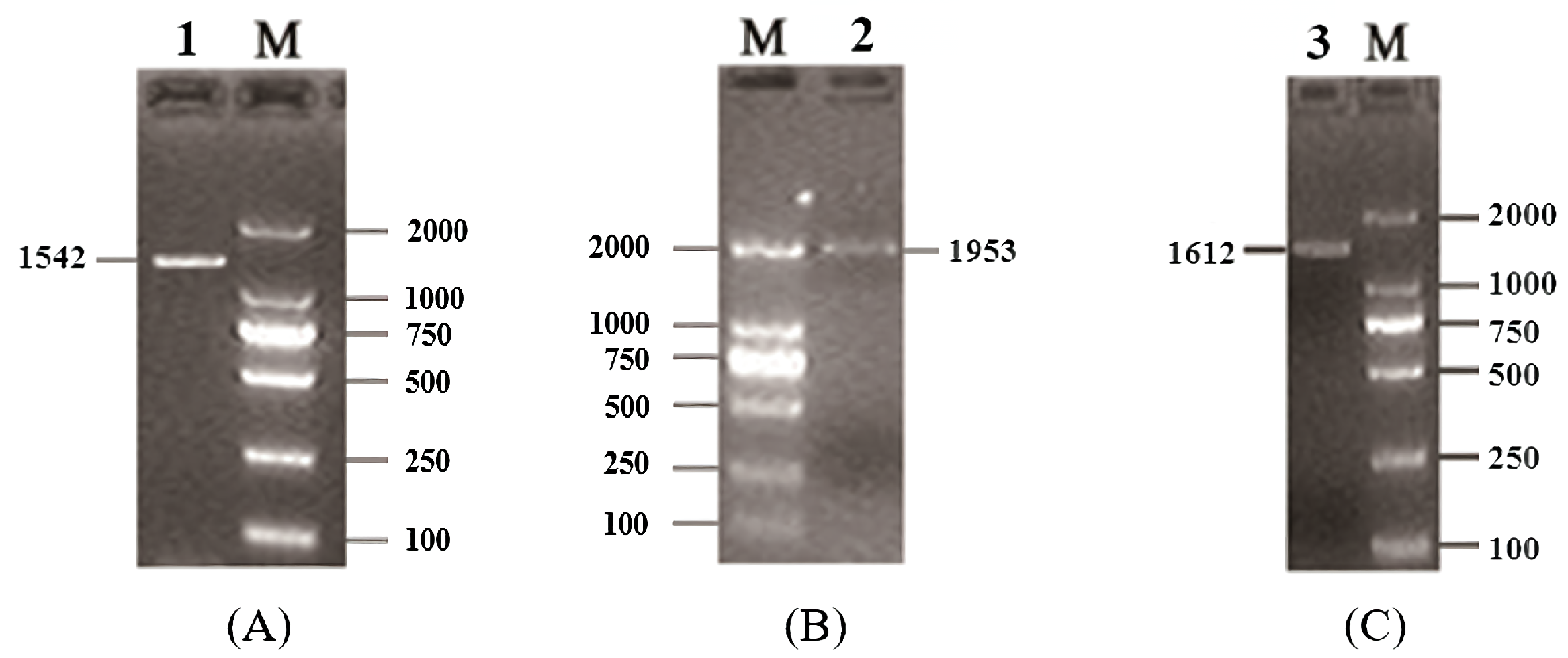
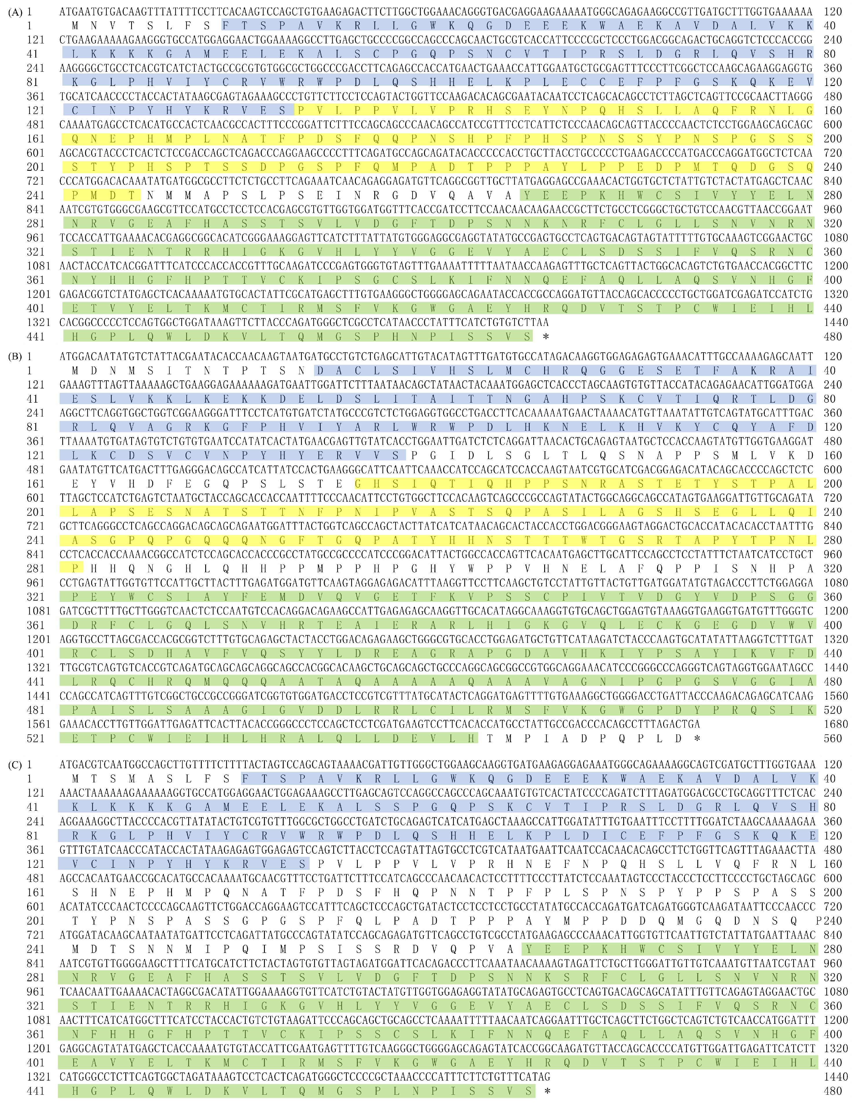
3.1. Isolation and Identification of Buffalo Smad1/4/5

Using the mixed cDNA as the templates, the full-length CDSs of buffalo Smads 1, 4, and 5 were isolated by PCR. The PCR products obtained were 1542 bp, 1953 bp, and 1612 bp in length, respectively (Figure 1). They, in turn, contain an open reading frame of 1398 bp, 1662 bp, and 1398 bp. The homology search showed that the CDSs from the three amplicons of 1542 bp, 1953 bp, and 1612 bp had high similarity with those of Smads 1, 4, and 5 of other common species of Bovidae (more than 97.85%). Therefore, the obtained sequences here are the sequences of buffalo Smads 1, 4, and 5. Then the CDSs of buffalo Smads 1, 4, and 5 were submitted to the NCBI database under the accession numbers KF472050, KF472051, and KF472052.

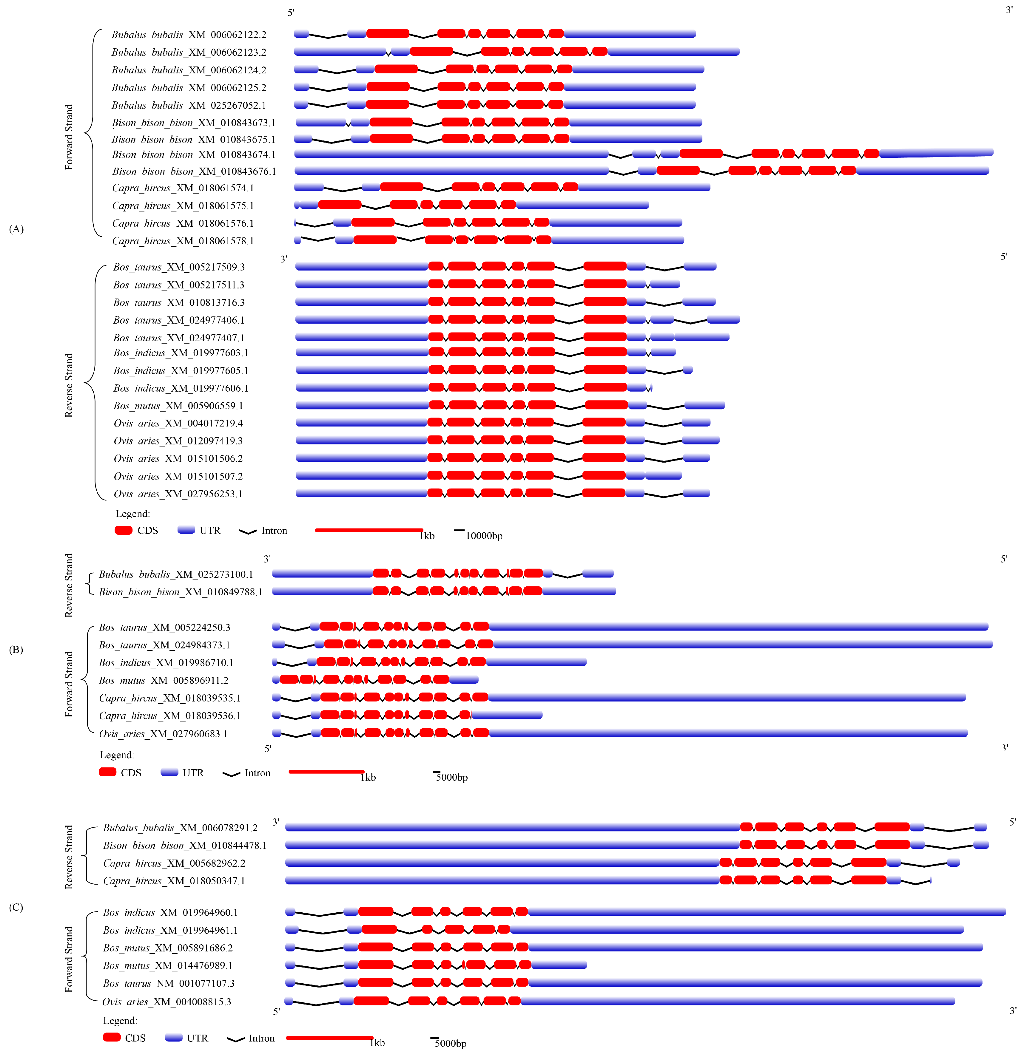
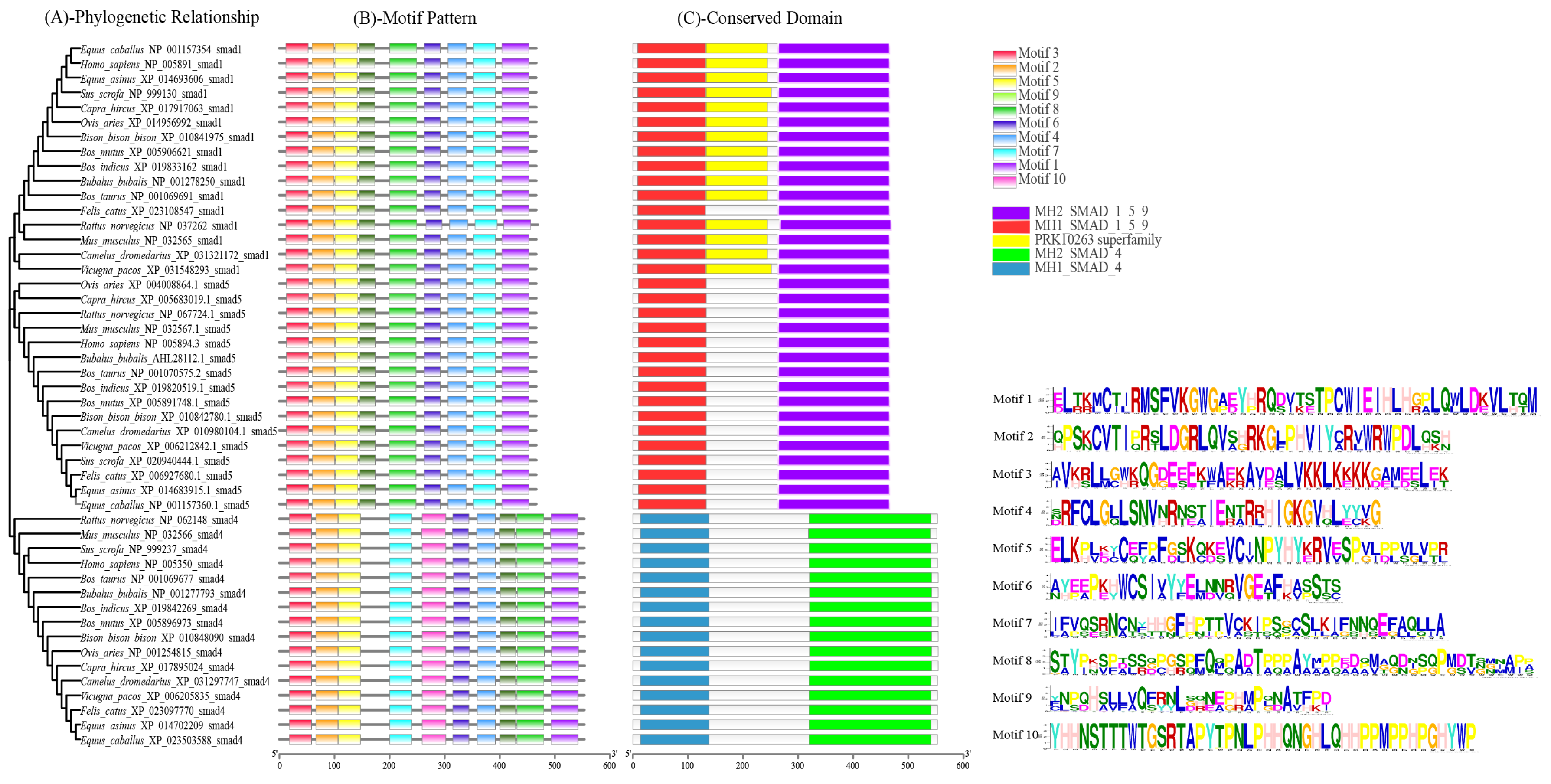
3.2. Gene Sequence and Structure Analysis

The CDSs of buffalo Smads 1, 4, and 5 and their amino acid sequences are presented in in Figure 2.
The results of codon usage bias analysis for buffalo Smads 1, 4, and 5 are shown in Tables S1–S6. The ENc (effective number of codons), contents of GC, and GC3s (the guanine or cytosine frequency at the third codon position of a synonymous codon, excluding methionine, tryptophan, and stop codons) for buffalo Smads 1, 4, and 5 in this study are closer to those of other species in Bovidae. The RSCUs (relative synonymous codon usage) of buffalo Smads 1, 4, and 5 are highly similar to those of cattle. The top three RSCU values for the buffalo Smad1 are CUG (2.33), AGC (2.08), and UGC (1.86), those for the buffalo Smad4 are AGU (2.05), GGA (2.05), and UUU (1.85), and for the buffalo Smad5 are AUU (2.12), AGC (1.80), and GCA (1.80). Codon CUA and AUA are not used in buffalo Smad1, and codon GCG is not used in buffalo Smad5, but there are no unused codons in buffalo Smad4.
The gene structures of buffalo Smads 1, 4, and 5 and their comparison with those of six other species of Bovidae are showed in Figure 3 and Tables S7–S9. For the Smads 1, 4, and 5, the length of CDS for each gene was almost the same across species of Bovidae. However, the lengths of the UTR (5′UTR and 3′UTR) and introns in them are inconsistent among different species (Figure 3). Among the species of Bovidae, the Smad1 gene commonly contains several alternative splicing transcripts, while the Smad4 and Smad5 contain a small number of the transcripts. In buffalo, Smad1 contains five kinds of alternative splicing transcripts with inconsistent 5’UTR, one of which has a codon deletion in exon 4 (this was also found in sheep), but only one alternative splicing transcript was found for Smad4 and Smad5 in buffalo. It is worth noting that the three genes can be located on different DNA strands in the same species. For example, Smads 1 and 5 are located on the positive strand of DNA in Bubalus bubalis, Bison bison bison, and Capra hircus, while Smad4 are located in the reverse strand of DNA in Bubalus bubalis and Bison bison bison.

3.3. Basic Physicochemical Properties of the Three Proteins

There were no significant differences in the basic physicochemical characteristics of Smads 1, 4, and 5 between buffalo and cattle (Smad1, NM_001076223.2; Smad4, NM_001076209.1; Smad5, NM_001077107.3) (Table 2). The Smads 1, 4, and 5 are all unstable proteins without transmembrane regions and signal peptides for both buffalo and cattle. According to the values of GRAVY, the Smads 1, 4, and 5 of buffalo and cattle are all hydrophilic (Table 2, Figure S1).

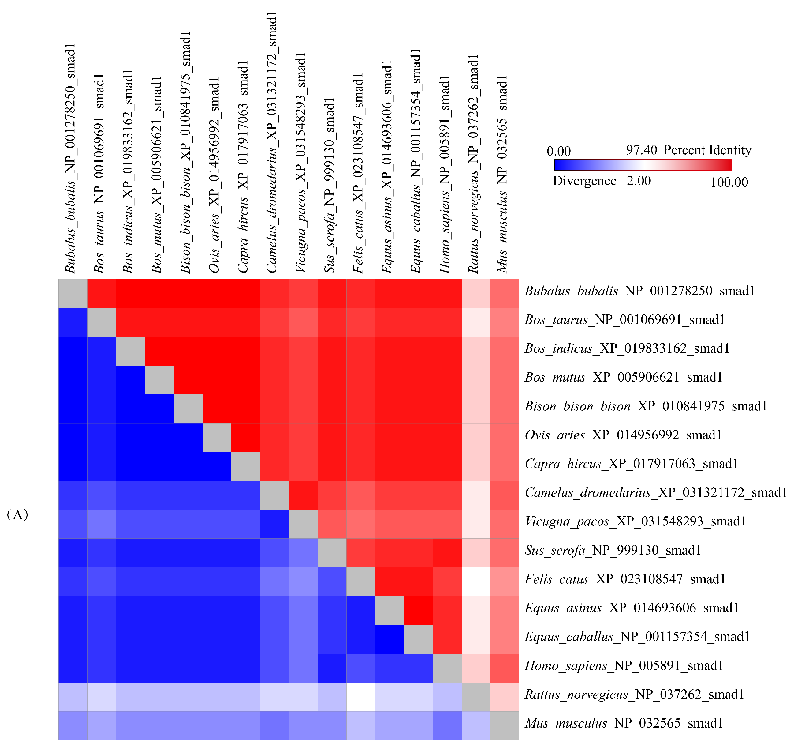
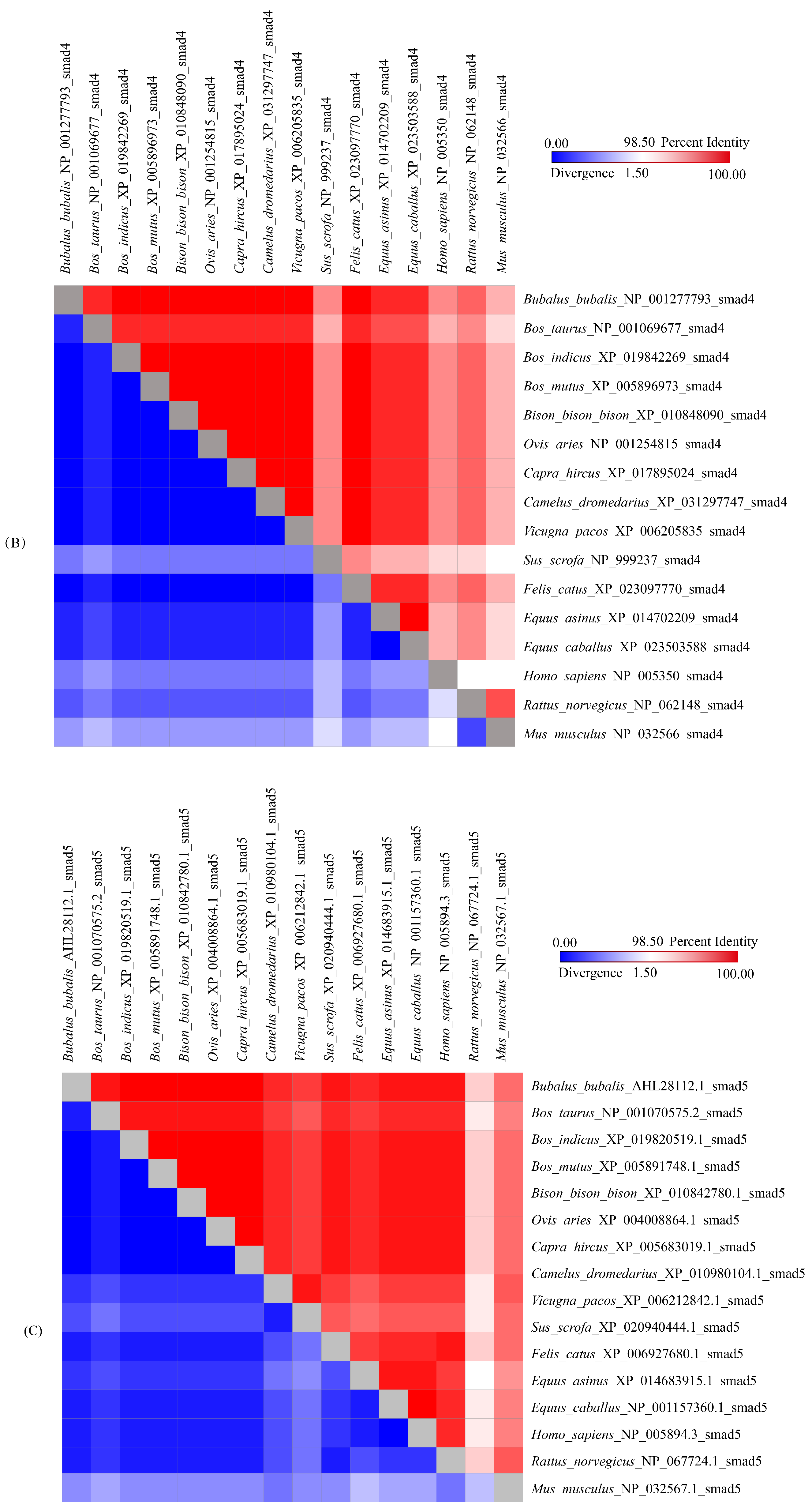
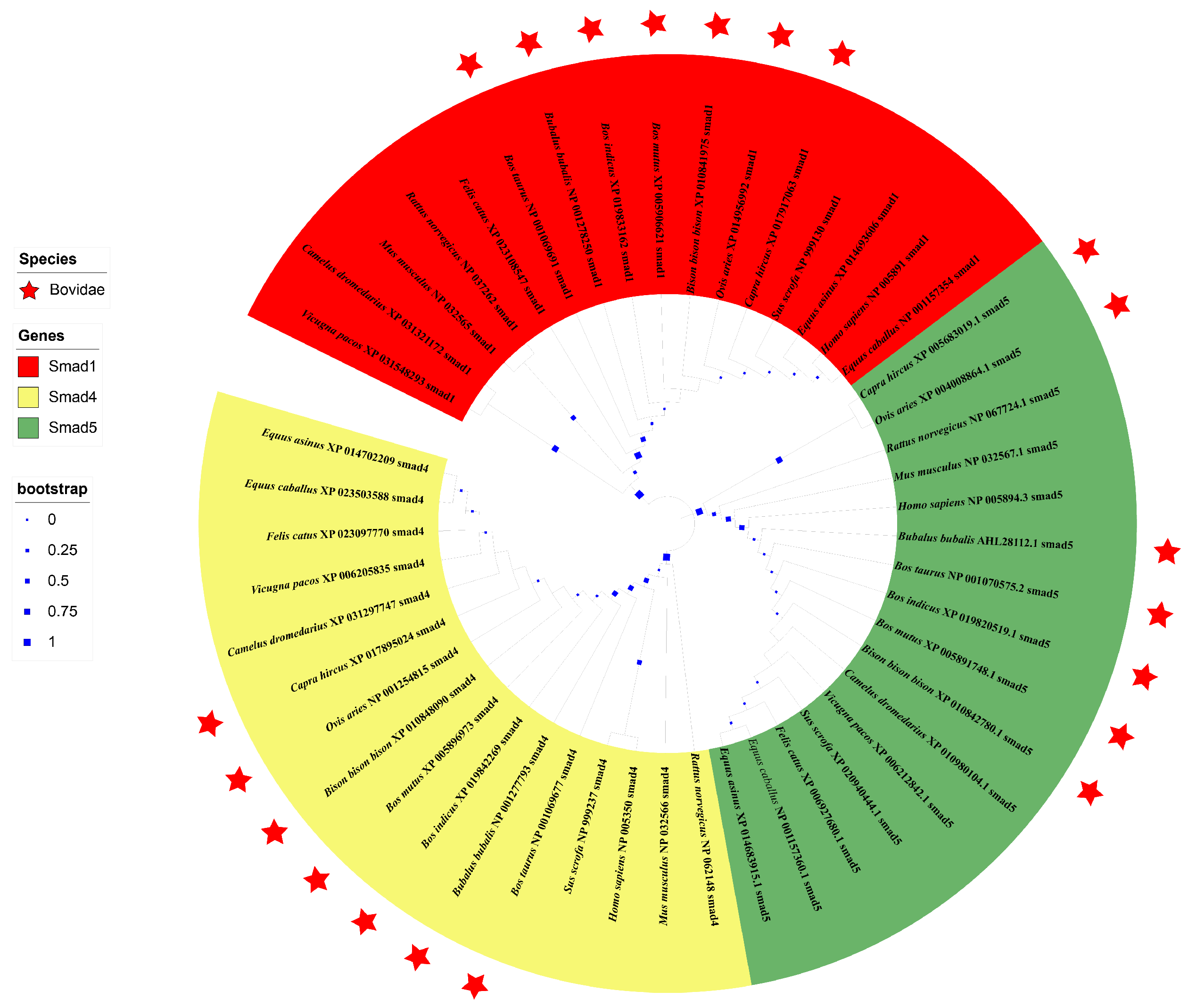
3.4. Sequence Identities and Phylogenetic Analysis

The alignment of amino acid sequences showed that buffalo Smads 1, 4, and 5 have high consistency with those of other species previously published in the NCBI database, especially with those of the species in Bovidae (Figure 4). The sequences of buffalo Smads 1, 4, and 5 all have more than 99.4% similarity with its homologous sequences of other species in the family Bovidae and more than 98.5% similarity with other mammalian species (Figure 4).
In the phylogenetic trees based on the amino acid of 15 species in mammals, Smads 1, 4, and 5 were clustered in their own clade, and the genetic relationship between buffalo and other species of Bovidae is relatively close (Figure 5).
To explore the common structural characteristics and functional similarities of the Smad 1, 4, and 5 proteins, the analyses of the motif patten and conserved domain were taken based on their amino acid sequences in the above species (Figure 6). In 15 mammalian species, nine to ten conserved motifs were found in the Smads 1, 4, and 5 (Figure 6B). Among 15 mammalian species, we found that similar motif patterns exist in Smad4, and in both of Smad1 and Smad5. The two ends of Smad4 had similar motifs to the two ends of Smad1 and Smad5, but the motifs in the middle of Smad4 and Smad1/5 were different (Figure 6B). Motif 9 only exists in Smad1 and Smad5, and motif 10 only exists in Smad4. For motif 6, there is only one copy in both Smad1 and Smad5, but for Smad4, only Sus scrofa and Bos taurus have one copy, and all the other 13 species have two copies (Figure 6B). More detail information about these motifs in the three Smads, such as E-value, sites, and window width is presented in Table 3.
The prediction showed that buffalo Smads 1, 4, and 5 all contain two conserved functional domains coupled at a link region, which are the same as in other species (Figure 6C): a N-terminal Mad Homology 1 domain (MH1, aa 9–132 for the Smad1, aa 14–138 for the Smad4, aa 10–133 for the Smad5), which belongs to MH1 superfamily, and a C-terminal Mad Homology 2 domain (MH2, aa 265–465 for the Smad1, aa 321–542 for the Smad4, aa 265–465 for the Smad5), which belongs to MH2 superfamily. In buffalo, Smads 1 and 5, a Ser-Ser-X-Ser (SSVS) motif was found at the end of the MH2 domain. In addition, there is a PRK10263 domain which belongs to PRK10263 superfamily in the Smad1, and a Pneumovirinae attachment membrane glycoprotein G domain which belongs to Pneumo_att_G superfamily in the Smad4.

3.5. Structure of Buffalo Smads 1, 4, and 5

Buffalo Smad1 consists of 22.58% α -helix (105 AAs), 17.85% extended strand (83 AAs), 5.38% β -turn (25 AAs), and 54.19% random coil (252 AAs); buffalo Smad4 contains 24.41% α -helix (135 AAs), 15.55% extended strand (86 AAs), 6.69% β -turn (37 AAs), and 53.35% random coil (295 AAs), and buffalo Smad5 is composed of 22.15% α -helix (103 AAs), 19.35% extended strand (90 AAs), 5.16% β -turn (24 AAs), and 53.33% random coil (248 AAs) (Figure S2).
Three-dimensional structures of buffalo Smad1/4/5 were built based on the method of homologous modeling.The sequence consistencies of buffalo Smads 1, 4, and 5 with their human counterparts (1khu. 1.a, 1DD1.b, and 1Khu. 1.b) were 99.54%, 100%, and 93.09%, respectively, and the coverage rates were 47%, 48%, and 47%, respectively. Three-dimensional structures of the three buffalo Smads are highly similar to those of cattle (Figure S3).

3.6. Subcellular Location and Molecular Function

NLS prediction showed that buffalo Smad1 contained an NLS in aa 12–42 with a score of 5.4, while the Smad5 contained an NLS in aa 13–43 with a score of 5.4. However, NLS was not found in the Smad4. The NLSs of Smads 1 and 5 are located in their MH1 domain. The cut-off score of 5.4 indicates that the Smads 1 and 5 are located in both nucleus and cytoplasm. Subcellular localization prediction also showed that buffalo Smads 1, 4, and 5 play roles in the cytoplasm and nucleus. The prediction showed that the Smads 1, 4, and 5 proteins are involved in the biological processes related to DNA-templated gene transcription regulation (GO:0006355) and TGF- β (transforming growth factor beta) receptor signaling pathway (GO:0007179). Their molecular function is mainly protein binding (GO:0005515), and their cellular component is the transcription regulator complex (GO:0005667).
The protein-protein interaction network was constructed by using the program SIRING (Figure S4). The three Smad proteins were found to interact with 17 other proteins, which included some TGF- β family receptors (such as BMPR1A, ACVR2A), SKI, and other Smads. In addition, the Smad4 and Smad5 can interact with a ubiquitin-protein ligase (SMURF2 or NEDD4L), the Smad1 and Smad5 can interact with ZFYVE9, the Smad1 can interact with YAP1, and the Smad4 can interact with BMP2 and SKIL.

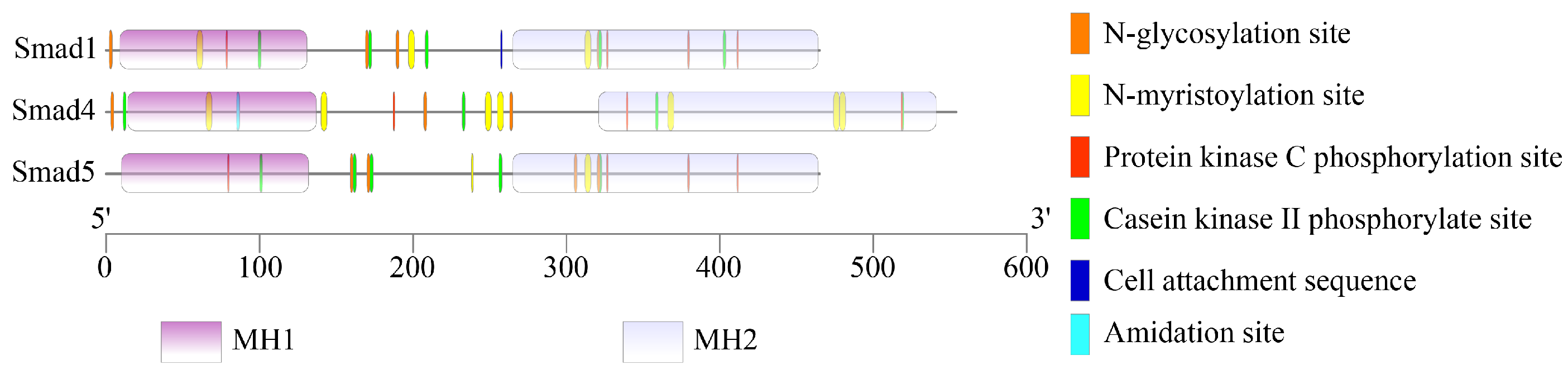
3.7. Amino Acid Modifications

The prediction of modification sites revealed that there were five, five, and four types of amino acid modification sites in the Smad1, Smad4, and Smad5, respectively (Figure 7), including N-glycosylation site, N-myristoylation site, protein kinase C phosphorylation site, and casein kinase II phosphorylate site (Table S10).

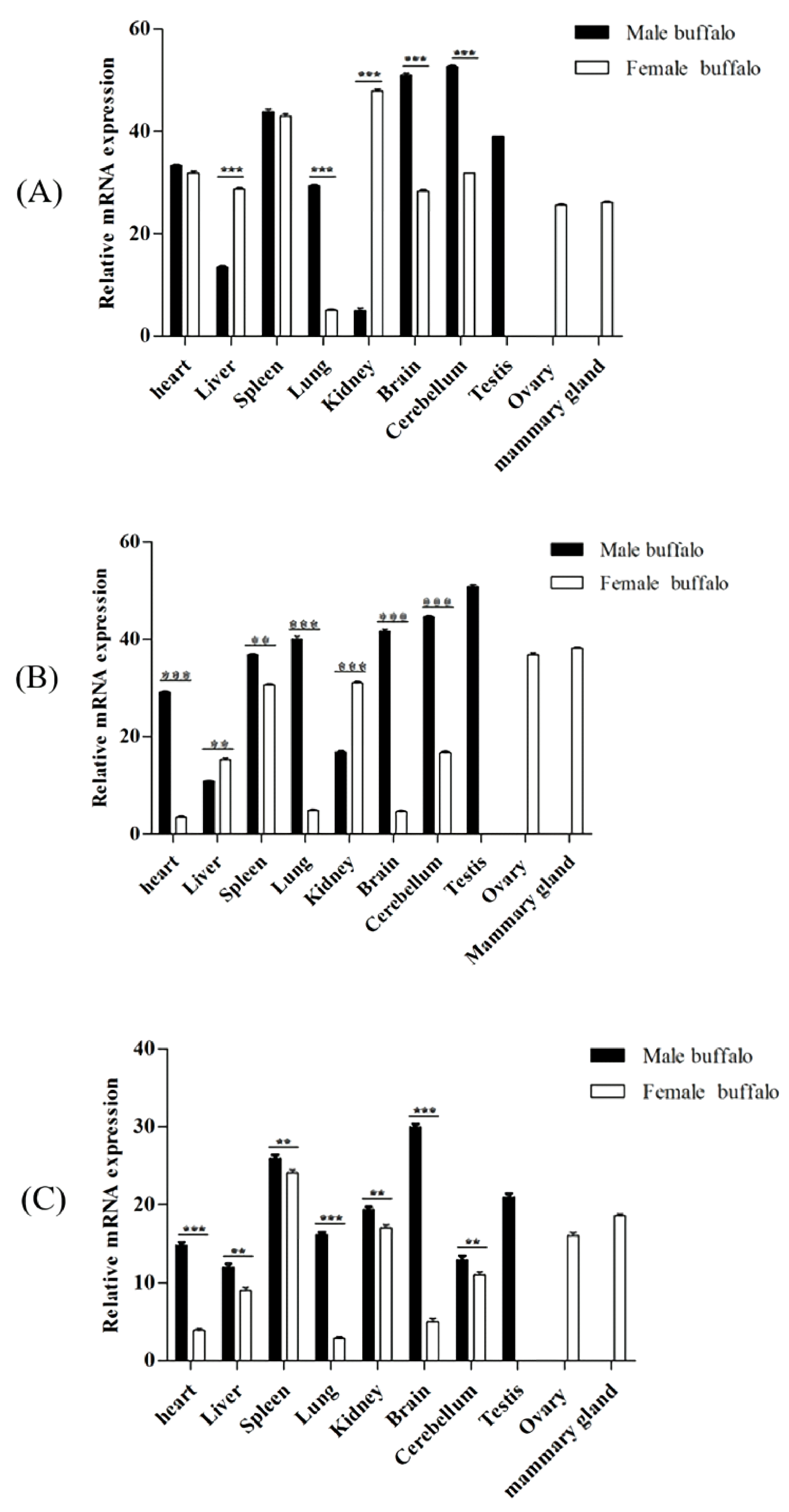
3.8. Tissue Expression Profile

The expressions of Smads 1, 4, and 5 genes were detected in 10 buffalo tissues in this study (Figure 8). For the male and female buffalo, the expression levels of Smads 1, 4, and 5 in most of the assayed tissues were inconsistent. For example, in male buffalo, the relative expression levels of Smad4 and Smad5 in the heart and spleen, Smad5 in the liver and kidney, Smads 1, 4, and 5 in the lung, brain, and cerebellum were significantly higher than those in female buffalo (p < 0.01), and the expression levels of Smad1 and Smad4 in the liver and kidney of male buffalo were significantly lower than those of female buffalo (p < 0.01). It was worth noting that the expression abundance of the three genes in the mammary gland, ovary, and testis was relatively high in all the tissues tested.

4. Discussion

A total of eight Smads are found in mammals, which are divided into three distinct subtypes: the first category includes Smads 1, 2, 3, 5, and 8, which are receptor-regulated Smads (R-Smads). The second type acts as common partner Smad (Co-Smad) for all R-Smads, including only Smad4. The third category is inhibitory Smads (I-Smads), including Smads 6 and 7 [29]. In this study, the CDS of Smads 1, 4, and 5 was isolated and identified from water buffalo. Only one type of CDS was identified in each of the three buffalo Smads. The length, base composition, and RSCU value of the CDS of buffalo Smads 1, 4, and 5 are highly consistent with that of their counterparts in other Bovidae species. In addition, the structure of the transcriptional regions of the three Smad genes of buffalo is also highly similar to that of other Bovidae animals. The physicochemical characteristics, motifs, conserved domains, and three-dimensional structures of buffalo Smads 1, 4, and 5 are also highly consistent with those of other mammalian species. The alignment of amino acid sequences showed that buffalo Smads 1, 4, and 5 have high consistency with those of other mammals. Phylogenetic analysis also displayed that the three buffalo Smads were clustered in their own clade, each of which showed that the buffalo has a close genetic relationship with other mammals. The above results indicate that buffalo Smads 1, 4, and 5 have similar functions with those of other mammals, especially the species in Bovidae, and that the three genes in mammals are functionally conserved.
Smads 1, 4, and 5, as signal transduction molecules, are an important part of the mammalian BMP signaling pathway, which can transmit signals from membrane receptors to the nucleus [30]. As members of the TGF-β superfamily, BMPs constitute a highly conservative multifunctional growth factor family. They have various functions, such as neurogenesis, skeletal formation, hematopoiesis, and embryogenesis [31]. Previous studies have shown that BMP-mediated receptor phosphorylation can initiate the BMP signaling [32]. After BMPs bind to the type II receptor, the type I receptor is phosphorylated, then it further phosphorylates the conserved C-terminal Ser-Ser-X-Ser (SSXS) motif of R-Smads (Smads 1, 5 and 8 in BMP pathway). The phosphorylated R-Smads further combine with Smad4 to form a transcriptional complex, which enters the nucleus, where the complex either binds to the specific enhancer/promoter sequence of the target gene alone, or cooperates with some DNA binding subunits to simultaneously activate or repress hundreds of target genes under tightly controlled conditions [33]. In this study, the conserved C-terminal SSVS motif has also been found in buffalo Smads 1 and 5, which also support the theory that buffalo Smads 1 and 5 can be phosphorylated by membrane receptors. Bioinformatic predictions indicate that the main functions of buffalo Smads 1, 4, and 5 are to interact with many proteins in the BMP signaling pathway, and ultimately form a transcriptional regulatory complex that participates in the regulation of target genes, suggesting that the three Smads probably play pivotal roles in buffalo BMP pathway.
The Smad complex mentioned above has a weak affinity for DNA, and its specificity is limited [30]. How they accurately regulate hundreds of BMP signal target genes in the same cell is the focus of current research. One mainstream view is that the key to precise control lies in the existence of post-translational modifications at several functional sites of Smad proteins [34]. Based on the function sites, the Smad complex can cooperate with other site-specific transcription factors and a variety of coactivators and corepressors to modify histones, modulate chromatin structure, stabilize their DNA binding, and further activate or repress target genes directly or indirectly [30]. In this study, some modification sites were predicted in each of the three buffalo proteins. It is speculated that they are closely related to the precise regulation of intracellular BMP signaling. Whether and how these function sites are involved in the precise control of buffalo Smads needs further study.
Previous studies have shown that Smad proteins mediate the signals of BMPs, which are involved in a variety of biological activities. It has been found that the BMP-Smad signaling pathway is involved in heart development [35], embryonic cerebellum development [36], and nervous system development [3]. During early embryonic development and reproductive system development, the genes involved in the BMP signaling pathway have been found to affect the early regulatory processes of porcine oogenesis [37]; in cattle and buffalo, BMP and its receptors have been found to regulate the process of follicle development through granulosa cell steroidogenesis [32,38], and are stage-specifically expressed in the corpus luteum of buffalo [39]. In addition, BMP signaling can induce specific primordial germ cells, which are further recruited to participate in several other germline-specific and somatic reproductive roles. In this study, we found that Smads are highly conserved in structure and function. From this, we infer that the three Smads are also involved in a variety of biological processes of buffalo.
Studies in cattle and Hu sheep have shown that Smads, including Smads 1, 4, and 5 are expressed in various tissues as the transcription factors in the BMP-Smad pathway [40,41]. In this study, the tissue differential expression analysis showed that the Smads 1, 4, and 5 were expressed in all male and female buffalo tissues assayed, indicating that they play a role in a variety of tissues. Furthermore, some research has suggested that the TGF-β/BMP pathway may be involved in immune response, and the spleen plays important roles in the immune system [42]. In this study, the Smads 1, 4, and 5 are highly expressed in the spleen of male and female buffalo, suggesting that the three buffalo Smads may also participate in the immune response. It is worth noting that buffalo Smads 1, 4, and 5 were relatively highly expressed in the mammary glands and gonads, which means that they may play an important role in these tissues, and they probably participate in the reproductive process of buffalo.

5. Conclusions

In this study, buffalo Smads 1, 4, and 5 were isolated and characterized. The physicochemical properties, motifs, conservative domains, and secondary and three-dimensional structures of buffalo Smads 1, 4, and 5 are highly similar to those of Smads 1, 4, and 5 in cattle. Furthermore, the three proteins are functionally conserved in mammals. Buffalo Smads 1, 4, and 5 can interact with BMPs and their receptors, and they probably serve as signal transducers to relay signals from membrane receptors to the nucleus in BMP signaling pathway. The three buffalo Smads were expressed in all examined tissues, especially with relatively high mRNA abundance in the mammary gland, gonad, and spleen. The results here suggests that buffalo Smads 1, 4, and 5 may play key roles in the regulation of gene transcription in a variety of tissues, and these roles may be closely related to the lactation, reproduction, and immune response processes of buffalo. This study will provide a basis for elucidating the genetic mechanism of buffalo production traits and high disease resistance.

Supplementary Materials

The following are available online at https://0-doi-org.brum.beds.ac.uk/10.5281/zenodo.5181340, Figure S1: Hydrophilicity prediction of buffalo Smad1/4/5; Figure S2: Predicted secondary structures of buffalo Smad1(A), Smad4(B) and Smad5(C); Figure S3: Predicted three-dimensional structures of buffalo and cattle Smad1(A), Smad4(B), and Smad5(C); Figure S4: Protein-protein interaction network of the Smad1/4/5; Table S1: ENc values, contents of GC and GC3s for Smad1 gene in different species; Table S2: ENc values, contents of GC and GC3s for Smad4 gene in different species; Table S3: ENc values, contents of GC and GC3s for Smad5 gene in different species; Table S4: RSCU values of Smad1 gene in buffalo and other species; Table S5: RSCU values of Smad4 gene in buffalo and other species; Table S6: RSCU values of Smad5 gene in buffalo and other species; Table S7: Compare of the gene structure of Smad1 between buffalo and some species of Bovidae; Table S8: Comparison of the gene structure of Smad4 between buffalo and other species of Bovidae; Table S9: Comparison of the gene structure of Smad5 between buffalo and other species of Bovidae; Table S10: Information on putative functional sites.

Author Contributions

Conceptualization, J.Z. and Y.M.; data curation, J.Z., G.Z. and Y.M.; formal analysis, J.Z. and G.Z.; funding acquisition, Y.M.; investigation, J.Z., G.Z. and Y.M.; project administration, Y.M.; resources, Y.M.; software, J.Z.; validation, J.Z., G.Z. and Y.M.; writing—original draft, J.Z. and Y.M.; writing—review & editing, J.Z. and Y.M. All authors have read and agreed to the published version of the manuscript.

Funding

This study was financially supported by the National Natural Science Foundation of China (Grant nos. 31760659 and 31460582) and the Natural Science Foundation Key Project of Yunnan Province, China (Grant nos. 2014FA032 and 2007C0003Z).

Institutional Review Board Statement

The procedures of sample collection were executed according to the Guide for Animal Care and Use of Experimental Animals and allowed by the Institutional Animal Care and Use Committee of Yunnan Agricultural University. Every effort has been made to minimize suffering.

Informed Consent Statement

Not applicable.

Data Availability Statement

Not applicable.

Conflicts of Interest

The authors declare that the study was conducted in the absence of any commercial or financial relationships that could be construed as a potential conflict of interest.

References

  1. Massagué, J.; Seoane, J.; Wotton, D. Smad transcription factors. Genes Dev. 2005, 19, 2783–2810. [Google Scholar] [CrossRef] [Green Version]
  2. Mullen, A.C.; Wrana, J.L. TGF-β Family Signaling in Embryonic and Somatic Stem-Cell Renewal and Differentiation. Cold Spring Harb. Perspect. Biol. 2017, 9, a022186. [Google Scholar] [CrossRef]
  3. Hegarty, S.V.; O’Keeffe, G.W.; Sullivan, A.M. BMP-Smad 1/5/8 signalling in the development of the nervous system. Prog. Neurobiol. 2013, 109, 28–41. [Google Scholar] [CrossRef]
  4. Rahman, M.S.; Akhtar, N.; Jamil, H.M.; Banik, R.S.; Asaduzzaman, S.M. TGF-β/BMP signaling and other molecular events: Regulation of osteoblastogenesis and bone formation. Bone Res. 2015, 3, 15005. [Google Scholar] [CrossRef] [Green Version]
  5. Lochab, A.K.; Extavour, C.G. Bone Morphogenetic Protein (BMP) signaling in animal reproductive system development and function. Dev. Biol. 2017, 427, 258–269. [Google Scholar] [CrossRef]
  6. McNatty, K.P.; Fidler, A.E.; Juengel, J.L.; Quirke, L.D.; Smith, P.R.; Heath, D.A.; Lundy, T.; O’Connell, A.; Tisdall, D.J. Growth and paracrine factors regulating follicular formation and cellular function. Mol. Cell. Endocrinol. 2000, 163, 11–20. [Google Scholar] [CrossRef]
  7. Kumar, S.; Lakshmi Devi, H.; Singh Jalmeria, N.; Punetha, M.; Pandey, Y.; Samad, H.A.; Singh, G.; Sarkar, M.; Chouhan, V.S. Expression and functional role of bone morphogenetic proteins (BMPs) in placenta during different stages of pregnancy in water buffalo (Bubalus bubalis). Gen. Comp. Endocrinol. 2020, 285, 113249. [Google Scholar] [CrossRef]
  8. Lee, K.B.; Folger, J.K.; Rajput, S.K.; Smith, G.W. Temporal regulation of mRNAs for select bone morphogenetic proteins (BMP), BMP receptors and their associated SMAD proteins during bovine early embryonic development: Effects of exogenous BMP2 on embryo developmental progression. Reprod. Biol. Endocrinol. 2014, 12, 67. [Google Scholar] [CrossRef] [Green Version]
  9. García, E.V.; Hamdi, M.; Barrera, A.D.; Sánchez-Calabuig, M.J.; Gutiérrez-Adán, A.; Rizos, D. Bovine embryo-oviduct interaction in vitro reveals an early cross talk mediated by BMP signaling. Reproduction 2017, 153, 631–643. [Google Scholar] [CrossRef] [Green Version]
  10. Neira, J.A.; Tainturier, D.; Peña, M.A.; Martal, J. Effect of the association of IGF-I, IGF-II, bFGF, TGF-β1, GM-CSF, and LIF on the development of bovine embryos produced in vitro. Theriogenology 2010, 73, 595–604. [Google Scholar] [CrossRef]
  11. Zhang, X.; Xu, S.; Gao, X.; Ren, H.; Chen, J. Cloning and bioinformatics analysis of cDNA encoding cattle Smad4 gene. Front. Agric. China 2008, 2, 493–497. [Google Scholar] [CrossRef]
  12. Zimin, A.V.; Delcher, A.L.; Florea, L.; Kelley, D.R.; Schatz, M.C.; Puiu, D.; Hanrahan, F.; Pertea, G.; Van Tassell, C.P.; Sonstegard, T.S.; et al. A whole-genome assembly of the domestic cow, Bos taurus. Genome Biol. 2009, 10, R42. [Google Scholar] [CrossRef] [Green Version]
  13. FAOSTAT. Food and Agriculture Organization of the United Nations Statistics Database. 2018. Available online: http://www.fao.org/faostat/en/ (accessed on 22 September 2021).
  14. Warriach, H.M.; McGill, D.M.; Bush, R.D.; Wynn, P.C.; Chohan, K.R. A Review of Recent Developments in Buffalo Reproduction. Asian-Australas. J. Anim. Sci. 2015, 28, 451–455. [Google Scholar] [CrossRef]
  15. Lalitha, S. Primer Premier 5. Biotech Softw. Internet Rep. 2000, 1, 270–272. [Google Scholar] [CrossRef]
  16. Chen, C.; Chen, H.; Zhang, Y.; Thomas, H.R.; Frank, M.H.; He, Y.; Xia, R. TBtools: An integrative toolkit developed for interactive analyses of big biological data. Mol. Plant 2020, 13, 1194–1202. [Google Scholar] [CrossRef]
  17. Hu, B.; Jin, J.; Guo, A.Y.; Zhang, H.; Luo, J.; Gao, G. GSDS 2.0: An upgraded gene feature visualization server. Bioinformatics 2015, 31, 1296–1297. [Google Scholar] [CrossRef] [Green Version]
  18. Petersen, T.N.; Brunak, S.; Von Heijne, G.; Nielsen, H. SignalP 4.0: Discriminating signal peptides from transmembrane regions. Nat. Methods 2011, 8, 785–786. [Google Scholar] [CrossRef]
  19. Krogh, A.; Larsson, B.; Von Heijne, G.; Sonnhammer, E.L. Predicting transmembrane protein topology with a hidden Markov model: Application to complete genomes. J. Mol. Biol. 2001, 305, 567–580. [Google Scholar] [CrossRef] [Green Version]
  20. Walker, J.M. The Proteomics Protocols Handbook; Springer: Berlin/Heidelberg, Germany, 2005. [Google Scholar]
  21. Larkin, M.A.; Blackshields, G.; Brown, N.P.; Chenna, R.; McGettigan, P.A.; McWilliam, H.; Valentin, F.; Wallace, I.M.; Wilm, A.; Lopez, R. Clustal W and Clustal X version 2.0. Bioinformatics 2007, 23, 2947–2948. [Google Scholar] [CrossRef] [Green Version]
  22. Kumar, S.; Stecher, G.; Tamura, K. MEGA7: Molecular evolutionary genetics analysis version 7.0 for bigger datasets. Mol. Biol. Evol. 2016, 33, 1870–1874. [Google Scholar] [CrossRef] [Green Version]
  23. Lu, S.; Wang, J.; Chitsaz, F.; Derbyshire, M.K.; Geer, R.C.; Gonzales, N.R.; Gwadz, M.; Hurwitz, D.I.; Marchler, G.H.; Song, J.S. CDD/SPARCLE: The conserved domain database in 2020. Nucleic Acids Res. 2020, 48, D265–D268. [Google Scholar] [CrossRef] [PubMed] [Green Version]
  24. Biasini, M.; Bienert, S.; Waterhouse, A.; Arnold, K.; Studer, G.; Schmidt, T.; Kiefer, F.; Cassarino, T.G.; Bertoni, M.; Bordoli, L. SWISS-MODEL: Modelling protein tertiary and quaternary structure using evolutionary information. Nucleic Acids Res. 2014, 42, W252–W258. [Google Scholar] [CrossRef] [PubMed]
  25. Kosugi, S.; Hasebe, M.; Entani, T.; Takayama, S.; Tomita, M.; Yanagawa, H. Design of peptide inhibitors for the importin alpha/beta nuclear import pathway by activity-based profiling. Chem. Biol. 2008, 15, 940–949. [Google Scholar] [CrossRef] [PubMed] [Green Version]
  26. Kosugi, S.; Hasebe, M.; Matsumura, N.; Takashima, H.; Miyamoto-Sato, E.; Tomita, M.; Yanagawa, H. Six classes of nuclear localization signals specific to different binding grooves of importin alpha. J. Biol. Chem. 2009, 284, 478–485. [Google Scholar] [CrossRef] [PubMed] [Green Version]
  27. Kosugi, S.; Hasebe, M.; Tomita, M.; Yanagawa, H. Systematic identification of cell cycle-dependent yeast nucleocytoplasmic shuttling proteins by prediction of composite motifs. Proc. Natl. Acad. Sci. USA 2009, 106, 10171–10176. [Google Scholar] [CrossRef] [Green Version]
  28. Chou, K.C.; Shen, H.B. A new method for predicting the subcellular localization of eukaryotic proteins with both single and multiple sites: Euk-mPLoc 2.0. PLoS ONE 2010, 5, e9931. [Google Scholar] [CrossRef]
  29. Hata, A.; Chen, Y.G. TGF-β Signaling from Receptors to Smads. Cold Spring Harb. Perspect. Biol. 2016, 8, a022061. [Google Scholar] [CrossRef]
  30. Hill, C.S. Transcriptional Control by the SMADs. Cold Spring Harb. Perspect. Biol. 2016, 8, a022079. [Google Scholar] [CrossRef] [Green Version]
  31. Carreira, A.C.; Alves, G.G.; Zambuzzi, W.F.; Sogayar, M.C.; Granjeiro, J.M. Bone morphogenetic proteins: Structure, biological function and therapeutic applications. Arch. Biochem. Biophys. 2014, 561, 64–73. [Google Scholar]
  32. Rajesh, G.; Mishra, S.R.; Paul, A.; Punetha, M.; Vidyalakshmi, G.M.; Narayanan, K.; Bag, S.; Bhure, S.K.; Singh Chouhan, V.; Maurya, V.P.; et al. Transcriptional and translational abundance of Bone morphogenetic protein (BMP) 2, 4, 6, 7 and their receptors BMPR1A, 1B and BMPR2 in buffalo ovarian follicle and the role of BMP4 and BMP7 on estrogen production and survival of cultured granulosa cells. Res. Vet. Sci. 2018, 118, 371–388. [Google Scholar] [CrossRef]
  33. Budi, E.H.; Duan, D.; Derynck, R. Transforming Growth Factor-β Receptors and Smads: Regulatory Complexity and Functional Versatility. Trends Cell Biol. 2017, 27, 658–672. [Google Scholar] [CrossRef]
  34. Xu, P.; Lin, X.; Feng, X.H. Posttranslational Regulation of Smads. Cold Spring Harb. Perspect. Biol. 2016, 8, a022087. [Google Scholar] [CrossRef] [Green Version]
  35. Koefoed, K.; Skat-Rørdam, J.; Andersen, P.; Warzecha, C.B.; Pye, M.; Andersen, T.A.; Ajbro, K.D.; Bendsen, E.; Narimatsu, M.; Vilhardt, F.; et al. The E3 ubiquitin ligase SMURF1 regulates cell-fate specification and outflow tract septation during mammalian heart development. Sci. Rep. 2018, 8, 9542. [Google Scholar] [CrossRef]
  36. Tong, K.K.; Ma, T.C.; Kwan, K.M. BMP/Smad signaling and embryonic cerebellum development: Stem cell specification and heterogeneity of anterior rhombic lip. Dev. Growth Differ. 2015, 57, 121–134. [Google Scholar] [CrossRef]
  37. Budna, J.; Rybska, M.; Ciesiółka, S.; Bryja, A.; Borys, S.; Kranc, W.; Wojtanowicz-Markiewicz, K.; Jeseta, M.; Sumelka, E.; Bukowska, D.; et al. Expression of genes associated with BMP signaling pathway in porcine oocytes before and after IVM—A microarray approach. Reprod. Biol. Endocrinol. RB&E 2017, 15. [Google Scholar] [CrossRef]
  38. Shimizu, T.; Magata, F.; Abe, Y.; Miyamoto, A. Bone morphogenetic protein 4 (BMP-4) and BMP-7 induce vascular endothelial growth factor expression in bovine granulosa cells. Anim. Sci. J. 2012, 83, 663–667. [Google Scholar] [CrossRef]
  39. Rajesh, G.; Paul, A.; Mishra, S.R.; Bharati, J.; Thakur, N.; Mondal, T.; Soren, S.; Harikumar, S.; Narayanan, K.; Chouhan, V.S.; et al. Expression and functional role of Bone Morphogenetic Proteins (BMPs) in cyclical corpus luteum in buffalo (Bubalus bubalis). Gen. Comp. Endocrinol. 2017, 240, 198–213. [Google Scholar] [CrossRef]
  40. Xu, Y.; Li, E.; Han, Y.; Chen, L.; Xie, Z. Differential expression of mRNAs encoding BMP/Smad pathway molecules in antral follicles of high- and low-fecundity Hu sheep. Anim. Reprod. Sci. 2010, 120, 47–55. [Google Scholar] [CrossRef]
  41. Steuernagel, L.; Meckbach, C.; Heinrich, F.; Zeidler, S.; Schmitt, A.O.; Gültas, M. Computational identification of tissue-specific transcription factor cooperation in ten cattle tissues. PLoS ONE 2019, 14, e0216475. [Google Scholar] [CrossRef] [Green Version]
  42. Yang, L.; Pang, Y.; Moses, H.L. TGF-β and immune cells: An important regulatory axis in the tumor microenvironment and progression. Trends Immunol. 2010, 31, 220–227. [Google Scholar] [CrossRef] [Green Version]
Figure 1. RT-PCR results for buffalo Smad1 (A), Smad4 (B), and Smad5 (C) genes. Lane 1, 2, and 3 are PCR products for the buffalo Smads 1, 4, and 5 genes, respectively. Lane M, DL2000 DNA marker.
Genes 12 01536 g001
Figure 2. Complete CDSs of buffalo Smads 1, 4, and 5 genes and AASs. (AC) exhibit buffalo Smad1, 4, and 5 genes, respectively. The shaded parts of blue/yellow/green represent putative conserved functional domains of MH1_SMAD_1_5_9, PRK10263, and MH2_SMAD_1_5_9 in Smad1, and those of MH1_SMAD_4, Pneumo_att_G, and MH1_SMAD_4 in Smad4, respectively. The shaded parts in blue/green represent putative conserved functional domains of MH1_SMAD_1_5_9 and MH2_SMAD_1_5_9 in Smad5, respectively. “*”, termination codon.
Genes 12 01536 g002
Figure 3. The gene structure of Smads 1, 4, and 5. (A) Smad1, (B) Smad4, (C) Smad5. The red boxes, blue boxes, and the black hat lines represented untranslated regions (UTRs), CDSs, and introns. The length of the exon and intron can be inferred from the legend at the bottom.
Genes 12 01536 g003
Figure 4. Divergences and percent identities according to the AASs of Smads 1, 4, and 5 between buffalo and other species. (A) Smad1, (B) Smad4, and (C) Smad5. The divergences and percent identities were shown in the upper and lower triangle of the matrix, respectively.
Genes 12 01536 g004aGenes 12 01536 g004b
Figure 5. Phylogenetic trees constructed based on the AASs of Smads 1, 4, and 5 in buffalo and other mammals. The bootstrap values of each branch in the tree were labelled with blue boxes of different sizes. The species in the family of Bovidae were labelled by a red star.
Genes 12 01536 g005
Figure 6. The phylogenetic relationships, motif patterns and conserved domains of Smads 1, 4, and 5 in 15 mammalian species. (A) Phylogenetic relationships; (B) motif pattern; (C) conserved domains. Ten conserved motifs and five kinds of conserved domains in Smads 1, 4, and 5 are labelled with different color boxes.
Genes 12 01536 g006
Figure 7. The putative modification sites in buffalo Smads 1, 4, and 5.
Genes 12 01536 g007
Figure 8. Multi-tissue expression profiles of buffalo Smads 1, 4, and 5 genes in male and female buffalo. (A) Smad1, (B) Smad4, (C) Smad5. The values are provided as means ± SE (** p < 0.01, *** p < 0.001).
Genes 12 01536 g008
Table 1. Primers used for isolating buffalo Smad1, Smad4, and Smad5 genes.
GenePrimers(5′–3′)Amplicon Length (bp)Annealing Temperature (°C)Purpose
Smad1F: AAGCCTCTTTCACTGTCCTTTCAC154260CDS isolation
R: GTCTGACTCGTCCATCCTTCAAGT
Smad4F: GCTGCCTCCGAAAGATCAAAAT195355CDS isolation
R: GCTCTGAGCCATGCCTGAAAAGT
Smad5F: TGGCACTTATGAAGATAAAGGTCA161255CDS isolation
R: CTGTACTGGAAGTTTCCCTAAAAT
Smad1F: CCCTCACTCTCCGACCAG23857.1Differential expression
R: ACCATCCACCAACACGCT
Smad4F: TCATTATCCACTGAAGGGCATT17354Differential expression
R: ATACTGGCGGGCTGACTTGT
Smad5F: TTTCCCTTATCTCCAAAT28651.9Differential expression
R: CGACAGGCTGAACATCTC
ACTBF: TGGGCATGGAATCCTG19657.9internal reference
R: GGCGCGATGATCTTGAT
RPS23R: CAGGAAGTGTGTCAGGGT15450.8internal reference
F: TCCAGGAATGTCACCAAC
RPS15R: GATGGTGGGCAGCATGGTT15958.6internal reference
F: AAGCGGGAGGAATGGGTGG
Table 2. Basic characteristics of Smad1/4/5 for buffalo and cattle.
ItemsSmad1Smad4Smad5
BuffaloCattleBuffaloCattleBuffaloCattle
Isoelectric point (PI)6.96.96.56.57.637.63
Molecular weight (MW)52.25 kDa52.22 kDa60.57 kDa60.54 kDa52.33 kDa52.25 kDa
Formula C 2318 H 3537 N 645 O 688 S 25 C 2317 H 3535 N 645 O 687 S 25 C 2675 H 4140 N 764 O 802 S 23 C 2674 H 4138 N 764 O 801 S 23 C 2334 H 3569 N 643 O 683 S 24 C 2328 H 3565 N 643 O 683 S 24
Strongly acidic amino acids (E, D)444451514242
Strongly basic amino acids (R, K)424244444343
Polar amino acids (S, T, Y, N, C, Q)148147160159145145
Hydrophobic amino acids (F, W, V, A, I, L)126126173173132132
Instability index (II)60.8460.2450.9550.8364.0164.42
Grand average of hydropathicity (GRAVY)−0.568−0.567−0.389−0.388−0.517−0.516
Aliphatic index (AI)65.3565.3576.0676.0668.0968.09
Table 3. E-value, sites, and widths of Smad1/4/5 conserved motifs.
Motif1Motif2Motif3Motif4Motif5Motif6Motif7Motif8Motif9Motif10
E-value3.0 × 10−24543.6 × 10−19579.4 ×10−17931.2 × 10−15442.1 × 10−17041.3 × 10−12711.5 × 10−13741.5 × 10−17221.1 × 10−9969.1 × 10−638
Sites48484848484845484816
Width50414134413041502944
Publisher’s Note: MDPI stays neutral with regard to jurisdictional claims in published maps and institutional affiliations.

