Article

A COL5A2 In-Frame Deletion in a Chihuahua with Ehlers-Danlos Syndrome

1
Institute of Genetics, Vetsuisse Faculty, University of Bern, 3001 Bern, Switzerland
2
Dermfocus, University of Bern, 3001 Bern, Switzerland
3
“Biology of the Neuromuscular System” Team, INSERM, IMRB, Univ Paris-Est Créteil, Ecole Nationale Vétérinaire d’Alfort, 94700 Maisons-Alfort, France
4
Unité de Dermatologie, ChuvA, Ecole Nationale Vétérinaire d’Alfort, 94700 Maisons-Alfort, France
5
Unité d’Histologie et d’Anatomie Pathologique, BioPôle Alfort, Ecole Nationale Vétérinaire d’Alfort, 94700 Maisons-Alfort, France
*
Author to whom correspondence should be addressed.
These authors contributed equally to this work.
Submission received: 2 May 2022 / Revised: 19 May 2022 / Accepted: 19 May 2022 / Published: 23 May 2022
(This article belongs to the Special Issue Advances in Canine Genetics)

Abstract

:
Ehlers-Danlos syndrome (EDS) is a group of heterogeneous, rare diseases affecting the connective tissues. The main clinical signs of EDS are skin hyperextensibility, joint hypermobility, and skin fragility. Currently, the classification of EDS in humans distinguishes 13 clinical subtypes associated with variants in 20 different genes, reflecting the heterogeneity of this set of diseases. At present, variants in three of these genes have also been identified in dogs affected by EDS. The purpose of this study was to characterize the clinical and histopathological phenotype of an EDS-affected Chihuahua and to identify the causative genetic variant for the disease. The clinical examination suggested a diagnosis of classical EDS. Skin histopathology revealed an abnormally thin dermis, which is compatible with classical EDS. Whole-genome sequencing identified a heterozygous de novo 27 bp deletion in the COL5A2 gene, COL5A2:c.3388_3414del. The in-frame deletion is predicted to remove 9 amino acids in the triple-helical region of COL5A2. The molecular analysis and identification of a likely pathogenic variant in COL5A2 confirmed the subtype as a form of classical EDS. This is the first report of a COL5A2-related EDS in a dog.

1. Introduction

Ehlers-Danlos syndrome (EDS) is a clinically and genetically heterogenous group of heritable connective tissue disorders. Typical clinical signs include joint hypermobility, skin hyperextensibility, and tissue fragility [1]. Different types of EDS can be distinguished according to the underlying pathomechanisms, such as defective primary structure and processing of collagen, collagen folding and cross-linking, structure and function of the myomatrix, glycosaminoglycan biosynthesis, intracellular processes, or complement pathway [2]. In humans, variants in 20 genes have so far been shown to cause different forms of EDS (Table 1) [2].
In dogs, EDS was first described as cutaneous asthenia more than 70 years ago [3]. To date, several reports of dogs with connective tissue disorders, such as EDS have been published [4,5,6,7,8,9,10,11,12,13,14,15,16,17]. However, only for a few of them, the underlying genetic variant has been identified. EDS in a Doberman Pinscher was caused by a homozygous nonsense variant in ADAMTS2 (OMIA 000328-9615) [18], which encodes the procollagen I N-proteinase that excises the N-propeptide of type I and type II procollagens [19].
Two compound heterozygous missense variants in TNXB were reported in a single mixed-breed dog with EDS. However, the evidence for pathogenicity was extremely weak and it is not fully clear whether this dog really suffered from a TNXB-related form of EDS (OMIA 002203-9615) [20]. The encoded tenascin-X is a large extracellular matrix protein, which is an essential regulator of collagen deposition by dermal fibroblasts [21,22,23].
Two independent variants in COL5A1 have been identified in a Labrador Retriever and a mixed-breed dog affected with EDS (OMIA 002165-9615) [24]. COL5A1 encodes the α1 chain of type V collagen, which is important for correct collagen fibrillogenesis [25].
In this study, we investigated the clinical and histopathological phenotype of an EDS-affected Chihuahua and the underlying causative genetic defect.

2. Materials and Methods

2.1. Animal Selection

This study included an EDS-affected Chihuahua and its unaffected parents. For the whole genome sequencing data analysis, we used 783 control dogs from different breeds and 9 wolves (Table S1). The control dogs and wolves had already been used in earlier studies, e.g., [26,27].

2.2. Clinical and Histopathological Examinations

The affected dog and both its parents underwent clinical examination. A piece of skin flap from a trauma-induced wound at the dorsum of the affected dog was fixed in 10% neutral-buffered formalin, processed routinely, and sections were stained with hematoxylin-eosin and saffron (HES).

2.3. DNA Extraction

Genomic DNA was isolated from EDTA blood of the affected dog using the Maxwell® 16 Blood DNA Purification Kit and from buccal cells of both parents using the Maxwell® 16 Buccal Swab LEV DNA Purification Kit, both using the Maxwell® 16 Instrument (Promega, Dübendorf, Switzerland). Genomic DNA was frozen at −20 °C until further use.

2.4. Whole-Genome Sequencing and Variant Calling

An Illumina TruSeq PCR-free DNA library with ~340 bp insert size of the affected dog was prepared and sequenced on a NovaSeq 6000 instrument with 25× coverage (Illumina, San Diego, CA, USA). The sequence data were submitted to the European Nucleotide Archive with the study accession PRJEB16012 and the sample accession SAMEA8797073. Mapping, alignment, and variant calling were performed as described [26]. Private variant filtering was performed with a hard filtering approach requiring the genotype 0/1 for heterozygous or 1/1 for homozygous variants in the affected dog and simultaneously a homozygous reference or missing genotype in the control dogs (0/0 or ./.).

2.5. Gene Analysis

All references within the canine COL5A2 gene correspond to the NCBI RefSeq accession numbers XM_005640393.3 (mRNA) and XP_005640450.1 (protein). We used the CanFam3.1 reference genome assembly and NCBI annotation release 105.

2.6. PCR and Sanger Sequencing

Sanger sequencing was used to validate the candidate variant COL5A2:c.3388_3414del in the affected dog and to genotype both parents. PCR products were amplified from 10 ng genomic DNA using GoTaq® G2 Flexi DNA Polymerase in a total volume of 25 µL including 0.2 µL Taq polymerase, 5 µL 5× buffer, 1.5 µL MgCl2 solution at 25 mM (Promega, Madison, WI, USA), 0.5 µL dNTP mix at 10 mM each (MP Biomedicals, Irvine, CA, USA) together with 0.5 µL each forward primer 5′-TAGCGTTCAGGCTTCCACTG-3′ and reverse primer 5′-CTCCAACACCTACGTGAGCC-3′ (primer concentrations were 10 µM). PCR amplification comprised 32 cycles (denaturation 30 s at 94 °C, annealing 40 s at 60 °C, and elongation 40 s at 72 °C) followed by 5 min at 72 °C. Electrophoresis was performed on a 2% agarose gel. After DNA gel extraction using the QIAquick Gel Extraction Kit (QIAGEN, Hilden, Germany), amplicons were sequenced on an ABI 3730XL DNA Analyzer (Thermo Fisher Scientific, Reinach, Switzerland). Sanger sequences were analyzed using the Chromas 2.6.6 software (Technelysium, Pty, Ltd., South Brisbane, Australia).

3. Results

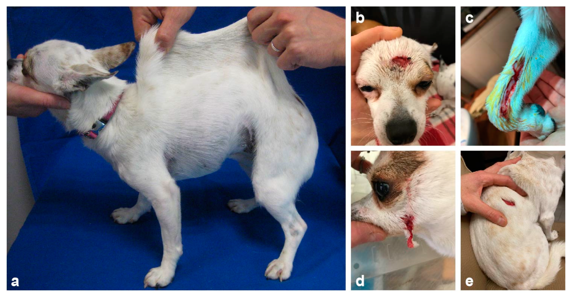
3.1. Clinical History and Examination

An 18-month-old spayed female Chihuahua was referred to the dermatology consultation for suspicion of EDS. The first clinical signs were noted on both eyes during puppyhood, at which time the dog was presented to the ophthalmologist with a diagnosis of corneal endothelial dystrophy. Moreover, its skin was hyperextensible (Figure 1a) and abnormalities were noted on the face around the eyes and on the extremities where the skin was fragile and tore easily after scratching or rubbing (Figure 1b–d). Several episodes of wounds on the trunk after minor injuries or scratching were also reported (Figure 1e). The healing process was difficult and long at each time. The dog had presented bilateral inguinal hernia. The dog lived with both parents and its two sisters and was the only one presenting these abnormalities. The dog wore a coat and a protective pet cone all day long in order to protect the skin and prevent any injuries and wound formation (Figure 2). At the time of consultation, the dog was in good general health condition. The skin was hyperextensible and no wounds were noted during clinical examination.

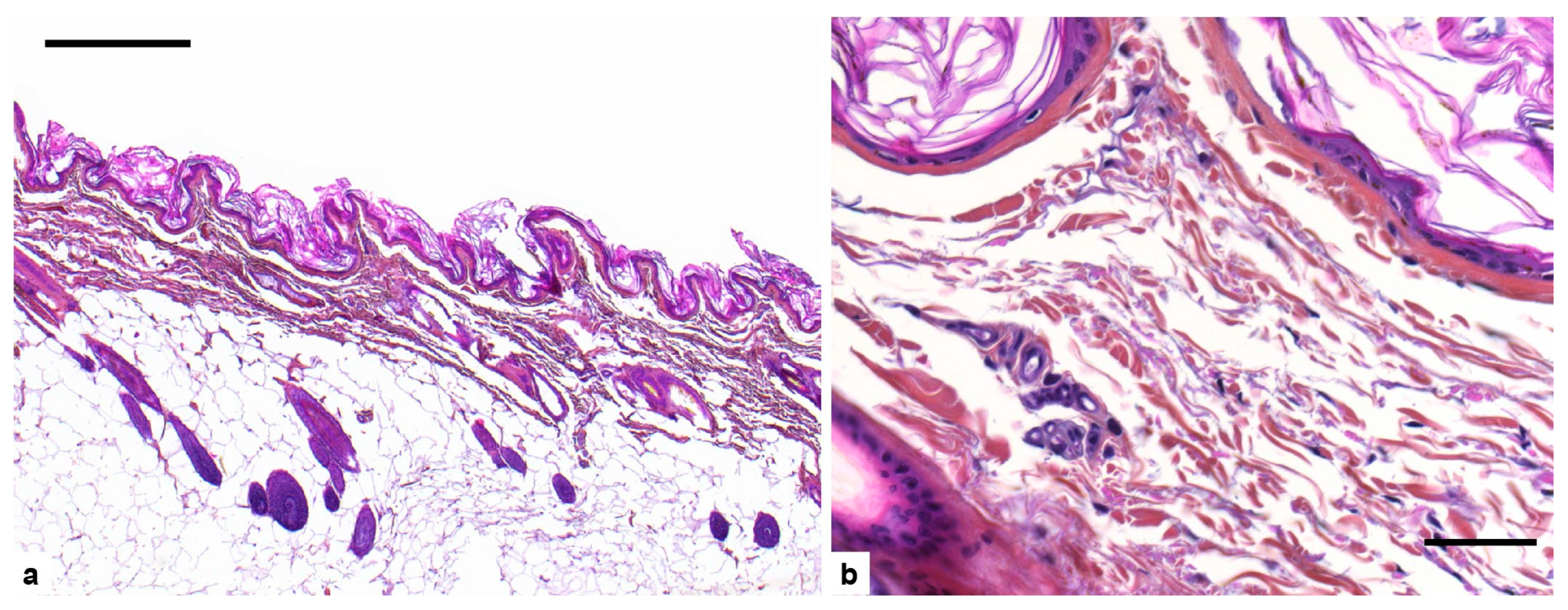
3.2. Histopathological Examination

Histopathological examination showed a marked reduction in dermis thickness (Figure 3a). The density of collagen fibers was markedly reduced giving a loose appearance of the dermis. There was moderate variation in collagen fibers size (Figure 3b). The epidermis and hypodermis were unremarkable.

3.3. Genetic Analysis

Whole-genome sequencing and private variant filtering were performed to identify the causative genetic variant. For this analysis, we compared the sequence data of the affected dog to the genomes of 783 control dogs of different breeds and 9 wolves (Table S1). We specifically looked for protein-changing variants in the 20 known EDS candidate genes (Table 1). The outcome of the various filtering steps is summarized in Table 2 and Table S2.
We identified a single private protein-changing variant within an EDS candidate gene, COL5A2. The variant can be designated as Chr36:30,548,697_30,548,723del. It is a heterozygous in-frame deletion of 27 bases (XM_005640393.3:c.3388_3414del), predicted to remove 9 amino acids, XP_005640450.1:p.(Lys1130_Asp1138del). The presence of the variant in a heterozygous state in the EDS-affected Chihuahua was confirmed by Sanger sequencing. The genotyping results of both parents revealed that COL5A2:c.3388_3414del represented a de novo variant as the mutant allele was absent from buccal cell DNA of both parents.

4. Discussion

In this study, we describe a Chihuahua with Ehlers-Danlos syndrome. EDS is a heterogeneous group of diseases affecting connective tissue. In human patients, 13 clinically different EDS subtypes with known causal variants in 20 different genes are recognized. A recently introduced additional clinical subtype has not yet been characterized at the molecular level [2].
In humans, the diagnosis of an EDS subtype is made by identifying clinical signs that can be categorized into major or minor criteria and by complementary molecular analysis. For the so-called classical EDS, the major and minor criteria are listed in Table 3.
Minimal criteria suggestive for classical EDS are the presence of either both major criterions or one major criterion and at least three minor criteria [1]. As this classification of EDS subtypes has been developed in humans, it must be used with caution in animals. Nevertheless, given the clinical, histopathological, and molecular similarities between animal models of EDS and human EDS, it is realistic to use it to define an animal EDS subtype.
The dog described here presented in its clinical history the first major criterion, namely (1) hyperextensibility of the skin and atrophic scarring, as well as three clinical signs listed as minor criteria, namely (1) easy bruising, (3) skin fragility and traumatic splitting, and (6) hernia. Thus, by referring to the human classification, the dog falls into the clinical category of classical EDS. As in humans, clinical diagnosis must be supplemented by molecular analysis. In our case, the genetic analysis confirmed the clinical hypothesis of a classical EDS.
Suspicion of EDS in animals usually leads to performing a skin biopsy and histopathological analysis of the dermis. Classically, histopathological descriptions report a dermis of normal thickness but present disorganized, smaller, curved collagen fibers of variable length and unequal diameter [9,16,24,28,29]. However, these characteristics are not always found and some affected animals show no abnormality on histological analysis. In some cases, a dermis of reduced thickness has been identified in affected animals, notably cats and dogs [5,29], as it is in our case.
Using a whole-genome sequencing approach, we identified a single heterozygous protein-changing variant in a known EDS candidate gene in the investigated EDS-affected Chihuahua. The variant is an in-frame deletion of 27 bases in COL5A2, encoding the α2 chain of type V collagen.
Type V collagen is present mainly as heterotrimers of two α1(V) chains (encoded by COL5A1) and one α2(V) chain [30,31]. Homotrimers of three α1(V) chains or heterotrimers comprised of an α1(V), α2(V), and α3(V) chain (encoded by COL5A3) also exist; however, their physiological function is largely unknown [2,32]. Type V collagen co-assembles with type I collagen into heterotypic fibrils in the extracellular matrix, with type V collagen being crucial for initial fibril formation [25]. Correct fibril formation and integrity play a key role in maintaining the physical properties of skin and other tissues [2].
The three chains of type V collagen are assembled into a triple helix. The sequence of each procollagen chain is characterized by extended Gly-Xaa-Yaa repeats. The presence of glycine (which has no side chain) in every third position permits the formation of the triple-helical structure. The Xaa and Yaa are often proline and hydroxyproline but can be any amino acid [2].
COL5A1-associated EDS mostly results from COL5A1 haploinsufficiency, as type V procollagen molecules cannot accommodate more than a single proα2(V) chain, and the reduction of available proα1(V) chains results in the production of about half the normal amount of type V collagen [33]. By contrast, proα1(V) chains can form functional homotrimers [34]. Thus, no heterozygous COL5A2 null alleles have been identified in EDS patients so far [35].
COL5A2-associated EDS is mostly related to structural variants located in the triple helix domain, resulting in the production of mutant proα2(V) chains which are expected to be incorporated in defective type V collagen molecules [35].
In accordance with [35], the COL5A2 variant identified in the EDS-affected dog from this study is predicted to remove three Gly-Xaa-Yaa triplet repeats and thus induce a structural alteration of the synthesized mutant proα2(V) chains. We assume that this causes aberrant heterotrimer formation and a defective type V collagen structure corresponding to a dominant-negative gain-of-function in the mutant allele.
In domestic animals, only one other COL5A2 variant, p.Gly789Val, has been reported to cause EDS in Holstein cattle [28].

5. Conclusions

To the best of our knowledge, our study represents the first report of a COL5A2-related EDS in dogs. The reported COL5A2 deletion arose by a de novo mutation event, which strongly supports its causality for the EDS phenotype.

Supplementary Materials

The following are available online at https://www.mdpi.com/article/10.3390/genes13050934/s1, Table S1: Public Genomes; Table S2: Private Variants.

Author Contributions

Conceptualization, L.C. and T.L.; investigation, S.K., L.C., A.B., N.C.-F. and E.R.-G.; data curation, V.J.; writing—original draft preparation, S.K. and L.C.; writing—review and editing, S.K., L.C., V.J., A.B., N.C.-F., E.R.-G. and T.L. All authors have read and agreed to the published version of the manuscript.

Funding

This research was funded by the Swiss National Science Foundation, grant number 310030_200354.

Institutional Review Board Statement

Ethical review and approval were waived for the investigation of the affected dog in this study as it was performed during clinical and pathological veterinary diagnostics. All animals in this study were examined with the consent of their owners and handled according to good ethical standards. The “Cantonal Committee for Animal Experiments” approved the collection of blood samples from control Chihuahuas (Canton of Bern; permit 71/19).

Informed Consent Statement

The owners of the dogs in this study gave informed consent to use samples and data for research.

Data Availability Statement

The accessions for the sequence data reported in this study are listed in Table S1.

Acknowledgments

The authors are grateful to the dog owners who donated samples and participated in the study. Authors are also thankful to Ambre Courtin for sample management and Cani-DNA biobank (http://dog-genetics.genouest.org, accessed on 23 May 2022), which is part of the French CRB-Anim infrastructure (ANR-11-INBS-0003), in the framework of the “Investing for the Future” program. We thank the Next Generation Sequencing Platform of the University of Bern for performing the high-throughput experiments and the Interfaculty Bioinformatics Unit of the University of Bern for providing high-performance computing infrastructure. We acknowledge the Dog10K genomes project and all researchers who deposited dog or wolf whole genome sequencing data into public databases.

Conflicts of Interest

The authors declare no conflict of interest.

References

  1. Malfait, F.; Francomano, C.; Byers, P.; Belmont, J.; Berglund, B.; Black, J.; Bloom, L.; Bowen, J.M.; Brady, A.F.; Burrows, N.P.; et al. The 2017 international classification of the Ehlers-Danlos syndromes. Am. J. Med. Genet. Part. C Semin. Med. Genet. 2017, 175, 8–26. [Google Scholar] [CrossRef] [PubMed] [Green Version]
  2. Malfait, F.; Castori, M.; Francomano, C.A.; Giunta, C.; Kosho, T.; Byers, P.H. The Ehlers–Danlos syndromes. Nat. Rev. Dis. Prim. 2020, 6, 64. [Google Scholar] [CrossRef] [PubMed]
  3. Arlein, M.S. Generalized acute cutaneous asthenia in a dog. J. Am. Vet. Med. Assoc. 1947, 111, 52. [Google Scholar] [PubMed]
  4. Hegreberg, G.A.; Padgett, G.A.; Gorham, J.R.; Henson, J.B. A Connective Tissue Disease of Dogs and Mink Resembling the Ehlers-Danlos Syndrome of Man. J. Hered. 1969, 60, 249–254. [Google Scholar] [CrossRef]
  5. Hegreberg, G.A.; Padgett, G.A.; Ott, R.L.; Henson, J.B. A Heritable Connective Tissue Disease of Dogs and Mink Resembling Ehlers-Danlos Syndrome of Man I. Skin Tensile Strength Properties. J. Investig. Dermatol. 1970, 54, 377–380. [Google Scholar] [CrossRef] [Green Version]
  6. Bellini, M.H.; Caldini, E.T.E.G.; Scapinelli, M.P.; Simões, M.J.; Machado, D.B.; Nürmberg, R. Increased elastic microfibrils and thickening of fibroblastic nuclear lamina in canine cutaneous asthenia. Vet. Dermatol. 2009, 20, 139–143. [Google Scholar] [CrossRef]
  7. Uri, M.; Verin, R.; Ressel, L.; Buckley, L.; McEwan, N. Ehlers–Danlos Syndrome Associated with Fatal Spontaneous Vascular Rupture in a Dog. J. Comp. Pathol. 2015, 152, 211–216. [Google Scholar] [CrossRef]
  8. Rasch, S.N. Surgical and medical treatment of ocular disease in a dog with Ehlers-Danlos syndrome. Clin. Case Rep. 2017, 5, 880–886. [Google Scholar] [CrossRef]
  9. Ueda, K.; Kawai, T.; Senoo, H.; Shimizu, A.; Ishiko, A.; Nagata, M. Histopathological and electron microscopic study in dogs with patellar luxation and skin hyperextensibility. J. Vet. Med. Sci. 2018, 80, 1309–1316. [Google Scholar] [CrossRef] [Green Version]
  10. Hegreberg, G.A.; Padgett, G.A.; Henson, J.B. Connective tissue disease of dogs and mink resembling Ehlers-Danlos syndrome of man. 3. Histopathologic changes of the skin. Arch. Pathol. 1970, 90, 159–166. [Google Scholar]
  11. Freeman, L.J.; Hegreberg, G.A.; Robinette, J.D. Ehlers-Danlos syndrome in dogs and cats. Semin. Vet. Med. Surg. (Small Anim). 1987, 2, 221–227. [Google Scholar] [PubMed]
  12. Barnett, K.C.; Cottrell, B.D. Ehlers-Danlos syndrome in a dog: Ocular, cutaneous and articular abnormalities. J. Small Anim. Pract. 1987, 28, 941–946. [Google Scholar] [CrossRef]
  13. Matthews, B.R.; Lewis, G.T. Ehlers-Danlos syndrome in a dog. Can. Vet. J. = La Rev. Vet. Can. 1990, 31, 389–390. [Google Scholar]
  14. Rodríguez, F.; Herráez, P.; Monteros, A.E.; de los Calabuig, P.; Rodríguez, J.L. Collagen Dysplasia in a Litter of Garaffiano Shepherd Dogs. J. Vet. Med. Ser. A 1996, 43, 509–512. [Google Scholar] [CrossRef]
  15. Jelínek, F.; Karban, J. Cutaneous Asthenia in One Dog. Acta Vet. Brno 1998, 67, 109–113. [Google Scholar] [CrossRef] [Green Version]
  16. Paciello, O.; Lamagna, F.; Lamagna, B.; Papparella, S. Ehlers-Danlos-like syndrome in 2 dogs: Clinical, histologic, and ultrastructural findings. Vet. Clin. Pathol. 2003, 32, 13–18. [Google Scholar] [CrossRef]
  17. Barrera, R.; Mañe, C.; Duran, E.; Vives, M.A.; Zaragoza, C. Ehlers-Danlos syndrome in a dog. Can. Vet. J. = La Rev. Vet. Can. 2004, 45, 355–356. [Google Scholar]
  18. Jaffey, J.A.; Bullock, G.; Teplin, E.; Guo, J.; Villani, N.A.; Mhlanga-Mutangadura, T.; Schnabel, R.D.; Cohn, L.A.; Johnson, G.S. A homozygous ADAMTS 2 nonsense mutation in a Doberman Pinscher dog with Ehlers Danlos syndrome and extreme skin fragility. Anim. Genet. 2019, 50, 543–545. [Google Scholar] [CrossRef] [Green Version]
  19. Colige, A.; Sieron, A.L.; Li, S.-W.; Schwarze, U.; Petty, E.; Wertelecki, W.; Wilcox, W.; Krakow, D.; Cohn, D.H.; Reardon, W.; et al. Human Ehlers-Danlos Syndrome Type VII C and Bovine Dermatosparaxis Are Caused by Mutations in the Procollagen I N-Proteinase Gene. Am. J. Hum. Genet. 1999, 65, 308–317. [Google Scholar] [CrossRef] [Green Version]
  20. Bauer, A.; de Lucia, M.; Leuthard, F.; Jagannathan, V.; Leeb, T. Compound heterozygosity for TNXB genetic variants in a mixed-breed dog with Ehlers-Danlos syndrome. Anim. Genet. 2019, 50, 546–549. [Google Scholar] [CrossRef]
  21. Bristow, J.; Tee, M.; Gitelman, S.; Mellon, S.; Miller, W. Tenascin-X: A novel extracellular matrix protein encoded by the human XB gene overlapping P450c21B. J. Cell Biol. 1993, 122, 265–278. [Google Scholar] [CrossRef] [PubMed]
  22. Burch, G.H.; Gong, Y.; Liu, W.; Dettman, R.W.; Curry, C.J.; Smith, L.; Miller, W.L.; Bristow, J. Tenascin–X deficiency is associated with Ehlers–Danlos syndrome. Nat. Genet. 1997, 17, 104–108. [Google Scholar] [CrossRef] [PubMed]
  23. Mao, J.R.; Taylor, G.; Dean, W.B.; Wagner, D.R.; Afzal, V.; Lotz, J.C.; Rubin, E.M.; Bristow, J. Tenascin-X deficiency mimics Ehlers-Danlos syndrome in mice through alteration of collagen deposition. Nat. Genet. 2002, 30, 421–425. [Google Scholar] [CrossRef] [PubMed]
  24. Bauer, A.; Bateman, J.F.; Lamandé, S.R.; Hanssen, E.; Kirejczyk, S.G.M.; Yee, M.; Ramiche, A.; Jagannathan, V.; Welle, M.; Leeb, T.; et al. Identification of Two Independent COL5A1 Variants in Dogs with Ehlers–Danlos Syndrome. Genes 2019, 10, 731. [Google Scholar] [CrossRef] [Green Version]
  25. Birk, D.E. Type V collagen: Heterotypic type I/V collagen interactions in the regulation of fibril assembly. Micron 2001, 32, 223–237. [Google Scholar] [CrossRef]
  26. Jagannathan, V.; Drögemüller, C.; Leeb, T.; Aguirre, G.; André, C.; Bannasch, D.; Becker, D.; Davis, B.; Ekenstedt, K.; Faller, K.; et al. A comprehensive biomedical variant catalogue based on whole genome sequences of 582 dogs and eight wolves. Anim. Genet. 2019, 50, 695–704. [Google Scholar] [CrossRef] [Green Version]
  27. Kiener, S.; Wiener, D.J.; Hopke, K.; Diesel, A.B.; Jagannathan, V.; Mauldin, E.A.; Casal, M.L.; Leeb, T. ABHD5 frameshift deletion in Golden Retrievers with ichthyosis. G3 (Bethesda) 2022, 12, jkab397. [Google Scholar] [CrossRef]
  28. Jacinto, J.G.P.; Häfliger, I.M.; Veiga, I.M.B.; Letko, A.; Benazzi, C.; Bolcato, M.; Drögemüller, C. A Heterozygous Missense Variant in the COL5A2 in Holstein Cattle Resembling the Classical Ehlers–Danlos Syndrome. Animals 2020, 10, 2002. [Google Scholar] [CrossRef]
  29. Gross, T.L.; Ihrke, J.P.; Walder, E.J.; Affolter, V.K. Degenerative, Dysplastic and Depositional Diseases of Dermal Connective Tissue. In Skin Diseases of the Dog and Cat: Clinical and Histopathologic Diagnosis; Blackwell Science Ltd.: Oxford, UK, 2005; pp. 386–389. [Google Scholar]
  30. Burgeson, R.E.; El Adli, F.A.; Kaitila, I.I.; Hollister, D.W. Fetal membrane collagens: Identification of two new collagen alpha chains. Proc. Natl. Acad. Sci. USA 1976, 73, 2579–2583. [Google Scholar] [CrossRef] [Green Version]
  31. Gay, S.; Kent Rhodes, R.; Gay, R.E.; Miller, E.J. Collagen molecules comprised of alpha 1(V)-chains (B-chains): An apparent localization in the exocytoskeleton. Coll. Relat. Res. 1981, 1, 53–58. [Google Scholar] [CrossRef]
  32. Imamura, Y.; Scott, I.C.; Greenspan, D.S. The pro-alpha3(V) collagen chain. Complete primary structure, expression domains in adult and developing tissues, and comparison to the structures and expression domains of the other types V and XI procollagen chains. J. Biol. Chem. 2000, 275, 8749–8759. [Google Scholar] [CrossRef] [PubMed] [Green Version]
  33. Wenstrup, R.J.; Florer, J.B.; Davidson, J.M.; Phillips, C.L.; Pfeiffer, B.J.; Menezes, D.W.; Chervoneva, I.; Birk, D.E. Murine model of the Ehlers-Danlos syndrome. col5a1 haploinsufficiency disrupts collagen fibril assembly at multiple stages. J. Biol. Chem. 2006, 281, 12888–12895. [Google Scholar] [CrossRef] [PubMed] [Green Version]
  34. Chanut-Delalande, H.; Bonod-Bidaud, C.; Cogne, S.; Malbouyres, M.; Ramirez, F.; Fichard, A.; Ruggiero, F. Development of a Functional Skin Matrix Requires Deposition of Collagen V Heterotrimers. Mol. Cell. Biol. 2004, 24, 6049. [Google Scholar] [CrossRef] [PubMed] [Green Version]
  35. Symoens, S.; Syx, D.; Malfait, F.; Callewaert, B.; De Backer, J.; Vanakker, O.; Coucke, P.; De Paepe, A. Comprehensive molecular analysis demonstrates type V collagen mutations in over 90% of patients with classic EDS and allows to refine diagnostic criteria. Hum. Mutat. 2012, 33, 1485–1493. [Google Scholar] [CrossRef]
Figure 1. Clinical signs of the classical Ehlers-Danlos-affected Chihuahua dog. (a) Skin hyperextensibility over the dorsum. (b) Skin laceration resulting from minimal trauma on the head, (c) on the forelimb, (d) on the lower jaw, and (e) on the dorsum.
Genes 13 00934 g001
Figure 2. The dog lives continuously with a protective suit and an Elizabethan collar to prevent wound formation.
Genes 13 00934 g002
Figure 3. Cutaneous histopathological findings of the dog affected with Ehlers-Danlos syndrome (EDS). (a) Dermis is abnormally thin while epidermis and hypodermis are normal. Hematoxylin-eosin and saffron stain (HES), bar 500 µm; (b) collagen fibers are moderately uneven in size and their density is markedly reduced. Hematoxylin-eosin and saffron stain (HES), bar 50 µm.
Genes 13 00934 g003
Table 1. Overview on genetic causes of human Ehlers-Danlos syndrome (EDS), adapted from [2].
GeneEDS TypeInheritance
ADAMTS2DermatosparaxisAR
AEBP1Classical-like type 2AR
B3GALT6Spondylo-dysplasticAR
B4GALT7Spondylo-dysplasticAD
CHST14MusculocontracturalAR
COL1A1Classical, Vascular, ArtrochalasiaAD
COL1A2Artrochalasia, Cardiac valvularAD
COL3A1VascularAD
COL5A1ClassicalAD
COL5A2ClassicalAD
COL12A1MyopathicAD
C1RPeriodontalAD
C1SPeriodontalAD
DSEMusculocontracturalAR
FKBP14KyphoscolioticAR
PRDM5Brittle cornea syndromeAR
PLOD1KyphoscolioticAR
SLC39A13Spondylo-dysplasticAR
TNXBClassical-likeAR
ZNF469Brittle cornea syndromeAR
Table 2. Results of variant filtering in the affected dog against 792 control genomes.
Filtering StepHeterozygous VariantsHomozygous Variants
All variants in the affected dog4,191,4112,457,482
Private variants18,9561258
Protein-changing private variants1042
Protein-changing private variants in 20 candidate genes10
Table 3. Classical EDS clinical criteria in humans, adapted from [1].
Major criteria1. Skin hyperextensibility and atrophic scarring
2. Generalized joint hypermobility
Minor criteria1. Easy bruising
2. Soft, doughy skin
3. Skin fragility (or traumatic splitting)
4. Molluscoid pseudotumors
5. Subcutaneous spheroids
6. Hernia (or history thereof)
7. Epicanthal folds
8. Complication of joint hypermobility (e.g., sprains, luxation/subluxation, pain, flexible flatfoot)
9. Family history of a first degree relative who meets clinical criteria
Publisher’s Note: MDPI stays neutral with regard to jurisdictional claims in published maps and institutional affiliations.

