Review

The Role of PET in the Diagnosis and Disease Activity Assessment in Large Vessel Vasculitis

1
Rheumatology Unit, Azienda Unità Sanitaria Locale-IRCCS di Reggio Emilia, 42122 Reggio Emilia, Italy
2
Surgical, Medical and Dental Department of Morphological Sciences Related to Transplant, Oncology and Regenerative Medicine Department, University of Modena and Reggio Emilia, 41100 Modena, Italy
3
Nuclear Medicine Unit, Azienda Unità Sanitaria Locale-IRCCS di Reggio Emilia, 42122 Reggio Emilia, Italy
*
Author to whom correspondence should be addressed.
These authors contributed equally to this work.
Hemato 2023, 4(4), 321-330; https://doi.org/10.3390/hemato4040026
Submission received: 31 August 2023 / Revised: 4 October 2023 / Accepted: 10 October 2023 / Published: 30 October 2023
(This article belongs to the Section Radiolabeled Blood Elements and Other Imaging Modalities)

Abstract

:
The role of 18F-fluorodeoxyglucose (FDG) positron emission tomography (18F-FDG PET) in the diagnosis of large vessel vasculitis (LVV) is well established. It permits us to assess the extent and the grade of vascular involvement and to rule out the other causes in clinical scenarios characterized by less specific symptoms. The advantages of 18F-FDG PET are far less clear in monitoring disease activity over time. Studies looking for the role of 18F-FDG PET as a potential biomarker had conflicting results and whether and when to repeat it during follow-up is based on clinical experience. A comprehensive assessment, including clinical, laboratory and morphological imaging is still required to monitor patients with large-vessel vasculitis over time. The aim of this review is to present more recent data about the utility of 18 F-FDG PET in the diagnosis and follow-up of LVV.

1. Introduction

Large-vessel vasculitides (LVVs), namely giant cell arteritis (GCA) and Takayasu’s Arteritis (TAK), are a group of heterogeneous diseases involving the aorta and its main branches [1]. They share similar clinical and radiological features. GCA affects elderly people over 50 years old, whereas TAK is more common among young female patients [1,2,3].
18F-fluorodeoxyglucose positron emission tomography (18F FDG-PET) scanning is a useful non-invasive metabolic imaging modality for evaluating patients with fever of unknown origin and tumors [4]. 18F FDG accumulates in hypermetabolic cells, and a high-grade uptake in the vascular wall is a typical sign of active LVV. Ample evidence suggests that it may be used as a surrogate of histological examination, especially in cases where obtaining biopsies is not feasible [5,6].
The assessment of FDG uptake may be qualitative or semiquantitative. The qualitative method visually compares the FDG uptake in the vessel wall to a background tissue, such as the liver. The Meller visual scale determines the inflammation, like the Deauville score for lymphoma, qualitatively: grade 1 for an FDG uptake < liver, grade 2 is similar to the liver, and grade 3 > liver [7,8]. Back in 2018, Grayson et al. developed a qualitative summary score based on global arterial FDG uptake, the PET Vascular Activity Score (PETVAS), which, using the grades from different arterial territories, reflects the global inflammatory burden of the disease [9].
On the other hand, the semiquantitative methods define regions of interest (ROI) to calculate the standardized uptake values (SUV) or ratios, such as the target-to-background ratio (TBR) that referenced SUV from arterial tissue to the liver or blood pool [10].
Advantages of the use of FDG-PET in the management of LVVs include the possibility to diagnose the aortic involvement without histologic confirmation, and especially in cases of diagnostic uncertainty, it may be helpful to exclude malignancy or infections. Additionally, advances in PET radiotracers may distinguish active vascular inflammation from other pathologies, such as atherosclerosis. Potential drawbacks of FDG-PET include limited access and high cost [11,12,13]. Further, vascular FDG uptake is attenuated rapidly following treatment initiation, making the use of arterial FDG uptake to guide treatment decisions and monitor disease activity less well-defined. In this review, we aim to describe the recent evidence about the accuracy and usefulness of FDG-PET in diagnosing and monitoring LVV.

2. FDG-PET in the Diagnosis and Activity Assessment of LVV

No validated diagnostic criteria exist for GCA or TAK. Historically, diagnosis of LVV was based on clinical signs and symptoms. These include specific signs and symptoms related to the particular vascular involvement, such as vascular bruits, claudication of the limbs, headache, carotidynia, visual loss and stroke, but also less specific symptoms like fever, myalgias and weight loss. In the latter case, 18F FDG-PET plays a crucial role not only in the diagnosis of LVV, but also in ruling out other conditions (e.g., malignancies, infections). In 2018 the EULAR recommendations proposed management recommendations for LVV and stated that every suspected diagnosis should be confirmed by imaging (either ultrasound or MR for temporal or other cranial arteries, ultrasound, CT, PET/CT, or MRI for the aorta/extracranial arteries) or by biopsy [14,15]. Further, in the recent update, 18F FDG-PET is recommended not only as the first imaging modality in the assessment of the large vessel involvement in GCA and TAK, but also as an alternative of ultrasound in the evaluation of cranial arteries in GCA (https://0-doi-org.brum.beds.ac.uk/10.1136/ard-2023-224543, access on 9 October 2023). Thus, imaging (and particularly 18F FDG-PET) has become mandatory in the clinical armamentarium to diagnose GCA and TAK.

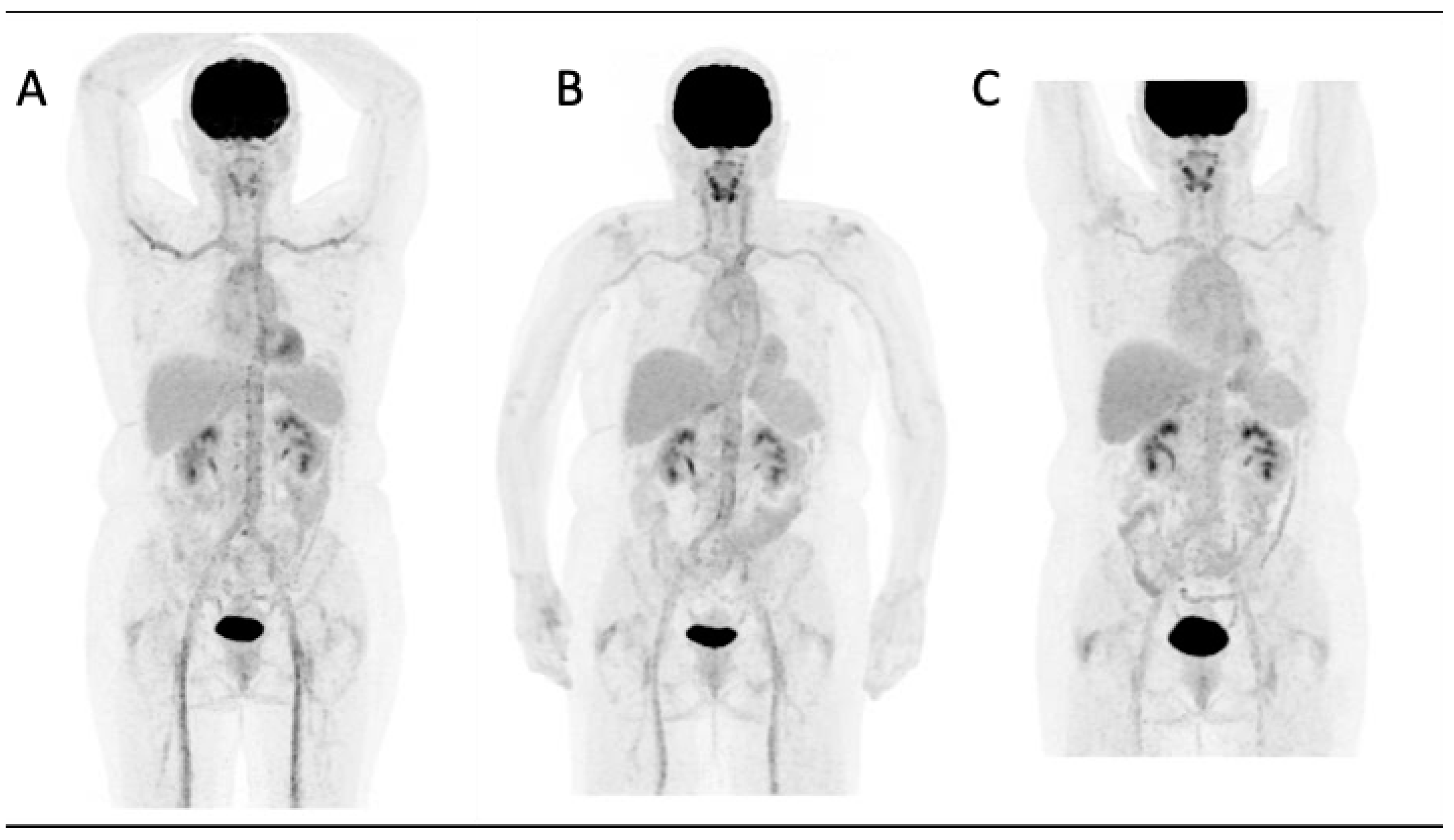
2.1. Large Vessel Giant Cell Arteritis

The use of FDG-PET in the diagnosis of LVV and polymyalgia rheumatica (PMR) is now well established. The first study in this regard was published in 1999 by Blockmans et al., who observed in the study group, consisting of patients with temporal arteritis and PMR, a significant increase in FDG uptake in the thoracic vessels, and arteries of the upper and lower limbs compared to the control group [16].
According to Blockmans et al., using temporal artery biopsy (TAB) as the reference standard, PET showed diagnostic sensitivity and specificity of 67% and 66%, respectively [17].
Lariviere et al., using the final diagnosis of GCA based on clinical judgment as the gold standard in a study of 24 patients, reported similar sensitivity values (77%), with a specificity of 100% [18]. It is likely that the low specificity reported by Blockmans underestimates the true diagnostic potential of PET, as TAB (used in this case as the reference standard) is often negative in the large vessel GCA (LV GCA) phenotype, where the temporal arteries can be spared from the inflammatory process.
Patients with a clinical suspicion of GCA should promptly start on glucocorticoids. In a retrospective study including ten patients with a new diagnosis of GCA, Nielsen highlighted that three days after the introduction of high-dose steroids (prednisone 60 mg orally), it was still possible to make a correct diagnosis of LV-GCA in 10 out of 10 patients, despite an inevitable reduction in FDG uptake on PET. However, at 10 days from the start of cortisone, only 5 out of 14 patients were correctly diagnosed with LV-GCA, and the diagnostic sensitivity of PET decreased to 36% [19]. Therefore, imaging should not be delayed once a clinical diagnosis of LV-GCA is made.

2.2. Takayasu’s Arteritis

The utility of 18F FDG-PET in the diagnosis of TAK is corroborated by multiple case series and case reports [20,21].
For years, angiography has been the gold standard in the diagnosis of TAK since it detects precisely vascular stenoses [22]. However, the presence of vascular lesions is already a sign of damage. The vascular uptake at 18F FDG-PET may assess the extent of vessel involvement before morphological abnormalities occur.
A recent meta-analysis included seven studies for a total of 191 patients with TAK (96 with active TAK, according to the National Institutes of Health (NIH) criteria in four studies). The FDG-PET parameters used to detect vascular inflammation were the visual analysis (n = 6 studies) and the semiquantitative analysis (n = 1). For the detection of large-vessel inflammation in TAK according to the disease activity status, FDG-PET showed a pooled sensitivity at 87% (95% CI, 78.0–92.6) and a pooled specificity at 73% (95% CI, 62.5–81.3). The specificity increased to 84% (95% CI, 72.5–91.5%) (p = 0.134, I2 = 46.3%) by combining the four studies assessing the disease activity state with the NIH scale [23]. These findings underline that in the absence of other clinical features of clinical active vasculitis, FDG-PET findings must be evaluated in the appropriate clinical context since different conditions (such as atherosclerosis or vascular remodeling) may mimic the presence of vasculitis.
FDG-PET seems more accurate in distinguishing clinical active disease in TAK than in GCA, as demonstrated by the work of our group evaluating the accuracy of FDG-PET in the assessment of active disease in LVV. Visual PET/CT grading scale better distinguished between clinically active and inactive disease in TAK (72% sensitivity, 78% specificity, AUC 0.75) than in LV GCA (51% sensitivity, 83% specificity, AUC 0.67) [24].
Further, incorporating 18F-FDG-PET in the definition of disease activity in TAK may also be useful in the standardization of recruitment of patients to be enrolled in randomized controlled trials. A survey among physicians indicated that FDG-PET data significantly improved agreement about enrolment decisions between raters (inter-rater reliability (IRR) = 0.68 (95% CI 0.67, 0.69) to IRR = 0.88 (95% CI 0.87, 0.89); p < 0.01) and were more strongly related to enrollment decisions than acute phase reactants levels [25]. These data indicate that 18F FDG-PET is one of the most important tools in the diagnosis of TAK and may be used as a complementary assessment in the evaluation of disease activity.

3. FDG-PET in the Follow-Up of LVV

Monitoring disease activity in LVV may be challenging. Patients often present with non-specific symptoms (such as fatigue and weight loss) and often the same symptoms may indicate either disease activity or vascular damage. Further acute phase reactants may be altered by treatment, especially Tocilizumab, which decreases the synthesis of ESR and CRP by blocking the interleukin 6 receptor. Novel biomarkers are currently understudy, including S100 proteins, pentraxin-3 and osteopontin. These molecules are independent of the interleukin 6 pathway and are less altered by treatment; however, their use in clinical practice has not been implemented yet. Other molecules are also being studied as markers of arterial remodeling (such as metalloproteinases 2 and 9, VEGF, YLK-40 and angiopoietins) or organ damage (such as pentraxin-3, for optic nerve ischemia) (https://0-doi-org.brum.beds.ac.uk/10.1007/s11926-021-00980-5 access on 9 October 2023).
The role of FDG-PET findings is still to be clarified: on one side, a positive FDG-PET imaging could detect active vasculitis and inform treatment decisions; on the other side, the evidence of active vasculitis during apparent clinical remission is still a complicating matter. Older autopsy studies have pointed out that vasculitis is often still present histologically in patients with LVV, despite being considered inactive at the time of death [26,27]. Further, patients with LVV can develop new arterial lesions on vascular imaging during periods of apparent clinical remission [9,15].
However, whether an increased FDG uptake without clinical signs of active disease requires adjustment of treatment is still unknown, and studies using it as a biomarker of disease activity have shown conflicting results.

3.1. Giant Cell Arteritis

A study involving 22 patients diagnosed with GCA found a notable decrease in overall basal FDG vascular uptake after 8 months of tocilizumab treatment [28]. All patients achieved clinical remission during the follow-up period. The researchers utilized a Total Vascular Score (TVS) ranging from 0 to 33 to quantitatively measure vascular inflammation. The study concluded that 18F FDG-PET/CT is valuable for monitoring the progression of LVV as it exhibits a strong correlation with clinically assessed disease activity. Nonetheless, the limited number of patients who underwent follow-up scans significantly hampers the broader applicability of the study’s results.
In an observational study called the RIGA study, researchers aimed to assess the response to treatment in patients with new-onset GCA using 18F FDG-PET/CT in combination with clinical and laboratory assessments [29]. The study included 88 patients with active, newly diagnosed LV GCA who met the inclusion criteria of the GiACTA trial [30]. Within two weeks of diagnosis, all participants underwent a baseline PET scan. Treatments included prednisolone alone for 27 patients, a combination of glucocorticoids and methotrexate for 42 patients and glucocorticoids with tocilizumab for 19 patients. Baseline PET scans indicated active vasculitis in 82 out of 88 patients. The researchers employed PETVAS to gauge treatment response in terms of vascular inflammation. The average PETVAS decreased from 18.9 at baseline to 8.0 during the follow-up period. All patients exhibited a decrease in global vascular uptake on follow-up scans, regardless of the treatment regimen. However, patients treated with methotrexate (reduction of about 12.3 units) or tocilizumab (reduction of about 11.7 units) experienced greater reductions in PETVAS compared to those receiving glucocorticoids alone (reduction of about 8.7 units). These PETVAS changes correlated with symptom resolution and a decline in acute-phase reactant levels.
On the other hand, a study outlined the discrepancy between disease activity assessments based on clinical evaluation and 18F FDG-PET/CT findings in GCA patients treated with tocilizumab [31]. The researchers utilized two imaging evaluation methods: the TVS and the TBR in 30 patients with PET evidence of extra-cranial large vessel involvement. At baseline, despite adequate glucocorticoid therapy, all patients were considered clinically active, leading to the initiation of tocilizumab treatment. After an average follow-up period of 10.8 ± 3.7 months, a notable reduction in vascular uptake was observed. The TBR at the thoracic aorta decreased from an average of 1.70 ± 0.52 to 1.48 ± 0.25 (p = 0.005), while the TVS dropped from an average of 4.97 ± 2.62 to 3.13 ± 1.89 (p < 0.001). At the end of the follow-up, approximately 83.3% of patients were considered clinically inactive. 18F FDG-PET/CT criteria for remission involved the complete normalization of vascular uptake during follow-up. Since only 30% and 10% of patients achieved normalization of TBR and TVS, respectively, complete agreement between clinical remission and PET normalization was lacking.
Regarding the role of 18F FDG-PET in predicting future relapses in GCA, data in the literature are contrasting. Bellan et al. retrospectively reviewed the medical records of 19 GCA patients who had a baseline 18 F FDG-PET/CT available [32]. Four relapses occurred over a median follow-up of 15 months (interquartile range of 4.5 to 26.5 months), but the authors did not find a significant difference in the baseline median SUVmax of patients who experienced a relapse [5.70 (IQR 5.34–5.89)] and those who did not [5.74 (IQR 4.98–7.99), p = 0.92]. The conclusion of the study was that the initial FDG vascular uptake was not effective in categorizing patients with an increased likelihood of experiencing subsequent relapses.
Similarly, in a prospective study, including 21 patients newly diagnosed with GCA who fulfilled the 1990 American College of Rheumatology classification criteria for GCA, all the participants had an 18F FDG-PET/CT scan performed within 72 h of starting prednisolone treatment and were prospectively followed-up over a 12-month period [33,34]. Since patients with a baseline TVS > 10 were not more likely to experience relapse than those with a baseline TVS < 10, the researchers concluded that PET is not a useful tool in predicting the risk of relapse. Nevertheless, it is important to consider that these conclusions are constrained by the small cohort size and the relatively short duration of follow-up.
On the contrary, results from a retrospective cohort including 254 patients with GCA showed that the large vessel involvement, the negativity of temporal artery biopsy, the male gender and the presence of musculoskeletal symptoms defined a clinical subset with a more relapsing disease (https://0-doi-org.brum.beds.ac.uk/10.1177/1759720X211009029 access on 9 October 2023). These results have recently been confirmed by a meta-analysis about the predictors of relapse in GCA. Thirty studies were included for a total of 2595 patients. Female sex and the large vessel involvement were predictors of relapse with an OR of 1.43 and 2.04, respectively (https://0-doi-org.brum.beds.ac.uk/10.1016/j.jbspin.2022.105494 access on 9 October 2023). Taken together, these data indicate that the assessment of large vessel involvement at baseline should be warranted in all patients with a new diagnosis of GCA to detect those at major risk of relapse who may potentially benefit from the early introduction of GC-sparing agents since the onset. In this context, 18F FDG-PET is fundamental as an initial imaging assessment. However, when and how often to repeat it during follow-up is still debated (https://0-doi-org.brum.beds.ac.uk/10.1136/ard-2023-224543 access on 9 October 2023).
Figure 1 illustrates a case of GCA at the time of diagnosis and its subsequent post-therapy follow-up.

3.2. Takayasu’s Arteritis

Similar to LV-GCA, also in TAK studies using 18F FDG-PET as a marker of response to treatment have shown conflicting results.
Twenty-one TAK patients enrolled in an observational cohort were evaluated every six months with clinical assessment and imaging. Treatment decisions were categorized as increased, decreased or unchanged. The FDG uptake was measured using PETVAS. The authors found that despite an improvement of PETVAS (21 vs. 16, p = 0.02 in seven patients treated with infliximab), rarely PET findings normalized, despite a significant improvement in the clinical evaluation [35].
A recent meta-analysis including eight studies (four longitudinal and four cross-sectional) found a moderate diagnostic accuracy to discriminate between patients with a clinical active LVV and those in clinical remission when treatment is initiated (sensitivity 77%, specificity 71%), indicating that 18F-FDG PET may aid in monitoring treatment response, but its findings need to be interpreted in the context of other clinical findings [36].
Different studies assessed the role of FDG-PET in predicting relapses in TAK. Specifically, Kwon et al., in a retrospective study involving 33 patients with inactive TAK according to the NIH criteria, evaluated the features associated with subsequent relapses, defined as the recurrence of clinically active disease following an inactive period and requiring therapy modification. Vascular FDG uptake was expressed using TBR. The mean observation period was 4.5 years. Eight (27.3%) patients had a relapse. The only two factors associated with multivariable analysis with an increased risk of relapse were a raised erythrocyte sedimentation rate (ESR) (hazard ratio (HR) 7) and TBR (HR 11.5) [37]. These findings suggest that TBR, together with the ESR, independently predicts the relapse risk and is thus useful to stratify clinically inactive patients for the risk of relapse.
Another retrospective study from a single center tried to predict not only relapses (primary outcome) but also other clinically relevant parameters (sustained remission, development of new angiographic lesions and changes in the therapeutic regimen) [37]. Thirty-two patients with TAK were assessed at baseline and subsequently at regular intervals over a median of 84 months. Sustained remission was defined as a lack of new manifestations of disease activity, no new angiographic lesions and no glucocorticoid treatment for at least six months.
Eleven (34.4%) patients at baseline were judged to have active disease according to the modified NIH criteria. Patients were stratified according to SUVmax values. This study demonstrated that patients with baseline SUVmax values > 1.3 (that better distinguished active disease from non-active) had an increased risk of relapses (odds ratio (OR) 5.66) and needed to change immunosuppressive agents (OR 7.93). However, at multivariate analysis, no associations were found with the development of disease relapses and with new angiographic lesions.

3.3. Giant Cell Arteritis and Takayasu’s Arteritis

An observational study from NIH used PETVAS to assess its role as a marker of relapsing disease. In 39 patients, longitudinal data about FDG-PET/CT and clinical assessment were available. Clinical relapse was defined as a recurrence of clinical symptoms attributable to LVV and the necessity to increase the prednisone dose to at least 10 mg daily and/or add another immunosuppressant. A PETVAS threshold of 20 could differentiate clinical active disease from remission and predict future relapse. The rate of relapse in patients with a PETVAS > 20 was 45% versus 11% in patients with a PETVAS < 20 (p = 0.03) [9]. Interestingly, patients with PETVAS ≥ 20 were more likely to have GCA than TAK, probably due to atherosclerosis in the former group contributing partly to vascular FDG uptake in LVV during clinical remission.
On the contrary, data about 81 patients with LVV who underwent an FDG PET/CT scan during a period of clinical remission were collected in a retrospective cohort. Relapse was defined by the reappearance of signs/symptoms attributable to vasculitis, resolution of signs/symptoms after increasing or restarting prednisone, increased acute phase reactants and exclusion of other causes. Thirty-four clinical relapses occurred (19 in TAK and 15 in GCA), but PETVAS was not a good predictor (age- and sex-adjusted HR 1.04 (95% CI 0.97, 1.11), p = 0.252), with a sensitivity of 47.1% and a specificity of 73.8% [24]. Of note, the retrospective collection of data may be biased by confounders, such as the GCs dose and more prospective studies are required to evaluate the role of 18F FDG-PET as a biomarker in LVV.
Whether the persistence of FDG-PET is associated with angiographic progression (stenoses, dilations or aneurysms) is another critical issue in monitoring disease activity.
In a retrospective study, 100 consecutive patients with LVV (49 with TAK and 51 with LV-GCA) with baseline PET/CT and morphological imaging (CT/MR angiography (CTA and MRA, respectively)) performed within three months were included [38]. A total of 1206 vascular segments were examined. Twenty-eight patients who underwent another CT/MR between 6 and 30 months from baseline were included in the per-segment and per-patient analyses. Seven new stenoses/dilations occurred, exclusively in TAK patients. The baseline PET score was strongly associated with incident stenoses/dilations (p = 0.001), while baseline wall thickening was not (p = 0.708), indicating that only a PET score was a good predictor of new vessel-wall damage during the follow-up.
Similarly, a prospective study evaluated 1091 arterial territories from 70 patients with LVV (TAK = 38; GCA = 32) [39]. All patients underwent baseline MRA or CTA of the aorta and primary branches as well as PET. All patients repeated the imaging assessment after at least six months per standardized imaging protocol. Over 1.6 years of median follow-up, new lesions (stenosis, occlusion, or aneurysm) developed only in eight arterial territories, always in TAK. At baseline, 298 territories were active on PET and only 24 (8%) presented an angiographic progression. A total of 80% of the arterial territories presenting progressive vascular damage were active on PET at baseline, confirming the results of the previous study. Furthermore, the analysis within a patient showed that an arterial territory with baseline PET activity had 20 times higher odds for angiographic change than a paired arterial territory without PET activity (p < 0.01).
These two studies reinforce the need to use PET and morphological imaging as complementary assessments to evaluate disease activity and vascular damage over time in patients with LVV. However, using PET scores to guide treatment in LVV is still open to question, and more prospective studies are needed.

4. Conclusions

18F FDG-PET has a clear role in diagnosing LVVs and has become a part of clinical tools to assess vascular involvement whenever a biopsy is not feasible. However, the increased availability of imaging has led to a more frequent diagnosis of vasculitis due to incidental findings. In this last context, imaging evaluation in the proper clinical setting becomes mandatory.
In the monitoring of vasculitis, the role of 18F-FDG PET is still interlocutory. On one side, it may inform treatment decisions if the disease is still clinically active. Conversely, the persistence of vascular uptake during periods of clinical remission may indicate subclinical vasculitis, remodeling or healing. Studies addressing these points and assessing the role of PET as a marker of relapsing or more aggressive disease are controversial. The decision about when to repeat a PET during follow-up should be tailored on an individual basis.

Funding

This research received no external funding.

Institutional Review Board Statement

Not applicable.

Informed Consent Statement

Not applicable.

Data Availability Statement

Not applicable.

Conflicts of Interest

The authors declare no conflict of interest.

References

  1. Jennette, J.C.; Falk, R.J.; Bacon, P.A.; Basu, N.; Cid, M.C.; Ferrario, F.; Flores-Suarez, L.F.; Gross, W.L.; Guillevin, L.; Hagen, E.C.; et al. 2012 Revised International Chapel Hill Consensus Conference Nomenclature of Vasculitides. Arthritis Rheum. 2012, 65, 1–11. Available online: https://pubmed.ncbi.nlm.nih.gov/23045170/ (accessed on 20 January 2022). [CrossRef]
  2. Ponte, C.; Grayson, P.C.; Robson, J.C.; Suppiah, R.; Gribbons, K.B.; Judge, A.; Luqmani, R.A. American College of Rheumatology/EULAR classification criteria for giant cell arteritis. Ann. Rheum. Dis. 2022, 81, 1647–1653. Available online: http://0-ard-bmj-com.brum.beds.ac.uk/ (accessed on 19 March 2023). [CrossRef]
  3. Grayson, P.C.; Ponte, C.; Suppiah, R.; Robson, J.C.; Gribbons, K.B.; Judge, A.; Craven, A.; Khalid, S.; Hutchings, A.; Danda, D.; et al. 2022 American College of Rheumatology/EULAR classification criteria for Takayasu arteritis. Ann. Rheum. Dis. 2022, 81, 1654–1660. Available online: https://pubmed.ncbi.nlm.nih.gov/36351705/ (accessed on 24 November 2022). [CrossRef]
  4. Dong, M.-J.; Zhao, K.; Liu, Z.-F.; Wang, G.-L.; Yang, S.-Y.; Zhou, G.-J. A meta-analysis of the value of fluorodeoxyglucose-PET/PET-CT in the evaluation of fever of unknown origin. Eur. J. Radiol. 2011, 80, 834–844. Available online: http://www.ejradiology.com/article/S0720048X10005632/fulltext (accessed on 8 August 2023). [CrossRef] [PubMed]
  5. Yamashita, H.; Kubota, K.; Mimori, A. Clinical value of whole-body PET/CT in patients with active rheumatic diseases. Arthritis Res. Ther. 2014, 16, 423. Available online: https://0-arthritis--research-biomedcentral-com.brum.beds.ac.uk/articles/10.1186/s13075-014-0423-2 (accessed on 8 August 2023). [CrossRef] [PubMed]
  6. Prieto-González, S.; Depetris, M.; García-Martínez, A.; Espígol-Frigolé, G.; Tavera-Bahillo, I.; Corbera-Bellata, M.; Planas-Rigol, E.; Alba, M.A.; Hernández-Rodríguez, J.; Grau, J.M.; et al. Positron emission tomography assessment of large vessel inflammation in patients with newly diagnosed, biopsy-proven giant cell arteritis: A prospective, case–control study. Ann. Rheum. Dis. 2014, 73, 1388–1392. Available online: https://0-ard-bmj-com.brum.beds.ac.uk/content/73/7/1388 (accessed on 8 August 2023). [CrossRef] [PubMed]
  7. Meller, J.; Strutz, F.; Siefker, U.; Scheel, A.; Sahlmann, C.O.; Lehmann, K.; Conrad, M.; Vosshenrich, R. Early diagnosis and follow-up of aortitis with [18F]FDG PET and MRI. Eur. J. Nucl. Med. 2003, 30, 730–736. Available online: https://0-link-springer-com.brum.beds.ac.uk/article/10.1007/s00259-003-1144-y (accessed on 9 August 2023). [CrossRef]
  8. Meignan, M.; Gallamini, A.; Haioun, C. Report on the First International Workshop on interim-PET scan in lymphoma. Leuk. Lymphoma 2009, 50, 1257–1260. Available online: https://0-www-tandfonline-com.brum.beds.ac.uk/doi/abs/10.1080/10428190903040048 (accessed on 9 August 2023). [CrossRef] [PubMed]
  9. Grayson, P.C.; Alehashemi, S.; Bagheri, A.A.; Civelek, A.C.; Cupps, T.R.; Kaplan, M.J.; Malayeri, A.A.; Merkel, P.A.; Novakovich, E.; Bluemke, D.A.; et al. 18 F-Fluorodeoxyglucose-Positron Emission Tomography As an Imaging Biomarker in a Prospective, Longitudinal Cohort of Patients With Large Vessel Vasculitis. Arthritis Rheumatol. 2018, 70, 439–449. Available online: https://pubmed.ncbi.nlm.nih.gov/29145713/ (accessed on 24 November 2022). [CrossRef]
  10. Hautzel, H.; Sander, O.; Heinzel, A.; Schneider, M.; Müller, H.-W. Assessment of Large-Vessel Involvement in Giant Cell Arteritis with 18F-FDG PET: Introducing an ROC-Analysis–Based Cutoff Ratio. J. Nucl. Med. 2008, 49, 1107–1113. Available online: https://jnm.snmjournals.org/content/49/7/1107 (accessed on 9 August 2023). [CrossRef]
  11. Einspieler, I.; Thürmel, K.; Pyka, T.; Eiber, M.; Wolfram, S.; Moog, P.; Reeps, C.; Essler, M. Imaging large vessel vasculitis with fully integrated PET/MRI: A pilot study. Eur. J. Nucl. Med. 2015, 42, 1012–1024. Available online: https://0-link-springer-com.brum.beds.ac.uk/article/10.1007/s00259-015-3007-8 (accessed on 9 August 2023). [CrossRef]
  12. Martin, O.; Schaarschmidt, B.M.; Kirchner, J.; Suntharalingam, S.; Grueneisen, J.; Demircioglu, A.; Heusch, P.; Quick, H.H.; Forsting, M.; Antoch, G.; et al. PET/MRI Versus PET/CT for Whole-Body Staging: Results from a Single-Center Observational Study on 1,003 Sequential Examinations. J. Nucl. Med. 2019, 61, 1131–1136. Available online: https://jnm.snmjournals.org/content/61/8/1131 (accessed on 9 August 2023). [CrossRef] [PubMed]
  13. Pugh, D.; Karabayas, M.; Basu, N.; Cid, M.C.; Goel, R.; Goodyear, C.S.; Dhaun, N. Large-vessel vasculitis. Nat. Rev. Dis. Primers 2022, 7, 93. Available online: https://0-www-nature-com.brum.beds.ac.uk/articles/s41572-021-00327-5 (accessed on 9 August 2023). [CrossRef] [PubMed]
  14. Hellmich, B.; Agueda, A.; Monti, S.; Buttgereit, F.; de Boysson, H.; Brouwer, E.; Cassie, R.; Cid, M.C.; Dasgupta, B.; Dejaco, C.; et al. 2018 Update of the EULAR recommendations for the management of large vessel vasculitis. Rheumatology 2019, 79, 19–30. Available online: https://pubmed.ncbi.nlm.nih.gov/31270110/ (accessed on 20 July 2023). [CrossRef]
  15. Dejaco, C.; Ramiro, S.; Duftner, C.; Besson, F.L.; Bley, T.A.; Blockmans, D.; Brouwer, E.; Cimmino, M.A.; Clark, E.; Dasgupta, B.; et al. EULAR recommendations for the use of imaging in large vessel vasculitis in clinical practice. Ann. Rheum. Dis. 2018, 77, 636–643. Available online: https://pubmed.ncbi.nlm.nih.gov/29358285/ (accessed on 30 March 2023). [CrossRef]
  16. Blockmans, D.; Maes, A.; Stroobants, S.; Nuyts, J.; Bormans, G.; Knockaert, D.; Bobbaers, H.; Mortelmans, L. New arguments for a vasculitic nature of polymyalgia rheumatica using positron emission tomography. Rheumatology 1999, 38, 444–447. [Google Scholar] [CrossRef]
  17. Blockmans, D.; Stroobants, S.; Maes, A.; Mortelmans, L. Positron emission tomography in giant cell arteritis and polymyalgia rheumatica: Evidence for inflammation of the aortic arch. Am. J. Med. 2000, 108, 246–249. Available online: http://www.amjmed.com/article/S0002934399004246/fulltext (accessed on 9 August 2023). [CrossRef] [PubMed]
  18. Lariviere, D.; Benali, K.; Coustet, B.; Pasi, N.; Hyafil, F.; Klein, I.; Sacre, K. Positron emission tomography and computed tomography angiography for the diagnosis of giant cell arteritis: A real-life prospective study. Medicine 2016, 95, e4146. Available online: https://journals.lww.com/md-journal/Fulltext/2016/07260/Positron_emission_tomography_and_computed.20.aspx (accessed on 9 August 2023). [CrossRef] [PubMed]
  19. Nielsen, B.D.; Gormsen, L.C.; Hansen, I.T.; Keller, K.K.; Therkildsen, P.; Hauge, E.-M. Three days of high-dose glucocorticoid treatment attenuates large-vessel 18F-FDG uptake in large-vessel giant cell arteritis but with a limited impact on diagnostic accuracy. Eur. J. Nucl. Med. 2018, 45, 1119–1128. Available online: https://link.springer.com/article/10.1007/s00259-018-4021-4 (accessed on 9 August 2023). [CrossRef] [PubMed]
  20. Kim, J.; Oh, M.-D. FDG PET-CT in the Diagnosis of Takayasu Arteritis Presenting as Fever of Unknown Origin: A Case Report. Infect. Chemother. 2015, 47, 190–193. [Google Scholar] [CrossRef]
  21. FDG-PET Finding in Early-Phase Takayasu Arteritis: Journal of Computer Assisted Tomography [Internet]. Available online: https://journals.lww.com/jcat/Abstract/1999/01000/FDG_PET_Finding_in_Early_Phase_Takayasu_Arteritis.4.aspx (accessed on 13 August 2023).
  22. Kerr, G.S.; Hallahan, C.W.; Giordano, J.; Leavitt, R.Y.; Fauci, A.S.; Rottem, M.; Hoffman, G.S. Takayasu Arteritis. Ann. Intern. Med. 1994, 120, 919–929. Available online: http://annals.org/article.aspx?doi=10.7326/0003-4819-120-11-199406010-00004 (accessed on 1 July 2019). [CrossRef]
  23. Soussan, M.; Nicolas, P.; Schramm, C.; Katsahian, S.; Pop, G.; Fain, O.; Mekinian, A. Management of large-vessel vasculitis with FDG-PET: A systematic literature review and meta-analysis. Medicine 2015, 94, e622. Available online: https://pubmed.ncbi.nlm.nih.gov/25860208/ (accessed on 24 November 2022). [CrossRef]
  24. Galli, E.; Muratore, F.; Mancuso, P.; Boiardi, L.; Marvisi, C.; Besutti, G.; Spaggiari, L.; Casali, M.; Versari, A.; Rossi, P.G.; et al. The role of PET/CT in disease activity assessment in patients with large vessel vasculitis. Rheumatology 2022, 61, 4809–4816. Available online: https://pubmed.ncbi.nlm.nih.gov/35258570/ (accessed on 24 November 2022). [CrossRef] [PubMed]
  25. A Quinn, K.; Alessi, H.D.; Ponte, C.; Rose, E.; A Ahlman, M.; Redmond, C.; Luo, Y.; Bolek, E.C.; A Langford, C.; A Merkel, P.; et al. Use of 18F-fluorodeoxyglucose positron emission tomography to standardize clinical trial recruitment in Takayasu’s arteritis. Rheumatology 2022, 61, 4047–4055. Available online: https://europepmc.org/articles/PMC9536789 (accessed on 24 November 2022). [CrossRef]
  26. Hamrin, B.; Jonsson, N.; Hellsten, S. “Polymyalgia arteritica”. Further clinical and histopathological studies with a report of six autopsy cases. Ann. Rheum. Dis. 1968, 27, 397–405. [Google Scholar] [CrossRef] [PubMed]
  27. Unizony, S.; Arias-Urdaneta, L.; Miloslavsky, E.; Arvikar, S.; Khosroshahi, A.; Keroack, B.; Stone, J.H. Tocilizumab for the treatment of large-vessel vasculitis (giant cell arteritis, Takayasu arteritis) and polymyalgia rheumatica. Arthritis Care Res. 2012, 64, 1720–1729. [Google Scholar] [CrossRef]
  28. Sebastian, A.; Kayani, A.; Prieto-Pena, D.; Tomelleri, A.; Whitlock, M.; Mo, J.; van der Geest, N.; Dasgupta, B. Efficacy and safety of tocilizumab in giant cell arteritis: A single centre NHS experience using imaging (ultrasound and PET-CT) as a diagnostic and monitoring tool. RMD Open 2020, 6, e001417. Available online: https://0-rmdopen-bmj-com.brum.beds.ac.uk/content/6/3/e001417 (accessed on 12 August 2023). [CrossRef] [PubMed]
  29. Schönau, V.; Roth, J.; Tascilar, K.; Corte, G.; Manger, B.; Rech, J.; Schmidt, D.; Cavallaro, A.; Uder, M.; Crescentini, F.; et al. Resolution of vascular inflammation in patients with new-onset giant cell arteritis: Data from the RIGA study. Rheumatology 2021, 60, 3851–3861. Available online: https://0-academic-oup-com.brum.beds.ac.uk/rheumatology (accessed on 11 March 2023). [CrossRef]
  30. Stone, J.H.; Klearman, M.; Collinson, N. Trial of Tocilizumab in Giant-Cell Arteritis. N. Engl. J. Med. 2017, 377, 1493–1495. Available online: http://www.nejm.org/doi/10.1056/NEJMc1711031 (accessed on 9 December 2018). [CrossRef]
  31. Peña, D.P.; Martínez-Rodríguez, I.; Atienza-Mateo, B.; Calderón-Goercke, M.; Banzo, I.; González-Vela, M.C.; Castañeda, S.; Llorca, J.; González-Gay, M.; Blanco, R. Evidence for uncoupling of clinical and 18-FDG activity of PET/CT scan improvement in tocilizumab-treated patients with large-vessel giant cell arteritis. Rheumatology 2021, 39, S69–S75. [Google Scholar]
  32. Bellan, M.; Puta, E.; Croce, A.; Sacchetti, G.M.; Orsini, F.; Zecca, E.; Soddu, D.; Gavelli, F.; Avanzi, G.C.; Castello, L.; et al. Role of positron emission tomography in the assessment of disease burden and risk of relapse in patients affected by giant cell arteritis. Clin. Rheumatol. 2019, 39, 1277–1281. Available online: https://iris.uniupo.it/handle/11579/108627 (accessed on 12 August 2023). [CrossRef] [PubMed]
  33. Sammel, A.M.; Hsiao, E.; Schembri, G.; Bailey, E.; Nguyen, K.; Brewer, J.; Schrieber, L.; Janssen, B.; Youssef, P.; Fraser, C.L.; et al. Cranial and large vessel activity on positron emission tomography scan at diagnosis and 6 months in giant cell arteritis. Int. J. Rheum. Dis. 2020, 23, 582–588. Available online: https://0-onlinelibrary-wiley-com.brum.beds.ac.uk/doi/full/10.1111/1756-185X.13805 (accessed on 12 August 2023). [CrossRef] [PubMed]
  34. Hunder, G.G.; Bloch, D.A.; Michel, B.A.; Stevens, M.B.; Arend, W.P.; Calabrese, L.H.; Edworthy, S.M.; Fauci, A.S.; Leavitt, R.Y.; Lie, J.T.; et al. The American College of Rheumatology 1990 criteria for the classification of giant cell arteritis. Arthritis Rheum. 1990, 33, 1122–1128. Available online: https://pubmed.ncbi.nlm.nih.gov/2202311/ (accessed on 20 July 2022). [CrossRef] [PubMed]
  35. Banerjee, S.; Quinn, K.A.; Gribbons, K.B.; Rosenblum, J.S.; Civelek, A.C.; Novakovich, E.; Merkel, P.A.; Ahlman, M.A.; Grayson, P.C. Effect of Treatment on Imaging, Clinical, and Serologic Assessments of Disease Activity in Large-vessel Vasculitis. J. Rheumatol. 2019, 47, 99–107. Available online: https://www.jrheum.org/content/47/1/99 (accessed on 20 March 2023). [CrossRef]
  36. van der Geest, K.S.M.; Treglia, G.; Glaudemans, A.W.J.M.; Brouwer, E.; Sandovici, M.; Jamar, F.; Gheysens, O.; Slart, R.H.J.A. Diagnostic value of [18F]FDG-PET/CT for treatment monitoring in large vessel vasculitis: A systematic review and meta-analysis. Eur. J. Nucl. Med. 2021, 48, 3886–3902. [Google Scholar] [CrossRef]
  37. Janes, A.L.F.; Castro, M.F.; Arraes, A.E.D.; Savioli, B.; Sato, E.I.; de Souza, A.W.S. A retrospective cohort study to assess PET-CT findings and clinical outcomes in Takayasu arteritis: Does 18F-fluorodeoxyglucose uptake in arteries predict relapses? Rheumatol. Int. 2020, 40, 1123–1131. Available online: https://0-link-springer-com.brum.beds.ac.uk/article/10.1007/s00296-020-04551-2 (accessed on 12 August 2023). [CrossRef]
  38. Besutti, G.; Muratore, F.; Mancuso, P.; Ferrari, M.; Galli, E.; Spaggiari, L.; Monelli, F.; Casali, M.; Versari, A.; Boiardi, L.; et al. Vessel inflammation and morphological changes in patients with large vessel vasculitis: A retrospective study. RMD Open 2022, 8, e001977. Available online: https://europepmc.org/articles/PMC8734042 (accessed on 24 November 2022). [CrossRef]
  39. Quinn, K.A.; Ahlman, M.A.; Malayeri, A.A.; Marko, J.; Civelek, A.C.; Rosenblum, J.S.; Grayson, P.C. Comparison of Magnetic Resonance Angiography and 18F-fluorodeoxyglucose Positron Emission Tomography in Large-Vessel Vasculitis. Ann. Rheum. Dis. 2018, 77, 1165. [Google Scholar] [CrossRef] [PubMed]
Figure 1. A 66-year-old woman diagnosed with giant cell arteritis. (A) FDG-PET whole-body MIP image shows increased FDG uptake in the aorta, carotid, subclavian and iliac arteries. FDG uptake is higher than liver uptake (grade 3 according to the Meller scale) and thus categorized as pathologic. After treatment with tocilizumab and steroids a progressive improvement was observed transitioning from grade 2 (B) to grade 1 (C).
Hemato 04 00026 g001
Disclaimer/Publisher’s Note: The statements, opinions and data contained in all publications are solely those of the individual author(s) and contributor(s) and not of MDPI and/or the editor(s). MDPI and/or the editor(s) disclaim responsibility for any injury to people or property resulting from any ideas, methods, instructions or products referred to in the content.

