Article

Portrait of an Etruscan Athletic Official: A Multi-Analytical Study of a Painted Terracotta Wall Panel

1
Getty Conservation Institute, Los Angeles, CA 90049, USA
2
The J. Paul Getty Museum, Los Angeles, CA 90049, USA
*
Author to whom correspondence should be addressed.
Submission received: 28 October 2021 / Revised: 2 December 2021 / Accepted: 4 December 2021 / Published: 9 December 2021
(This article belongs to the Special Issue Polychromy in Ancient Sculpture and Architecture)

Abstract

:
The Getty’s Etruscan painted terracotta wall panel, Athletic Official, recently has been speculated to be associated with a Caeretan wall panel depicting a Discobolus based on a shared iconography. To better understand the materials and techniques used to create the Getty panel and investigate its relation to extant Etruscan painted terracotta panels, a multi-analytical study was conducted, using broadband visible, IR, and UV imaging, along with scanning MA-XRF, FORS, Raman, SEM-EDS, and XRD analytical techniques. The analytical results together with PCA analysis suggest the clay support of the Getty panel is most similar in composition to that of panels from Cerveteri. A manganese black was identified in the decorative scheme; not commonly employed, this appears to be an important marker for the workshop practice in Cerveteri. Most significantly, the use of MA-XRF scanning allowed for invisible ruling lines on the Athletic Official, presumably laid down at the earliest stages of the creation of the panel, to be visualized. Taken together, the results of this study provide new insights into Caeretan workshop practice as well as provide a framework for better understanding the design and execution of Etruscan polychromy.

1. Introduction

In the mid-6th century BCE, Etruscans began to produce a characteristic and highly original series of painted terracotta panels. They devised innovative techniques of painting with clay, applying polychrome earth pigments to unbaked tiles that were then fired to create vivid mythological, ceremonial, and heroic narratives. Less well known than the funerary frescoes of the necropolises at Tarquinia and Chiusi [1,2], the rectangular plaques were mounted side-by-side on the walls of elite residences, temples, and occasionally tombs to create continuous pictorial panoramas. Precedents for this approach to building decorative programs can be traced to the early Greek tradition of painting on ceramic and whitened wood tablets (pinakes leleukoménoi), of which very few have survived [3]. Used predominantly in Southern Etruria in the leading cities of Caere (modern-day Cerveteri),Veii, and Falerii, these monuments of Archaic Etruscan architectural ornament and contemporary painted tombs constitute the largest corpus of paintings in the Mediterranean prior to the Roman period [4,5].
In 2016, 45 cases of antiquities, looted from Italian sites and stored for more than 20 years at the free port in Geneva, were repatriated to Italy. The recovered artifacts included an extraordinary group of 1779 fragments of polychromed terracotta slabs and revetments—the so-called “Geneva group”—dating to between 530 and 480 BCE and exhibiting clear stylistic analogies with Caeretan plaques found in earlier excavations [6,7]. This recuperation sparked a renewed interest in these panels. Archaeometric investigations, by means of spectroscopic- and minero-petrological analyses, were carried out on a subset of the Geneva group [6,8,9], namely a tile with an armed warrior from Quartaccio di Ceri [10,11,12] and several slabs in the Museo Nazionale Etrusco di Villa Giulia in Rome [13], with the aim of elucidating the manufacturing technology, transmission of technical skills among local craft ateliers, and trading networks.
The recovery of the Geneva material also prompted the study presented here of an Etruscan painted panel in collection of the J. Paul Getty Museum [14] (Figure 1a). Dating to 520–500 BCE, the panel depicts a standing draped male figure with ruddy flesh and curly brown hair. Held in the man’s left hand at an oblique angle is a forked knotty staff, an emblem of authority that identifies him as an athletic trainer or official, the Etruscan equivalent of a Greek agonothetes, a judge or superintendent of the sacred games. Advancing to the right, he pauses in an exaggerated twist of the body, with his legs in profile, right hand resting on his frontal chest, and head looking back to the left. Fastened on his left shoulder, he wears a brown mantle with a red and white zigzag border over a yellow-orange tunic. Across the top of the tile, an unusual floral frieze consisting of a double row of dot-edged red and black palmettes alternating with black ivy leaves connected by slanted black s-curved tendrils is bordered on the top and bottom by bands of black, red, and black dots [15]. Based on the presence of an identical floral pattern, the sports iconography, and the partial overlap of the man’s head with the lower bands [9], the Athletic Official has been recently described as being associated with another panel of the same scale, depicting a nude blond youth preparing to throw a discus (Discobolus), which was reconstructed from fragments in the Geneva group (fragment L10, group G) [16,17,18].
The study of the Getty’s Athletic Official panel described here employed a suite of imaging and analytical techniques to address unresolved questions about the provenance of the raw materials used to make the terracotta, the selection of pigments, and methods of applying the pictorial layers to the slab. The overall integrity and relative flatness of the panel offered a valuable opportunity to apply scanning macro X-ray fluorescence (MA-XRF) spectroscopy, a non-invasive technique that can generate maps showing the distribution of individual chemical elements across the entire surface of the panel. The use of MA-XRF for the study of paintings and manuscripts is well established [19,20,21,22], but its application to archaeological, and specifically Etruscan, objects is much less common [23]. From the element distribution maps generated by MA-XRF, combined with complementary analyses that provided more detailed information about the physical context and chemical environment of the elements mapped (using fiber optic reflectance spectroscopy (FORS), scanning electron microscopy with energy dispersive spectroscopy (SEM-EDS), Raman spectroscopy, and powder X-ray diffraction (XRD)), it was possible to better characterize the materials used in the production of the Athletic Official panel, offering new material evidence that supports its attribution to a Caeretan workshop.

2. Experimental

Scanning macro X-ray fluorescence (MA-XRF) spectroscopy was performed using a Bruker M6 Jetstream spectrometer equipped with a 30 W Rh tube and polycapillary focusing optics. The scans were carried out using the following parameters: 530 μm spot size, 530 μm step size, and 25 ms per pixel dwell time. The X-ray tube was operated at 50 kV and 600 μA. Spectra were collected over a 40 keV energy range and 130 kcps throughput. The raw map data were calibrated, fit, and mosaicked using PyMca and Datamuncher [24,25,26].
Point-based X-ray fluorescence (XRF) spectroscopy was performed using a Bruker Tracer III-V handheld spectrometer, outfitted with a 4 W Rh tube operated at 40 kV and 15 μA. The experiments were conducted under vacuum without any filter, with 60-s accumulations.
Principal component analysis (PCA) was performed on the XRF spectra from the point-based analyses over the range 3 to 8 keV using the Solo 8.5 software package (Eigenvector Research, Wenatchee, WA, USA). Prior to analysis, pre-processing consisting of normalization between 0 and 1, followed by standard normal variate (SNV) normalization and mean centering, was applied.
In situ fiber optic reflectance spectroscopy (FORS) measurements were collected using a FieldSpec 4 Hi-Res spectrophotometer (Malvern Panalytical, Malvern, UK) with a standoff distance of approximately 1 cm, producing a measurement spot ~5 mm in diameter on the surface of the object. The raw data were splice-corrected using the built-in ViewSpec software. Additional spectral processing following the procedure outlined by Scheinost was carried out for analyzing the spectra obtained from iron earths [27]. The splice-corrected reflectance spectra were transformed using the Kubelka–Munk remission function f(R) using OPUS (Bruker Corp., Billerica, MA, USA) and exported to OriginPro (OriginLab Corp., Northampton, MA, USA, ver. 2021) for fitting and differentiation. The spectra were smoothed using a Savitzky–Golay algorithm followed by interpolation using a third-order polynomial cubic B-spline function. Here, the second derivative of the spline-corrected spectra was calculated to determine the position of the absorption minima.
Near-infrared reflected (IRR) imaging was performed using a modified Phase One XF Camera System (IQ3 100 MP digital back with internal IR filter removed) paired with an 80 mm macro lens and B+W 093 Infrared Filter 830, with IR transmission 900 nm and above, under continuous irradiation (Broncolor tungsten halogen modeling lights). Broadband ultraviolet-induced visible fluorescence (UVF) imaging was carried out using a Phase One XF Camera System (IQ3 100 MP digital back) paired with an 80 mm macro lens and Tiffen 2E filter under continuous irradiation using a Wildfire 250 W UV Light 250W WF-250-BL 120V Ultra-Violet Technology; λmax= 365 nm). No post-processing was undertaken.
Multilayer fragments removed from select locations across the object were mounted in Technovit LC2000 light-curing acrylic resin (Kulzer GmbH, Hanau, Germany) and dry-polished using MicroMesh abrasive cloths (Micro-Surface Finishing Products Inc., Wilton, IA, USA). The mounted and polished samples were analyzed in cross-section by light microscopy using a Leica DM4000 microscope outfitted with a Flex camera (Diagnostics Instruments, Sterling Heights, MI, USA).
Scanning electron microscopy coupled with energy dispersive spectroscopy (SEM-EDS) was performed using a Zeiss GeminiSEM 300 field emission scanning electron microscope operating in backscatter electron mode in variable pressure (30 Pa N2 environment). EDS analysis was carried out in point- and map-based acquisitions using the AZtec software (Oxford Instruments, Abingdon, UK) with a working distance of 10.5 mm, 20 kV voltage, and 100 μs per pixel dwell time.
Raman spectroscopy was performed using a Renishaw InVia Raman microscope using 785 nm excitation and a L50× objective. Laser power and other collection parameters varied between measurements to optimize the signal while avoiding sample degradation. Overall, the power density was kept at 3.5 × 10⁻² mW/μm2 or lower.
Powder X-ray diffraction (XRD) patterns were collected using a Rigaku MiniFlex 600 benchtop X-ray diffractometer outfitted with a Cu tube operating at 40 kV, 15 mA, and 0.03 mm Ni filter, with a DTex Ultra detector. Randomly oriented powder was scanned in spin mode from 2° to 80° 2θ, with a 0.01° 2θ step size and a counting time of 2 deg/s. Data analysis was carried out using Rigaku PDXL2 software and Profex [28].

3. Results and Discussion

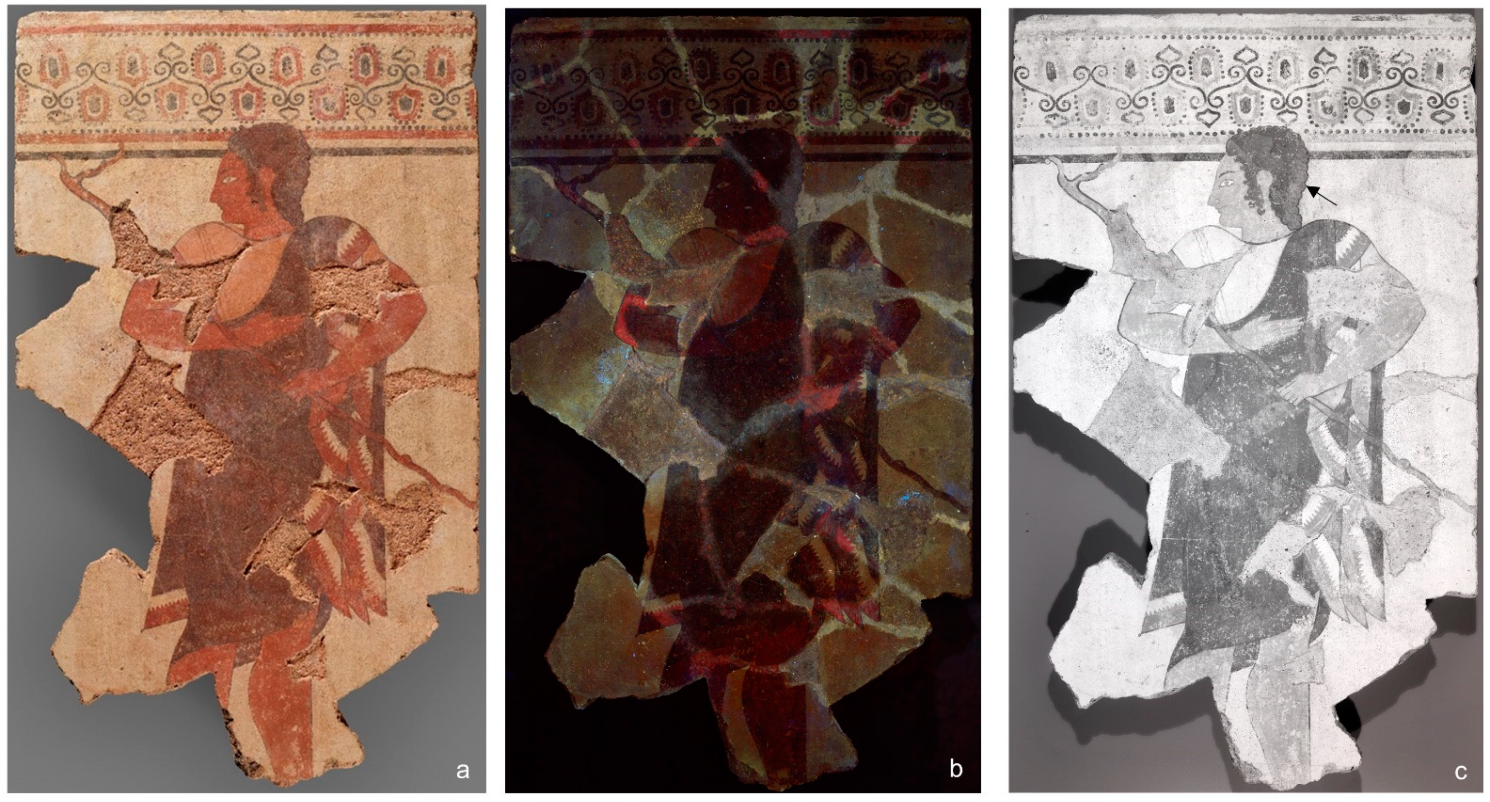
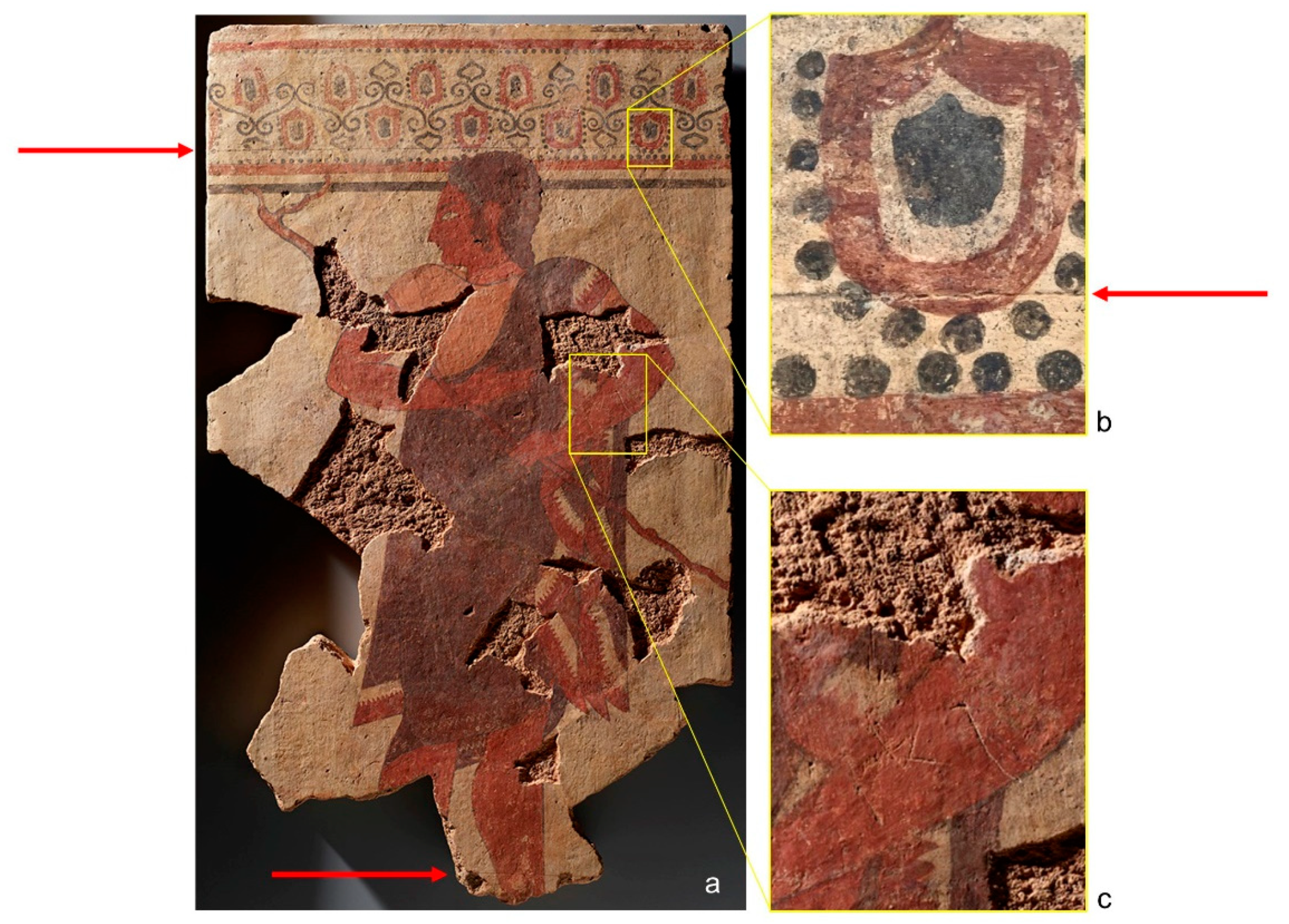
3.1. Multimodal Broadband Imaging

Overall, the Athletic Official panel (Figure 1a) measures 88 × 52.5 cm and is composed of 33 fragments that were reassembled before entering the Getty collection in 1996. Despite the loss of the bottom of the panel, the overall dimensions of the assembled extant fragments are similar to other intact panels of this type [4,12,29]. Losses throughout the panel were compensated using a fill material that closely matched the color of the ancient terracotta and was toned to integrate it with the original painted design, preventing areas of restoration from being easily identified by eye. However, the ancient areas are easily distinguished from the imitative restorations that exhibit intense fluorescence under UV radiation (Figure 1b). Additionally, reflected infrared imaging (Figure 1c) revealed that areas of the hair, back of the head (as indicated by the black arrow), floral frieze, proper right elbow, and the upper portion of the staff were retouched and strengthened using a black pigment exhibiting differential infrared absorption, e.g., showing a more saturated, darker appearance, when compared to the original polychromy in the palmettes and ivy leaves in the upper decorative frieze.
Visible raking light imaging (Figure 2a) revealed two thin horizontal lines incised into the terracotta substrate: one located just below the upper decorative freeze (see red arrows in Figure 2a,b), and the second cutting across the figure’s lower leg near the bottom edge of the fragment. Faint contour incisions are also visible around the face, back of the left thigh, waist, lower end of the forked staff, and across the drapery to the left arm. These incisions were presumably marked on the unfired terracotta surface as preliminary sketches to guide the design of the figure scene [30]. If the function of the incised horizontal lines was to assist with the positioning of the figure within the terracotta panel, it remains unclear why the lower line cuts through the figure’s legs. Additionally, imaging revealed the presence of three graffito characters of unknown function that were incised on the left forearm after firing (see Figure 2c) [31].

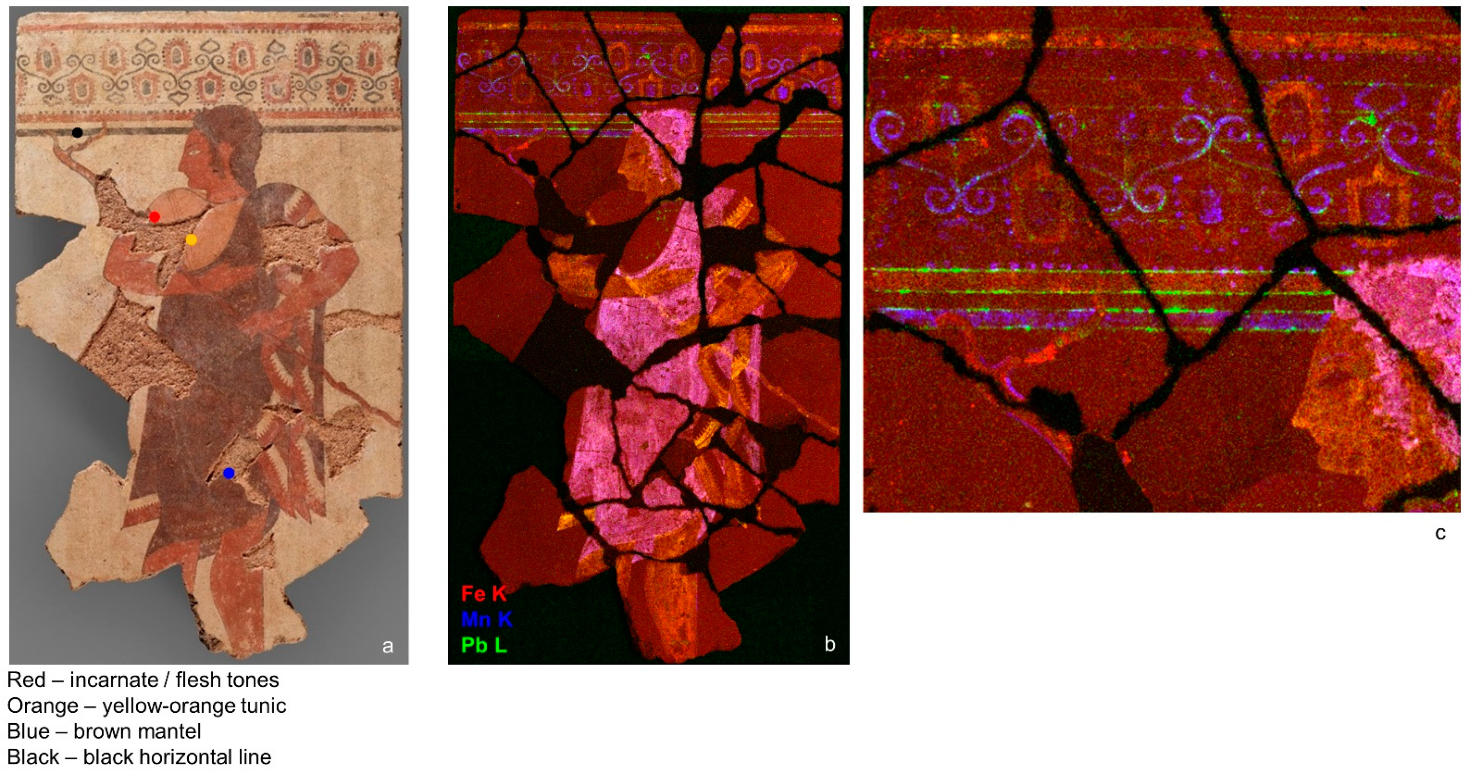
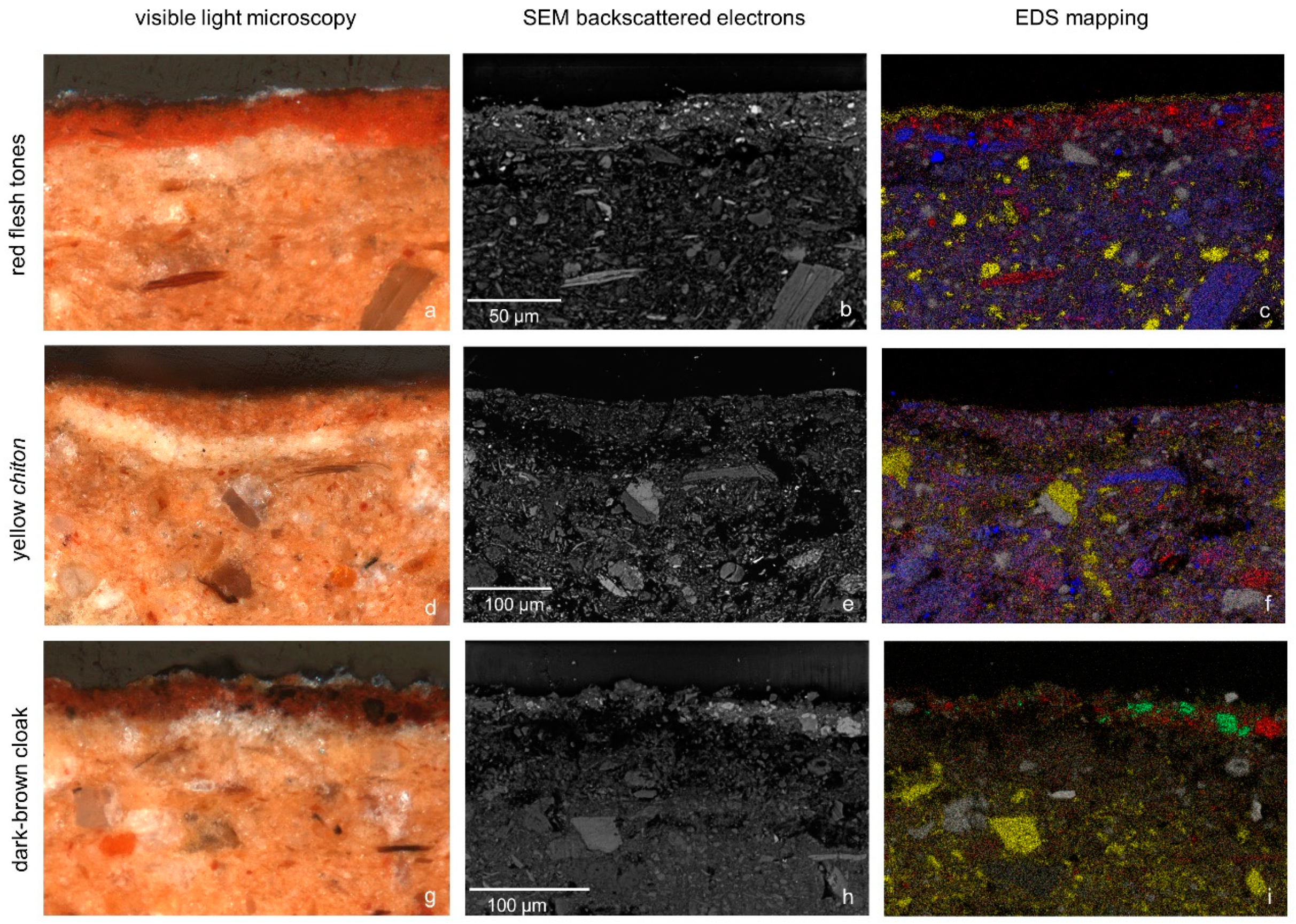
3.2. Pigment Analysis

Scanning macro X-ray fluorescence (MA-XRF) spectroscopy provided information about the elemental composition and distribution of the colorants used in the pictorial decoration. Unsurprisingly, iron—most likely in the form of iron earth compounds— is found throughout the panel, being present in the compositional features that comprise the figure, parts of the decorative background, as well as the terracotta substrate. The different iron-containing materials present are best visualized through a RGB false-color distribution map (Figure 3b), in which iron is shown in red, manganese in blue, and lead in green. The red flesh tones appear yellow in the false-color map, indicating the presence of both iron and lead compounds. This same iron- and lead-containing colorants were used for the lower part of the staff and red decorative designs on the official’s mantle, as well as the red decoration and horizontal bands in the upper frieze. The dark brown cloak and hair appear bright pink in the false-color map, corresponding to areas where the distributions of iron and manganese are co-present, perhaps suggesting the use of umbers. Somewhat surprisingly, the yellow-orange tunic does not appear to contain any elements that distinguish it from the iron-rich terracotta background.
The greatest variation in colorant use was found in the lines and tendrils that comprise the upper ornamental frieze. Along with the iron- and lead-containing compounds used for the red in the palmettes and horizontal bands, a black manganese-rich colorant, appearing blue in the false-color map, was used to create the lower wide horizontal band and slanted s-curved tendrils.
Unexpectedly, the false-color map also revealed the presence of four very thin (less than 1 mm in width), lead-rich (appearing green in the false-color map, see Figure 3c) horizontal lines outlining the red and black bands at the base of the decorative frieze. The MA-XRF map data also showed the presence of additional thin lead-rich horizontal lines marking the upper and lower limits of the curls in the s-curved tendrils and red palmettes. These evenly spaced lines appear to be rulings applied to help guide the laying out of the different design elements that comprise the ornamental frieze. Unlike the two horizontal lines bounding the main figure (Figure 2), these lines are not incised and not visible except through XRF mapping. It has been suggested that guidelines may have been added by snapping taught, pigment-laden, strings onto the surface of the unfired terracotta [32]. However, the thinness of the ruling lines, as observed by the MA-XRF, might suggest the use of a finer tool. To date similar rulings on other Etruscan panels have not been reported in the literature.
To further characterize the iron compounds detected by MA-XRF, fiber optic reflectance spectroscopy (FORS) was carried out across the panel. Diffuse reflectance spectra were collected from areas representative of the yellow-orange, red, and dark-brown areas (star symbols in Figure S1a). As expected, the reflectance spectra (Figure S1b) all share features associated with iron oxides: broad absorption bands near 750 nm and 850–900 nm, and an inflection point in the range of 575 to 590 nm [33]. The results suggest hematite is the most intense chromophore in all three areas [27,34]. The presence of additional iron oxide(s) cannot be excluded, although their identification cannot be determined by FORS due to insufficient separation of the electron pair transition (EPT) bands [27,34,35].
Together, MA-XRF and FORS provided information about the major classes of materials associated with the polychromy as well as their distributions across the pictorial design. To examine the paint stratigraphy and composition of the decorative polychromy and the terracotta substrate, samples of the yellow-orange, red, and dark-brown pigments were removed for additional sample-based analysis (locations from which samples were removed are indicated by the colored dots in Figure 3a). Each of the samples was found to consist of three layers (see Figure 4): the terracotta substrate over which a discontinuous white layer (likely a preparatory layer to level uneven surfaces due to the manufacturing and nature of the terracotta substrate) was applied, followed by a top pigmented layer. SEM backscattered electron images and EDS maps (showing the distributions of calcium (yellow), iron (red), silicon (white), aluminum (blue), and manganese (green) across the region of interest) are shown in the second and third column of images, respectively, in Figure 4.
The white preparatory layer in all three samples appears to have a higher average porosity than either the terracotta base or the overlying pigment layer; dark voids are visible throughout this layer in the BSE images (Figure 4b,e,h). Raman analysis indicates the presence of anatase (TiO2) together with α-quartz and calcite. Together, the detection of these three minerals often is used as proxy for the identification of the presence of kaolin, given that kaolinite (Al2Si2O5(OH)4), the main component of kaolin, has a weak Raman signal [12,36]. The use of kaolin, quarried locally at Monte Sughereto (Sasso di Furbara) [11], has been previously reported on Caeretan terracotta panels [9,11,12]. A similar preparatory layer was also identified in some samples from the Geneva group produced in Cerveteri during the 5th century BCE [6,9]. The presence of this preparatory layer in the Getty panel thus offers additional material evidence to support the close relationship between the Getty and Cerveteri panels.
The pictorial layer in the sample taken from the red skin tone (Figure 4a–c) is compact, bright red in color, and measures approximately 20 μm in thickness. The EDS map shows this layer to be iron rich, and Raman analysis confirms the presence of hematite (Fe2O3) throughout this layer. The thin, discontinuous calcium- and sulfur-rich layer on the surface of the sample is modern, added in a recent conservation treatment.
The pigment layer from the sample taken from the yellow-orange tunic (Figure 4d–f) is similar in color to the terracotta substrate, although more uniform in size and more finely grained. Except for grain size, the BSE and EDS maps do not show any notable differences in the composition of the two layers, perhaps suggesting the use of a material derived from that used for the terracotta substrate. The process of levigation could have been employed to remove the heavier fractions and help produce a finer-grain clay. Recall that the MA-XRF maps likewise found no discernable compositional differences between the yellow-orange painted areas and the underlying terracotta matrix, and Raman spectroscopy did not allow for a clear identification of the colorant in this layer, being dominated by a large fluorescence background. Although the colorant used in the yellow-orange tunic could not be identified, the use of iron oxides, including goethite [37,38] and lepidocrocite [12,38], for the yellow and orange tones has been previously reported both in Etruscan wall paintings in tomb contexts [37,38] and on painted terracotta slabs [12].
The pigment layer in the sample from the dark-brown iron- and manganese-containing cloak (Figure 4g–i) is approximately 20 μm thick and appears to be primarily composed of a reddish-brown matrix with black particles scattered throughout. These black particles range in size from 5 to 20 μm and are characterized by a higher average atomic density in the backscattered electron image than the surrounding matrix. The EDS map shows iron-rich grains (shown in red), which comprise the matrix (identified as hematite by Raman spectroscopy), as well as the manganese-containing particles (shown in green). Raman analysis of these manganese-rich grains show a sharp band at 658 cm−1 together with two weaker bands at 368 and 320 cm−1 (Figure 5), consistent with the presence of a manganese oxide, likely hausmannite (Mn3O4) [39,40,41,42]. Although many manganese compounds undergo laser-induced reduction, hausmannite is highly stable [39] and therefore readily detected by Raman spectroscopy. Notably, Raman spectroscopy also identified hausmannite in a sample taken from the black horizontal band (sample location indicated by black dot in Figure 3a) in the upper decorative frieze, suggesting it was used as the black colorant for the black dots as well as in the palmettes and ivy leaves.
In Etruscan painted objects, brown polychromy typically consists of mixtures based on iron earths. For example, the wall paintings of the Tarquinia tombs [38,43,44] were found to be mixtures of a red ochre mixed with a carbon-based black pigment. Similarly, the brown painted passage in the Ceri warrior [12] was reported to contain a mixture of hematite mixed with other iron oxides, such as wustite (FeO) or maghemite (γ-Fe2O3). In the case of the Athletic Official panel, however, the use of hematite mixed with manganese oxide appears to be most similar to the brown polychromy previously analyzed in fragments from the Geneva group. Those studies showed the presence of browns based on the presence of hematite mixed with an unidentified manganese oxide, suggesting this mixture may be a marker of Caeretan workshop practice [9].

3.3. Terracotta Substrate

In cross-section, visually, the terracotta substrate appears light pink in color and consists of a compact clay paste with numerous small- to medium-sized brown, dark red, white, and black inclusions, and a few large dark, glassy, and sharp inclusions, likely of volcanic origin (see Figure 4). EDS mapping (Figure 4c,f,i) shows the presence of alumino-silicates, calcium inclusions, and other accessory minerals rich in potassium and aluminum. The inclusions were likely added to the terracotta paste as temper, to reduce shrinkage and cracking of the panel during firing, a practice commonly observed in large-scale ceramic panels such as this [45,46,47,48].
The identification of the specific mineralogical phases in ancient terracotta—in particular calcite, dolomite and illite-type clays—is an established method for estimating the firing temperature [49,50]. Previous studies on Etruscan terracotta suggest firing occurred between 800 and 900 °C [6,9,12]. Dolomite decomposes between 700 and 800 °C [50], forming calcite and magnesia [50,51,52]. Calcite undergoes complete thermal degradation between 850 and 900 °C [50,53,54], triggering the formation of new phases, including diopside, anorthite, gehlenite, and wollastonite [55,56,57]. Illite-type clays, such as illite and muscovite, decompose at higher temperatures (950–1050 °C), triggering the formation of new phases, including mullite and spinel [53].
Powder X-ray diffraction (XRD) was carried out on bulk terracotta samples removed from two different locations on the Athletic Official. Both samples gave similar patterns, in which the presence of quartz, muscovite, calcite, dolomite, plagioclase (albite), k-feldspar (orthoclase), anatase, and hematite were identified (see Figure S2). The presence of calcite and apparent absence of any of the mineral phases known to form at higher temperatures as a result of degradation suggests the panel was fired at a temperature not exceeding 850°C, consistent with what has been previously suggested for other fragments belonging to the Geneva group [6].
The elemental composition of a large corpus of Etruscan terracotta panels, including fragments with known archaeological provenance from the Banditaccia Necropolis in Cerveteri, Temple B of Uni-Astarte in Pyrgi, the site of Veii, as well as fragments belonging to the Geneva group were measured by Barone et al., using portable XRF spectroscopy [6,9]. From their results they concluded there were three groups of terracotta: CaO < 9%, CaO > 17 wt%, and intermediate CaO concentrations between 9 and 17 wt% [6,9].
To examine whether the Athletic Official could be linked to any of the above-mentioned archaeological contexts, and thus whether terracotta composition may be a useful figure of merit for separating different Etruscan workshops, point XRF data were collected from undecorated areas of terracotta on the Getty panel. The data were analyzed using principal component analysis (PCA), with Barone’s data serving as the reference set. A plot of PC1 vs. PC3 (Figure 6) yielded three distinct clusters, separated along PC1 (which accounts for the 93.88% of the total variance) by calcium concentration, and along PC3 (which accounts for the 1.00% of the total variance) by manganese and iron content; the loadings plots are shown in Figure 6. Group A (yellow squares, lower left quadrant) has the lowest calcium concentration and is comprised entirely of those samples from the Geneva group previously characterized as being low in calcium and similar in composition to the materials from the Cerveteri area. Group B (green diamonds, lower right quadrant) shows the highest levels of calcium and includes specimens from Veii and Pyrgi, as well as two samples from the Geneva group, the Discobolus being one. Group C (pink triangles, upper quadrant) has an intermediate calcium concentration but higher iron and manganese concentrations than both groups A and B, and contains samples from ancient Caere, the Banditaccia Necropolis, and one sample from the Geneva group.
The results from the Getty panel (blue dots) most closely align with group C: high iron and manganese content and intermediate calcium levels, relative to groups A and B. This match of the elemental composition with the archaeological samples excavated in Caere and the Banditaccia Necropolis offers a likely provenance for the Athletic Official, locating its production in the Cerveteri area.
The finding that the terracotta substrate in the Discobolus and Athletic Official panels are compositionally different may simply reflect a variation in the sourcing of the clay for the substrate and does not necessarily exclude the likelihood that the figures were executed by the same painter. Support for the hypothesis that the panels were created by the same artist would be strengthened, however, if similar ruling lines to those shown here for the Athletic Official were to be found on the Discobolus (through a MA-XRF scan).

4. Conclusions

This multi-analytical study, combining both non-invasive and sample-based analyses, provided new insight into the materials and techniques used to create the Athletic Official panel. Examination of the stratigraphy provided evidence the panel was made by first applying a thin, white kaolin-rich preparatory layer to the unfired calcium-rich alumino-silicate clay slab, most likely to smooth out the irregular surface of the terracotta prior to the application of the decorative polychromy. Parallel, horizontal lines were incised near the top and bottom of the unfired terracotta, presumably to delimit the extents of the decorated surface. While the upper incision appears related to the positioning of the Athletic Official’s head, the exact function of the lower line, cutting across the figure’s shin, remains unclear.
Perhaps the most signficant finding from this study was the discovery of a second set of preparatory guides: MA-XRF scaning revealed now-invisible horizontal lead-rich ruling lines in the ornamental frieze at top of the panel, apparently applied as a guide for creating the floral and geometric decorations behind the figure. Highly contrasted pigmented preparatory lines have been previously reported in Etruscan panels and tomb depictions, but the application of MA-XRF allows for their visualization even when they are no longer visible, as is the case of the Getty panel.
The decorative scheme is based on a limited palette, consisting predominantly of iron earths and, notably, manganese black. Placed within the larger context of the Caeretan painted terracotta panels, the presence of manganese black, and its use in combination with hematite for the brown shades, appears to be an important marker for the workshop practice in ancient Caere. Additional evidence supporting a local production is offered by the examination of the terracotta base: the clay used to fabricate the Getty panel is most similar to other panels from Cerveteri. Interestingly, however, these results also revealed that the Athletic Official and Discobolus plaques do not share a similar terracotta composition. Currently, these panels are hypothesized to be linked based on the similar iconography and presence of an identical upper ornamental frieze. Additional research on the Discobolus is therefore necessary to evaluate whether there is material evidence to support this hypothesis. Similar material and art historical studies on a wider range of similar Etruscan pinakes with known archaeological provenance would help to better understand both the raw materials used and the different workshop production practices across Etruria, facilitating the creation of a more robust model for classifying unknown fragments within these contexts.

Supplementary Materials

The following are available online at https://0-www-mdpi-com.brum.beds.ac.uk/article/10.3390/heritage4040253/s1, Figure S1: Fiber optics reflectance spectroscopy (FORS) analysis; Figure S2: XRD results.

Author Contributions

Conceptualization, M.G., D.M. and M.S.; methodology and data analysis, M.G., D.M. and K.T.; writing—original draft preparation, M.G. and D.M.; writing—review and editing, M.G., D.M., M.S., C.L. and K.T. All authors have read and agreed to the published version of the manuscript.

Funding

This research received no external funding.

Institutional Review Board Statement

Not Applicable.

Informed Consent Statement

Not Applicable.

Data Availability Statement

Data generated and analyzed during this study are available from the corresponding author upon reasonable request.

Acknowledgments

The authors would like to thank Germana Barone and Paolo Mazzoleni from the University of Catania for sharing the raw data from relevant comparanda. We also thank Nathan Daly for a productive discussion on PCA. The authors thank the GCI Technical Studies Research group and Tahnee Cracchiola of the J. Paul Getty Museum Imaging Services and Preparations departments.

Conflicts of Interest

The authors declare no conflict of interest.

References

  1. Steingräber, S.; Ridgway, D.; Serra Ridgway, F.R. Etruscan Painting: Catalogue Raisonné of Etruscan Wall Paintings; Harcourt Brace Jovanovich: New York, NY, USA, 1986. [Google Scholar]
  2. Steingräber, S. Abundance of Life: Etruscan Wall Painting; Getty Publications: Los Angeles, CA, USA, 2006. [Google Scholar]
  3. Bellelli, V. Il guerriero di Ceri. In Il Guerriero di Ceri: Tecnologie per far Rivivere e Interpretare un Capolavoro Della Pittura Etrusca su Terracotta; Guidi, F., Bellelli, V., Trojsi, G., Eds.; ENEA: Roma, Italy, 2006; pp. 59–99. [Google Scholar]
  4. Roncalli, F. Le Lastre Dipinte da Cerveteri; Sansoni: Firenze, Italy, 1966. [Google Scholar]
  5. Roncalli, F. Painted Plaques. In Caere; De Grummond, N., Pieraccini, L., Eds.; University of Texas Press: Austin, TX, USA, 2016; pp. 231–239. [Google Scholar]
  6. Barone, G.; Fugazzotto, M.; Mazzoleni, P.; Raneri, S.; Russo, A. Color and painting techniques in Etruscan architectural slabs. Dye. Pigment. 2019, 171, 107766. [Google Scholar] [CrossRef]
  7. Russo, A. Introduzione. In Pittura di Terracotta: Mito e Immagine Nelle Lastre Dipinte di Cerveteri; Russo, A., Cosentino, R., Zaccagnini, R., Bochicci, L., Eds.; Gangemi Editore: Rome, Italy, 2018; pp. 11–19. [Google Scholar]
  8. Falcucci, C. Le indagini Scientifiche sulle lastre recuperate: Falsi o patrimonio per lo studio di una tecnica antica? In Pittura di Terracotta: Mito e Immagine Nelle Lastre Dipinte di Cerveteri; Russo, A., Cosentino, R., Zaccagnini, R., Bochicchio, L., Eds.; Gangemi Editore: Rome, Italy, 2018; pp. 47–56. [Google Scholar]
  9. Barone, G.; Mazzoleni, P.; Fugazzotto, M.; Raneri, S. Caratterizzazione dei pigmenti e ipotesi di proveneinza delle lastre dipinte di Cerveteri mediante analisi mineralogiche e chimiche non distruttive: Un progetto in corso. In Pittura di Terracotta: Mito e Immagine Nelle Lastre Dipinte di Cerveteri; Russo, A., Cosentino, R., Zaccagnini, R., Bochiccio, L., Eds.; Gangemi Editore: Rome, Italy, 2018; pp. 57–77. [Google Scholar]
  10. Tabasso Laurenzi, M.; Capasso Carola, A. Studio chimico e fisico sulla tecnica di esevuzione di alcune lastre dipinte di terracotta provenienti da Cerveteri. In Proceedings of the Atti della XLIX Riunione della Societa’ Italiana per il Progresso delle Scienze, Siena, Italy, 23–27 September 1967; pp. 997–1009. [Google Scholar]
  11. Bordignon, F.; Postorino, P.; Nucara, A.; Dore, P.; Trojsi, G.; Bellelli, V. The white colour in Etruscan polychromes on terracotta: Spectroscopic identification of kaolin. J. Cult. Herit. 2008, 9, 23–29. [Google Scholar] [CrossRef]
  12. Bordignon, F.; Postorino, P.; Dore, P.; Guidi, G.; Trojsi, G.; Bellelli, V.J.A. In search of Etruscan colours: A spectroscopic study of a painted terracotta slab from Ceri. Archaeometry 2007, 49, 87–100. [Google Scholar] [CrossRef]
  13. Bordignon, F.; Postorino, P.; Dore, P.; Trojsi, G. Raman identification of green and blue pigments in Etruscan polychromes on architectural terracotta panels. J. Raman Spectrosc. 2007, 38, 255–259. [Google Scholar] [CrossRef]
  14. Painted Wall Panel with an Athletic Official. 520–500 B.C. Terracotta and pigment, 88 × 52.5 × 4.5 cm (34 5/8 × 20 11/16 × 1 3/4 in.), acc. 96.AD.140. The J. Paul Getty Museum, Villa Collection, Malibu, California, Gift of Barbara and Lawrence Fleischman. Available online: http://www.getty.edu/art/collection/objects/29613/ (accessed on 27 September 2021).
  15. Pontelli, E. Painted Wall Panel with an Athletic Official. In Etruscan and Italic Art in the J. Paul Getty Museum; Lyons, C., Ed.; Getty Trust Publications: Los Angeles, CA, USA, forthcoming.
  16. Bochicchio, L. Sezione 3. L’atletismo. Cat. 83.86. In Pittura di Terracotta. Mito e Immagine Nelle Lastre Dipinte di Cerveteri. Catalogo della Mostra (Santa Marinella, 22 Giugno–22 Dicembre 2018); Russo, A., Cosentino, R., Zaccagnini, R., Eds.; Gangemi Editore: Rome, Italy, 2018; p. 192. [Google Scholar]
  17. Bochicchio, L. Il mito greco e le altre tematiche nelle lastre dipinte da Cerveteri. In Colori degli Etruschi: Tesori di Terracotta alla Centrale Montemartini, Catalogo della Mostra (Roma, 11 Luglio 2019–28 Giugno 2020); Agnoli, N., Bochiccio, L., Maras, D.F., Zaccagnini, R., Eds.; Gangemi Editore: Rome, Italy, 2019; pp. 89–101. [Google Scholar]
  18. Lyons, C. Etruscan and Italic Art in the J. Paul Getty Museum; Getty Trust Publications: Los Angeles, CA, USA, forthcoming.
  19. Alfeld, M.; Janssens, K.; Dik, J.; de Nolf, W.; van der Snickt, G. Optimization of mobile scanning macro-XRF systems for the in situ investigation of historical paintings. J. Anal. At. Spectrom. 2011, 26, 899–909. [Google Scholar] [CrossRef]
  20. Alfeld, M.; De Nolf, W.; Cagno, S.; Appel, K.; Siddons, D.P.; Kuczewski, A.; Janssens, K.; Dik, J.; Trentelman, K.; Walton, M.; et al. Revealing hidden paint layers in oil paintings by means of scanning macro-XRF: A mock-up study based on Rembrandt’s “An old man in military costume”. J. Anal. At. Spectrom. 2013, 28, 40–51. [Google Scholar] [CrossRef]
  21. Van der Snickt, G.; Legrand, S.; Caen, J.; Vanmeert, F.; Alfeld, M.; Janssens, K.J.M.J. Chemical imaging of stained-glass windows by means of macro X-ray fluorescence (MA-XRF) scanning. Microchem. J. 2016, 124, 615–622. [Google Scholar] [CrossRef]
  22. Hložek, M.; Trojek, T.; Prokeš, R.; Linhart, V.J.R.P. Mediaeval metal threads and their identification using micro-XRF scanning, confocal XRF, and X-ray micro-radiography. Radiat. Phys. Chem. 2019, 155, 299–303. [Google Scholar] [CrossRef]
  23. Alfeld, M.; Baraldi, C.; Gamberini, M.C.; Walter, P. Investigation of the pigment use in the Tomb of the Reliefs and other tombs in the Etruscan Banditaccia Necropolis. X-ray Spectrom. 2019, 48, 262–273. [Google Scholar] [CrossRef]
  24. Solé, V.A.; Papillon, E.; Cotte, M.; Walter, P.; Susini, J. A multiplatform code for the analysis of energy-dispersive X-ray fluorescence spectra. Spectrochim. Acta Part B At. Spectrosc. 2007, 62, 63–68. [Google Scholar] [CrossRef]
  25. Alfeld, M.; Janssens, K. Strategies for processing mega-pixel X-ray fluorescence hyperspectral data: A case study on a version of Caravaggio’s painting Supper at Emmaus. J. Anal. At. Spectrom. 2015, 30, 777–789. [Google Scholar] [CrossRef]
  26. Wolff, T.; Malzer, W.; Mantouvalou, I.; Hahn, O.; Kanngießer, B. A new fundamental parameter based calibration procedure for micro X-ray fluorescence spectrometers. Spectrochim. Acta Part B At. Spectrosc. 2011, 66, 170–178. [Google Scholar] [CrossRef]
  27. Scheinost, A.; Chavernas, A.; Barrón, V.; Torrent, J. Use and limitations of second-derivative diffuse reflectance spectroscopy in the visible to near-infrared range to identify and quantify Fe oxide minerals in soils. Clays Clay Miner. 1998, 46, 528–536. [Google Scholar] [CrossRef]
  28. Doebelin, N.; Kleeberg, R. Profex: A graphical user interface for the Rietveld refinement program BGMN. J. Appl. Crystallogr. 2015, 48, 1573–1580. [Google Scholar] [CrossRef] [Green Version]
  29. Pieraccini, L.C. The Colors of Caere in California. Etruscan Stud. 2011, 14, 55–70. [Google Scholar] [CrossRef]
  30. Brøns, C.; Hedegaard, S.B.; Rasmussen, K.L. The real Thing?: Polychromy research employed in authenticity stidies of Etruscan pinakes in the Ny Carlsberg Glyptotek. Studi Etruschi 2018, 79, 195–223. [Google Scholar]
  31. Wallace, R. Painted Wall Panel with an Atheltic Official. In Etruscan and Italic Art in the J. Paul Getty Museum; Lyons, C., Ed.; Getty Trust Publications: Los Angeles, CA, USA, forthcoming.
  32. Cecchini, A.; Barone, G.; Fugazzotto, M.; Mazzoleni, P.; Russo, A. An Archaeometric Approach to the Pigments of the Etruscan Tombs at Tarquinia. In Proceedings of the 2021 Archaeological Institute of America Meeting, Chicago, IL, USA, 7–10 January 2021. [Google Scholar]
  33. Aceto, M.; Agostino, A.; Fenoglio, G.; Idone, A.; Gulmini, M.; Picollo, M.; Ricciardi, P.; Delaney, J.K. Characterisation of colourants on illuminated manuscripts by portable fibre optic UV-visible-NIR reflectance spectrophotometry. Anal. Methods 2014, 6, 1488–1500. [Google Scholar] [CrossRef]
  34. Cornell, R.M.; Schwertmann, U. The Iron Oxides: Structure, Properties, Reactions, Occurrence and Uses; VCH: Weinheim, Germany, 1996. [Google Scholar]
  35. Sherman, D.M.; Waite, T.D. Electronic spectra of Fe3+ oxides and oxide hydroxides in the near IR to near UV. Am. Mineral. 1985, 70, 1262–1269. [Google Scholar]
  36. Murad, E. Identification of minor amounts of anatase in kaolins by Raman spectroscopy. Am. Mineral. 1997, 82, 203–206. [Google Scholar] [CrossRef]
  37. Klempan, B.; Helwig, K.; Colivicchi, F. Examination and analysis of Etruscan wall paintings at Caere, Italy. Archaeometry 2017, 59, 1082–1094. [Google Scholar] [CrossRef]
  38. Sodo, A.; Artioli, D.; Botti, A.; De Palma, G.; Giovagnoli, A.; Mariottini, M.; Paradisi, A.; Polidoro, C.; Ricci, M.A. The colours of Etruscan painting: A study on the Tomba dell’Orco in the necropolis of Tarquinia. J. Raman Spectrosc. 2008, 39, 1035–1041. [Google Scholar] [CrossRef]
  39. Bernard, M.C.; Hugot-Le Goff, A.; Thi, B.V.; de Torresi, S.C. Electrochromic reactions in manganese oxides I. Raman analysis. J. Electrochem. Soc. 1993, 140, 3065–3070. [Google Scholar] [CrossRef]
  40. Strohmeier, B.R.; Hercules, D.M. Surface spectroscopic characterization of manganese/aluminum oxide catalysts. J. Phys. Chem. 1984, 88, 4922–4929. [Google Scholar] [CrossRef]
  41. Mironova-Ulmane, N.; Kuzmin, A.; Grube, M. Raman and infrared spectromicroscopy of manganese oxides. J. Alloys Compd. 2009, 480, 97–99. [Google Scholar] [CrossRef]
  42. Zahn, D.T. Vibrational spectroscopy of bulk and supported manganese oxides. Phys. Chem. Chem. Phys. 1999, 1, 185–190. [Google Scholar]
  43. Barone, G.; Mazzoleni, P.; Cecchini, A.; Russo, A. In situ Raman and pXRF spectroscopic study on the wall paintings of Etruscan Tarquinia tombs. Dye. Pigment. 2018, 150, 390–403. [Google Scholar] [CrossRef]
  44. Klempan, B.; Corbeil, M.-C.; Poulin, J.; Cook, P. A technical and scientific study of two A. Y. Jackson paintboxes. J. Can. Assoc. Conserv. 2009, 34, 29–38. [Google Scholar]
  45. Del Chiaro, M.A. Two Etruscan Painted Terracotta Panels. J. Paul Getty Mus. J. 1983, 11, 129–134. [Google Scholar]
  46. Barone, G.; Mazzoleni, P.; Raneri, S.; Spagnolo, G.; Santostefano, A. Coroplastic Art in Sicily: An Investigation on Provenance and Manufacturing Technology of Greek Architectural Terracotta from Gela (Italy). Mediterr. Archaeol. Archaeom. 2017, 17, 89–101. [Google Scholar]
  47. Barone, G.; Mazzoleni, P.; Raneri, S.; Monterosso, G.; Santostefano, A.; Spagnolo, G.; Vasta, V. Exploring the Coroplasts’ ‘Techne’ in Greek Architectural Terracottas from Sicily: An Archaeometric Approach. Archaeometry 2018, 60, 986–1001. [Google Scholar] [CrossRef]
  48. Rice, P.M. Pottery Analysis: A Sourcebook; University of Chicago Press: Chicago, IL, USA, 2015. [Google Scholar]
  49. Tite, M.S. Determination of the Firing Temperature of Ancient Ceramics by Measurement of Thermal Expansion: A Reassessment. Archaeometry 1969, 11, 131–143. [Google Scholar] [CrossRef]
  50. Gliozzo, E. Ceramic technology. How to reconstruct the firing process. Archaeol. Anthropol. Sci. 2020, 12, 1–35. [Google Scholar] [CrossRef]
  51. Trindade, M.; Dias, M.; Coroado, J.; Rocha, F. Mineralogical transformations of calcareous rich clays with firing: A comparative study between calcite and dolomite rich clays from Algarve, Portugal. Appl. Clay Sci. 2009, 42, 345–355. [Google Scholar] [CrossRef]
  52. Trindade, M.J.; Dias, M.I.; Coroado, J.; Rocha, F. Firing tests on clay-rich raw materials from the Algarve basin (southern Portugal): Study of mineral transformations with temperature. Clays Clay Miner. 2010, 58, 188–204. [Google Scholar] [CrossRef]
  53. Cultrone, G.; Rodriguez-Navarro, C.; Sebastian, E.; Cazalla, O.; De La Torre, M.J. Carbonate and silicate phase reactions during ceramic firing. Eur. J. Mineral. 2001, 13, 621–634. [Google Scholar] [CrossRef] [Green Version]
  54. Papachristodoulou, C.; Oikonomou, A.; Ioannides, K.; Gravani, K. A study of ancient pottery by means of X-ray fluorescence spectroscopy, multivariate statistics and mineralogical analysis. Anal. Chim. Acta 2006, 573, 347–353. [Google Scholar] [CrossRef] [PubMed]
  55. Maggetti, M. Phase analysis and its significance for technology and origin. In Archaeological Ceramics; Smithsonian Institution Press: Washington, DC, USA, 1982; pp. 121–133. [Google Scholar]
  56. Riccardi, M.; Messiga, B.; Duminuco, P. An approach to the dynamics of clay firing. Appl. Clay Sci. 1999, 15, 393–409. [Google Scholar] [CrossRef]
  57. Barone, G.; Crupi, V.; Galli, S.; Longo, F.; Majolino, D.; Mazzoleni, P.; Spagnolo, G. Archaeometric analyses on ‘Corinthian B’ transport amphorae found at Gela (Sicily, Italy). Archaeometry 2004, 46, 553–568. [Google Scholar] [CrossRef]
Figure 1. Painted wall panel with an Athletic Official, 520–500 B.C., terracotta and pigment, 88 × 52.5 × 4.5 cm (34 5/8 × 20 11/16 × 1 3/4 in.), accession number: 96.AD.140. The J. Paul Getty Museum, Villa Collection, Malibu, California, Gift of Barbara and Lawrence Fleischman. (a) visible light, (b) UV-induced visible fluorescence (UVF) image, and (c) infrared reflected (IRR) image. Digital image in visible light courtesy of the Getty’s Open Content Program.
Heritage 04 00253 g001
Figure 2. (a) Painted wall panel with an Athletic Official in raking light. The two details at larger magnification show (b) the horizontal incised line cutting through the palmette and black dots, and (c) three graffito characters incised on the left forearm after firing.
Heritage 04 00253 g002
Figure 3. (a) Spot map showing sampling locations on the Etruscan slab; (b) MA-XRF tri-color channel with Fe K in red, Mn K in blue, and Pb in green, and (c) inset showing a detail at larger magnification of the now-obscured, lead-rich preparatory lines. For easier reading of the maps, iron only is shown as red, manganese only as blue, lead only as green, while a mixture of iron + manganese appears pink, and iron + lead is yellow. Digital image in visible light courtesy of the Getty’s Open Content Program.
Heritage 04 00253 g003
Figure 4. Light microscopy (crossed polarizing filters), backscatter electron images, and EDS maps (red = iron; yellow = calcium, white = silicon, blue = aluminum, and green = manganese) obtained from multi-layer fragments removed from the red flesh tone (ac), the yellow tunic (df), and the dark-brown mantle (gi).
Heritage 04 00253 g004
Figure 5. Representative Raman spectrum for the dark-brown mantel (black trace), and reference spectrum of hausmannite (blue trace) from the RRUFF database.
Heritage 04 00253 g005
Figure 6. Principal component analysis, showing PC1 and PC3 scores (left) and loadings (right) plots.
Heritage 04 00253 g006
Publisher’s Note: MDPI stays neutral with regard to jurisdictional claims in published maps and institutional affiliations.

