Article

Biological Control of Pear Valsa Canker Caused by Valsa pyri Using Penicillium citrinum

Zhengzhou Fruit Research Institute, Chinese Academy of Agricultural Sciences, Zhengzhou 450009, China
*
Authors to whom correspondence should be addressed.
Submission received: 17 June 2021 / Revised: 13 July 2021 / Accepted: 15 July 2021 / Published: 16 July 2021
(This article belongs to the Section Plant Pathology and Disease Management (PPDM))

Abstract

:
Valsa canker caused by Valsa pyri is one of the most destructive diseases of commercial pear. For the present analysis, 29 different endophytic fungal strains were isolated from the branches of a healthy pear tree. In dual culture assays, strain ZZ1 exhibited robust antifungal activity against all tested pathogens including Valsa pyri. Microscopic analyses suggested that following co-culture with ZZ1, the hyphae of V. pyri were ragged, thin, and ruptured. ZZ1 also induced significant decreases in lesion length and disease incidence on detached pear branches inoculated with V. pyri. ZZ1 isolate-derived culture filtrates also exhibited antifungal activity against V. pyri, decreasing mycelial growth and conidium germination and inhibiting V. pyri-associated lesion development on pear branches. These results suggest that the ZZ1 isolate has the potential for use as a biological control agent against V. pyri. The strain was further identified as Penicillium citrinum based on its morphological characteristics and molecular analyses. Overall, these data highlight a potentially valuable new biocontrol resource for combating pear Valsa canker.

1. Introduction

Pear is one of the most important fruits in China, which is the leading global producer of pear. However, pear trees are susceptible to a number of microbial pathogens, with Valsa canker being among the most destructive of these diseases, posing a major threat to pear production [1,2]. Pear Valsa canker is caused by Valsa pyri [3,4], which can infect plants through bark injuries, leading infected tissue to turn reddish-brown and to soften and decay, potentially resulting in the death of the entire tree in some cases [2,3]. As such, Valsa canker disease severely impacts the pear industry. Currently, chemical control measures are the most common and effective approaches to controlling this disease, but environmental and food safety concerns can markedly constrain the use of these fungicides. In addition, the prolonged use of chemical fungicides has the potential to select for fungicide-resistant V. pyri isolates. There is thus an urgent need for the development of alternative or complementary approaches capable of controlling this economically important plant disease.
The biological control of plant diseases using endophytes or derivatives thereof has been shown to be safe and largely nontoxic, and as such, this is a major focus of active research efforts. A diverse array of microorganisms have been identified as potential biocontrol resources to date [5,6,7], some of which have been implemented in commercial production [8,9]. However, there have been relatively few studies evaluating the biological control of pear Valsa canker using antagonistic endophytes, limiting the available biocontrol options for this disease. Song et al. (2020) recently demonstrated that dipicolinic acid (DPA) derived from endophytic Bacillus subtilis exhibited antifungal activity against different canker pathogens including V. pyri. DPA was able to suppress chitin biosynthesis and cause V. pyri cell lysis [10]. Two endophytic Bacillus velezensis strains isolated from pear branches were also shown to be antagonistic against pear Valsa canker [11]. However, overall, endophytes or bioactive molecules derived there from exhibiting antifungal activity against V. pyri remain rare.
Plant endophytic fungi are natural plant colonizers that live within plant tissues without causing any apparent damage [7]. The use of fungal endophytes for the biological control of plant diseases has been a focus of growing research in recent years [5,12], highlighting the promise of this approach. These endophytes can both directly inhibit pathogen growth and can indirectly prevent disease development by inducing host resistance mechanisms [5,13,14].
As there have been few reported endophytic fungi exhibiting antagonistic activity against V. pyri-induced pear Valsa canker disease to date [10,11], the present study was designed to identify novel biocontrol agents for this pear disease in order to expand the available biocontrol supply. To that end, we isolated endophytic fungi from the bark of healthy branches of pear trees from Xinjiang province, China, and demonstrated their ability to antagonize V. pyri and to prevent pear Valsa canker disease.

2. Materials and Methods

2.1. Pathogenic Fungal Isolates

The pathogens Valsa pyri, Valsa mali, Botryosphaeria dothidea and Colletotrichum gloeosporioideswere used in this study and stored at Zhengzhou Fruit Research Institute, Chinese Academy of Agricultural Sciences, Zhengzhou, China [15,16]. The fungal strains used in this study were cultured in potato dextrose agar (PDA) (200 g potato extracts L−1, 2% glucose and 2% agar) at 25 °C.

2.2. Isolation of Endophytic Fungi from the Branches of Pear

One-year-old branches (about 1 cm in diameter) without any canker disease symptoms were collected from ten-year-old pear tree at Korlaheung city, Xingjiang province, China in June 2020. The endophytic fungi were isolated using a tissue separation method according to a previous study [17]. In brief, the bark of pear branches was peeled off and cut into small pieces (5 mm × 5 mm) and put into 75% ethyl alcohol for 1 min. The small pieces were then placed into 1% sodium hypochlorite for 5 min. After disinfection, the small pieces were washed with sterilized ddH2O five times and put onto sterilized filter paper to absorb water. Subsequently, the small pieces were put onto PDA medium. The plates were incubated at 25 °C for 2–3 weeks, after which emerged fungal colonies were sub-cultured onto new plates to isolate individual colonies.

2.3. Dual Culture Test for Screening Potential Biocontrol Fungi

In total, 29 different endophytic fungal strains were isolated from the branches of a healthy pear tree and were screened for antagonistic activity against V. pyri strain lfl-XJ by dual culture test. After the preliminary screening, the antagonistic strains were further tested antifungal activity against V. mali, B. dothidea and C. gloeosporioides. The method of dual culture for screening potential biocontrol fungi was based on a pervious study with some modifications [18]. Four mycelial plugs (5 mm in diameter) of endophytic fungi were placed at each side 2 cm from the center on PDA medium. After inoculation for 2 days, a mycelial plug (5 mm in diameter) of the pathogenic fungal isolate was placed in the center on PDA medium. Plates were incubated at 25 °C for 6 days. Then, the colony diameter of the pathogenic fungal isolate was measured. The negative control was just inoculated with the pathogenic fungal isolate. Inhibition = (colony diameter of control − colony diameter of treated)/(colony diameter of control − 0.5 cm) × 100%.

2.4. Assessment of the Impact of Antagonistic Strains on V. pyri Hyphal Morphology

Following two days of dual culture, the morphological characteristics of hyphae were assessed using an ultra-depth three-dimensional microscope (KEYENCE), with hyphal diameters being measured. A total of three replicates were analyzed per treatment, with a minimum of 10 hyphae per replicate. The assay was repeated two times.

2.5. Assessment of Antagonistic Strain-Derived Culture Filtrates on V. pyri Mycelial Growth

Three 5 mm diameter mycelial plugs from antagonistic fungi were added to 100 mL of potato dextrose broth (PDB) (200 g potato extracts L−1, 2% glucose) for 7 days (25 °C, 200 rpm). Culture filtrates were collected by centrifuging these samples for 10 min at 5000 rpm and then passing the supernatant through a filter with a 0.22 μm pore size to remove remaining hyphal fragments and spores. After sterilization, the PDA medium was then mixed with different culture filtrate volumes to yield PDA containing 10% or 20% culture filtrate. Each plate was inoculated with one 5 mm diameter plug of V. pyri. Following a 6-day inoculation period, V. pyri colony diameters were measured, with untreated PDA medium serving as a control.

2.6. Assessment of Antagonistic Strain-Derived Culture Filtrates on V. pyti Conidial Germination

V. pyri conidia were isolated as in prior studies [19] and counted with a hemocytometer, after which they were diluted to 106 conidia/mL. This conidial suspension was mixed with the culture filtrate derived from the antagonistic endophytic strain at a 50% concentration (v/v), with an equivalent volume of PB media being used as a control. Next, 15 μL volumes of this liquid were added to 1 cm2 of PDA medium on glass slides in Petri dishes. Samples were incubated at 25 °C for 36 h. Numbers of conidia were then counted with an optical microscope, with three replicates and a minimum of 100 conidia being counted per replicate. The assay was repeated twice.

2.7. Disease Suppression Assay

One-year-old detached branches from a healthy pear tree (Zhongli no.1) were utilized to assess the ability of isolated strains to suppress pear Valsa canker disease development. Branches were cut into 10 cm lengths, washed with sterilized water, disinfected with 75% ethanol, and punched at the midpoint of each branch (5 mm in diameter) using a puncher that had been burned with an alcohol burner. Conidial suspensions (108 conidia/mL) or culture filtrates prepared from antagonistic strains were sprayed evenly onto the surface of these branches using a small spray bottle (1 mL/branch), after which the branches were inoculated with V. pyri mycelial plugs immediately after culture filtrate treatment or two days following conidial suspension treatment. V. pyri mycelial plugs were obtained from the edge of PDA medium after 4 days’ inoculation. The branches were sprayed with sterile water or tebuconazole (86 μg/mL) (Anhui Huilong Group Youngsun Pesticides Co., Ltd, China) as negative and positive controls, respectively. Mycelial plugs were covered with medical absorbent cotton (about 1 cm × 2 cm) (Wellday) wetted with sterile water to maintain appropriate moisture levels. The inoculated branches were kept horizontal in a transparent plastic box. Samples were then incubated for 7 days at 25 °C, after which vernier calipers were used to measure lesion length. Assays were repeated three times, with 10 inoculation sites per experiment. Disease incidence was calculated as follows: (number of symptomatic inoculation sites)/total number of inoculation sites × 100%.

2.8. Antagonistic Strain Identification

The endophytic ZZ1 fungal strain, which exhibited the most robust antagonistic activity against V. pyri, was identified via morphological and molecular identification.
For morphological identification, isolate ZZ1 was inoculated onto PDA medium and cultured for 7 days at 25 °C, after which colony morphology including colony color and colony shape were visually observed. In addition, conidia were eluted from the colony using sterile water and the conidial morphology was observed with a light microscope (NIKON). For molecular identification, DNA from the ZZ1 isolate was extracted with a DNA extraction kit (Omega), and the ITS-rDNA,β-tubulin, nuclear small subnit rDNA (ssu) and elongation factor 1-a (EF-1a) sequences were amplified with appropriate primers. ITS1: 5′-CCGTAGGTGAACCTGCGG-3′, ITS4: 5′-TCCTCCGCTTATTGATATGC-3′ [20]; Bt2a: 5′-GGTAACCAAATCGGTGCTGCTTTC-3′, Bt2b: 5′-ACCCTCAGTGTAGTGACCCTTGGC-3′ [21]; NS1: 5′-GTAGTCATATGCTTGTCTC-3′, NS6: 5′-GCATCACAGACCTGTTATTGCCTC-3′ [20]; EF6: 5′-CTTSTYCCARCCCTTGTACCA-3′, EF-1b: 5′-CACATCAACATCGTCGTTAT-3′ [22]. All PCR reactions were conducted in a reaction containing 1.5 μL of 10 × Taq buffer, 1 μL of 2.5 mM dNTPs, 1 μL of 100 mM Mg2+, 0.25 μL of 5 U/μL Taq DNA polymerase, 0.25 μL of each primer (10 μM), 1 μL of 10 ng/μL fungal DNA, and ddH2O to a final 15 μL volume. Thermo cycler settings were: 3 min at 94 °C; 30 cycles of 94 °C for 30 s, 55 °C for 30 s, and 72 °C for 30 s; 72 °C for 5 min. PCR products were sequenced by Bgi Genomics Co., Ltd., China, and the resultant sequences were blasted against the NCBI nucleotide collection database. Highly homologous sequences were selected for multiple sequence alignment with the MEGA 7.0 software. A tree was constructed via a Neighbor-Joining approach, with 1000 replicate Bootstrap analyses being used to calculate node support.

3. Results

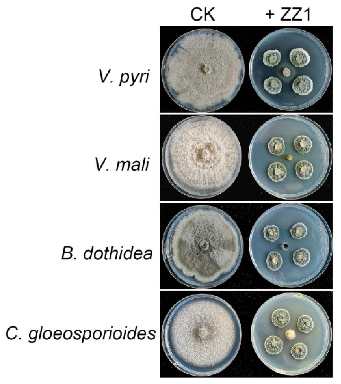
3.1. Isolation and Assessment of Antifungal Activity of ZZ1 against Phytopathogenic Fungi

In total, 29 different endophytic fungal strains were isolated from the bark of branches of a healthy pear tree, and were screened for antagonistic activity against V. pyri train lfl-XJ in vitro. The inhibition of endophytic fungal strains against V. pyri is shown in Table 1. Of these, strain ZZ1 exhibited the most pronounced antagonistic activity, inhibiting ~95% of V. pyri mycelial growth in a dual culture assay (Figure 1, Table 1). Most of the endophytes did not show clear inhibition zone (Supplementary Figure S1), which indicated that these endophytes did not have antibiotic activity to V. pyri.
The ability of strain ZZ1 to inhibit the growth of other fungal fruit tree pathogens was additionally assessed. In a dual culture assay system, isolate ZZ1 was able to strongly inhibit all tested pathogens including Valsa mali, Botryosphaeria dothidea, and Colletotrichum gloeosporioides, with respective inhibition of 96%, 94% and 92% (Figure 1). Together, these data indicated that isolate ZZ1 possessed broad-spectrum antifungal activity.

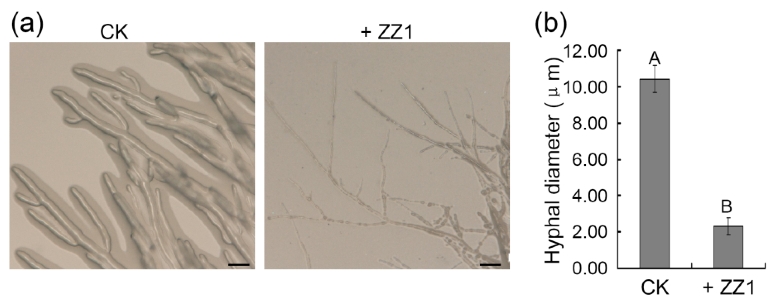
3.2. Assessment of the Antagonistic Effects of Isolate ZZ1 on V. pyri Hyphal Morphology

To further examine the antifungal activity of endophytic strain ZZ1, we utilized V. pyri as a model pathogen in follow-up experiments. We examined the antagonistic effects of isolate ZZ1 on V. pyri hyphae. Under control conditions, V. pyri hyphae appeared smooth and transparent with branches, whereas following ZZ1 isolate exposure these hyphae were rough, tenuous, and ruptured (Figure 2a). Average V. pyri hyphal diameter following ZZ1 exposure (2.3 μm) was significantly smaller than for the control treatment (Figure 2b).

3.3. Analysis of the Ability of ZZ1 Conidial Suspensions to Control Pear Valsa Canker Disease

Conidial suspensions prepared from the ZZ1 isolate were next used to pre-treat detached branches from healthy pear trees to assess the resultant suppression of V. pyri-induced Valsa canker disease development. As of day 7 post-V. pyri inoculation, ZZ1 conidial suspensions had significantly suppressed disease development (Figure 3a), reducing disease incidence to 32% as compared to the 95% incidence observed under control conditions (Figure 3b). Branches treated with tebuconazole as a positive control remained disease-free (Figure 3a,b). The average lesion length on detached branches was also significantly reduced following treatment with a ZZ1 conidial suspension as compared to the control, with these reductions being comparable to those observed following tebuconazole treatment (Figure 3c). These data thus indicated that strain ZZ1 was able to suppress the incidence of pear Valsa canker disease caused by V. pyri.

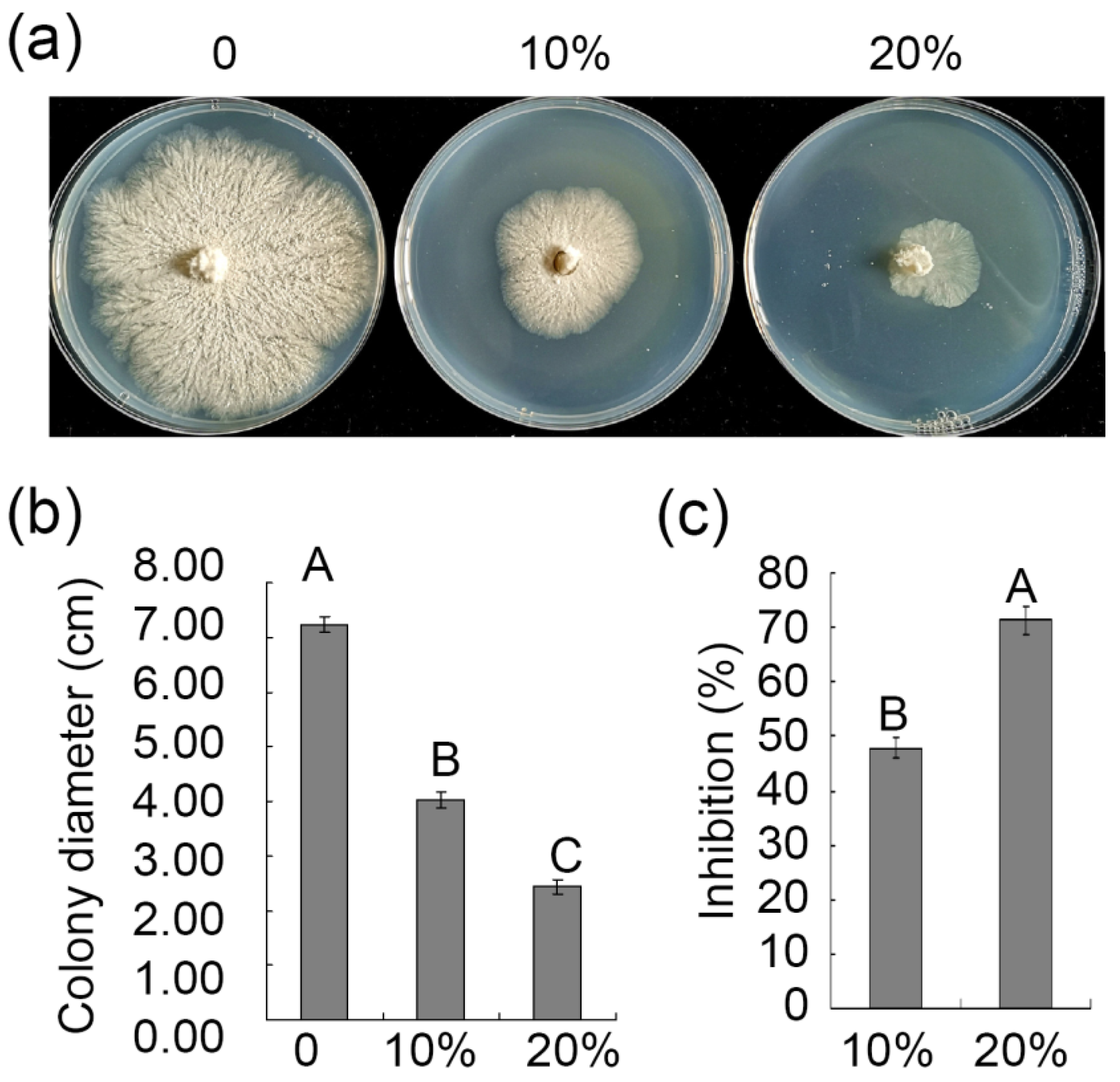
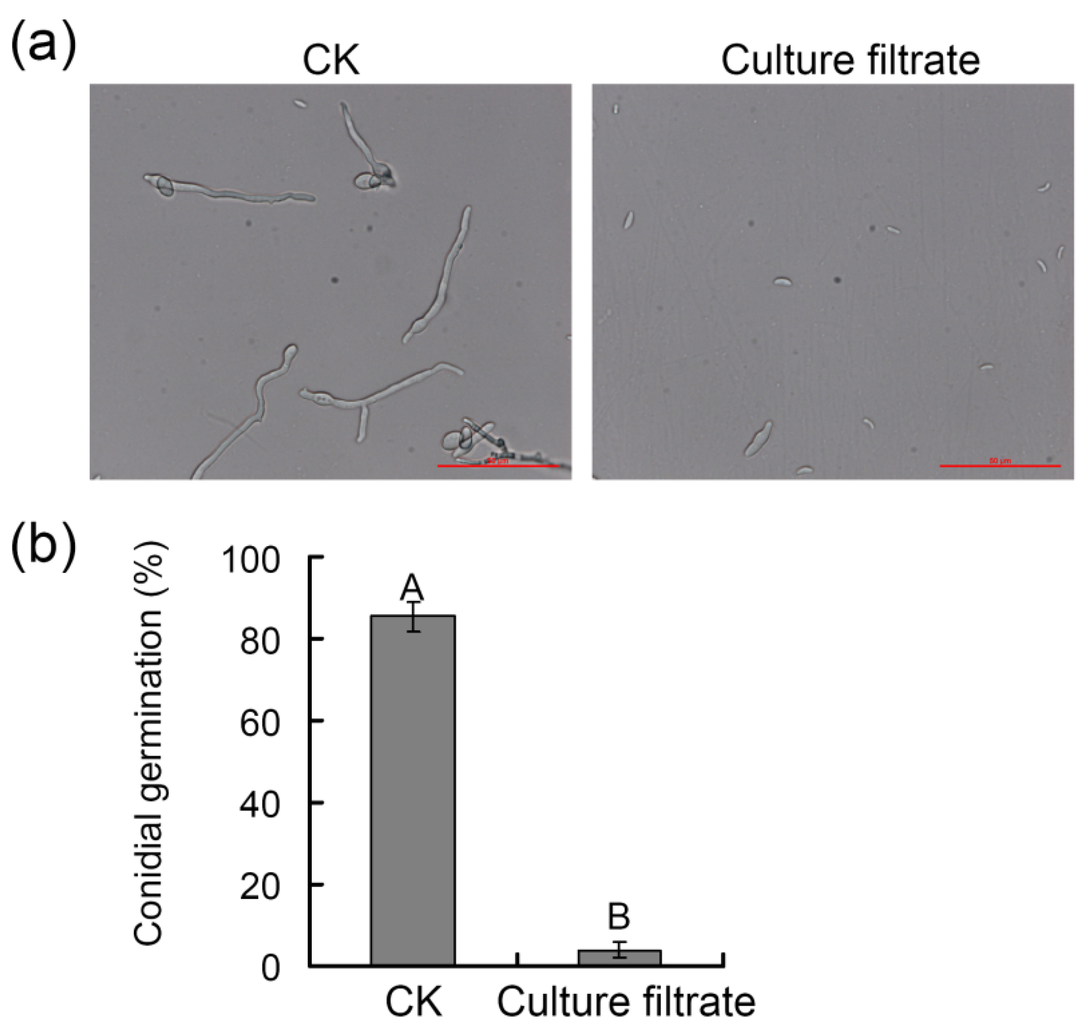
3.4. Assessment of the Ability of ZZ1 Culture Filtrates to Suppress V. pyri Mycelial Growth and Spore Germination

We next collected culture filtrates from isolate ZZ1 and applied these filtrates to PDA medium prior to V. pyri mycelial plug inoculation to determine whether these culture filtrates retained the antifungal activity of the strain from which they were derived. The growth of V. pyri mycelia was significantly suppressed by 47.9% and 71.3% on PDA medium containing 10% and 20% ZZ1 culture filtrate, respectively, relative to control medium (Figure 4a–c). These ZZ1 culture filtrates similarly suppressed V. pyri conidial germination (Figure 5a), with conidial germination in the control and ZZ1 culture filtrate-treated groups of 85% and 4%, respectively (Figure 5b). These results indicated that ZZ1 culture filtrates possess antifungal activity against V. pyri.

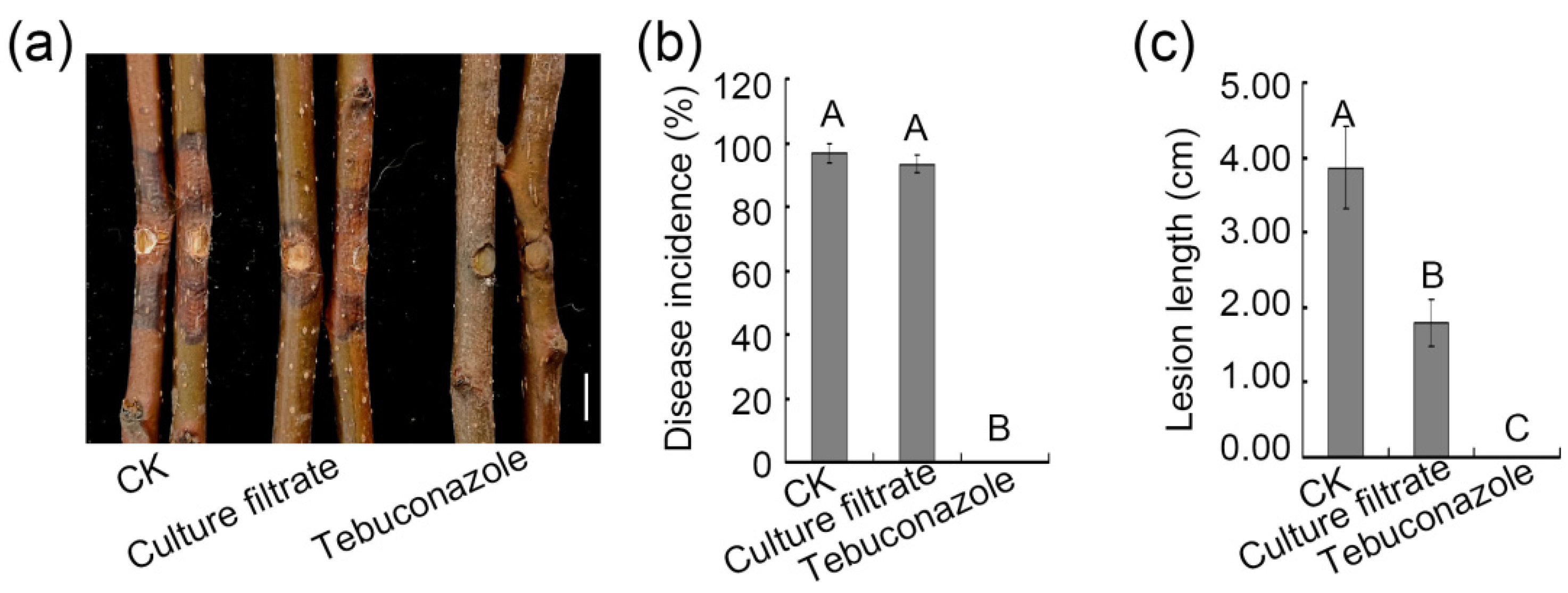
3.5. Assessment of the Ability of ZZ1 Culture Filtrates to Control Pear Valsa Canker Disease

To determine whether ZZ1 culture filtrates were able to inhibit pear Valsa canker disease, detached branches from healthy pear trees were sprayed with these culture filtrates, followed by inoculation with a V. pyri mycelial plug. The results indicated that ZZ1 culture filtrates strongly impacted disease development (Figure 6a). Specifically, while culture filtrates had no effect on disease incidence, the average lesion length decreased significantly relative to that observed following control treatment (Figure 6b,c). This thus suggested that ZZ1culture filtrates were able to inhibit V. pyri-induced lesion development on pear branches.

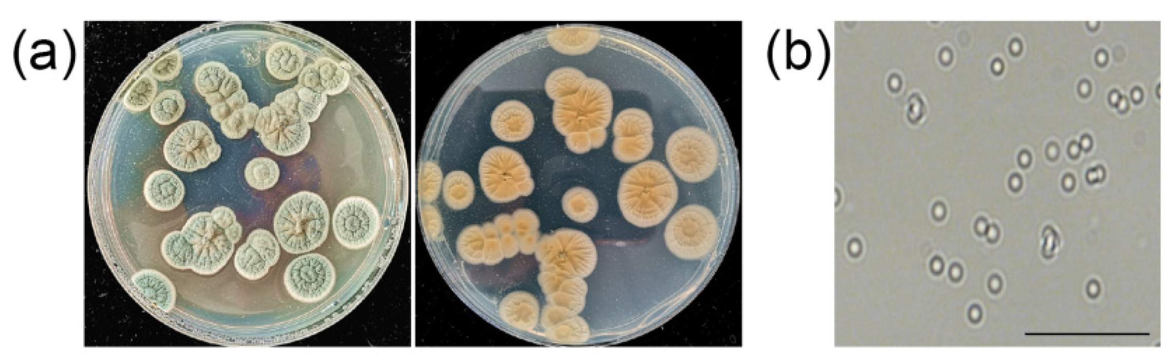
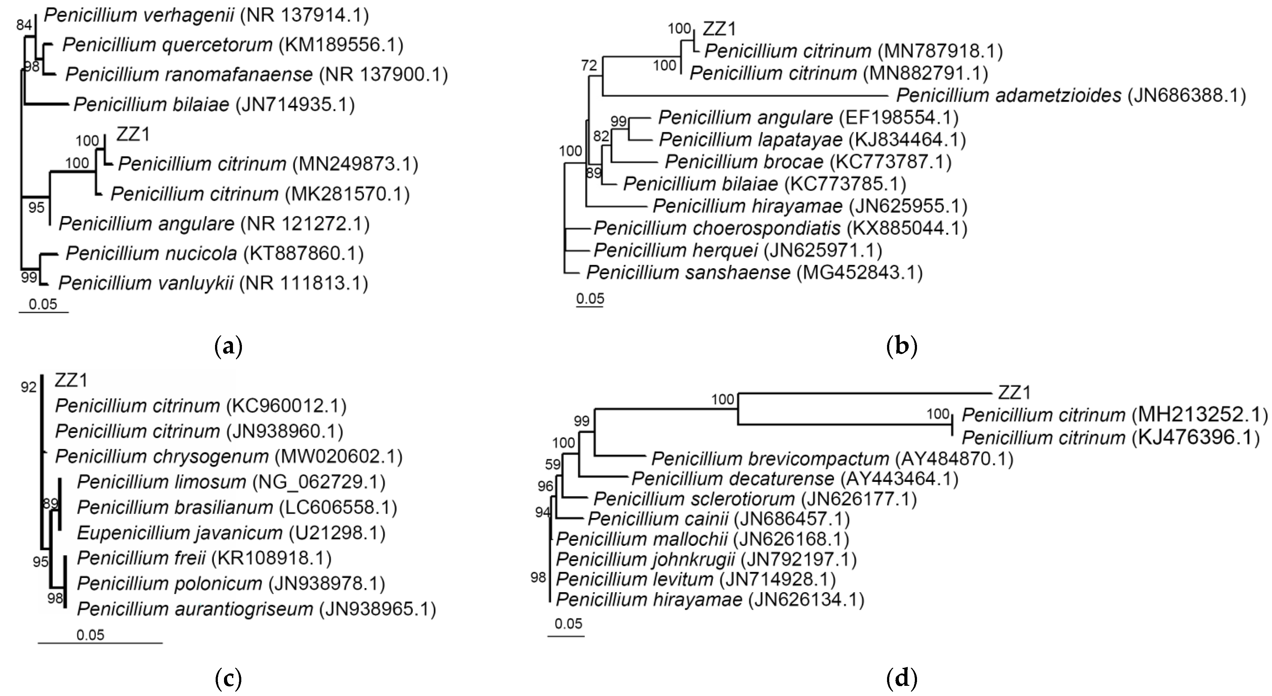
3.6. Identification of Strain ZZ1

When grown on PDA, ZZ1 exhibited gray-olive colored colonies containing distinctive folds. These colonies had white margins, and appeared grayish-yellow from the underside (Figure 7a). Colonies exhibited numerous ovoid conidia (Figure 7b). These morphological characteristics were consistent with those of Penicillium sp.
ITS sequences from isolate ZZ1 were next amplified and used to construct a phylogenetic tree with other closely related sequences using the MEGA 7.0 software. In this analysis, isolate ZZ1 was included in the same branch as P. citrinum strains (Figure 8a). When we similarly constructed a tree based upon the β-tubulin sequence, ssu or EF-1a,isolate ZZ1 similarly exhibited the greatest homology to P. citrinum strains (Figure 8b–d).
Based upon these morphological and molecular analysis results, strain ZZ1 was thus identified as P. citrinum. strain ZZ1 has been preserved at the China General Microbiological Culture Collection Center (CGMCC), a typical culture preservation center in China, under preservation number CGMCC No. 22436; the deposit date is 26 May 2021.

4. Discussion

Valsa canker caused by V. pyri is one of the greatest threats to global pear production. Endophyte-based biocontrol of plant diseases has emerged as a viable and sustainable alternative to chemical fungicides [23]. However, there have been relatively few endophytes reported to date that exhibit antagonistic activity against pear Valsa canker, thus necessitating the use of agrochemicals to control the spread of this disease. Nowadays, many chemical fungicides have been registered for control of Valsa canker, such as tebuconazole, carbendazim and thiophanate-methyl, while only one biological control agent (Bacillus methylotrophicus) is registered for this disease in China. Herein, we isolated 29 endophytic fungal strains from the branches of healthy pear trees and determined that strain ZZ1 possessed significant antagonistic activity against a range of pathogenic fungi. Notably, strain ZZ1 was able to inhibit the development of V. pyri-induced pear Valsa canker disease, suggesting that this strain represents a promising resource for the biocontrol of this destructive disease.
Through a series of morphological and molecular analyses, the isolate ZZ1 was ultimately identified as a strain of Penicillium citrinum. Penicillium species are common fungi that are often studied in the context of biocontrol, with many such isolates having been shown to exhibit antifungal activities against a range of plant pathogens [24,25]. For example, P. striatisporum isolate Pst10 exhibited robust antagonistic activity against Phytophthora spp. and was able to significantly suppress chili pepper Phytophthora root rot disease incidence [26]. Similarly, a P. citrinum isolate derived from Egyptian henbane was able to significantly inhibit a range of fungi, secreting specific extracellular antifungal compounds [27]. P. citrinum isolate BTF08 from wild bananas was also shown to elicit host defenses against Fusarium oxysporum f. sp. cubense in banana plantlets [28]. However, there have been no prior reports regarding the biocontrol of Valsa canker using Penicillium species, and as such, this study is the first to describe the use of a pear tree-derived P. citrinum isolate as a resource capable of suppressing V. pyri-induced pear Valsa canker. Although there are a few studies showing that some P. citrinum strains were pathogenic [29,30,31], our result revealed that P. citrinum strain ZZ1 did not cause disease symptom on pear branch (Supplementary Figure S2). This indicated that strain ZZ1 is an endophyte, not a pathogen on pear. We further found that strain ZZ1 exhibited broad-spectrum antifungal activity against other fruit tree pathogens such as V.mali, B. dothidea and C. gloeosporioides, suggesting that it represents a valuable resource with the potential to be leveraged for the biocontrol of a range of fruit tree diseases.
As biocontrol agents, endophytes can produce an array of bioactive secondary metabolites that can enhance the growth of plants under conditions of biotic stress [6,32,33]. Recently, there have been several research efforts aimed at isolating and identifying antimicrobial compounds derived from Penicillium species. Three are certain compounds with antagonistic activity against plant pathogen species, including citrininand emodin derived from the fermentation of P. citrinum [32], and ent-homocyclopiamine B derived from P. concentricum [34]. Our data indicated that V. pyri mycelial growth and conidium germination were significantly reduced following treatment with ZZ1 culture filtrates. Additionally, we found that ZZ1 culture filtrates were able to significantly inhibit the development of V. pyri lesion on pear branches. As such, these data suggested that ZZ1-derived compounds with antimicrobial activity against V. pyri were present within these culture filtrates. Therefore, isolating and identifying the bioactive compounds in these culture filtrates will be an important focus of our future research.
Herein, detached pear branches were used to evaluate the biocontrol potential of the ZZ1 isolate as an inhibitor of V. pyri-induced pear Valsa canker disease. This analysis revealed that both conidium suspensions and culture filtrates from the ZZ1 isolate were able to suppress such disease, with the biocontrol efficiency of conidial suspensions being comparable to that of the fungicide tebuconazole. As such, these results offer new evidence that the ZZ1 isolate may be used as an effective new antifungal agent for controlling pear Valsa canker disease.

5. Conclusions

In summary, we successfully isolated and identified a P. citrinum isolate ZZ1 from the bark of a pear tree as a potential resource for the biocontrol of pear Valsa canker caused by V. pyri. ZZ1-derived culture filtrates retained antifungal activity against V. pyri, inhibiting associated mycelial growth and conidium germination. Both ZZ1 conidium suspensions and culture filtrates were additionally able to suppress pear Valsa canker caused by V. pyri.

Supplementary Materials

The following are available online at https://0-www-mdpi-com.brum.beds.ac.uk/article/10.3390/horticulturae7070198/s1, Figure S1: The endophyte strain ZZ5 did not show antagonistic activity to V. pyri, Figure S2: Strain ZZ1 did not cause disease symptoms on pear branch.

Author Contributions

H.Y., B.S. and T.H. carried out experiments; Z.Z. gave instructions in experiments; H.Y. designed experiments and wrote the manuscript; L.W., H.H. and H.T. supervised the project. All authors have read and agreed to the published version of the manuscript.

Funding

This work was funded by Major Scientific and Technological Project of Xinjiang Corps (2019AA004); Central Public-Interest Scientific Institution Basal Research Fund (No. 1610192020105).

Institutional Review Board Statement

Not applicable.

Informed Consent Statement

Not applicable.

Data Availability Statement

Not applicable.

Conflicts of Interest

The authors declare no conflict of interest.

References

  1. Kange, A.M.; Xia, A.; Si, J.; Li, B.; Zhang, X.; Ai, G.; He, F.; Dou, D. The fungal-specific transcription factor VpFSTF1 is required for virulence in Valsa pyri. Front. Microbiol. 2020, 10, 2945. [Google Scholar] [CrossRef] [PubMed]
  2. Yin, Z.; Liu, H.; Li, Z.; Ke, X.; Dou, D.; Gao, X.; Song, N.; Dai, Q.; Wu, Y.; Xu, J.-R.; et al. Genome sequence of Valsa canker pathogens uncovers a potential adaptation of colonization of woody bark. New Phytol. 2015, 208, 1202–1216. [Google Scholar] [CrossRef] [PubMed]
  3. Wang, X.; Zang, R.; Yin, Z.; Kang, Z.; Huang, L. Delimiting cryptic pathogen species causing apple Valsa canker with multilocus data. Ecol. Evol. 2014, 4, 1369–1380. [Google Scholar] [CrossRef]
  4. Dong, R.; Wei, S.; Ran, K.; Wang, H.; Dong, X.; Wang, S. The identification and the pathogenicity of analysis for the pathogen of pear Valsa canker. J. Fruit Sci. 2018, 35, 135–138. [Google Scholar]
  5. Latz, M.A.C.; Jensen, B.; Collinge, D.B.; Jørgensen, H.J.L. Endophytic fungi as biocontrol agents: Elucidating mechanisms in disease suppression. Plant Ecol. Divers. 2018, 11, 555–567. [Google Scholar] [CrossRef] [Green Version]
  6. Zheng, R.; Li, S.; Zhang, X.; Zhao, C. Biological activities of some new secondary metabolites isolated from endophytic fungi: A review study. Int. J. Mol. Sci. 2021, 22, 959. [Google Scholar] [CrossRef]
  7. Rodriguez, R.J.; White, J.F., Jr.; Arnold, A.E.; Redman, R.S. Fungal endophytes: Diversity and functional roles. New Phytol. 2009, 182, 314–330. [Google Scholar] [CrossRef]
  8. Liu, Y.; Zhou, S.; Zhang, Y.; Wang, Z.; Liu, G.; Dao, L.; Kang, X.; Li, S. Field control effects of 5 fungicides on diseases of silage maize. Plant Prot. 2019, 45, 280–284. [Google Scholar]
  9. Rusita, I.; Sasongko, H. Effectivity of Trichoderma harzianum as bio-fungicide against moler disease and bio-stimulator of shallot growth. J. Agri-Food Sci. Technol. 2020, 1, 12–17. [Google Scholar] [CrossRef]
  10. Song, X.; Han, M.; He, F.; Wang, S.; Li, C.; Wu, G.; Huang, Z.; Liu, D.; Liu, F.; Laborda, P.; et al. Antifungal mechanism of dipicolinic acid and its efficacy for the biocontrol of pear Valsa canker. Front. Microbiol. 2020, 11, 958. [Google Scholar] [CrossRef]
  11. Gulizige, M.; Xu, L.; Li, Y.; Han, J.; Yu, F.; Luo, M. Antagonistic bacteria against Korla fragrant pear canker: Screening and control effects. Chin. Agric. Sci. Bull. 2020, 36, 127–134. [Google Scholar]
  12. Savita, S.A. Fungi as biological control agents. In Biofertilizers for Sustainable Agriculture and Environment; Giri, B., Prasad, R., Wu, Q.-S., Varma, A., Eds.; Springer International Publishing: Cham, Switzerland, 2019; Volume 55, pp. 395–411. [Google Scholar]
  13. Zhao, X.; Liu, X.; Zhao, H.; Ni, Y.; Lian, Q.; Qian, H.; He, B.; Liu, H.; Ma, Q. Biological control of Fusarium wilt of sesame by Penicillium bilaiae 47M-1. Biol. Control 2021, 158, 104601. [Google Scholar] [CrossRef]
  14. Chaibub, A.A.; de Sousa, T.P.; de Araújo, L.G.; de Filippi, M.C.C. Cladosporium cladosporioides C24G modulates gene expression and enzymatic activity during leaf blast suppression in rice plants. J. Plant Growth Regul. 2019, 39, 1140–1152. [Google Scholar] [CrossRef]
  15. Yuan, H.; Hou, H.; Zhou, Z.; Wang, L.; Tu, H. Isolation and identification of endophytic fungi JK2 antagonistic against pear Valsa canker caused by Valsa pyri. J. Fruit Sci. 2021. [Google Scholar] [CrossRef]
  16. Hou, H.; Zhang, H.; Zhou, Z.; Wang, L.; Yan, Z.; Wang, S. Evaluation of resistance to apple ring rot in Malus Germplasms. Acta Hortic. Sin. 2017, 44, 1559–1568. [Google Scholar]
  17. Xu, T.; Hu, T.; Wang, Y.; Wang, S.; Cao, K. Isolation of endophytic fungi from apple bark and their potential for biological control of Valsa ceratosperma. Acta Phytophylacica Sinica 2012, 39, 327–333. [Google Scholar]
  18. Wang, Y.; Li, Y.; Chang, L.; Yu, H.; Zhou, Z.; Qu, L. Isolation and identification of endophytic fungi resistant to three apple pathogens from the branches of Malus sieversii. J. Fruit Sci. 2020, 37, 390–396. [Google Scholar]
  19. Dai, P.; Zong, Z.; Ma, Q.; Wang, Y. Isolation, evaluation and identification of rhizosphere actinomycetes with potential application for biocontrol of Valsa mali. Eur. J. Plant Pathol. 2018, 153, 119–130. [Google Scholar] [CrossRef]
  20. White, T.; Bruns, T.; Lee, S.; Taylor, F.; White, T.; Lee, S.H.; Taylor, L.; Shawetaylor, J. Amplification and direct sequencing of fungal ribosomal RNA genes for phylogenetics. In PCR Protocols; Elsevier; Academic Press: New York, NY, USA, 1990; pp. 315–322. [Google Scholar]
  21. Lousie, G.N.; Donaldson, G.C. Development of primer sets designed for use with the PCR to amplify conserved genes from filamentous ascomycetes. Appl. Environ. Microbiol. 1995, 61, 1320–1330. [Google Scholar]
  22. Peterson, S.W. Multilocus DNA sequence analysis shows that Penicillium biourgeianum is a distinct species closely related to P. brevicompactum and P. olsonii. Mycol. Res. 2004, 108, 434–440. [Google Scholar] [CrossRef]
  23. Haggag, W.M. The role of entophytic microorganisms in biocontrol of plant diseases. Life Sci. J. 2010, 7, 57–62. [Google Scholar]
  24. Toghueo, R.M.K.; Boyom, F.F. Endophytic Penicillium species and their agricultural, biotechnological, and pharmaceutical applications. 3 Biotech 2020, 10, 1–35. [Google Scholar] [CrossRef] [PubMed]
  25. Sreevidya, M.; Gopalakrishnan, S.; Melø, T.M.; Simic, N.; Bruheim, P.; Sharma, M.; Srinivas, V.; Alekhya, G. Biological control of Botrytis cinerea and plant growth promotion potential by Penicillium citrinum in chickpea (Cicer arietinum L.). Biocontrol. Sci. Technol. 2015, 25, 739–755. [Google Scholar] [CrossRef]
  26. Ma, Y.; Chang, Z.Z.; Zhao, J.T.; Zhou, M.G. Antifungal activity of Penicillium striatisporum Pst10 and its biocontrol effect on Phytophthora root rot of chilli pepper. Biol. Control 2008, 44, 24–31. [Google Scholar] [CrossRef]
  27. Ab De L-Motaal, F.F.; Nassar, M.S.M.; El-Zayat, S.A.; El-Sayed, M.A.; Ito, S.I. Antifungal activity of endophytic fungi isolated from Egyptian henbane (Hyoscyamus muticus L.). Pak. J. Bot. 2010, 42, 2883–2894. [Google Scholar]
  28. Ting, A.; Mah, S.W.; Tee, C.S. Evaluating the feasibility of induced host resistance by endophytic isolate Penicillium citrinum BTF08 as a control mechanism for Fusarium wilt in banana plantlets. Biol. Control 2012, 61, 155–159. [Google Scholar] [CrossRef]
  29. González-Estrada, R.R.; de Jesus Ascencio-Valle, F.; Ragazzo-Sánchez, J.A.; Santoyo, M.C. Use of a marine yeast as a biocontrol agent of the novel pathogen Penicillium citrinum on Persian Lime. Emir. J. Food Agric. 2017, 29, 114–122. [Google Scholar] [CrossRef] [Green Version]
  30. Mohammadiani, E.; Aliakbarlu, J.; Ownagh, A.; Kaboudari, A. Antifungal interactions of Persian shallot (Allium hirtifolium) extracts and potassium sorbate against Aspergillus flavus and Penicillium citrinum. Flavour Fragr. J. 2021, 36, 332–338. [Google Scholar] [CrossRef]
  31. Thamara, C.C.; Mariana, C.F.; Luiz, H.R.; Ana, M.; Enio, N. Penicillium citrinum and Penicillium mallochii: New phytopathogens of orange fruit and their control using chitosan. Carbohydr. Polym. 2020, 234, 115918. [Google Scholar]
  32. Luo, H.; Zhen, Q.; Deng, Y.; Deng, Z.; Lin, W. Two polyketides produced by endophytic Penicillium citrinum DBR-9 from medicinal plant Stephania kwangsiensis and their antifungal activity against plant pathogenic fungi. Nat. Prod. Commun. 2019, 14. [Google Scholar] [CrossRef] [Green Version]
  33. Vinale, F.; Nicoletti, R.; Lacatena, F.; Marra, R.; Sacco, A.; Lombardi, N.; d’Errico, G.; Digilio, M.C.; Lorito, M.; Woo, S.L. Secondary metabolites from the endophytic fungus Talaromyces pinophilus. Nat. Prod. Res. 2017, 31, 1778–1785. [Google Scholar] [CrossRef] [PubMed] [Green Version]
  34. Ali, T.; Pham, T.M.; Ju, K.S.; Rakotondraibe, H.L. Ent-homocyclopiamine B, a prenylated indole alkaloid of biogenetic interest from the endophytic fungus Penicillium concentricum. Molecules 2019, 24, 218. [Google Scholar] [CrossRef] [PubMed] [Green Version]
Figure 1. Antifungal activity of strain ZZ1 against different phytopathogenic fungi. After inoculation with ZZ1 for 2 days, a mycelial plug (5 mm in diameter) of the pathogenic fungal isolate was placed in the center on PDA medium. The pictures were taken at 6 days after inoculation.
Horticulturae 07 00198 g001
Figure 2. Effect of ZZ1 isolate on hyphal morphology of V. pyri. (a) Hyphae morphology of V. pyri after antagonism with ZZ1. The picture was taken at 2 days after antagonism with ZZ1 with an ultra-depth three-dimensional microscope. Bar = 10 μm. (b) Hyphal diameter of V. pyri after antagonism with ZZ1. Values are means ± SD from ten replicates. Letters above the bars indicate statistical significance (p < 0.01) based on Tukey’s test.
Horticulturae 07 00198 g002
Figure 3. Effect of conidial suspension of ZZ1 on pear Valsa canker disease caused by V. pyri. (a) Conidial suspension of ZZ1 suppressed pear Valsa canker. Bar, 1 cm. The picture was taken at 7 days after inoculation. (b) Statistical analysis of the disease incidence. (c) Statistical analysis of disease lesion length. Values are means ± SD from ten replicates. Letters above the bars indicate statistical significance (p < 0.01) based on Tukey’s test.
Horticulturae 07 00198 g003
Figure 4. Effect of culture filtrate of ZZ1 on mycelial growth of V. pyri. (a) Inhibition of culture filtrate of ZZ1 on mycelial growth of V. pyri. The picture was taken at 6 days after inoculation on PDA medium. (b) Statistical analysis of colony diameter. (c) Inhibition of culture filtrate of ZZ1 on mycelial growth of V. pyri. Values are means ± SD from three replicates. Letters above the bars indicate statistical significance (p < 0.01) based on Tukey’s test.
Horticulturae 07 00198 g004
Figure 5. Effect of culture filtrate of ZZ1 on conidial germination of V. pyri. (a) Optical photomicrograph of conidial germination of V. pyri after treatment with culture filtrate of ZZ1 isolate. Bar, 50 μm. The picture was taken at 36 h after inoculation (b) Inhibition of culture filtrate of ZZ1 on conidial germination of V. pyri. Conidial germination was counted at 36 h after inoculation. Values are means ± SD from three replicates. Letters above the bars indicate statistical significance (p < 0.01) based on Tukey’s test.
Horticulturae 07 00198 g005
Figure 6. Effect of culture filtrate of ZZ1 on pear Valsa canker disease caused by V. pyri. (a) Culture filtrate of ZZ1 could antagonize pear Valsa canker. Bar, 1 cm. The picture was taken at 7 days after inoculation. (b) Statistical analysis of the disease incidence. (c) Statistical analysis of disease lesion length. Values are means ± SD from ten replicates. Letters above the bars indicate statistical significance (p < 0.01) based on Tukey’s test.
Horticulturae 07 00198 g006
Figure 7. Morphological characteristics of the antagonistic strain ZZ1. (a) Colony characteristics of ZZ1 isolate. The picture was taken at 7 days after inoculation on PDA at 25 °C. (b) Conidia of ZZ1 isolate. Bar, 20 μm.
Horticulturae 07 00198 g007
Figure 8. Phylogenetic analysis of strain ZZ1 and its relatives based on the nucleotide sequences of ITS (a) the genes encoding β-tubulin (b), ssu (c) and EF-1a (d).
Horticulturae 07 00198 g008
Table 1. The inhibition of endophytic fungal strains against V. pyri.
Endophytic StrainsInhibition (%)
ZZ195.2 ± 2.1
ZZ27.4 ± 0.8
ZZ33.9 ± 0.7
ZZ45.7 ± 1.1
ZZ52.7 ± 0.8
ZZ63.5 ± 1.2
ZZ712.3 ± 1.4
ZZ832.5 ± 3.1
ZZ913.5 ± 2.3
ZZ105.3 ± 1.6
ZZ113.5 ± 0.5
ZZ1255.0 ± 2.6
ZZ1322.3 ± 3.1
ZZ1415.4 ± 2.4
ZZ155.9 ± 1.5
ZZ1614.6 ± 2.4
ZZ1758.7 ± 3.2
ZZ188.6 ± 0.9
ZZ195.4 ± 0.4
ZZ2023.5 ± 3.5
ZZ2116.5 ± 2.6
ZZ227.4 ± 1.8
ZZ238.9 ± 1.1
ZZ243.3 ± 0.8
ZZ255.4 ± 1.3
ZZ268.4 ± 2.1
ZZ274.9 ± 0.5
ZZ289.4 ± 1.7
ZZ2910.5 ± 2.1
Note: Values are means ± SD from three replicates.
Publisher’s Note: MDPI stays neutral with regard to jurisdictional claims in published maps and institutional affiliations.

