Article

Variability of Polyphenolic Compounds and Biological Activities among Perilla frutescens var. crispa Genotypes

1
Department of Industrial Plant Science and Technology, Chungbuk National University, Cheongju 28644, Korea
2
College of Agriculture & Life Sciences, SARI, Jeju National University, Jeju-si 63243, Korea
3
Gangwon Forest Science Institute, Chuncheon 24207, Korea
*
Author to whom correspondence should be addressed.
Submission received: 17 September 2021 / Revised: 11 October 2021 / Accepted: 14 October 2021 / Published: 15 October 2021

Abstract

:
Perilla frutescens var. crispa (Pfc) of the family Lamiaceae is used as a medicinal plant due to its pharmacological properties. Although Pfc is an important resource for the medical nutrition industry, the variability in phytonutrients and biological activities among genotypes of Pfc is not well understood. The effects of genotype on the phytochemical composition, antioxidant activities, antimelanogenic principles, and anti-inflammatory effects of Pfc were determined using eight Pfc genotypes. Using HPLC analysis, we identified 30 polyphenolic compounds from Pfc, although variation was observed in the polyphenolic composition of Pfc genotypes. Pfc 5 exhibited antimelanogenic activity in B16F10 melanoma cells via inhibition of tyrosinase activity. In addition, Pfc 2 strongly inhibited lipopolysaccharide-induced nitric oxide production through translational downregulation of inducible NOS in RAW264 murine macrophages. Taken together, the results of our study reveal the significant impacts of genotype on phytonutrients and biological activities. This finding will assist in the breeding and genetic engineering of Pfc in order to meet future phytonutrition and health challenges.

1. Introduction

Over the past three decades, the use of herbal medicines and phytonutrients (often referred to as phytochemicals) or nutraceuticals has increased tremendously across the world, with many people resorting to them as a primary form of healthcare [1]. In general, medicinal plant-based products consist of unpurified extracts obtained from single plants or various plants that are believed to work together synergistically, indicating that the phytonutrient content and quality of plants can have a significant impact on the quality of derived products. The composition and content of phytonutrients are affected by several environmental factors as well as by genetic diversity [2,3]. For example, Curcuma longa ecotypes contain various levels of polyphenols and curcumin, resulting in different levels of antioxidant and anti-inflammatory activities [4] and suggesting that understanding genetic diversity is important for quality control of medicinal plant-based products.
Perilla (Perilla frutescens), an annual herbaceous crop belonging to the mint family (Lamiaceae), has been used for flavor and fragrance and as an oil, vegetable, and medicine in Asian countries such as China, Japan, and Korea [5]. Among the five varieties of perilla, P. frutescens var. crispa (Pfc) is used as a medicinal herb due to its medicinal properties [6]. Since various polyphenolic compounds, including derivatives of cinnamic acid, apigenin, luteolin, scutellarein, and anthocyanins, were isolated from Pfc and the structure was determined, pharmaceutical applications of Pfc, including antioxidant, antimicrobial, anti-inflammatory, antitumor, and antiarthritic effects, have been developed [5,7,8,9]. Due to its pharmacological functions, Pfc has been used as a functional source in the cosmetic, functional food, and pharmaceutical industries. However, while numerous studies have reported the genetic diversity of Pfc [10,11], there are limited studies on the variation in the chemical composition and biological activities of Pfc among its genotypes.
In this study, we analyzed the antioxidant, antimelanogenic, and anti-inflammatory effects of 70% EtOH extracts obtained from eight different genotypes of Pfc and suggested the genetic variability of biological activities. In addition, polyphenolic contents determined using HPLC indicated that the variation in the biological activities of Pfc genotypes was driven by different levels of polyphenolic compounds. This study is expected to motivate further interest in the use of selected Pfc genotypes as plant breeding and genetic resources in order to improve the phytonutrient contents of Pfc for enhanced utilization in the cosmetics, functional food, and pharmaceutical industries.

2. Materials and Methods

2.1. Plant Materials and Extract Preparations

Eight genotypes of Pfc (Figure 1a and Table S1) were obtained from the National Agrobiodiversity Center. Under natural climate conditions, 20 seedlings from each genotype were grown under field conditions in the research farm at the Chungbuk National University, Republic of Korea (36°37′27.7′′ N, 127°27′3′′ E). Leaves (50 leaves per plant) were harvested from three-month-old plants (August 2019) and lyophilized. The ground materials were soaked in 70% (v/v) ethanol for 24 h and subjected to ultrasonication as described by Lim et al. [12]. Filtrated extracts were evaporated and kept at −20 °C until further usage.

2.2. Determination of Phytochemical Contents

Total phenolic (TPC), flavonoid (TFC), and anthocyanin contents (TAC) were determined as described by Jin et al. [13]. TPC in each extract was calculated in micrograms of gallic acid equivalents (µg GAE/mg extract) using a calibration curve prepared with gallic acid. The TFC was calculated similarly using quercetin (µg QE/mg extract) as the standard, while TAC was calculated as follows: TAC = (OD530 − 0.25 × OD675)/10 mg of extract.
The compositions and contents of thirty polyphenolic compounds were analyzed using HPLC coupled with an ultraviolet–visible detector as described by Yoo et al. [14].

2.3. Analysis of Antioxidant Activities

The free radical scavenging activity of Pfc extracts was determined using 1,1-diphenyl-2-picryl-hydrazyl (DPPH) radicals as described by Ju et al. [15]. DPPH-free radical scavenging activity was expressed as the half maximal reduction concentration of DPPH radicals (RC50).
To analyze the reducing power of each extract, 300 µg/mL of extract was mixed with 200 µL reaction buffer containing 0.2 M sodium phosphate buffer (pH6.6) and 1% potassium ferricyanide as described by Ju et al. [15]. Butylated hydroxytoluene (BHT) was used as a positive control for the DPPH-free radical scavenging assay and calculation of reducing power.
An oxygen radical antioxidant capacity (ORAC) assay was performed using the method described in a previous study [16]. The net area under the curve (AUC) values of the standard and samples were calculated, and the values were expressed as µM of trolox equivalents (µM TE).

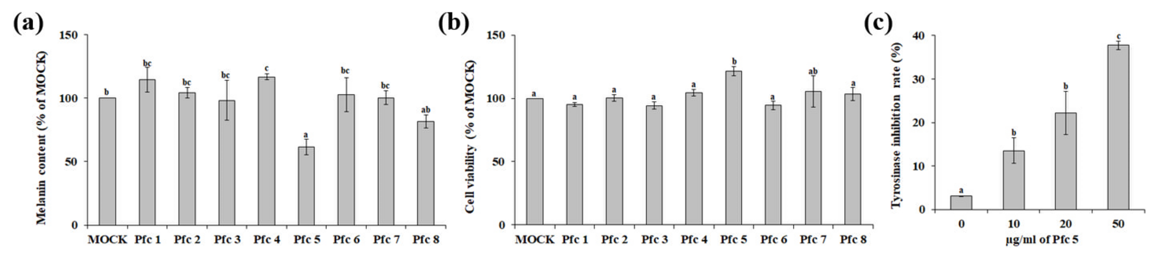
2.4. Determination of Antimelanogenic Effect

To analyze the inhibitory effect of extracts on melanin production, B16F10 melanoma cells (1 × 10⁵ cells/well in 6-well plates) were cultured and treated with each extract together with 1 nM α-melanocyte-stimulating hormone (α-MSH) and 100 µM isobutylmethylxanthine (IBMX), which were used to induce melanogenesis, as described by Jin et al. [17]. After 48 h incubation in a CO2 incubator at 37 °C, cells were harvested by centrifugation. Then, cell pellets were re-suspended in 10% DMSO containing 1 M NaOH and incubated at 65 °C for 1 h. The melanin content was determined by the absorbance at 490 nm.
The in vitro tyrosinase inhibitory activity of selected extracts was determined using a Tyrosinase Inhibition Screening Kit (BioVision, Milpitas, CA, USA).

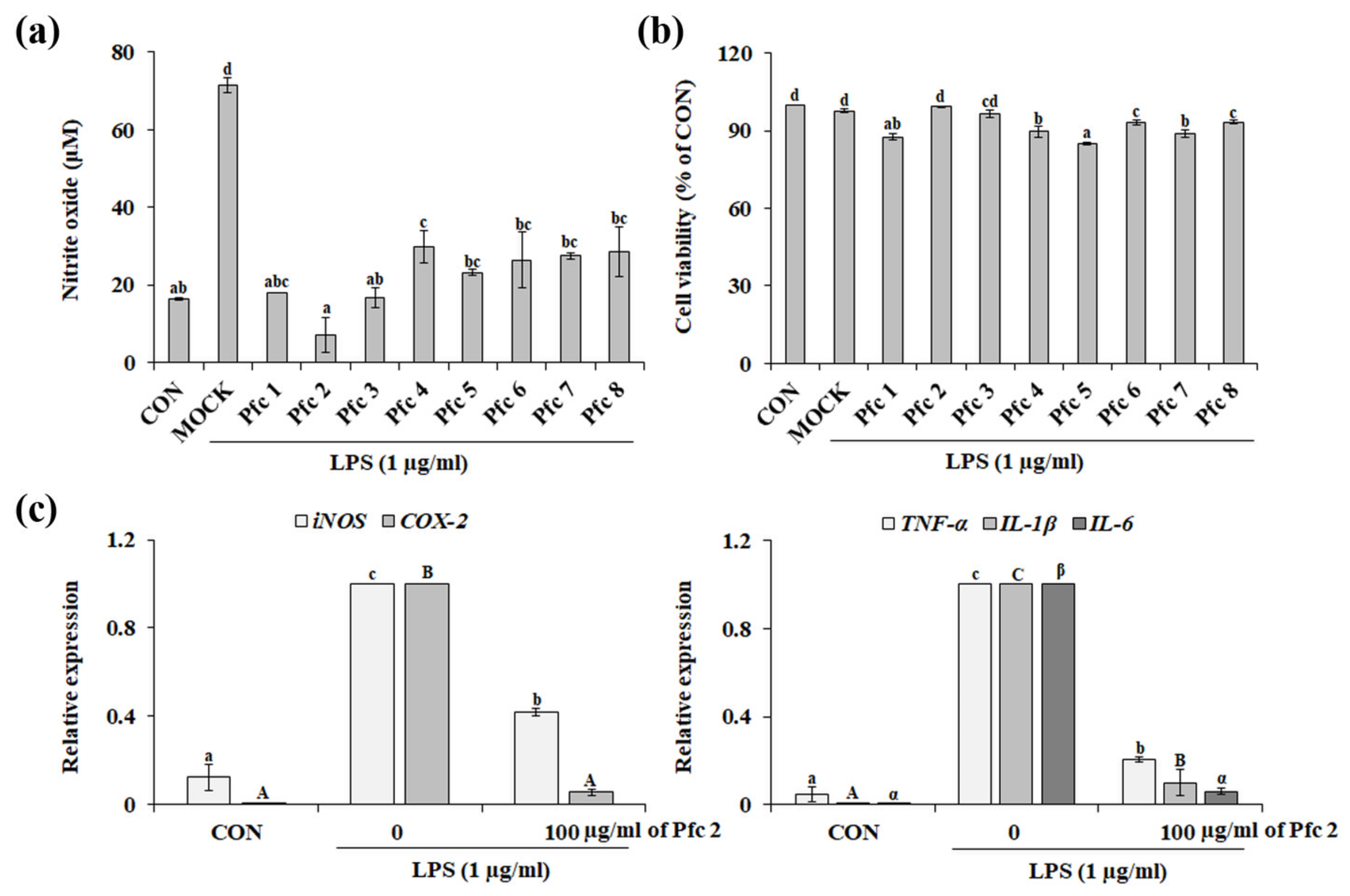
2.5. Determination of Nitric Oxide (NO) Production

RAW 264.7 murine macrophage cells (1.5 × 10⁵ cells/mL in 96-well plates) were cultured and treated with each extract together with 1 μg/mL lipopolysaccharide (LPS) to induce NO production as described by Yoo et al. [18]. After 24 h incubation, NO secretion in the cultured medium was determined using Griess reagent, and NO concentration was determined using the standard curve of sodium nitrite.

2.6. Cell Viability Assay

Cultured B16F10 or RAW 264.7 cells were treated with each extract together with α-MSH/IBMX or LPS. Cell viability was determined using an MTT solution as described by Yoo et al. [14], and the absorbance was measured at 520 nm.

2.7. Quantitative Real-Time PCR (qRT-PCR)

After the treatment with the selected extracts, the TRI reagent (Invitrogen, Carlsbad, CA, USA) was used to isolate total RNA from cells. cDNA was synthesized using the ReverTra Ace® qPCR RT Master Mix with gDNA Remover (TOYOBO, Co., Ltd., Osaka, Japan), and SYBR® Green Real-time PCR Master Mix (TOYOBO, Co., Ltd., Osaka, Japan) was used to run a qRT-PCR analysis using the CFX96TM Real-Time PCR Detection System (Bio-Rad, Hercules, CA, USA). GAPDH was used as an internal standard, and the specific qRT-PCR primers used are listed in Table S2.

2.8. Statistical Analysis

Data were analyzed using one-way ANOVAs, followed by Duncan multiple range tests. A p value < 0.05 was considered to be significant.

3. Results and Discussion

3.1. Phytochemical Compounds and Antioxidant Activities of Pfc Genotypes

In plants, phytochemicals play a significant role in a variety of physiological functions, including defense against herbivorous predators, protection from microbial infections and environmental pollutants, and survival responses to environmental stresses [19]. Among various phytochemicals, polyphenolic compounds are considered to be a key bioactive compound due to their various potential biological activities, including antioxidant, antimelanogenic, tumor cell inhibition, and anti-inflammatory activities [20]. Eight Pfc genotypes (Figure 1a and Table S1) grown in the same environmental conditions were tested for their polyphenolic contents and antioxidant activities. As shown in Figure 1b, the mean values of TPC, TFC, and TAC varied among the Pfc genotypes. TPC ranged from 133.1 to 168.5 µg GAE/mg in the Pfcs, and the TFC values could be ordered as follows: Pfc 6 (31.6 µg QE/mg) > Pfc 1 (30.9 µg QE/mg) > Pfc 8 (30.1 µg QE/mg) > Pfc 5 (28.9 µg QE/mg) > Pfc 4 (28.5 µg QE/mg) (Figure 1b). Anthocyanin is known as a key factor for distinguishing Pfc plants divided into the red and green forms [21]. As expected, the phenotype of purple leaves in Pfcs is caused by high levels of TAC (Figure 1a,b). To further analyze the variation of the chemical composition in Pfc genotypes, polyphenolic compounds were analyzed using HPLC. We quantified thirty polyphenolic compounds, including narirutin, hesperidin, rutin, kaempferol, rhamnetin, gallic acid, vanillic acid, caffeic acid, p-coumaric acid, and sinapinic acid (Table 1). The major flavonoid compound in Pfc 1, 4, 5, 7, and 8 was narirutin, whereas kaempferol was found to be the main flavonoid compound in Pfc 2 (Table 1). Similar to the flavonoid composition, variation was observed in the phenolic compounds of the Pfc genotypes (Table 1), indicating that genotype is an important determinant factor affecting phytochemical compositions and contents.
Polyphenols are known as antioxidant compounds due to their ability to donate hydrogen or electrons as well as to form stable radical intermediates [22]. To determine the antioxidant properties of the Pfc extracts, we analyzed their antioxidant activities using the DPPH free radical scavenging, reducing power, and ORAC assays. As shown in Figure 1c, the strongest DPPH free radical scavenging activity was detected in Pfc 1 (RC50 = 75.2 ± 0.7 μg/mL extract), followed by Pfc 5 (RC50 = 86.4 ± 0.9 μg/mL extract) and Pfc 8 (RC50 = 88.2 ± 2.3 μg/mL extract). In addition, Pfc 1 revealed the highest reducing power compared with the other genotypes, although no significant difference was observed between Pfc 1, Pfc 5, Pfc 7, and Pfc 8. Notably, Pfc 7 (ORAC value = 25.6 ± 1.0 μM TE) exhibited a better ORAC value than Pfc 1 (ORAC value = 16.3 ± 1.1 μM TE). Antioxidant compounds react with free radicals through different mechanisms: hydrogen atom transfer (HAT, e.g., ORAC assay), single electron transfer (SET, e.g., reducing power assay), or the combination of both HAT and SET (e.g., DPPH free radical scavenging assay) [23], indicating that Pfc 1 prevents free-radical-mediated oxidative damage by stabilizing reactive oxygen species though the SET mechanism. In this study, variables such as soil nutrient status, environmental conditions, and geographical locations were minimized by cultivating genotypes at the same location, suggesting that the large observed variations in TPC, TFC, TAC, polyphenolic compounds, and antioxidant activities in the Pfcs are attributable to genotype.

3.2. Variation in Antimelanogenic Principles of Pfc Genotypes

Although melanin production is an essential physiological process to protect the skin from ultraviolet light damage, over-accumulation of melanin leads to hyperpigmentation disorders such as melanocytic nevus, seborrheic keratosis, solar lentigo, and melanoma [13]. So far, a number of melanin synthesis inhibitor compounds or extracts have been introduced as skin whitening agents [24]. Among them, Pfc extract has been exhibited the marked inhibitory effect on melanin production in B16F10 melanoma cells [25]. This indicates the potential Pfc as a skin whitening agent, although variation in the antimelanogenic properties of Pfc genotypes is unknown. To investigate the antimelanogenic activity of the Pfc genotypes, the inhibitory effects of each extract on α-MSH/IBMX-stimulated melanin accumulation in B16F10 cells were determined. As shown in Figure 2a, 25 μg/mL Pfc 5 markedly inhibited α-MSH/IBMX-induced melanin synthesis in B16F10 cells, whereas treatment with the other Pfc extracts exhibited no or low inhibitory effect on α-MSH/IBMX-induced melanin accumulation. To investigate whether the antimelanogenic activity of the Pfc extracts was mediated by cell viability, the cytotoxic effect of the Pfc extracts on the α-MSH/IBMX-stimulated B16F10 cells was determined by MTT assay. No significant reduction in viability was observed for any of the tested extracts (Figure 2b), suggesting that the antimelanogenic activity of Pfc 5 was not due to cytotoxicity. Taken together, this indicates that Pfcs have potential to be developed as a whitening agent, but the skin-whitening-related activities of the Pfcs vary among genotypes.
Melanin accumulation induced by α-MSH/IBMX is mainly mediated by tyrosinase activity [17]. In particular, apigenin and sinensetin, which were highly present in Pfc 5 compared to the other extracts (Table 1), have been identified as tyrosinase inhibitors [26,27], indicating that the stronger antimelanogenic effect of Pfc 5 might be mediated by inhibition of tyrosinase activity. To investigate the direct effect of Pfc 5 on tyrosinase activity, tyrosinase was incubated with different concentrations of Pfc 5 (10–50 µg/mL). The tyrosinase inhibitory activity of Pfc 5 was observed to be dose-dependent, suggesting that the antimelanogenic activity of Pfc 5 in B16F10 melanoma cells is mediated by inhibition of the L-DOPA oxidation activity of tyrosinase.

3.3. Variation in Anti-Inflammatory Effects among Genotypes of Pfc

Inflammation is the immune response to various external stimuli, such as pathogens, damaged cells, trauma, and chemicals; however, uncontrolled inflammation can lead to increased risk of chronic inflammatory diseases [18,28]. Various studies have introduced the anti-inflammatory effects of Perilla extracts in immune cells, such as macrophages and mast cells [29,30,31]. In addition, Pfc extract has been found to inhibit the activation of Src family kinases and intracellular Ca2+ mobilization, resulting in reduced inflammatory activities of N-formyl-Met-Leu-Phe-stimulated human neutrophils [8]. However, to our knowledge, no study has assessed the impact of genetic variation on the anti-inflammatory properties of Pfc. As shown in Figure 3a, LPS-induced NO production in RAW 264.7 macrophage cells was significantly inhibited by all of the tested Pfc extracts. In addition, none of the tested Pfc extracts affected the viability of RAW 264.7 cells regardless of the presence of LPS (Figure 3b). Although there is variation in polyphenolic contents and compositions between Pfc extracts, the presence of various polyphenolic compounds known as anti-inflammatory compounds (ferullic acid, chlorogenic acid, etc., Table 1) indicates that Pfc extracts exhibit promising anti-inflammatory activity.
Pro-inflammatory mediators, including NO and prostaglandin E2 (PGE2), play a significant role in the clinical inflammatory response and lead to inflammatory symptoms and pain [32]. In LPS-stimulated RAW 264.7 cells, NO and PGE2 are mainly synthesized by inducible NOS (iNOS) and cyclooxygenase-2 (COX-2), respectively [33]. In addition, tumor necrosis factor alpha (TNF-α), together with interleukin (IL)-1β and -6, is known as a pro-inflammatory cytokine, which is produced by activated macrophages and involved in inflammatory reactions [34]. To investigate the effect of Pfc on the production of these pro-inflammatory molecules, the expression levels of pro-inflammatory genes under stimulation of LPS plus Pfc 2, which exhibited the strongest anti-inflammatory activity (Figure 3a), were analyzed using qRT-PCR. LPS treatment significantly induced the transcription of pro-inflammatory genes compared to non-treated cells (CON), and the expression of LPS-induced pro-inflammatory genes was significantly decreased by Pfc 2 treatment (Figure 3c). This indicates that the anti-inflammatory activity of Pfc 2 is mediated by the inhibition of pro-inflammatory mediators and cytokines.

4. Conclusions

The results of the present study indicate the effects of genotype on the phytochemical composition, antioxidant activities, antimelanogenic activities, and anti-inflammatory effects of Pfc and suggest that genetic factors also play an essential role in phytonutrient concentrations and biological activities. Of the studied genotypes, Pfc 5 exhibited the best antimelanogenic activity, whereas Pfc 2 had the highest anti-inflammatory activity, suggesting the importance of selecting the appropriate genotype to obtain maximum health benefits.

Supplementary Materials

The following are available online at https://0-www-mdpi-com.brum.beds.ac.uk/article/10.3390/horticulturae7100404/s1, Table S1: Information of the Perilla frutescens var. crispa genotypes used in this study, Table S2: Primer sequences for qRT-PCR analysis.

Author Contributions

Conceptualization, H.K., J.-S.K. and T.K.H.; investigation, H.J.J. and K.C.K.; writing—original draft preparation, H.J.J., H.K., J.-S.K. and T.K.H.; writing—review and editing, H.J.J., H.K., J.-S.K. and T.K.H. All authors have read and agreed to the published version of the manuscript.

Funding

This work was supported by “Regional Innovation Strategy (RIS)” through the National Research Foundation of Korea (NRF), funded by the Ministry of Education (MOE).

Institutional Review Board Statement

Not applicable.

Informed Consent Statement

Not applicable.

Data Availability Statement

Not applicable.

Conflicts of Interest

The authors declare no conflict of interest.

References

  1. Ekor, M. The growing use of herbal medicines: Issues relating to adverse reactions and challenges in monitoring safety. Front. Pharmacol. 2014, 4, 177. [Google Scholar] [CrossRef] [Green Version]
  2. Maharjan, P.; Penny, J.; Partington, D.L.; Panozzo, J.F. Genotype and environment effects on the chemical composition and rheological properties of field peas. J. Sci. Food Agric. 2019, 99, 5409–5416. [Google Scholar] [CrossRef] [PubMed]
  3. Mohammadi Bazargani, M.; Falahati-Anbaran, M.; Rohloff, J. Comparative analyses of phytochemical variation within and between congeneric species of willow herb, Epilobium hirsutum and E. parviflorum: Contribution of environmental factors. Front. Plant Sci. 2021, 11, 595190. [Google Scholar] [CrossRef] [PubMed]
  4. Balada, C.; Castro, M.; Fassio, C.; Zamora, A.; Marchant, M.J.; Acevedo, W.; Guzmán, L. Genetic diversity and biological activity of Curcuma longa ecotypes from Rapa Nui using molecular markers. Saudi J. Biol. Sci. 2021, 28, 707–716. [Google Scholar] [CrossRef] [PubMed]
  5. Ju, H.J.; Bang, J.-H.; Chung, J.-W.; Hyun, T.K. Variation in essential oil composition and antimicrobial activity among different genotypes of Perilla frutescens var. crispa. J. Appl. Biol. Chem. 2021, 64, 127–131. [Google Scholar] [CrossRef]
  6. Ahmed, H.M. Ethnomedicinal, phytochemical and pharmacological investigations of Perilla frutescens (L.) Britt. Molecules 2019, 24, 102. [Google Scholar] [CrossRef] [PubMed] [Green Version]
  7. Saita, E.; Kishimoto, Y.; Tani, M.; Iizuka, M.; Toyozaki, M.; Sugihara, N.; Kondo, K. Antioxidant activities of Perilla frutescens against low-density lipoprotein oxidation in vitro and in human subjects. J. Oleo Sci. 2012, 61, 113–120. [Google Scholar] [CrossRef] [PubMed] [Green Version]
  8. Chen, C.Y.; Leu, Y.L.; Fang, Y.; Lin, C.F.; Kuo, L.M.; Sung, W.C.; Tsai, Y.F.; Chung, P.J.; Lee, M.C.; Kuo, Y.T.; et al. Anti-inflammatory effects of Perilla frutescens in activated human neutrophils through two independent pathways: Src family kinases and calcium. Sci. Rep. 2015, 5, 18204. [Google Scholar] [CrossRef] [PubMed] [Green Version]
  9. Jin, C.H.; So, Y.; Kim, H.Y.; Han, S.N.; Kim, J.B. Anti-Arthritic activities of supercritical carbon dioxide extract derived from radiation mutant Perilla frutescens var. crispa in collagen antibody-induced arthritis. Nutrients 2019, 11, 2959. [Google Scholar] [CrossRef] [PubMed] [Green Version]
  10. Sa, K.J.; Choi, I.Y.; Park, K.C.; Lee, J.K. Genetic diversity and population structure among accessions of Perilla frutescens (L.) Britton in East Asia using new developed microsatellite markers. Genes Genom. 2018, 40, 1319–1329. [Google Scholar] [CrossRef]
  11. Ma, S.J.; Sa, K.J.; Hong, T.K.; Lee, J.K. Genetic diversity and population structure analysis in Perilla crop and their weedy types from northern and southern areas of China based on simple sequence repeat (SSRs). Genes Genom. 2019, 41, 267–281. [Google Scholar] [CrossRef]
  12. Lim, H.J.; Woo, K.W.; Lee, K.R.; Lee, S.K.; Kim, H.P. Inhibition of proinflammatory cytokine generation in lung inflammation by the leaves of Perilla frutescens and its constituents. Biomol. Ther. 2014, 22, 62–67. [Google Scholar] [CrossRef] [PubMed] [Green Version]
  13. Jin, S.; Eom, S.H.; Kim, J.S.; Jo, I.H.; Hyun, T.K. Influence of ripening stages on phytochemical composition and bioavailability of ginseng berry (Panax ginseng C.A. Meyer). J. Appl. Bot. Food Qual. 2019, 92, 130–137. [Google Scholar]
  14. Yoo, T.K.; Kim, J.S.; Hyun, T.K. Polyphenolic composition and anti-melanoma activity of white forsythia (Abeliophyllum distichum Nakai) organ extracts. Plants 2020, 9, 757. [Google Scholar] [CrossRef]
  15. Ju, H.J.; Yoo, T.K.; Jin, S.; Kim, H.; Hyun, T.K. In vitro evaluation of the pharmacological properties of crude methanol extract and its fractions of Aconitum austrokoreense aerial parts. Rev. Mex. Ing. Quim. 2020, 19, 1341–1350. [Google Scholar] [CrossRef]
  16. Jin, S.; Hyun, T.K. Ectopic expression of production of anthocyanin pigment 1 (PAP1) improves the antioxidant and anti-melanogenic properties of ginseng (Panax ginseng C.A. Meyer) hairy roots. Antioxidants 2020, 9, 922. [Google Scholar] [CrossRef] [PubMed]
  17. Jin, M.L.; Park, S.Y.; Kim, Y.H.; Park, G.; Son, H.J.; Lee, S.J. Suppression of α-MSH and IBMX-induced melanogenesis by cordycepin via inhibition of CREB and MITF, and activation of PI3K/Akt and ERK-dependent mechanisms. Int. J. Mol. Med. 2012, 29, 119–124. [Google Scholar] [PubMed] [Green Version]
  18. Yoo, T.K.; Jeong, W.T.; Kim, J.G.; Ji, H.S.; Ahn, M.A.; Chung, J.W.; Lim, H.B.; Hyun, T.K. UPLC-ESI-Q-TOF-MS-Based metabolite profiling, antioxidant and anti-inflammatory properties of different organ extracts of Abeliophyllum distichum. Antioxidants 2021, 10, 70. [Google Scholar] [CrossRef] [PubMed]
  19. Leonov, A.; Arlia-Ciommo, A.; Piano, A.; Svistkova, V.; Lutchman, V.; Medkour, Y.; Titorenko, V.I. Longevity extension by phytochemicals. Molecules 2015, 20, 6544–6572. [Google Scholar] [CrossRef] [Green Version]
  20. Abbaszadeh, H.; Keikhaei, B.; Mottaghi, S. A review of molecular mechanisms involved in anticancer and antiangiogenic effects of natural polyphenolic compounds. Phytother. Res. 2019, 33, 2002–2014. [Google Scholar] [CrossRef]
  21. Fukushima, A.; Nakamura, M.; Suzuki, H.; Saito, K.; Yamazaki, M. High-throughput sequencing and de novo assembly of red and green forms of the Perilla frutescens var. crispa transcriptome. PLoS ONE 2015, 10, e0129154. [Google Scholar] [CrossRef] [PubMed] [Green Version]
  22. Nićiforović, N.; Mihailović, V.; Masković, P.; Solujić, S.; Stojković, A.; Pavlović Muratspahić, D. Antioxidant activity of selected plant species; potential new sources of natural antioxidants. Food Chem. Toxicol. 2010, 48, 3125–3130. [Google Scholar] [CrossRef] [PubMed]
  23. Liang, N.; Kitts, D.D. Antioxidant property of coffee components: Assessment of methods that define mechanisms of action. Molecules 2014, 19, 19180–19208. [Google Scholar] [CrossRef] [PubMed] [Green Version]
  24. Qian, W.; Liu, W.; Zhu, D.; Cao, Y.; Tang, A.; Gong, G.; Su, H. Natural skin-whitening compounds for the treatment of melanogenesis (Review). Exp. Ther. Med. 2020, 20, 173–185. [Google Scholar] [CrossRef] [PubMed] [Green Version]
  25. Hwang, J.-H.; Lee, B.M. Inhibitory effects of plant extracts on tyrosinase, L-DOPA oxidation, and melanin synthesis. J. Toxicol. Environ. Health Part A 2007, 70, 393–407. [Google Scholar] [CrossRef]
  26. Ha, T.-J.; Hwang, S.-W.; Jung, H.-J.; Park, K.-H.; Yang, M.-S. Apigenin, tyrosinase inhibitor isolated from the flowers of Hemisteptia lyrata Bunge. J. Appl. Biol. Chem. 2002, 445, 170–172. [Google Scholar]
  27. Yoon, H.S.; Ko, H.-C.; Kim, S.-J.; Kim, S.S. Stimulatory effects of a 5,6,7,3′,4′-pentamethoxyflavone, sinensetin, on melanogenesis in B16/F10 murine melanoma cells. Lat. Am. J. Pharm. 2015, 34, 1087–1092. [Google Scholar]
  28. Chen, L.; Deng, H.; Cui, H.; Fang, J.; Zuo, Z.; Deng, J.; Li, Y.; Wang, X.; Zhao, L. Inflammatory responses and inflammation associated diseases in organs. Oncotarget 2018, 9, 7204–7218. [Google Scholar] [CrossRef] [PubMed] [Green Version]
  29. Shin, T.Y.; Kim, S.H.; Kim, S.H.; Kim, Y.K.; Park, H.J.; Chae, B.S.; Jung, H.J.; Kim, H.M. Inhibitory effect of mast cell-mediated immediate-type allergic reactions in rats by Perilla frutescens. Immunopharmacol. Immunotoxicol. 2000, 22, 489–500. [Google Scholar] [CrossRef] [PubMed]
  30. Huang, B.P.; Lin, C.H.; Chen, Y.C.; Kao, S.H. Anti-inflammatory effects of Perilla frutescens leaf extract on lipopolysaccharide-stimulated RAW264.7 cells. Mol. Med. Rep. 2014, 10, 1077–1083. [Google Scholar] [CrossRef] [Green Version]
  31. Park, S.H.; Shin, D.; Lim, S.S.; Lee, J.Y.; Kang, Y.H. Purple perilla extracts allay ER stress in lipid-laden macrophages. PLoS ONE 2014, 9, e110581. [Google Scholar]
  32. Toriyabe, M.; Omote, K.; Kawamata, T.; Namiki, A. Contribution of interaction between nitric oxide and cyclooxygenases to the production of prostaglandins in carrageenan-induced inflammation. Anesthesiology 2004, 101, 983–990. [Google Scholar] [CrossRef] [PubMed]
  33. Zhang, D.D.; Zhang, H.; Lao, Y.Z.; Wu, R.; Xu, J.W.; Murad, F.; Bian, K.; Xu, H.X. Anti-inflammatory effect of 1,3,5,7-tetrahydroxy-8-isoprenylxanthone isolated from twigs of Garcinia esculenta on stimulated macrophage. Mediat. Inflamm. 2015, 2015, 350564. [Google Scholar] [CrossRef] [Green Version]
  34. Zhang, J.M.; An, J. Cytokines, inflammation, and pain. Int. Anesthesiol. Clin. 2007, 45, 27–37. [Google Scholar] [CrossRef] [PubMed] [Green Version]
Figure 1. Effects of genotype on phytochemical concentrations and antioxidant activities. (a) Typical leaves of eight genotypes of Perilla frutescens var. crispa (Pfc). Scale bar = 1 cm. (b) Total phenolic content, total flavonoid content, and total anthocyanin content of eight Pfc genotypes. (c) Antioxidant activities of eight Pfc genotypes. The antioxidant activities were determined using DPPH free radical scavenging, reducing power, and ORAC assays. Different letters indicate that means are significantly difference at p < 0.05 (means ± SE of three independent experiments).
Horticulturae 07 00404 g001
Figure 2. Comparison of antimelanogenic activity among Perilla frutescens var. crispa genotypes. (a) The effect of extracts obtained from eight genotypes on melanin production was analyzed in α-MSH/IBMX-stimulated B16F10 cells. (b) The effect of each extract on the viability of B16F10 cells. (c) Inhibitory effect of Pfc 5 on tyrosinase activity. The mean of three independent experiments is plotted with SE. Different letters indicate significant differences among the groups (p < 0.05).
Horticulturae 07 00404 g002
Figure 3. Comparison of anti-inflammatory activities among Perilla frutescens var. crispa (Pfc) genotypes. Effects of Pfc genotype on NO production (a) and cell viability (b) in LPS-treated RAW 264.7 cells. (c) Effect of Pfc 2 on the expression of LPS-induced iNOS, COX-2, TNF-α, IL-1β, and IL-6 in RAW 264.7 cells. The expression levels for each gene were calculated relative to its expression in LPS-stimulated RAW 264.7 cells (0 µg/mL of Pfc 2). The mean of three independent experiments is plotted with SE. Different letters indicate significant differences among the groups (p < 0.05).
Horticulturae 07 00404 g003
Table 1. Polyphenolic compounds in different Perilla frutescens var. crispa genotypes.
Compound
(µg/g of Extract)
Pfc 1Pfc 2Pfc 3Pfc 4Pfc 5Pfc 6Pfc 7Pfc 8
Rutin2.33 ± 0.14 b,(1)0 a2.60 ± 0.02 bc8.31 ± 1.47 e0 a4.06 ± 0.10 cd5.23 ± 0.30 d2.23 ± 0.01 b
Taxifolin0.99 ± 0.03 d0 a0.62 ± 0.01 c0 a1.15 ± 0.02 d0.34 ± 0.17 b0.07 ± 0.01 a1.44 ± 0.01 e
Narirutin47.98 ± 0.11 c2.43 ± 0.03 a2.43 ± 0.10 a18.68 ± 8.77 b44.44 ± 0.07 c5.85 ± 0.18 a11.45 ± 6.32 ab45.56 ± 1.47 c
Naringin2.27 ± 0.06 a2.73 ± 0.01 a,b0.94 ± 0.06 a5.59 ± 2.67 b0.65 ± 0.0.004 a1.12 ± 0.24 a1.91 ± 0.02 a2.04 ± 1.10 a
Hesperidin0.86 ± 0.02 a1.36 ± 0.01 a9.01 ± 3.23 b1.99 ± 0.12 a1.22 ± 0.02 a13.78 ± 0.03 c2.81 ± 0.02 a15.78 ± 0.06 c
Neohesperidin4.92 ± 0.02 f0.87 ± 0.01 b0.76 ± 0.01 b1.32 ± 0.02 c1.59 ± 0.01 d0.51 ± 0.01 a2.07 ± 0.02 e1.20 ± 0.03 c
Myricetin1.24 ± 0.01 cd1.18 ± 0.01 c1.06 ± 0.07 b1.29 ± 0.02 d0 a1.71 ± 0.01 e1.21 ± 0.01 cd0 a
Quercetin0 a0 a0 a1.16 ± 0.01 b1.71 ± 0.01 d0 a1.55 ± 0.03 c0 a
Luteolin0 a0 a0 a1.02 ± 0.01 b0 a0 a0 a0 a
Naringenin0.49 ± 0.07 ab0.50 ± 0.05 ab0.48 ± 0.05 ab0.54 ± 0.01 ab0.36 ± 0.03 a0.62 ± 0.12 b0.485 ± 0.005 ab0.43 ± 0.02 ab
Hesperetin2.23 ± 0.01 b7.62 ± 1.39 c1.14 ± 0.57 ab0 a0 a0 a0.28 ± 0.003 a0 a
Apigenin0 a0 a0 a0.63 ± 0.003 b2.41 ± 0.11 d0.77 ± 0.39 b1.76 ± 0.01 c0.36 ± 0.004 ab
Kaempferol15.09 ± 0.07 b45.61 ± 9.13 c5.33 ± 2.19 ab3.35 ± 0.01 a13.26 ± 1.02 ab2.68 ± 0.10 a3.35 ± 0.05 a9.35 ± 0.69 ab
Isorhaemnetin2.09 ± 1.16 ab4.68 ± 0.54 b2.81 ± 1.62 ab4.59 ± 0.03 b2.30 ± 0.69 ab3.54 ± 0.27 ab3.71 ± 0.55 ab1.76 ± 0.37 a
Rhamnetin1.86 ± 0.08 a2.64 ± 0.20 a7.24 ± 2.76 b1.93 ± 0.02 a12.96 ± 0.28 c1.93 ± 0.97 a1.73 ± 0.13 a13.63 ± 0.06 c
Sinensetin1.01 ± 0.16 a1.50 ± 0.32 ab1.09 ± 0.01 a1.07 ± 0.06 a3.15 ± 0.15 c1.74 ± 0.04 b1.96 ± 0.16 b1.07 ± 0.04 a
Nobiletin0.39 ± 0.20 b1.14 ± 0.22 c0 a0 a1.82 ± 0.12 d0 a0 a0.71 ± 0.004 b
Tangeretin6.79 ± 0.12 d0.54 ± 0.09 b0 a0 a1.07 ± 0.14 c0 a0 a0 a
Gallic acid5.64 ± 0.15 bc4.10 ± 2.05 b5.77 ± 0.02 bc0 a6.65 ± 0.02 c0 a5.62 ± 0.04 bc0 a
Protocatecuic acid1.52 ± 0.76 a2.42 ± 0.02 a2.32 ± 0.05 a1.48 ± 0.74 a2.24 ± 0.01 a2.24 ± 0.03 a2.36 ± 0.05 a1.46 ± 0.73 a
p-Hydroxybenzoic acid0.69 ± 0.35 a1.07 ± 0.06 abc1.52 ± 0.08 cd1.55 ± 0.08 d0.98 ± 0.02 ab1.23 ± 0.09 bcd1.25 ± 0.08 bcd1.24 ± 0.06 bcd
Vanillic acid5.76 ± 2.89 a16.23 ± 0.25 c10.87 ± 0.13 b10.69 ± 0.10 b8.68 ± 0.11 ab12.65 ± 2.48 bc11.79 ± 0.37 b6.28 ± 0.07 a
Chlorogenic acid2.47 ± 1.24 a3.65 ± 0.17 a3.27 ± 0.03 a3.65 ± 0.11 a3.50 ± 0.04 a3.32 ± 0.02 a3.40 ± 0.03 a3.31 ± 0.02 a
Caffeic acid3.22 ± 1.61 a6.37 ± 0.13 b4.947 ± 0.26 ab5.35 ± 0.01 b5.54 ± 0.06 b5.08 ± 0.48 ab5.25 ± 0.10 b13.00 ± 0.28 c
Syringic acid1.86 ± 0.93 a2.61 ± 0.02 ab2.66 ± 0.39 ab2.75 ± 0.05 ab2.56 ± 0.01 ab3.78 ± 0.42 b7.76 ± 0.05 c3.02 ± 0.01 ab
p-Coumaric acid3.57 ± 1.78 a10.38 ± 0.05 b12.11 ± 0.11 bc17.29 ± 0.10 e17.86 ± 0.05 e4.08 ± 0.53 a16.11 ± 0.11 de13.87 ± 1.01 cd
Benzoic acid1.88 ± 0.94 b0 a1.75 ± 0.05 b2.51 ± 0.03 b0 a1.23 ± 0.61 ab2.30 ± 0.01 b2.41 ± 0.04 b
Ferullic acid2.14 ± 0.01 a2.04 ± 0.007 ab2.55 ± 0.2 b2.13 ± 0.02 ab4.14 ± 0.02 c2.26 ± 0.05 ab2.73 ± 0.03 b3.65 ± 0.005 c
Sinapinic acid4.94 ± 0.50 bc6.19 ± 0.02 d5.75 ± 0.02 cd6.04 ± 0.02 d3.86 ± 0.003 a5.70 ± 0.52 c,d7.36 ± 0.02 e4.13 ± 0.31 ab
Cinnamic acid1.21 ± 0.13 a1.44 ± 0.02 bc1.71 ± 0.12 d1.34 ± 0.005 ab1.74 ± 0.1 d1.28 ± 0.08 ab1.41 ± 0.004 abc1.63 ± 0.01 cd
(1) Different letters indicate significant differences among the groups (p < 0.05).
Publisher’s Note: MDPI stays neutral with regard to jurisdictional claims in published maps and institutional affiliations.

