
Fatigue Increases Muscle Activations but Does Not Change Maximal Joint Angles during the Bar Dip

Abstract
:1. Introduction
2. Materials and Methods
2.1. Participants
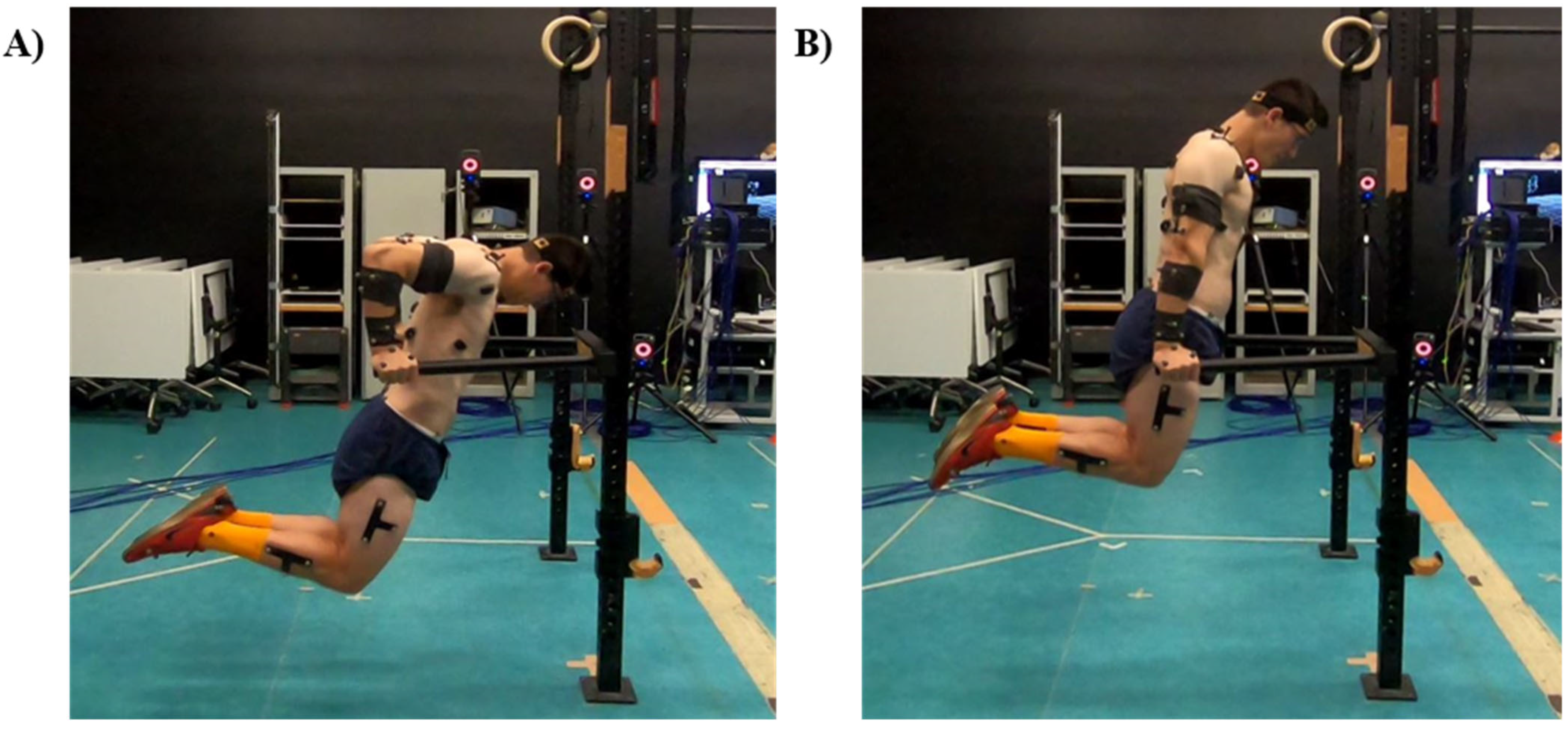
2.2. Procedures
- (i)
- Potential agonists of shoulder flexion and elbow extension: PM clavicular head, anterior deltoid (AD), and TB long head,
- (ii)
- Potential scapula stabilizers: upper trapezius (UT), serratus anterior (SA), and lower trapezius (LT), and
- (iii)
- Potential glenohumeral stabilizers: biceps brachii (BB), infraspinatus (IS), and latissimus dorsi (LD).
2.3. Data Collection
2.4. Data Analysis
2.5. Statistical Analysis
3. Results
3.1. Repetition Characteristics
3.2. Kinematics
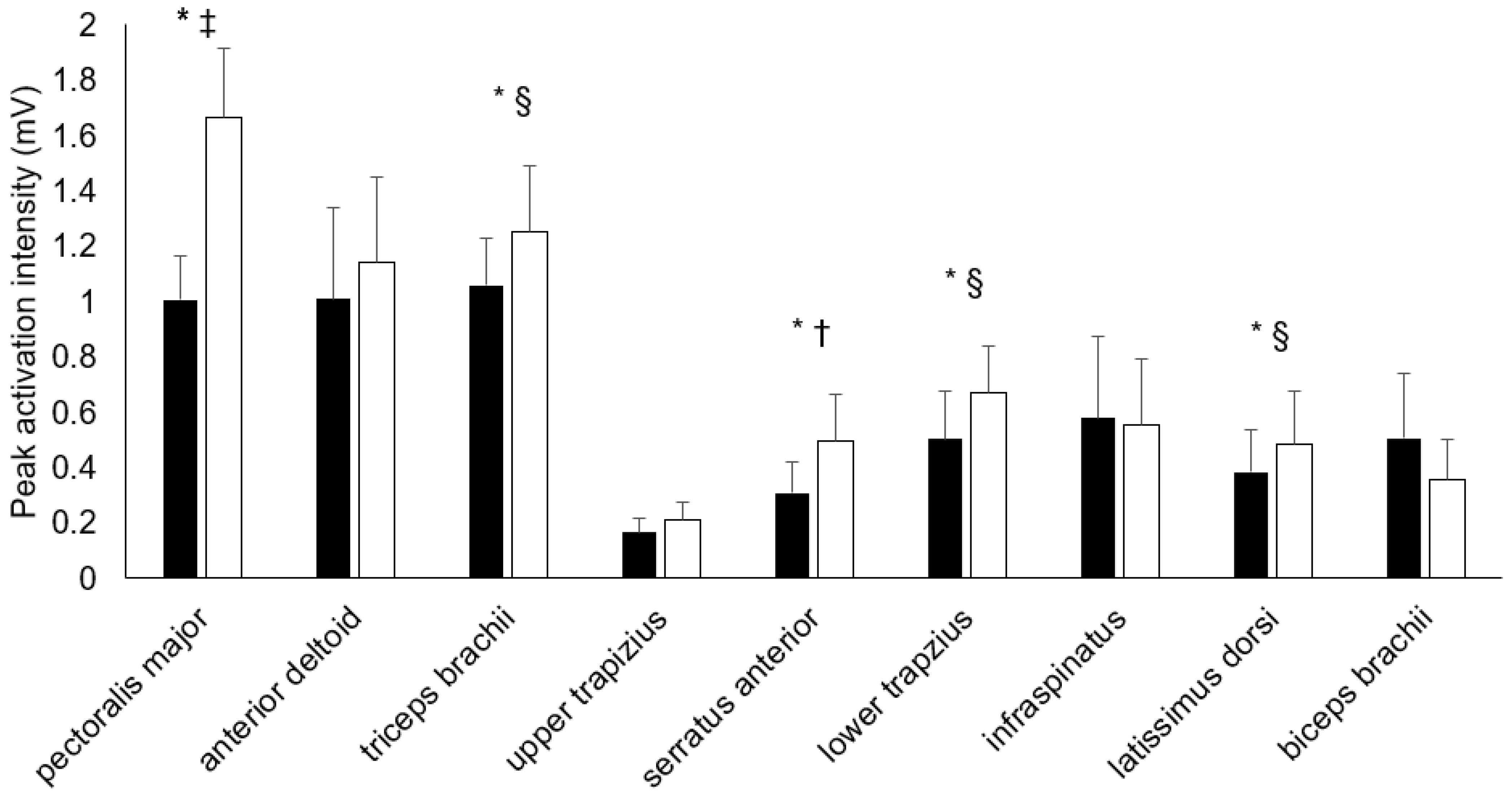
3.3. Muscle Electromyography (Activations)
4. Discussion
5. Conclusions
Author Contributions
Funding
Institutional Review Board Statement
Informed Consent Statement
Data Availability Statement
Conflicts of Interest
References
- Ciccantelli, P. Strength exercise: The dip. Strength Cond. J. 1991, 13, 53–54. [Google Scholar] [CrossRef]
- Hurd, J.; Goldenburg, L.; Bliss, S. Teaching techniques# 13: Dips. Strength Cond. J. 1991, 13, 66–69. [Google Scholar]
- McKenzie, A.; Crowley-McHattan, Z.; Meir, R.; Whitting, J.; Volschenk, W. Bench, Bar, and Ring Dips: Do Kinematics and Muscle Activity Differ? Int. J. Environ. Res. Public Health 2022, 19, 13211. [Google Scholar] [CrossRef] [PubMed]
- Coyne, J.O.; Tran, T.T.; Secomb, J.L.; Lundgren, L.; Farley, O.R.; Newton, R.U.; Sheppard, J.M. Reliability of pull up & dip maximal strength tests. J. Aust. Strength Cond. 2015, 23, 21–27. [Google Scholar]
- Collins, S.M.; Silberlicht, M.; Perzinski, C.; Smith, S.P.; Davidson, P.W. The relationship between body composition and preseason performance tests of collegiate male lacrosse players. J. Strength Cond. Res. 2014, 28, 2673–2679. [Google Scholar] [CrossRef]
- Binkley, H.; Schroyer, T. Aquatic therapy in the treatment of upper extremity injuries. Int. J. Athl. Ther. Train. 2002, 7, 49–54. [Google Scholar] [CrossRef] [Green Version]
- Hoppes, C.W.; Ross, M.D.; Moore, J.H. Undetected pectoralis major tendon rupture in a patient referred to a physical therapist in a combat environment: A case report. Phys. Ther. 2013, 93, 1225–1233. [Google Scholar] [CrossRef]
- Jaggi, A.; Lambert, S. Rehabilitation for shoulder instability. Br. J. Sports Med. 2010, 44, 333–340. [Google Scholar] [CrossRef]
- Kocialkowski, C.; Carter, R.; Peach, C. Triceps tendon rupture: Repair and rehabilitation. Shoulder Elb. 2017, 10, 62–65. [Google Scholar] [CrossRef] [Green Version]
- Vincent, H.K.; Vincent, K.R. Rehabilitation and prehabilitation for upper extremity in throwing sports: Emphasis on lacrosse. Curr. Sport Med. Rep. 2019, 18, 229–238. [Google Scholar] [CrossRef]
- Newton, R.U.; Jones, J.; Kraemer, W.J.; Wardle, H. Strength and power training of Australian Olympic swimmers. Strength Cond. J. 2002, 24, 7–15. [Google Scholar] [CrossRef]
- Enoka, R.M.; Duchateau, J. Muscle fatigue: What, why and how it influences muscle function: Muscle Fatigue. J. Physiol. 2008, 586, 11–23. [Google Scholar] [CrossRef] [PubMed]
- Brennecke, A.; Guimarães, T.M.; Leone, R.; Cadarci, M.; Mochizuki, L.; Simão, R.; Amadio, A.C.; Serrão, J.C. Neuromuscular Activity During Bench Press Exercise Performed With and Without the Preexhaustion Method. J. Strength Cond. Res. 2009, 23, 1933–1940. [Google Scholar] [CrossRef] [PubMed]
- Duffey, M.J.; Challis, J.H. Fatigue effects on bar kinematics during the bench press. J. Strength Cond. J. 2007, 21, 556. [Google Scholar]
- van den Tillaar, R.; Saeterbakken, A. Effect of Fatigue Upon Performance and Electromyographic Activity in 6-RM Bench Press. J. Hum. Kinet. 2014, 40, 57. [Google Scholar] [CrossRef] [Green Version]
- Hsu, H.H.; Chou, Y.L.; Huang, Y.P.; Huang, M.J.; Lou, S.Z.; Chou, P.P.H. Effect of push-up speed on upper extremity training until fatigue. J. Med. Biol. Eng. 2011, 31, 289–293. [Google Scholar] [CrossRef]
- Myers, J.B.; Guskiewicz, K.M.; Schneider, R.A.; Prentice, W.E. Proprioception and neuromuscular control of the shoulder after muscle fatigue. J. Athl. Train. 1999, 34, 362. [Google Scholar]
- Sánchez-Medina, L.; González-Badillo, J.J. Velocity Loss as an Indicator of Neuromuscular Fatigue during Resistance Training. Med. Sci. Sports Exerc. 2011, 43, 1725–1734. [Google Scholar] [CrossRef]
- van den Tillaar, R. Comparison of kinematics and muscle activation between push-up and bench press. Sports Med. Int. Open. 2019, 3, E74. [Google Scholar]
- Iida, N.; Kaneko, F.; Aoki, N.; Shibata, E. The effect of fatigued internal rotator and external rotator muscles of the shoulder on the shoulder position sense. J. Electromyogr. Kinesiol. 2014, 24, 72–77. [Google Scholar] [CrossRef]
- Kallenberg, L.A.; Schulte, E.; Disselhorst-Klug, C.; Hermens, H.J. Myoelectric manifestations of fatigue at low contraction levels in subjects with and without chronic pain. J. Electromyogr. Kinesiol. 2007, 17, 264–274. [Google Scholar] [CrossRef] [PubMed]
- McKenzie, A.K.; Crowley-McHattan, Z.J.; Meir, R.; Whitting, J.W.; Volschenk, W. Glenohumeral Extension and the Dip: Considerations for the Strength and Conditioning Professional. Strength Cond. J. 2020, 43, 93–100. [Google Scholar] [CrossRef]
- Carek, P.J.; Hawkins, A.L. Rupture of pectoralis major during parallel bar dips: Case report and review. Med. Sci. Sports Exerc. 1998, 30, 335–338. [Google Scholar] [CrossRef] [PubMed]
- Giordano, B.D.; Weisenthal, B.M. Common injuries and conditions in CrossFit participation. In Endurance Sports Medicine; Springer: Berlin, Germany, 2016; pp. 147–158. [Google Scholar]
- Potter, B.K.; Lehman, R.A., Jr.; Doukas, W.C. Simultaneous bilateral rupture of the pectoralis major tendon: A case report. J. Bone Jt. Surg. 2004, 86, 1519–1521. [Google Scholar] [CrossRef] [PubMed]
- CrossFit Regionals Saw 36 Pectoral Tears This Year. What Went Wrong? Available online: https://www.yahoo.com/lifestyle/crossfit-regionals-saw-36-pectoral-tears-year-went-wrong-194942403.html (accessed on 19 September 2022).
- Pec Tears in CrossFit Regionals: Lessons on Training Volume and Injury Prevention. Available online: https://thebarbellphysio.com/pec-tears-crossfit-regionals-lessons-training-volume-injury-prevention/ (accessed on 15 December 2021).
- Campbell, A.; Lloyd, D.; Alderson, J.; Elliott, B. MRI development and validation of two new predictive methods of glenohumeral joint centre location identification and comparison with established techniques. J. Biomech. 2009, 42, 1527–1532. [Google Scholar] [CrossRef] [PubMed]
- Lloyd, D.G.; Alderson, J.; Elliott, B. An upper limb kinematic model for the examination of cricket bowling: A case study of Mutiah Muralitharan. J. Sports Sci. 2000, 18, 975–982. [Google Scholar] [CrossRef]
- Naperalsky, M.E.; Anderson, J.-H. An Upper Extremity Active Dynamic Warm-Up for Sport Participation. Strength Cond. J. 2012, 34, 51–54. [Google Scholar] [CrossRef]
- Sawilowsky, S.S. New effect size rules of thumb. J. Mod. Appl. Stat. Methods 2009, 8, 26. [Google Scholar] [CrossRef]
- Madsen, N.; McLaughlin, T. Kinematic factors influencing performance and injury risk in the bench press exercise. Med. Sci. Sports Exerc. 1984, 16, 376–381. [Google Scholar] [CrossRef]
- Hales, M.E.; Johnson, B.F.; Johnson, J.T. Kinematic analysis of the powerlifting style squat and the conventional deadlift during competition: Is there a cross-over effect between lifts? J. Strength Cond. Res. 2009, 23, 2574–2580. [Google Scholar] [CrossRef] [Green Version]
- van den Tillaar, R.; Andersen, V.; Saeterbakken, A.H. The Existence of a Sticking Region in Free Weight Squats. J. Hum. Kinet. 2014, 42, 63. [Google Scholar] [CrossRef] [PubMed] [Green Version]
- Kompf, J.; Arandjelović, O. Understanding and Overcoming the Sticking Point in Resistance Exercise. Sports Med. 2016, 46, 751–762. [Google Scholar] [CrossRef] [PubMed] [Green Version]
- Kompf, J.; Arandjelović, O. The Sticking Point in the Bench Press, the Squat, and the Deadlift: Similarities and Differences, and Their Significance for Research and Practice. Sports Med. 2016, 47, 631–640. [Google Scholar] [CrossRef] [PubMed] [Green Version]
- van den Tillaar, R.; Saeterbakken, A.H.; Ettema, G. Is the occurrence of the sticking region the result of diminishing potentiation in bench press? J. Sports Sci. 2012, 30, 591–599. [Google Scholar] [CrossRef]
- Wilson, G.J.; Elliott, B.C.; Kerr, G.K. Bar Path and Force Profile Characteristics for Maximal and Submaximal Loads in the Bench Press. Int. J. Sport Biomech. 1989, 5, 390–402. [Google Scholar] [CrossRef]
- Carpenter, J.E.; Blasier, R.B.; Pellizzon, G.G. The Effects of Muscle Fatigue on Shoulder Joint Position Sense. Am. J. Sports Med. 1998, 26, 262–265. [Google Scholar] [CrossRef]
- Emery, K.; Côté, J.N. Repetitive arm motion-induced fatigue affects shoulder but not endpoint position sense. Exp. Brain Res. 2011, 216, 553–564. [Google Scholar] [CrossRef]
- Allen, T.; Leung, M.; Proske, U. The effect of fatigue from exercise on human limb position sense. J. Physiol. 2010, 588, 1369–1377. [Google Scholar] [CrossRef]
- Kibler, B.W.; McMullen, J. Scapular Dyskinesis and Its Relation to Shoulder Pain. J. Am. Acad. Orthop. Surg. 2003, 11, 142–151. [Google Scholar] [CrossRef] [Green Version]
- Struyf, F.; Nijs, J.; Baeyens, J.-P.; Mottram, S.; Meeusen, R. Scapular positioning and movement in unimpaired shoulders, shoulder impingement syndrome, and glenohumeral instability. Scand. J. Med. Sci. Sports 2011, 21, 352–358. [Google Scholar] [CrossRef]
- Biel, A. Trail Guide to the Body; Books of Discovery: Boulder CO, USA, 2005. [Google Scholar]


| Peak Joint Angles () | Joint Angle at the Starting Positions () | Joint Angles at the Point of Maximal Shoulder Extension () | Joint Angles at the End Position () | |||||
|---|---|---|---|---|---|---|---|---|
| Joint Movement | Non-Fatigued | Fatigue | Non-Fatigued | Fatigue | Non-Fatigued | Fatigue | Non-Fatigued | Fatigue |
| Shoulder extension | 67.66 ± 11.34 | 67.18 ± 10.06 | 2.99 ± 8.21 *† | 7.18 ± 8.67 *† | 67.66 ± 11.34 | 67.18 ± 10.06 | 3.51 ± 7.97 *† | 8.07 ± 8.59 *† |
| Abduction | 31.25 ± 3.56 | 32.84 ± 9.32 | 12.07 ± 4.09 | 12.66 ±5.24 | 27.86 ±11.35 | 26.78 ±5.91 | 11.88 ± 4.02 *† | 14.29 ± 5.32 *† |
| External rotation | 9.03 ± 14.30 | 8.83 ± 13.90 | −41.96 ± 14.29 *§ | −46.61 ± 9.94 *§ | 8.98 ± 14.29 | 8.75 ± 13.92 | −42.32 ± 10.70 *† | −49.93 ± 11.11 *† |
| Anterior thoracic lean | 37.25 ± 9.78 | 35.93 ± 7.38 | 18.49 ± 12.54 | 13.24 ±9.13 | 30.34 ± 21.06 | 34.61 ± 5.89 | 17.74 ± 11.87 | 12.76 ± 8.92 |
| Elbow flexion | 114.63 ± 8.59 | 112.20 ± 10.25 | 18.20 ± 11.04 | 15.55 ± 12.57 | 115.88 ± 9.72 | 111.59 ± 10.05 | 17.37 ± 11.35 | 17.72 ± 14.13 |
| Muscle | Non-Fatigued | Fatigued | p-Value (Cohen’s d) |
|---|---|---|---|
| Activation intensity at the point of maximal shoulder extension (displayed as % of peak activation during the non-fatigued condition) | |||
| Pectoralis major | 64.80 ± 9.55 * | 105.68 ± 36.17 * | <0.001 (1.55) |
| Anterior deltoid a | 66.04 ± 22.96 | 59.35 ± 31.32 | 0.552 (0.24) |
| Triceps brachii | 79.53 ± 12.38 | 78.37 ± 20.71 | 0.832 (0.07) |
| Upper trapezius | 74.77 ± 12.38 | 89.93 ± 68.62 | 0.352 (0.30) |
| Serratus anterior | 65.30 ± 22.78 * | 145.17 ± 125.81 * | 0.032 (0.88) |
| Lower trapezius | 77.39 ± 17.16 * | 101.52 ± 31.77 * | 0.010 (0.95) |
| Infraspinatus | 67.85 ± 20.79 | 78.38 ± 40.41 | 0.291 (0.33) |
| Latissimus dorsi | 67.86 ± 24.33 | 72.59 ± 27.39 | 0.427 (0.18) |
| Biceps brachii a | 69.01 ± 32.07 * | 61.01 ± 41.46 * | 0.046 (0.22) |
Publisher’s Note: MDPI stays neutral with regard to jurisdictional claims in published maps and institutional affiliations. |
© 2022 by the authors. Licensee MDPI, Basel, Switzerland. This article is an open access article distributed under the terms and conditions of the Creative Commons Attribution (CC BY) license (https://creativecommons.org/licenses/by/4.0/).
Share and Cite
McKenzie, A.; Crowley-McHattan, Z.; Meir, R.; Whitting, J.; Volschenk, W. Fatigue Increases Muscle Activations but Does Not Change Maximal Joint Angles during the Bar Dip. Int. J. Environ. Res. Public Health 2022, 19, 14390. https://0-doi-org.brum.beds.ac.uk/10.3390/ijerph192114390
McKenzie A, Crowley-McHattan Z, Meir R, Whitting J, Volschenk W. Fatigue Increases Muscle Activations but Does Not Change Maximal Joint Angles during the Bar Dip. International Journal of Environmental Research and Public Health. 2022; 19(21):14390. https://0-doi-org.brum.beds.ac.uk/10.3390/ijerph192114390
Chicago/Turabian StyleMcKenzie, Alec, Zachary Crowley-McHattan, Rudi Meir, John Whitting, and Wynand Volschenk. 2022. "Fatigue Increases Muscle Activations but Does Not Change Maximal Joint Angles during the Bar Dip" International Journal of Environmental Research and Public Health 19, no. 21: 14390. https://0-doi-org.brum.beds.ac.uk/10.3390/ijerph192114390