Article

Melatonin Promotes Ubiquitination of Phosphorylated Pro-Apoptotic Protein Bcl-2-Interacting Mediator of Cell Death-Extra Long (BimEL) in Porcine Granulosa Cells

College of Animal Science and Technology, Yangzhou University, Yangzhou 225009, China
*
Author to whom correspondence should be addressed.
Int. J. Mol. Sci. 2018, 19(11), 3431; https://0-doi-org.brum.beds.ac.uk/10.3390/ijms19113431
Submission received: 12 October 2018 / Revised: 27 October 2018 / Accepted: 27 October 2018 / Published: 1 November 2018
(This article belongs to the Section Biochemistry)

Abstract

:
Melatonin (N-acetyl-5-methoxytryptamine) is found in ovarian follicular fluid, and its concentration is closely related to follicular health status. Nevertheless, the molecular mechanisms underlying melatonin function in follicles are uncertain. In this study, melatonin concentration was measured in porcine follicular fluid at different stages of health. The melatonin concentration decreased as the follicles underwent atresia, suggesting that melatonin may participate in the maintenance of follicular health. The molecular pathway through which melatonin may regulate follicular development was further investigated. The pro-apoptotic protein BimEL (Bcl-2-interacting mediator of cell death-Extra Long), a key protein controlling granulosa cell apoptosis during follicular atresia, was selected as the target molecule. BimEL was downregulated when porcine granulosa cells were cultured in medium containing 10−9 M melatonin and isolated cumulus oocyte complexes (COCs) or follicle stimulating hormone (FSH). Interestingly, ERK-mediated phosphorylation was a prerequisite for the melatonin-induced decline in BimEL, and melatonin only promoted the ubiquitination of phosphorylated BimEL, and did not affect the activities of the lysosome or the proteasome. Moreover, the melatonin-induced downregulation of BimEL was independent of its receptor and its antioxidant properties. In conclusion, melatonin may maintain follicular health by inducing BimEL ubiquitination to inhibit the apoptosis of granulosa cells.

1. Introduction

During mammalian follicular development, only a limited number of follicles are selected to ovulate, with the remainder undergoing atresia at different stages. Follicle atresia is triggered by the apoptosis of granulosa cells, and several apoptotic-signaling molecules, such as hormones/growth factors/cytokines, the death ligand-receptor system, and B cell lymphoma/leukemia 2 (Bcl-2) family members, are involved in this process [1,2,3,4]. The Bcl-2 family, which includes both anti-apoptotic (Bcl-2, B cell lymphoma/leukemia X (Bcl-X)) and pro-apoptotic (Bcl-2 interacting domain (Bid), Bim, Bax, Bak) proteins, are key regulators of apoptosis, and members of the Bcl-2 protein family play pivotal roles in follicular growth and atresia [2,5,6].
Bim (Bcl-2 interacting mediator of cell death), a BH3-only family member, binds with high affinity to anti-apoptotic Bcl-2 family members and regulates apoptotic signaling through Bax and Bak [7]. Moreover, Bim can be phosphorylated by several MAP kinases to regulate its activity [8]. BimEL is the predominant isoform of Bim in several mammalian tissues, as determined using Western blotting analysis [9]. Our previous studies showed that BimEL participates in porcine follicular atresia through regulating granulosa cell apoptosis, and FSH and GDF9 regulate its expression [9,10]. In addition to its transcriptional regulation, the post-translational modification of BimEL is also important for its function [11,12]. However, the regulation of BimEL modification during mammalian follicular development is still not clearly defined.
Melatonin (N-acetyl-5-methoxytryptamine) can be synthesized from tryptophan in different cells, tissues, and organs, and mainly for local utilization (autocrine and paracrine actions). However, circulating melatonin is largely secreted rhythmically by the pineal gland [13,14]. Melatonin is found in follicular fluid and its concentration is higher than that in blood [15,16]. Granulosa cells, cumulus cells, and oocyte have been reported as able to synthesize melatonin [17,18,19] and melatonin receptors (MT1, MT2) expressed in human granulosa cells [20]. So far, the function of melatonin as an antioxidant and free radical scavenger in follicular development is well-established [21,22]. Moreover, melatonin has been reported to exhibit anti-apoptotic effects in different cells [23,24]. However, previous studies indicated that melatonin could also induce apoptosis under the regulation of Bim in many different cancer cell lines, including human hepatocellular carcinoma cells, breast carcinoma MDA-MB231 cells, and renal cancer Caki cells [11,25,26]. These findings imply that BimEL has diverse functions in different cell types. To date, the exact mechanism of melatonin on follicular development is still not clear and requires further study. In this study, we explored the relationship between melatonin and BimEL in porcine follicle granulosa cells.

2. Results

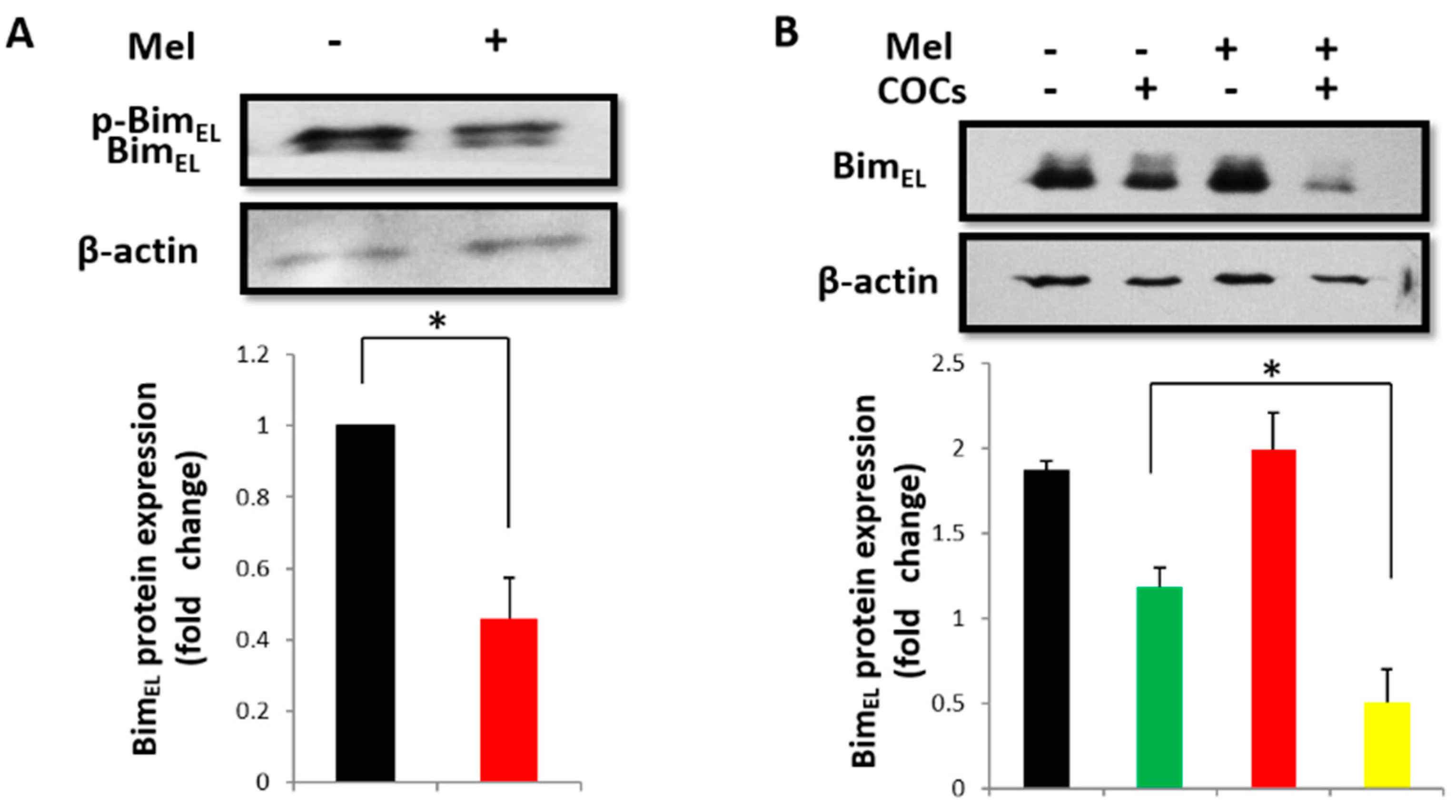
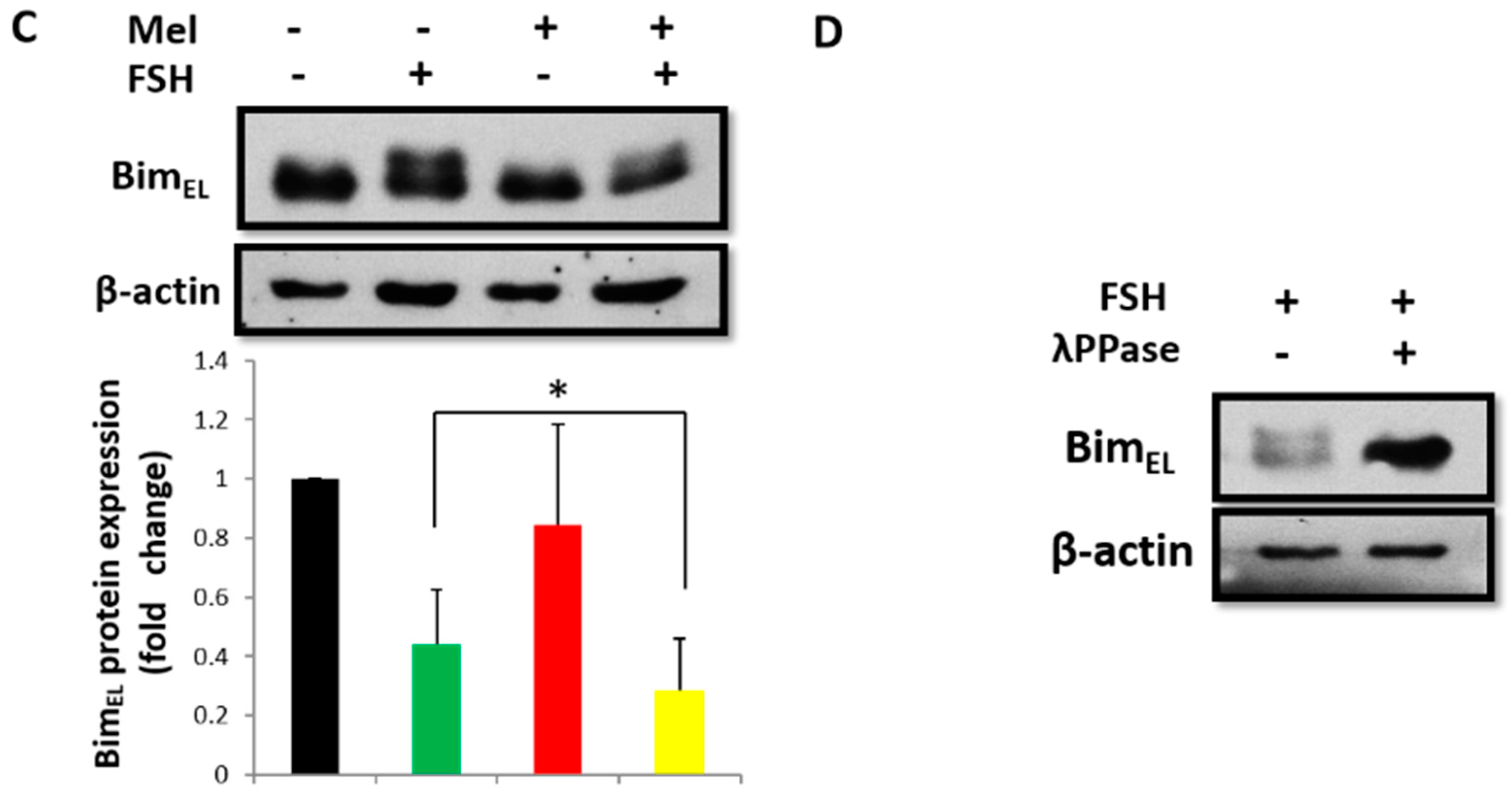
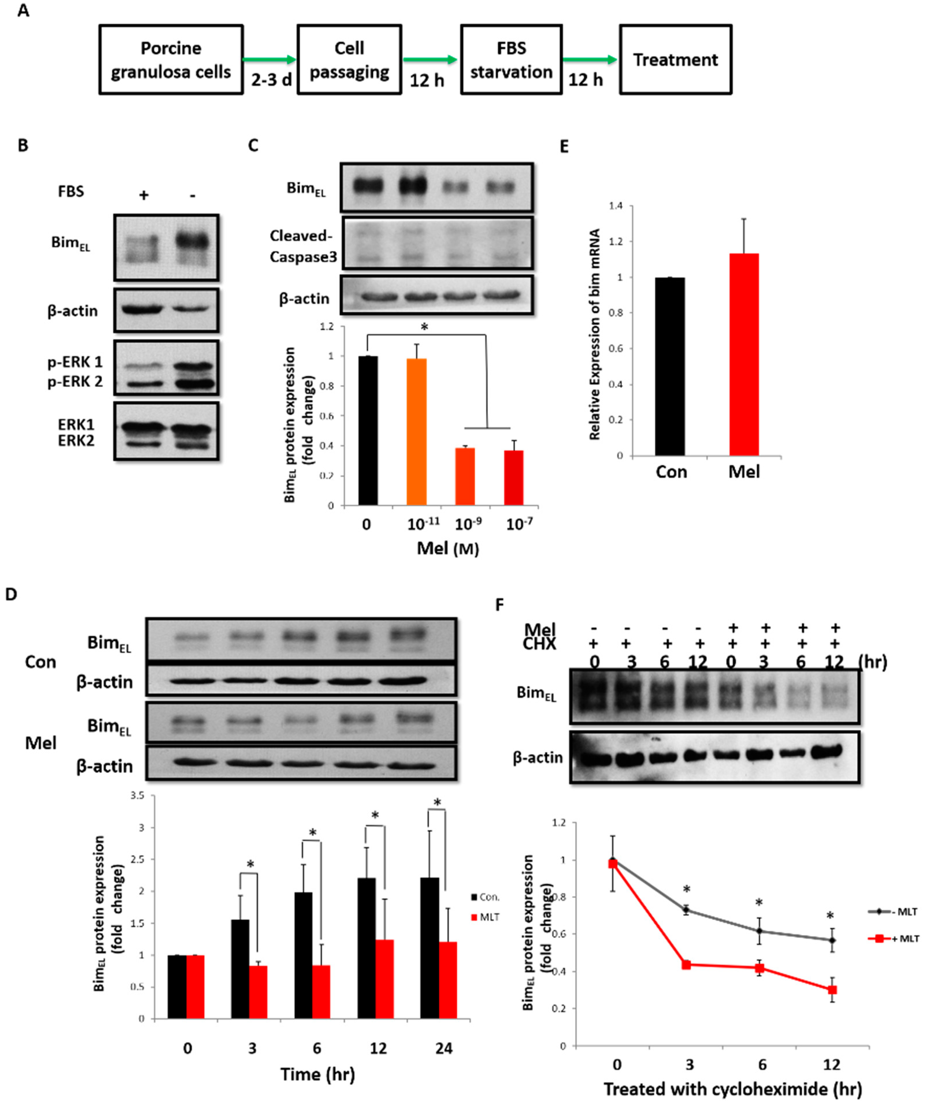
2.1. Melatonin Downregulates BimEL in Porcine Granulosa Cells

The BimEL protein in the surrounding cumulus granulosa cells decreased significantly after porcine COCs were treated with 10−9 M melatonin for 42–44 h (Figure 1A). However, there was no change in the BimEL level in primary granulosa cells after treatment with 10−9 M melatonin for 24 h (Figure 1B). Interestingly, melatonin significantly decreased the BimEL level when primary granulosa cells were cocultured with COCs (Figure 1B). Similar results were obtained after granulosa cells were treated with FSH (Figure 1C). Furthermore, COCs exposure and FSH treatment resulted in a BimEL protein mobility shift (Figure 1A–C), and this shift was inhibited when the lysates from FSH-treated primary granulosa cells were incubated with λ phosphatase (Figure 1D), indicating that the observed BimEL mobility shift was caused by its phosphorylation. These results also indicated that COCs or FSH could induce BimEL phosphorylation in granulosa cells. Thus, these data suggest that the function of melatonin in the downregulation of BimEL may depend on its phosphorylation level.

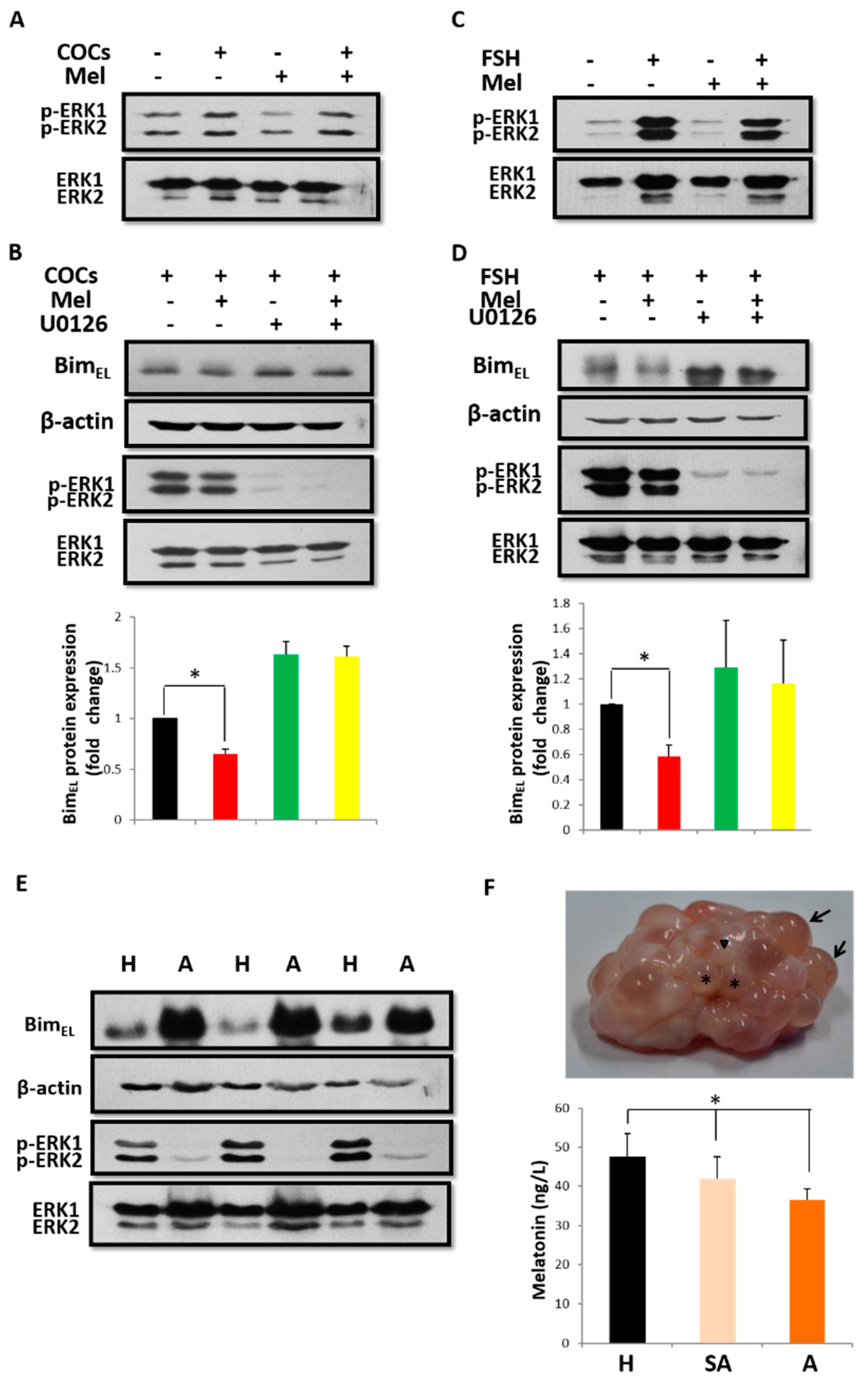
2.2. Melatonin Downregulates BimEL Depending on ERK Activation and BimEL Phosphorylation

Phosphorylation of BimEL by extracellular signal-regulated kinase 1/2 (ERK1/2) has been shown to induce the degradation of BimEL [27], so we first determined the status of ERK activation. As shown in Figure 2A,C, ERK1/2 in granulosa cells was activated after treatment with COCs or FSH, but it was not induced by melatonin, revealing that melatonin could not directly promote the degradation of BimEL by phosphorylation. To determine the precise role of melatonin in this process, primary granulosa cells cultured in the presence of COCs or FSH were pretreated with U0126, an MEK1/2 inhibitor, before melatonin treatment. As expected, U0126 abolished the induction of BimEL phosphorylation by COCs or FSH in parallel with the abrogation of BimEL downregulation caused by melatonin (Figure 2B,D). These results confirmed that the phosphorylation of BimEL induced by ERK was a prerequisite for BimEL reduction induced by melatonin. To further confirm whether this phenomenon exists in follicles in vivo, the lysates from granulosa cells obtained from healthy or atretic follicles were subjected to SDS-PAGE to detect ERK activation and BimEL expression. As shown in Figure 2E, the level of activated ERK1/2 was higher, whereas the BimEL level was lower, in granulosa cells of healthy follicles compared to atretic follicles. Furthermore, melatonin concentration decreased with the atresia of porcine ovarian follicles. The concentrations of melatonin in healthy, slightly atretic, and atretic follicles were 47.47 ± 6.03 ng/L, 41.97 ± 5.66 ng/L, and 36.50 ± 2.84 ng/L, respectively, and the difference between healthy follicles and slightly atretic or atretic ones was significant (p < 0.05, Figure 2F). These results suggest that ERK activation is responsible for the induction of BimEL phosphorylation by COCs or FSH, and it promotes melatonin-induced BimEL downregulation in porcine granulosa cells. This process is likely to play a vital role in maintaining follicle health.

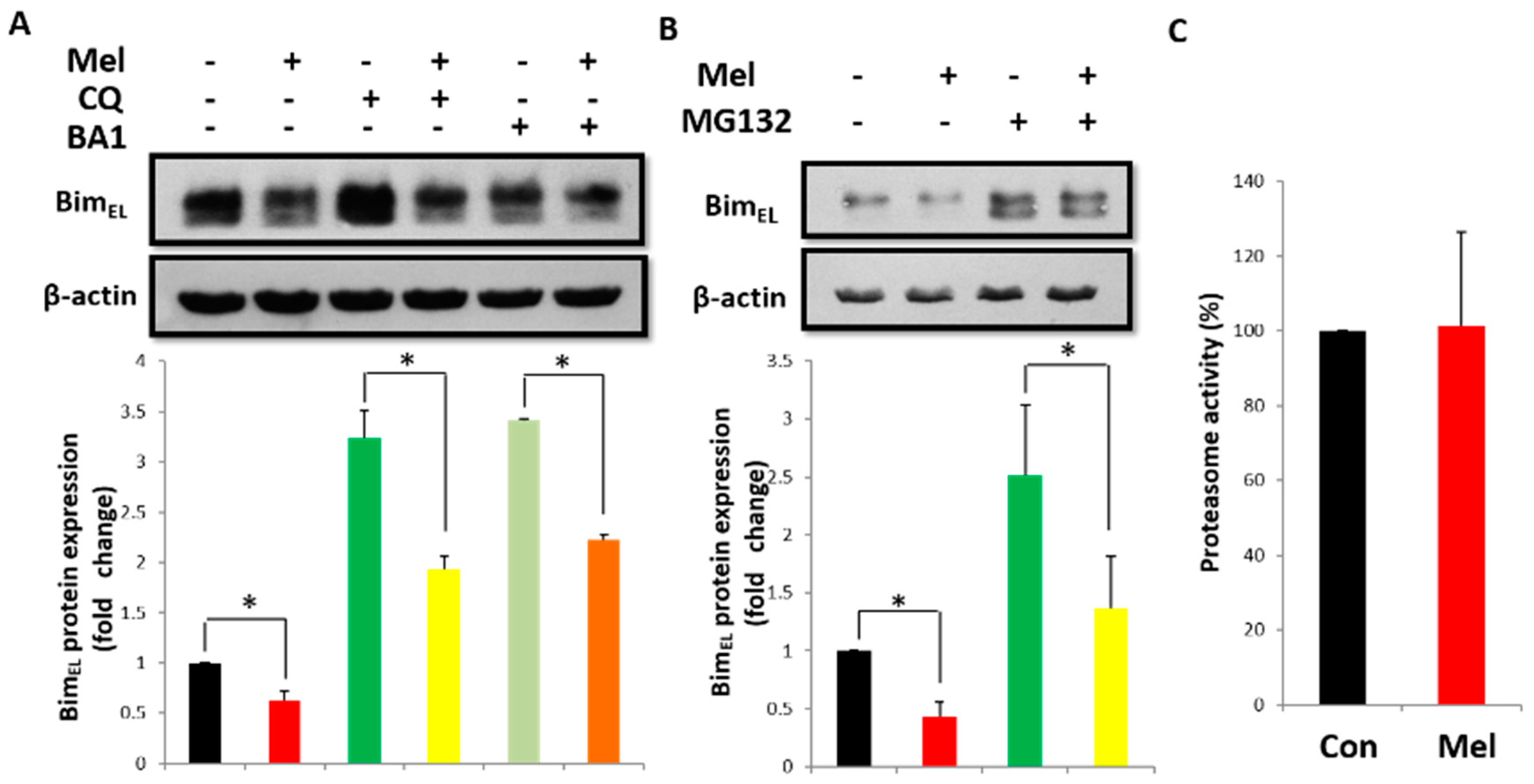
2.3. Post-Translational Pathway Is Involved in Melatonin-Induced Downregulation of BimEL

The molecular mechanism of melatonin-induced downregulation of BimEL was systemically investigated using porcine adherent granulosa cells with the experimental protocol shown in Figure 3A. After 12 h of serum withdrawal, a significant increase in phosphorylated BimEL was observed (Figure 3B), accompanied by a robust activation of ERK1/2, which was similar to that in primary granulosa cells treated with COCs or FSH. To determine whether melatonin could downregulate the BimEL protein in porcine adherent granulosa cells, cells were treated with melatonin at different concentrations (0, 10−11, 10−9, 10−7 M) for 24 h. As shown in Figure 3C, the levels of BimEL and Cleaved Caspase3 significantly decreased after 10−9 M melatonin treatment, and this effect was evident within 3 h after treatment (Figure 3D).
Because the BimEL protein expression level can be regulated by transcriptional and post-translational pathways, our next experiments aimed to determine the mechanism responsible for this change. As shown in Figure 3E, there was no difference in the mRNA expression of bim 3 h post melatonin treatment compared to the control group. Therefore, we hypothesized that the downregulation of BimEL was controlled by post-translational modifications. To address this, porcine adherent granulosa cells were incubated with cycloheximide (CHX) alone or co-treated with CHX and melatonin for indicated time periods (Figure 3F). The combination of CHX and melatonin induced a rapid decrease in the BimEL level within 3 h after treatment compared with CHX alone. These results indicate that the melatonin-mediated BimEL decline is regulated at the post-translational level, and BimEL is actively degraded.

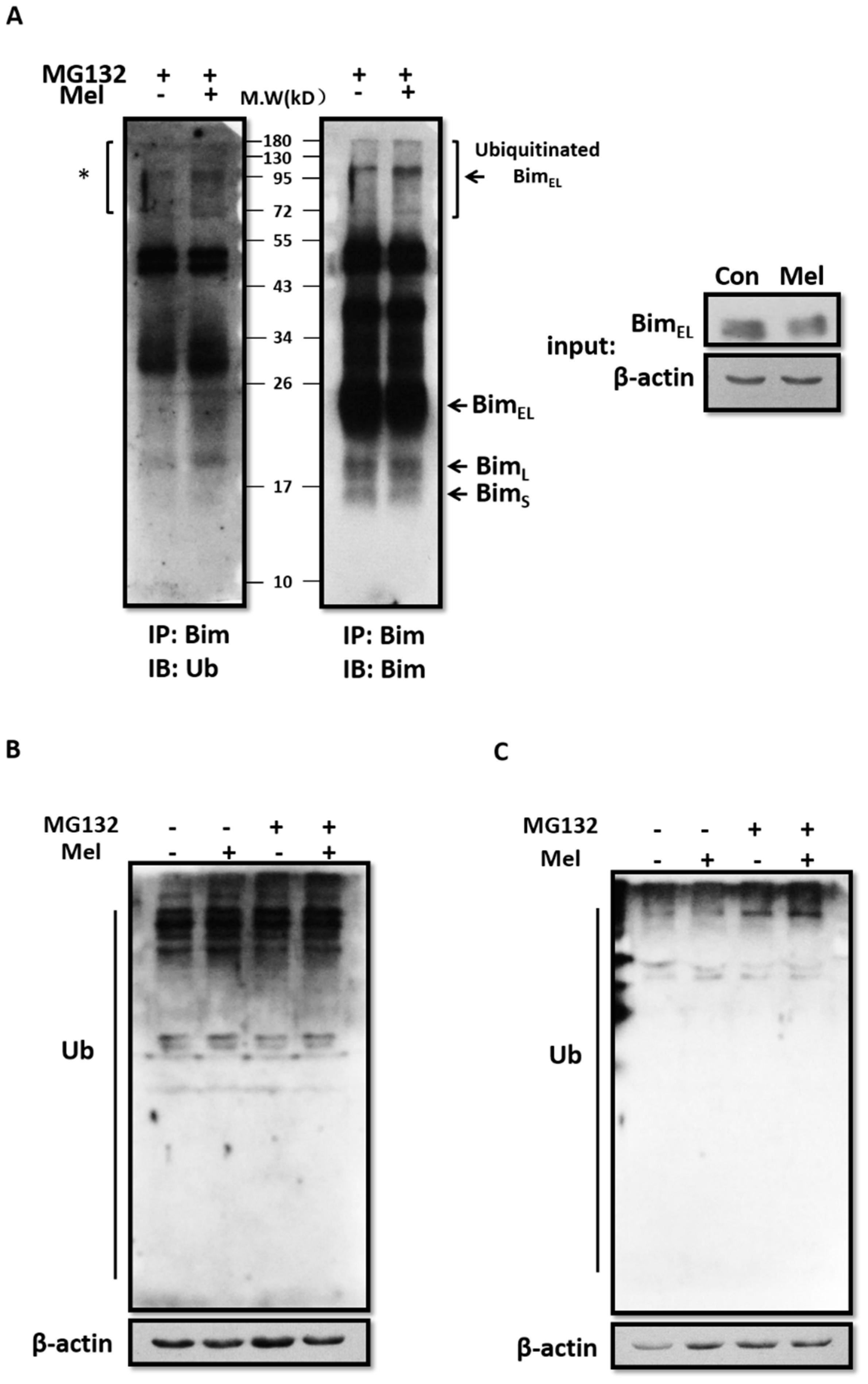
2.4. Melatonin Promotes BimEL Ubiquitination

Proteasomes and lysosomes comprise two major intracellular proteolytic systems in mammalian cells. Hence, we investigated whether BimEL was increasingly degraded by lysosomes or proteasomes following melatonin treatment. First, porcine adherent granulosa cells were treated with melatonin in the presence of a potent inhibitor of the vacuolar type H+-ATPase, Bafilomycin A1, or chloroquine, which accumulated in lysosomes and raised the intralysosomal pH value. The inhibitors induced the accumulation of LC3-II (data not shown) to manifest its inhibition of lysosomal proteolysis. As shown in Figure 4A, Bafilomycin A1 and chloroquine failed to block BimEL downregulation by melatonin, indicating that the proteasomal pathway was responsible for BimEL degradation. Protein phosphorylation is required to prepare Bim for ubiquitination and proteasomal degradation. Porcine adherent granulosa cells were treated with melatonin in the presence of the proteasome inhibitor MG132 for 3 h. However, MG132 could also not counteract the BimEL repression by melatonin (Figure 4B). To confirm whether melatonin could influence the proteasomal activity in granulosa cells, we measured chymotrypsin-like activity using a commercially-available proteasome 20S assay kit. Melatonin did not change the proteasomal activity in granulosa cells (Figure 4C). These results indicate that neither lysosomes nor proteasomes directly participate in BimEL reduction by melatonin.
It has been shown that activation of the ERK pathway promotes the phosphorylation of BimEL, which serves to mark BimEL for ubiquitination [28]. Immunoprecipitation of BimEL, followed by immunoblot analysis using an anti-ubiquitin antibody, demonstrated that melatonin enhanced the level of BimEL poly-ubiquitination in porcine adherent granulosa cells supplemented with MG132 (Figure 5A, left lane). We found that the level of ubiquitinated BimEL increased, whereas levels of BimEL, BimL, and BimS decreased after melatonin treatment (Figure 5B, right lane). Western blot analysis of whole-cell lysates showed that melatonin also elevated the overall level of ubiquitination in both porcine primary and adherent granulosa cells (Figure 5B,C). Taken together, our data suggest that melatonin activates the ubiquitination of BimEL.

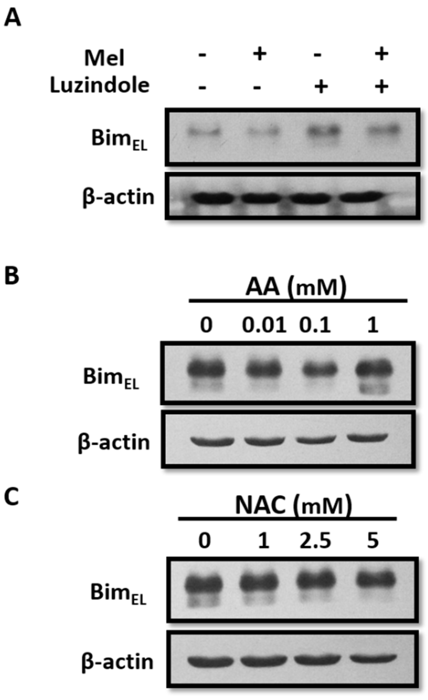
2.5. Reduction of BimEL by Melatonin Does Not Associate with Its Receptor or Antioxidant Properties
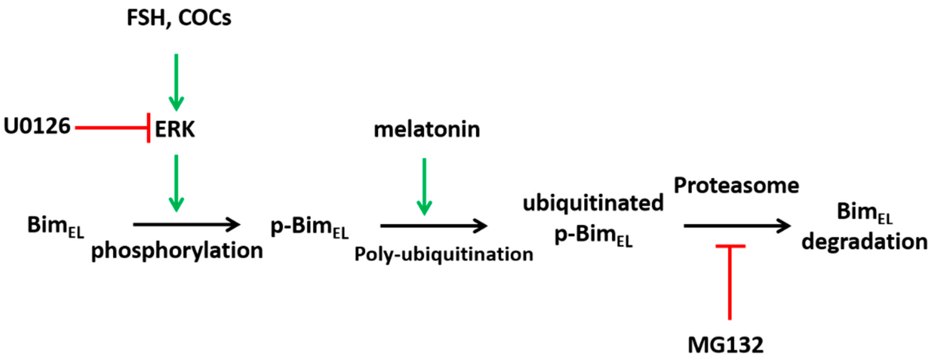
To classify the underlying mechanisms of the melatonin-induced decrease in BimEL, we investigated the potential role of melatonin receptors because various physiological effects of melatonin can be mediated by its two G-protein-coupled MT1 and MT2 receptors. Porcine adherent granulosa cells were treated with melatonin in the presence of luzindole, a melatonin receptor antagonist, and then BimEL protein was examined. As shown in Figure 6A, luzindole failed to block the BimEL downregulation by melatonin. In addition, melatonin is known to be a powerful antioxidant. To determine the relevance of antioxidant activity on BimEL degradation, we tested the effects of two other antioxidant reagents, N-acetylcysteine (NAC) and ascorbic acid (AA), on the BimEL protein in porcine adherent granulosa cells. The western blot showed no change in the level of BimEL after treatment compared to the control (Figure 6B,C). Based on these data, we conclude that melatonin-mediated BimEL downregulation is independent of the melatonin receptor-mediated pathway or its antioxidant function.

3. Discussion

Melatonin plays a pivotal role in female reproduction, including puberty, ovarian follicle growth, ovulation, and luteinization [29,30,31]. Several studies have demonstrated that melatonin has beneficial effects on oocyte maturation and subsequent embryo development in many species, such as mice [32], cattle [33], and pigs [15]. Except for its antioxidant properties, however, the role of melatonin in female reproduction remains largely unknown. Throughout the reproductive life span, more than 99% of germ cells are eliminated from the ovary through follicular atresia, and granulosa cells play a major role in this process. It is well-established that the initial step of follicular atresia is granulosa cell apoptosis [34]. Our previous study showed that the pro-apoptotic protein BimEL plays an important role in porcine granulosa cell apoptosis [9]. In this study, we demonstrated that melatonin decreases the BimEL protein via inducing its ubiquitination through the post-translational pathway in porcine granulosa cells.
It has been shown that the supplementation of porcine maturation medium with 10−9 M melatonin is beneficial for in vitro maturation (IVM) of porcine oocytes and subsequent embryo development [15]. Moreover, our previous study showed that BimEL-mediated apoptosis in cumulus cells accelerates oocyte aging and degeneration [35]. In this study, we showed that melatonin decreased the BimEL protein in cumulus granulosa cells during IVM. Granulosa cells play an important role in supporting oocyte maturation, which prompted us to focus on the effects of melatonin on BimEL protein expression in porcine primary granulosa cells. However, melatonin treatment alone failed to decrease the BimEL level. Conversely, a combination of melatonin with COCs or FSH downregulated the BimEL protein, although melatonin can induce bim expression in several cancer cell lines [11,25,26]. Thus, these results suggested that the mechanism of regulating bim expression may be different between cancer cells and normal tissue cells or these contradictory results are from using different melatonin concentrations.
Our results demonstrated that ERK1/2 was activated in granulosa cells by COCs and FSH rather than by melatonin. These results were consistent with those of a previous report by Baumgarten et al., who found that FSH could activate the ERK pathway in human cumulus granulosa cells [36]. Oocyte-secreted factors such as GDF9 also activate ERK in human granulosa cells. A recent study reported that melatonin activated ERK1/2 in HEK293 cells in a concentration-dependent manner after 5 min of treatment [37]. Moreover, the activation of ERK1/2 induced by melatonin antagonized UVB-induced apoptosis in U937 cells [38]. Thus, melatonin appears to activate ERK1/2 in different cells. ERK1/2 kinase was responsible for BimEL phosphorylation because the MEK1/2 inhibitor U0126 blocked COC- and FSH-induced BimEL phosphorylation. However, U0126 can partially inhibit MEK5 and ERK5 activation [39]; thus, the phosphorylation of BimEL may also be partially due to ERK5 activation. Moreover, accumulating data indicate that multiple phosphorylated isoforms of BimEL are regulated by different kinase pathways, including ERK, JNK, and p38 MAP kinases, which may result in different apoptotic end-points [8]. For example, UV-mediated JNK activation results in the phosphorylation of BimEL on Thr-112, potentiating its apoptotic activity [40], and sodium arsenite-induced apoptosis causes the phosphorylation of BimEL at Ser-65 by p38 in PC12 cells [41]. In contrast, phosphorylation by ERK on Ser-55/65/73 targeted BimEL for degradation via the ubiquitin-proteasome pathway and promoted cell survival [28]. In the present study, melatonin-induced downregulation of BimEL was abolished by U0126, indicating that BimEL phosphorylation by ERK was essential for this process; however, the specific phosphorylated sites in BimEL induced by COCs or FSH should be confirmed in a further study.
In this study, we observed correlations among ERK1/2 activation, the BimEL level, and follicle health status. An inverse relationship between ERK activation and BimEL level was observed in porcine follicles, with healthy follicles displaying a higher level of phosphorylated ERK than atretic ones. Similarly, higher levels of phosphorylated ERK in dominant follicles were detected compared with subordinate follicles in sheep [42]. These data suggest that ERK activation plays an important role, not only in melatonin-induced downregulation of BimEL, but also in the process of dominant follicle selection.
We showed that serum starvation of porcine adherent granulosa cells significantly activated ERK and enhanced BimEL phosphorylation, which provided an ideal model to mimic the effects of COCs and FSH in primary granulosa cells. The same phenomenon of ERK activation by serum starvation also exists in human colon carcinoma cells [43]. Following the activation of ERK, melatonin downregulated the BimEL protein and Cleaved Caspase 3 level in porcine adherent granulosa cells (Figure 3C). It is well-established that gene transcription, mRNA stability, and post-translational modifications regulate the BimEL level by different stimuli [8,44,45]. The transcriptional factors FOXO3a, Runx3, E2F1, c-Jun, and SP1 have been shown to regulate bim transcription. During follicular development, FSH regulates BimEL expression through FoxO3a in granulosa cells [9]. It was demonstrated that melatonin induces the expression of transcription factors of Sp1 and E2F1, coinciding with the induction of BimEL in renal cancer Caki cells [11]. However, we did not detect an obvious mRNA change of bim in porcine granulosa cells by real-time PCR after melatonin treatment (Figure 3E). Moreover, a significant decline of BimEL was observed when granulosa cells were treated with melatonin in the presence of CHX (Figure 3F), implying that the downregulation of BimEL in granulosa cells by melatonin was closely associated with its post-translational modification.
Proteasomes or lysosomes play imperative roles in controlling intracellular protein quantity and quality [46]. Choi et al. reported that melatonin increased the degradation of TGFBlp via activating autophagy and counteracted the inhibition of autophagy by Bafilomycin A1 in corneal fibroblasts [47]. Previous studies have shown that melatonin can inhibit proteasome activity [48]. Nevertheless, neither a lysosomal-degradation inhibitor nor a proteasomal inhibitor could inhibit the decline in BimEL induced by melatonin in our study. Unexpectedly, melatonin markedly increased the ubiquitination of BimEL. According to our results, melatonin may also have induced the ubiquitination of other proteins in granulosa cells. Therefore, melatonin decreased the BimEL level by increasing its ubiquitination. Although melatonin was previously regarded as an inhibitor of the ubiquitin-proteasome system [49], our contradictory results may be attributed to different cells with different experimental conditions. Park et al. reported that melatonin could increase the level of BimEL by inhibiting the activity of proteasomes to induce apoptosis of human renal cancer cells [11]. In their experiment, cancer cells were treated with melatonin at the concentration from 0.1 to 1 mM, but the concentration was just 1 nM in our experiment. Above all, according to previous studies, melatonin presented toxicity to the oocyte during maturation when its concentration reached at 10−5 M [15]. In addition, the effect of melatonin on proteasome activity may be different between cancer cells and granulosa cells. It has been shown that mitochondrial-associated Bcl-2 family proteins, including Bax, Bcl-2, and Bim, are regulated through ubiquitin/proteasomal degradation during apoptosis [50]. Wan et al. reported that APCCdc20 acts as an E3 ubiquitin ligase to promote Bim ubiquitination and destruction [51]. Additional studies are required to delineate the precise mechanisms involved in melatonin-induced protein ubiquitination in granulosa cells.
Generally, melatonin participates in various physiological processes via its receptors or antioxidant properties [52,53,54]. However, our results showed that melatonin downregulated BimEL through neither a receptor-mediated nor antioxidant pathway. Therefore, the detailed mechanism of BimEL downregulation by melatonin in granulosa cells waits further investigation.
A hypothetical model of BimEL regulation by melatonin in granulosa cells is presented in Figure 7. Although there are some details that need to be further pursued, our results demonstrate that melatonin downregulates phosphorylated BimEL via a ubiquitin-proteasome pathway in porcine granulosa cells to maintain follicle health. Future studies should investigate how melatonin precisely regulates protein ubiquitination.

4. Materials and Methods

All chemicals used in this study were purchased from Sigma Chemical Co. (St. Louis, MO, USA) unless otherwise specified.

4.1. Classification of Follicles and Measurement of Melatonin in Porcine Follicular Fluid

Follicles were classified as healthy or atretic according to previously established morphological criteria [53]. In brief, healthy follicles had vascularized theca interna and clear amber follicular fluid with no debris. The slightly atretic and atretic follicles had gray theca interna and flocculent follicular fluid of different degrees. The concentration of melatonin in follicular fluid was assessed using the porcine melatonin ELISA kit (Shanghai MLBIO Biotechnology Co., Ltd., Shanghai, China) with a measurement range for melatonin from 1.5–65 ng/L.

4.2. In Vitro Maturation of Oocytes

In vitro maturation (IVM) of oocytes was performed as previously described [35]. In brief, porcine ovaries were collected at a local abattoir and transported to the laboratory within 2–3 h after collection. Cumulus oocyte complexes (COCs) were aspirated, and those with several layers of unexpanded cumulus cells were cultured in maturation medium for 42–44 h. The maturation medium was Tissue Culture Medium 199 with Earle’s salts (TCM199; Invitrogen, Carlsbad, CA, USA) supplemented with 10% porcine follicular fluid, 10 IU/mL hCG (Chorulon, Intervet Australia Pty Ltd., Victoria, Australia), 10 IU/mL eCG (Folligon, Intervet Australia Pty Ltd.), 10 ng/mL EGF, 0.6 mM cysteine, 75 mg/L penicillin, and 50 mg/L streptomycin. After IVM, cumulus cells were separated from oocytes and lysates were used for Western blotting.

4.3. Cell Culture and Treatment

Porcine primary or adherent granulosa cells were cultured as previously described [9]. In brief, porcine ovaries were collected at a local abattoir and transported to the laboratory within 2–3 h after collection. Ovaries were washed thrice with sterile 0.9% saline (37 °C) containing 100 IU/L penicillin and 100 mg/L streptomycin. Granulosa cells were then isolated by puncturing healthy follicles (2–5 mm in diameter) with a 25-gauge hypodermic needle and gently washing thrice with DMEM/F12 supplemented with 1% fetal bovine serum, 100 IU/L penicillin, and 100 mg/L streptomycin. Primary granulosa cells were selected under a microscope for different treatments according to the experimental design. In addition, granulosa cells were isolated by puncturing healthy follicles (2–5 mm in diameter) with a 25-gauge hypodermic needle and gently washing thrice with DMEM containing 10% fetal bovine serum, 100 IU/L penicillin and 100 mg/L streptomycin. Cells were then incubated at 37 °C in a humidified atmosphere of 5% CO2/95% air for 24 h. The cells were passaged upon reaching confluence. The culture medium was replaced with DMEM containing 100 IU/L penicillin and 100 mg/L streptomycin at 12 h after passaging, and the cells were cultured for an additional 12 h. Thereafter, the cells were treated with melatonin and/or other compounds for the indicated time periods. The cells were pretreated with 10 μM LY294002, 1 μM cycloheximide, 10 μM choloroquine, 100 nM Bafilomycin A1, or 5 μM MG132 at 1 h before melatonin treatment.

4.4. Western Blotting

The cells were lysed in Laemmli sample buffer (Bio-Rad, Hercules, CA, USA). An equal amount of protein was separated by sodium dodecyl sulfate polyacrylamide gel electrophoresis (SDS-PAGE; 12% acrylamide gel), and proteins were transferred to nitrocellulose membranes (Millipore, Billerica, MA, USA). After blocking with 5% non-fat milk Tris-buffered saline containing 0.1% Tween-20 (TBST), the membranes were incubated with primary antibodies overnight at 4 °C. After washing with TBST, the membranes were incubated with the appropriate secondary antibodies conjugated to horseradish peroxidase at a dilution of 1:2000 for 1 h. The protein bands were visualized using an enhanced chemiluminescence detection system (Applygen Technologies Inc., Beijing, China). The western blotting images were processed using Image J software (National Institutes of Health, Bethesda, MD, USA).

4.5. Lambda Phosphatase Treatment

Whole-cell extracts (30 μg) were incubated with or without 1 μL of lambda protein phosphatase (400 U/μL; New England BioLabs, Ipswich, MA, USA) for 2 h at 30 °C. The samples were boiled for 5 min after adding 10 μL of 5× SDS sample buffer.

4.6. Proteasome Activity Assay

Chymotrypsin-like protease activity was measured using the Amplite™ Fluorometric Proteasome 20S Assay Kit (AAT Bioquest Inc., Sunnyvale, CA, USA) according to the manufacturer’s instructions. In brief, 100 μL of proteasome assay loading solution was added to each well after granulosa cells were treated with 10−9 M melatonin for 3 h and the tissue culture plate was then incubated at 37 °C for 2 h. The assay was performed by monitoring the fluorescence at 480 nm EX/520 nm EM.

4.7. Immunoprecipitation

To detect ubiquitinated Bim in whole-cell extracts, granulosa cells were homogenized in RIPA buffer containing a broad-spectrum protease inhibitor cocktail (Roche, Basel, Switzerland). The protein concentrations of the cell extracts were measured, and equal amounts of protein were incubated overnight at 4 °C with a polyclonal Bim antibody (Cell Signaling Technology, Beverly, MA, USA). The antibody-antigen complex was then incubated with protein A/G PLUS-agarose beads (Santa Cruz Biotechnology, Santa Cruz, CA, USA). The immunoprecipitated proteins were resolved by SDS-PAGE (12% acrylamide gel) and processed for Western blotting using specific antibodies to detect ubiquitin and Bim.

4.8. Real-Time quantitative PCR

A reverse transcription polymerase chain reaction was used to determine if melatonin regulated the mRNA expression of bim. First-strand cDNA synthesis was carried out with the SuperScript™ First-Strand Synthesis System for RT-PCR (Invitrogen) according to the manufacturer’s instructions. The real-time PCR primers for bim were: 5′-AGGCTGAACCCGCAGATA-3′ (forward) and 5′-GCATTAAATTCGTCTCCAATACG-3′ (reverse). The real-time PCR primers for β-actin were: 5′-ATCGTGCGGGACATAAG-3′ (forward) and 5′-CTCGTTGCCGATGGTGAT-3′ (reverse). The mRNA expression of bim was normalized to that of the endogenous control β-actin. Real-time PCR reactions were performed using the ABI 7900 Sequence Detection System (Applied Biosystems, Foster City, CA, USA).

4.9. Statistical Analysis

Data are presented as the means ± standard deviation (S.D.) of at least three independent replicates. Data were analyzed by one-way and two-way analysis of variance (ANOVA) and Duncan’s test using SAS software (SAS Institute, Cary, NC, USA). Differences were considered statistically significant at p-values < 0.05.

Author Contributions

Conceptualization, Y.W. and S.Z.; Funding acquisition, S.Z.; Project administration, Y.W.; Writing—original draft, Y.W.; Writing—review & editing, S.Z.

Funding

This research was funded by National Natural Science Foundation of China (No. 31470077, 31672419) and the Priority Academic Program Development of Jiangsu Higher Education Institution.

Conflicts of Interest

The authors declare no conflict of interest.

References

  1. Inoue, N.; Matsuda, F.; Goto, Y.; Manabe, N. Role of cell-death ligand-receptor system of granulosa cells in selective follicular atresia in porcine ovary. J. Reprod. Dev. 2011, 57, 169–175. [Google Scholar] [CrossRef] [PubMed]
  2. Matsuda-Minehata, F.; Inoue, N.; Goto, Y.; Manabe, N. The regulation of ovarian granulosa cell death by pro- and anti-apoptotic molecules. J. Reprod. Dev. 2006, 52, 695–705. [Google Scholar] [CrossRef] [PubMed]
  3. Nakayama, M.; Manabe, N.; Nishihara, S.; Miyamoto, H. Species-specific differences in apoptotic cell localization in granulosa and theca interna cells during follicular atresia in porcine and bovine ovaries. J. Reprod. Dev. 2000, 46, 147–156. [Google Scholar] [CrossRef]
  4. Matsuda, F.; Inoue, N.; Manabe, N.; Ohkura, S. Follicular growth and atresia in mammalian ovaries: Regulation by survival and death of granulosa cells. J. Reprod. Dev. 2012, 58, 44–50. [Google Scholar] [CrossRef] [PubMed]
  5. Liew, S.H.; Vaithiyanathan, K.; Cook, M.; Bouillet, P.; Scott, C.L.; Kerr, J.B.; Strasser, A.; Findlay, J.K.; Hutt, K.J. Loss of the proapoptotic BH3-only protein BCL-2 modifying factor prolongs the fertile life span in female mice. Biol. Reprod. 2014, 90, 77. [Google Scholar] [CrossRef] [PubMed]
  6. Hsu, S.Y.; Lai, R.J.; Finegold, M.; Hsueh, A.J. Targeted overexpression of Bcl-2 in ovaries of transgenic mice leads to decreased follicle apoptosis, enhanced folliculogenesis, and increased germ cell tumorigenesis. Endocrinology 1996, 137, 4837–4843. [Google Scholar] [CrossRef] [PubMed]
  7. Strasser, A. The role of BH3-only proteins in the immune system. Nature reviews. Immunology 2005, 5, 189–200. [Google Scholar] [PubMed]
  8. Hubner, A.; Barrett, T.; Flavell, R.A.; Davis, R.J. Multisite phosphorylation regulates Bim stability and apoptotic activity. Mol. Cell 2008, 30, 415–425. [Google Scholar] [CrossRef] [PubMed]
  9. Wang, X.L.; Wu, Y.; Tan, L.B.; Tian, Z.; Liu, J.H.; Zhu, D.S.; Zeng, S.M. Follicle-stimulating hormone regulates pro-apoptotic protein Bcl-2-interacting mediator of cell death-extra long (BimEL)-induced porcine granulosa cell apoptosis. J. Boil. Chem. 2012, 287, 10166–10177. [Google Scholar] [CrossRef] [PubMed]
  10. Wang, X.L.; Wang, K.; Zhao, S.; Wu, Y.; Gao, H.; Zeng, S.M. Oocyte-secreted growth differentiation factor 9 inhibits BCL-2-interacting mediator of cell death-extra long expression in porcine cumulus cell. Biol. Reprod. 2013, 89, 56. [Google Scholar] [CrossRef] [PubMed]
  11. Park, E.J.; Woo, S.M.; Min, K.J.; Kwon, T.K. Transcriptional and post-translational regulation of Bim controls apoptosis in melatonin-treated human renal cancer Caki cells. J. Pineal Res. 2014, 56, 97–106. [Google Scholar] [CrossRef] [PubMed]
  12. Thi, H.T.H.; Lim, H.-S.; Kim, J.; Kim, Y.-M.; Kim, H.-Y.; Hong, S. Transcriptional and post-translational regulation of Bim is essential for TGF-β and TNF-α-induced apoptosis of gastric cancer cell. Biochim. Biophys. Acta Gener. Subj. 2013, 1830, 3584–3592. [Google Scholar] [CrossRef] [PubMed]
  13. Acuna-Castroviejo, D.; Escames, G.; Venegas, C.; Diaz-Casado, M.E.; Lima-Cabello, E.; Lopez, L.C.; Rosales-Corral, S.; Tan, D.X.; Reiter, R.J. Extrapineal melatonin: Sources, regulation, and potential functions. Cell. Mol. Life Sci. CMLS 2014, 71, 2997–3025. [Google Scholar] [CrossRef] [PubMed]
  14. Venegas, C.; Garcia, J.A.; Escames, G.; Ortiz, F.; Lopez, A.; Doerrier, C.; Garcia-Corzo, L.; Lopez, L.C.; Reiter, R.J.; Acuna-Castroviejo, D. Extrapineal melatonin: Analysis of its subcellular distribution and daily fluctuations. J. Pineal Res. 2012, 52, 217–227. [Google Scholar] [CrossRef] [PubMed]
  15. Shi, J.M.; Tian, X.Z.; Zhou, G.B.; Wang, L.; Gao, C.; Zhu, S.E.; Zeng, S.M.; Tian, J.H.; Liu, G.S. Melatonin exists in porcine follicular fluid and improves in vitro maturation and parthenogenetic development of porcine oocytes. J. Pineal Res. 2009, 47, 318–323. [Google Scholar] [CrossRef] [PubMed]
  16. Brzezinski, A.; Seibel, M.M.; Lynch, H.J.; Deng, M.H.; Wurtman, R.J. Melatonin in human preovulatory follicular fluid. J. Clin. Endocrinol. Metab. 1987, 64, 865–867. [Google Scholar] [CrossRef] [PubMed]
  17. Sakaguchi, K.; Itoh, M.T.; Takahashi, N.; Tarumi, W.; Ishizuka, B. The rat oocyte synthesises melatonin. Reprod. Fertile. Dev. 2013, 25, 674–682. [Google Scholar] [CrossRef] [PubMed]
  18. El-Raey, M.; Geshi, M.; Somfai, T.; Kaneda, M.; Hirako, M.; Abdel-Ghaffar, A.E.; Sosa, G.A.; El-Roos, M.E.; Nagai, T. Evidence of melatonin synthesis in the cumulus oocyte complexes and its role in enhancing oocyte maturation in vitro in cattle. Mol. Reprod. Dev. 2011, 78, 250–262. [Google Scholar] [CrossRef] [PubMed]
  19. Amireault, P.; Dube, F. Serotonin and its antidepressant-sensitive transport in mouse cumulus-oocyte complexes and early embryos. Biol. Reprod. 2005, 73, 358–365. [Google Scholar] [CrossRef] [PubMed]
  20. Niles, L.P.; Wang, J.; Shen, L.; Lobb, D.K.; Younglai, E.V. Melatonin receptor mRNA expression in human granulosa cells. Mol. Cell. Endocrinol. 1999, 156, 107–110. [Google Scholar] [CrossRef]
  21. Tamura, H.; Takasaki, A.; Taketani, T.; Tanabe, M.; Kizuka, F.; Lee, L.; Tamura, I.; Maekawa, R.; Asada, H.; Yamagata, Y. Melatonin as a free radical scavenger in the ovarian follicle. Endocr. J. 2013, 60, 1–13. [Google Scholar] [CrossRef] [PubMed]
  22. Tamura, H.; Takasaki, A.; Taketani, T.; Tanabe, M.; Kizuka, F.; Lee, L.; Tamura, I.; Maekawa, R.; Aasada, H.; Yamagata, Y. The role of melatonin as an antioxidant in the follicle. J. Ovar. Res. 2012, 5, 5. [Google Scholar] [CrossRef] [PubMed] [Green Version]
  23. Dong, Y.; Fan, C.; Hu, W.; Jiang, S.; Ma, Z.; Yan, X.; Deng, C.; Di, S.; Xin, Z.; Wu, G.; et al. Melatonin attenuated early brain injury induced by subarachnoid hemorrhage via regulating NLRP3 inflammasome and apoptosis signaling. J. Pineal Res. 2016, 60, 253–262. [Google Scholar] [CrossRef] [PubMed]
  24. Sainz, R.M.; Mayo, J.C.; Rodriguez, C.; Tan, D.X.; Lopez-Burillo, S.; Reiter, R.J. Melatonin and cell death: Differential actions on apoptosis in normal and cancer cells. Cell. Mol. Life Sci. CMLS 2003, 60, 1407–1426. [Google Scholar] [CrossRef] [PubMed]
  25. Woo, S.M.; Min, K.J.; Kwon, T.K. Melatonin-mediated Bim up-regulation and cyclooxygenase-2 (COX-2) down-regulation enhances tunicamycin-induced apoptosis in MDA-MB-231 cells. J. Pineal Res. 2015, 58, 310–320. [Google Scholar] [CrossRef] [PubMed]
  26. Carbajo-Pescador, S.; Steinmetz, C.; Kashyap, A.; Lorenz, S.; Mauriz, J.L.; Heise, M.; Galle, P.R.; Gonzalez-Gallego, J.; Strand, S. Melatonin induces transcriptional regulation of Bim by FoxO3a in HepG2 cells. Br. J. Cancer 2013, 108, 442–449. [Google Scholar] [CrossRef] [PubMed]
  27. Luciano, F.; Jacquel, A.; Colosetti, P.; Herrant, M.; Cagnol, S.; Pages, G.; Auberger, P. Phosphorylation of Bim-EL by Erk1/2 on serine 69 promotes its degradation via the proteasome pathway and regulates its proapoptotic function. Oncogene 2003, 22, 6785–6793. [Google Scholar] [CrossRef] [PubMed] [Green Version]
  28. Ley, R.; Balmanno, K.; Hadfield, K.; Weston, C.; Cook, S.J. Activation of the ERK1/2 signaling pathway promotes phosphorylation and proteasome-dependent degradation of the BH3-only protein, Bim. J. Boil. Chem. 2003, 278, 18811–18816. [Google Scholar] [CrossRef] [PubMed]
  29. Tamura, H.; Takasaki, A.; Taketani, T.; Tanabe, M.; Lee, L.; Tamura, I.; Maekawa, R.; Aasada, H.; Yamagata, Y.; Sugino, N. Melatonin and female reproduction. J. Obstet. Gynaecol. Res. 2014, 40, 1–11. [Google Scholar] [CrossRef] [PubMed]
  30. Rocha, R.; Lima, L.; Alves, A.; Celestino, J.; Matos, M.; Lima-Verde, I.; Bernuci, M.; Lopes, C.; Báo, S.; Campello, C. Interaction between melatonin and follicle-stimulating hormone promotes in vitro development of caprine preantral follicles. Domest. Anim. Endocrinol. 2013, 44, 1–9. [Google Scholar] [CrossRef] [PubMed]
  31. De Holanda, F.S.; Tufik, S.; Bignotto, M.; Maganhin, C.G.; Vieira, L.H.; Baracat, E.C.; Soares, J.M., Jr. Evaluation of melatonin on the precocious puberty: A pilot study. Gynecol. Endocrinol. 2011, 27, 519–523. [Google Scholar] [CrossRef] [PubMed]
  32. Fernandes, H.; Schefer, L.; Castro, F.; Leal, C. Melatonin on in vivo and in vitro maturation of mouse oocytes. Reprod. Fertil. Dev. 2015, 27, 246. [Google Scholar] [CrossRef]
  33. Tian, X.; Wang, F.; He, C.; Zhang, L.; Tan, D.; Reiter, R.J.; Xu, J.; Ji, P.; Liu, G. Beneficial effects of melatonin on bovine oocytes maturation: A mechanistic approach. J. Pineal Res. 2014, 57, 239–247. [Google Scholar] [CrossRef] [PubMed]
  34. Tripathi, A.; Shrivastav, T.G.; Chaube, S.K. An increase of granulosa cell apoptosis mediates aqueous neem (Azadirachta indica) leaf extract-induced oocyte apoptosis in rat. Int. J. Appl. Basic Med. Res. 2013, 3, 27–36. [Google Scholar] [PubMed]
  35. Wu, Y.; Wang, X.-L.; Liu, J.-H.; Bao, Z.-J.; Tang, D.-W.; Wu, Y.; Zeng, S.-M. BIMEL-mediated apoptosis in cumulus cells contributes to degenerative changes in aged porcine oocytes via a paracrine action. Theriogenology 2011, 76, 1487–1495. [Google Scholar] [CrossRef] [PubMed]
  36. Baumgarten, S.C.; Convissar, S.M.; Zamah, A.M.; Fierro, M.A.; Winston, N.J.; Scoccia, B.; Stocco, C. FSH Regulates IGF-2 Expression in Human Granulosa Cells in an AKT-Dependent Manner. J. Clin. Endocrinol. Metab. 2015, 100, E1046–E1055. [Google Scholar] [CrossRef] [PubMed]
  37. Chen, L.; He, X.; Zhang, Y.; Chen, X.; Lai, X.; Shao, J.; Shi, Y.; Zhou, N. Melatonin receptor type 1 signals to extracellular signal-regulated kinase 1 and 2 via Gi and Gs dually coupled pathways in HEK-293 cells. Biochemistry 2014, 53, 2827–2839. [Google Scholar] [CrossRef] [PubMed]
  38. Luchetti, F.; Betti, M.; Canonico, B.; Arcangeletti, M.; Ferri, P.; Galli, F.; Papa, S. ERK MAPK activation mediates the antiapoptotic signaling of melatonin in UVB-stressed U937 cells. Free Radic. Biol. Med. 2009, 46, 339–351. [Google Scholar] [CrossRef] [PubMed]
  39. Kamakura, S.; Moriguchi, T.; Nishida, E. Activation of the protein kinase ERK5/BMK1 by receptor tyrosine kinases. Identification and characterization of a signaling pathway to the nucleus. J. Boil. Chem. 1999, 274, 26563–26571. [Google Scholar] [CrossRef]
  40. Lei, K.; Davis, R.J. JNK phosphorylation of Bim-related members of the Bcl2 family induces Bax-dependent apoptosis. Proc. Natl. Acad. Sci. USA 2003, 100, 2432–2437. [Google Scholar] [CrossRef] [PubMed] [Green Version]
  41. Cai, B.; Chang, S.; Becker, E.B.; Bonni, A.; Xia, Z. p38 MAP kinase mediates apoptosis through phosphorylation of Bimel at Ser65. J. Biol. Chem. 2006, 281, 25215–25222. [Google Scholar] [CrossRef] [PubMed]
  42. Evans, A.C.; Martin, F. Kinase pathways in dominant and subordinate ovarian follicles during the first wave of follicular development in sheep. Anim. Reprod. Sci. 2000, 64, 221–231. [Google Scholar] [CrossRef]
  43. Jung, Y.D.; Nakano, K.; Liu, W.; Gallick, G.E.; Ellis, L.M. Extracellular signal-regulated kinase activation is required for up-regulation of vascular endothelial growth factor by serum starvation in human colon carcinoma cells. Cancer Res. 1999, 59, 4804–4807. [Google Scholar] [PubMed]
  44. Matsui, H.; Asou, H.; Inaba, T. Cytokines direct the regulation of Bim mRNA stability by heat-shock cognate protein 70. Mol. Cell 2007, 25, 99–112. [Google Scholar] [CrossRef] [PubMed]
  45. Sunters, A.; Fernandez de Mattos, S.; Stahl, M.; Brosens, J.J.; Zoumpoulidou, G.; Saunders, C.A.; Coffer, P.J.; Medema, R.H.; Coombes, R.C.; Lam, E.W. FoxO3a transcriptional regulation of Bim controls apoptosis in paclitaxel-treated breast cancer cell lines. J. Boil. Chem. 2003, 278, 49795–49805. [Google Scholar] [CrossRef] [PubMed]
  46. Wang, X.; Robbins, J. Proteasomal and lysosomal protein degradation and heart disease. J. Mol. Cell Cardiol. 2014, 71, 16–24. [Google Scholar] [CrossRef] [PubMed] [Green Version]
  47. Choi, S.I.; Kim, K.S.; Oh, J.Y.; Jin, J.Y.; Lee, G.H.; Kim, E.K. Melatonin induces autophagy via an mTOR-dependent pathway and enhances clearance of mutant-TGFBIp. J. Pineal Res. 2013, 54, 361–372. [Google Scholar] [CrossRef] [PubMed]
  48. Vriend, J.; Reiter, R.J. Melatonin and ubiquitin: What’s the connection? Cell. Mol. Life Sci. CMLS 2014, 71, 3409–3418. [Google Scholar] [CrossRef] [PubMed]
  49. Vriend, J.; Reiter, R.J. Melatonin feedback on clock genes: A theory involving the proteasome. J. Pineal Res. 2015, 58, 1–11. [Google Scholar] [CrossRef] [PubMed]
  50. Karbowski, M.; Youle, R.J. Regulating mitochondrial outer membrane proteins by ubiquitination and proteasomal degradation. Curr. Opin. Cell Biol. 2011, 23, 476–482. [Google Scholar] [CrossRef] [PubMed] [Green Version]
  51. Wan, L.; Tan, M.; Yang, J.; Inuzuka, H.; Dai, X.; Wu, T.; Liu, J.; Shaik, S.; Chen, G.; Deng, J.; et al. APCCdc20 suppresses apoptosis through targeting Bim for ubiquitination and destruction. Dev. Cell. 2014, 29, 377–391. [Google Scholar] [CrossRef] [PubMed]
  52. Rocha, C.S.; Rato, L.; Martins, A.D.; Alves, M.G.; Oliveira, P.F. Melatonin and male reproductive health: Relevance of darkness and antioxidant properties. Curr. Mol. Med. 2015, 15, 299–311. [Google Scholar] [CrossRef] [PubMed]
  53. Jarzynka, M.J.; Passey, D.K.; Johnson, D.A.; Konduru, N.V.; Fitz, N.F.; Radio, N.M.; Rasenick, M.; Benloucif, S.; Melan, M.A.; Witt-Enderby, P.A. Microtubules modulate melatonin receptors involved in phase-shifting circadian activity rhythms: In vitro and in vivo evidence. J. Pineal Res. 2009, 46, 161–171. [Google Scholar] [CrossRef] [PubMed]
  54. Jolly, P.D.; Smith, P.R.; Heath, D.A.; Hudson, N.L.; Lun, S.; Still, L.A.; Watts, C.H.; McNatty, K.P. Morphological evidence of apoptosis and the prevalence of apoptotic versus mitotic cells in the membrana granulosa of ovarian follicles during spontaneous and induced atresia in ewes. Biol. Reprod. 1997, 56, 837–846. [Google Scholar] [CrossRef] [PubMed] [Green Version]
Figure 1. Melatonin downregulates the BimEL protein in porcine cumulus granulosa cells and primary granulosa cells treated with COCs or FSH. (A) BimEL level decreased in porcine cumulus granulosa cells after COCs were treated with 10−9 M melatonin (Mel) for 42–44 h. (B) BimEL level decreased in porcine primary granulosa cells treated with 10−9 M melatonin in the presence of COCs for 24 h. (C) BimEL level decreased in porcine primary granulosa cells treated with 10−9 M melatonin and 0.01 IU/mL FSH for 24 h. (D) Western blot analyses of BimEL phosphorylation after porcine primary granulosa cells were treated with 0.01 IU/mL FSH for 24 h. Whole-cell lysates were obtained and incubated with or without λ phosphatase (λ PPase). Values are expressed as the means ± S.D of three separate experiments. * p < 0.05.
Ijms 19 03431 g001aIjms 19 03431 g001b
Figure 2. Melatonin downregulates BimEL protein by COCs or FSH-mediated, activating the ERK pathway in porcine primary granulosa cells. (A) Phosphorylated ERK level increased in porcine primary granulosa cells treated with 10−9 M melatonin (Mel) in the presence of COCs for 24 h. (B) Inhibition of ERK phosphorylation by 20 μM U0126 prevented the decrease in BimEL level induced by melatonin and COCs, coinciding with the decrease in phosphorylated ERK. (C) phosphorylated ERK level increased in porcine primary granulosa cells treated with 10−9 M melatonin in the presence of FSH for 24 h. (D) Inhibition of ERK phosphorylation by 20 μM U0126 prevented the decrease in BimEL level induced by melatonin and FSH, coinciding with the decrease in phosphorylated ERK. (E) There was an inverse relationship between levels of BimEL and phosphorylated ERK in porcine granulosa cells from healthy or atretic follicles. (F) Melatonin concentration decreased in follicles with progressive atresia. H, healthy follicles (arrows); SA, slightly atretic follicle (arrowhead); A, atretic follicles (asterisks). Data are representative of three independent experiments. Values are expressed as the means ± S.D. of three separate experiments. * p < 0.05.
Ijms 19 03431 g002
Figure 3. Melatonin decreases BimEL protein in porcine adherent granulosa cells. (A) Experimental protocol. Porcine primary granulosa cells were cultured for two to three days, passaged, and cultured for an additional 12 h, and then incubated with serum-free medium for 12 h. Thereafter, different treatments were performed. (B) Levels of phosphorylated BimEL and ERK increased in porcine adherent granulosa cells after culturing in serum-free medium for 12 h. (C) BimEL decreased in porcine adherent granulosa cells 24 h after melatonin treatment. (D) BimEL decreased in porcine adherent granulosa cells within 3 h of melatonin treatment. (E) Melatonin did not affect bim mRNA expression in porcine adherent granulosa cells. (F) Melatonin accelerated BimEL degradation in porcine adherent granulosa cells treated with cycloheximide (CHX). Values are expressed as the means ± S.D. of three separate experiments. * p < 0.05.
Ijms 19 03431 g003
Figure 4. Neither lysosomal activity nor proteasomal activity is involved in downregulation of BimEL induced by melatonin. (A) Lysosomal inhibitor chloroquine (CQ) and Bafilomycin A1 (BA1) did not block the downregulation of BimEL caused by melatonin in porcine adherent granulosa cells. (B) Proteasomal inhibitor MG132 did not affect the downregulation of BimEL in porcine adherent granulosa cells. (C) Melatonin did not affect proteasomal activity. Values are expressed as the means ± S.D. of three separate experiments. * p < 0.05.
Ijms 19 03431 g004
Figure 5. Melatonin induces the ubiquitination of BimEL and other proteins. (A) BimEL ubiquitination increased in porcine adherent granulosa cells treated with 10−9 M melatonin in the presence of 5 μM MG132 for 3 h. Bim was immunoprecipitated, and Western blotting was performed using anti-Bim and ubiquitin (Ub) antibodies. The asterisk indicates ubiquitinated BimEL appearing as a smear of bands with higher molecular weights. The level of protein ubiquitination increased in porcine adherent granulosa cells (B) and porcine primary granulosa cells (C) treated with 10−9 M melatonin in the presence of MG132 (5 μM) for 3 h. Western blotting was performed with an anti-ubiquitin antibody. Data are representative of three independent experiments.
Ijms 19 03431 g005
Figure 6. Melatonin receptors and melatonin antioxidant properties are not involved in BimEL downregulation. (A) BimEL degradation caused by melatonin was not affected by luzidole, a melatonin receptor antagonist, in porcine adherent granulosa cells. (B,C) Antioxidants AA and NAC had no effects on the BimEL level in porcine adherent granulosa cells. Data are representative of three independent experiments.
Ijms 19 03431 g006
Figure 7. Diagram of BimEL degradation as regulated by melatonin in porcine granulosa cells.
Ijms 19 03431 g007

