Biocompatibility and Osteogenic Capacity of Mg-Zn-Ca Bulk Metallic Glass for Rabbit Tendon-Bone Interference Fixation

 , ,
, , 
Abstract
:1. Introduction
2. Result
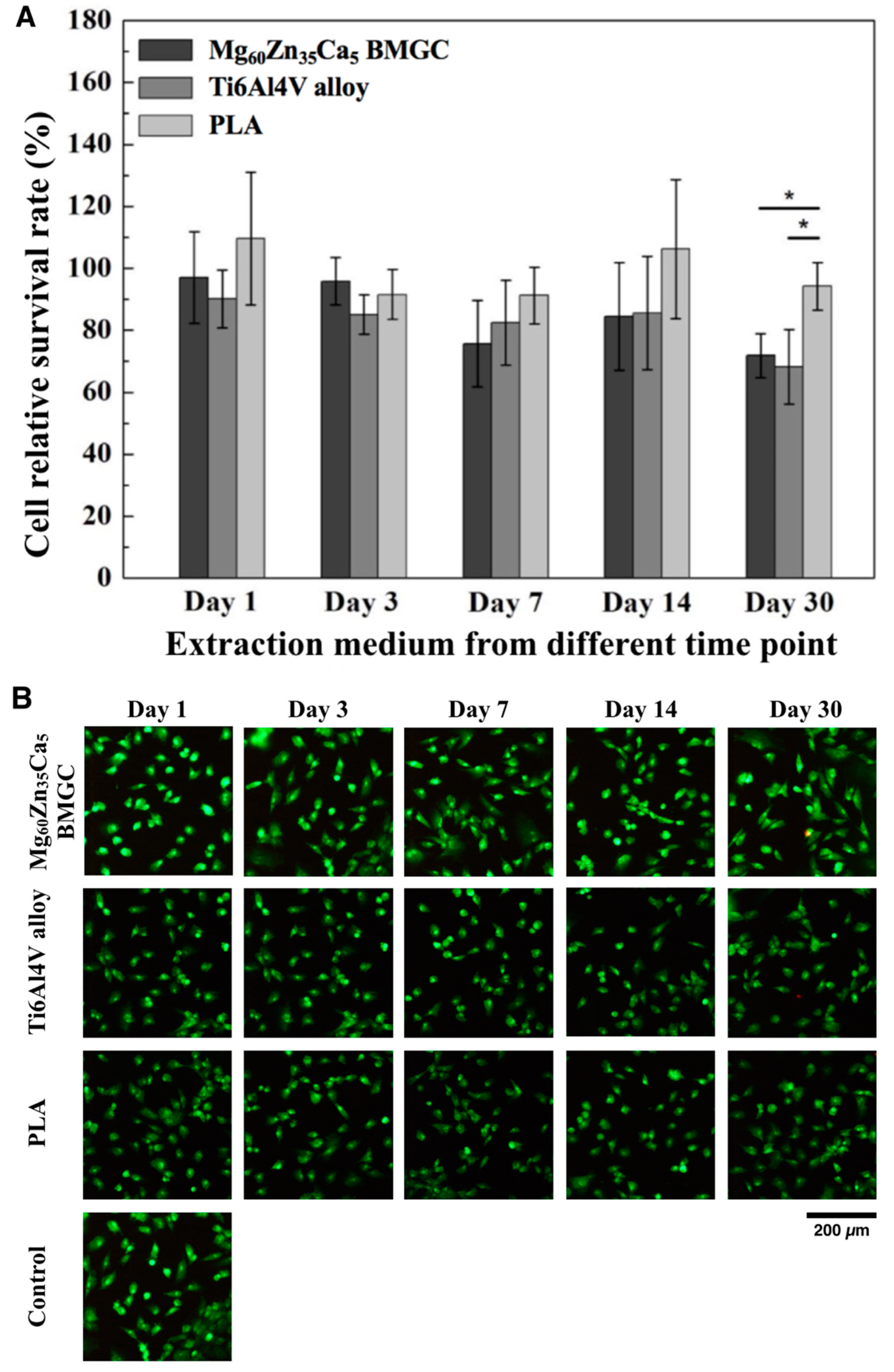
2.1. Cell Viability
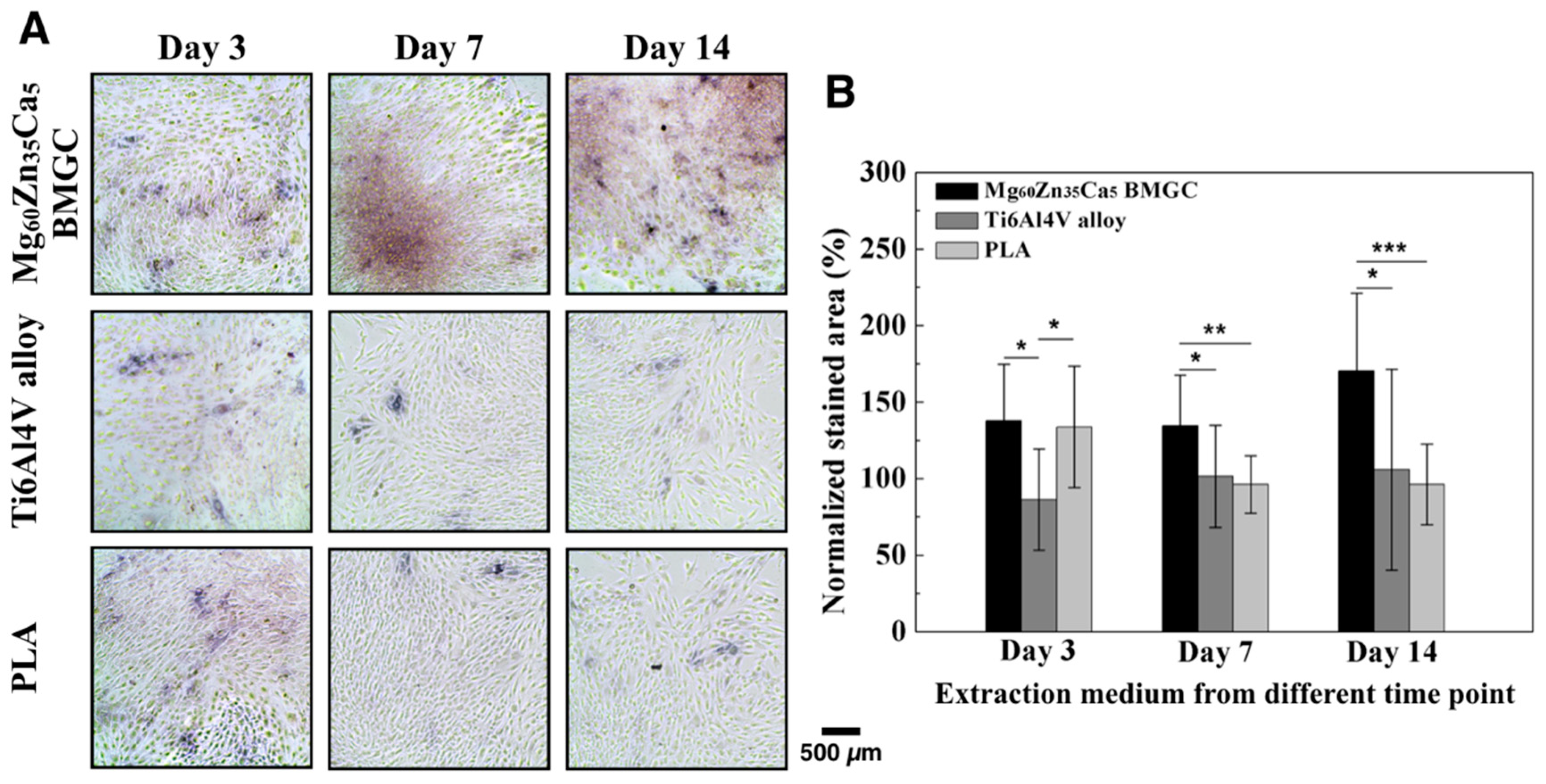
2.2. ALP Activity
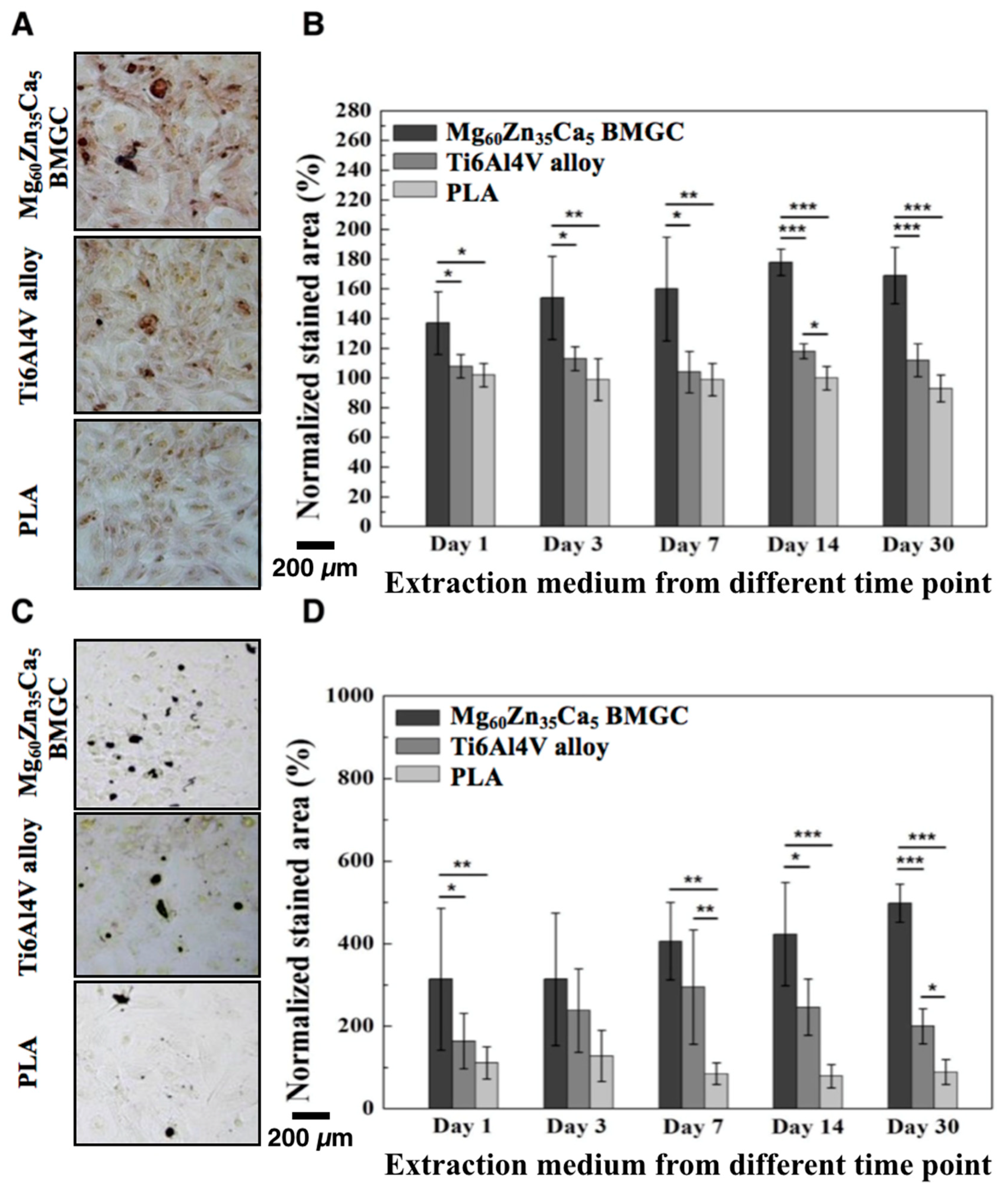
2.3. Extracellular Matrix Calcium Deposition
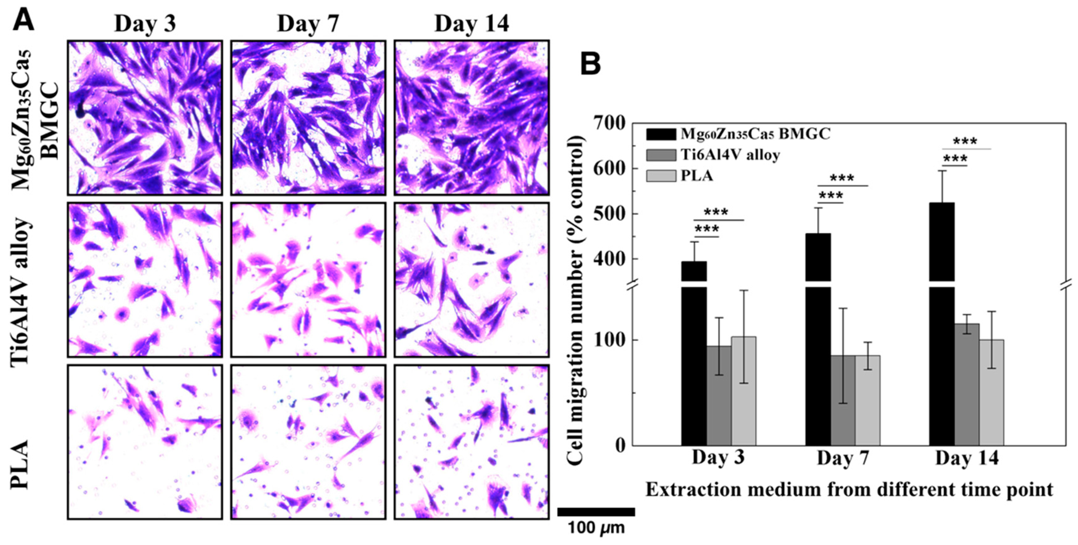
2.4. Migration Capacity
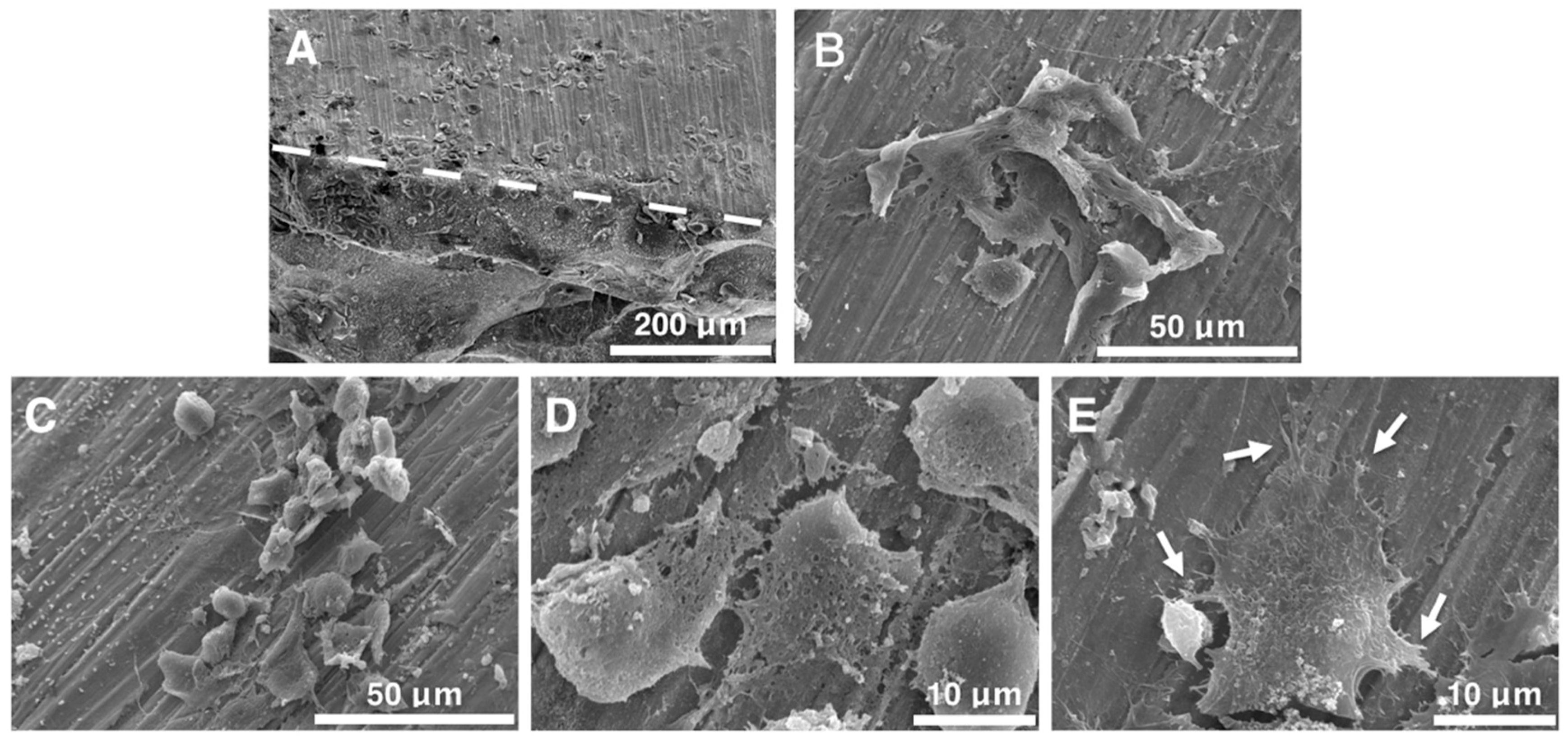
2.5. Cell Morphology Observation
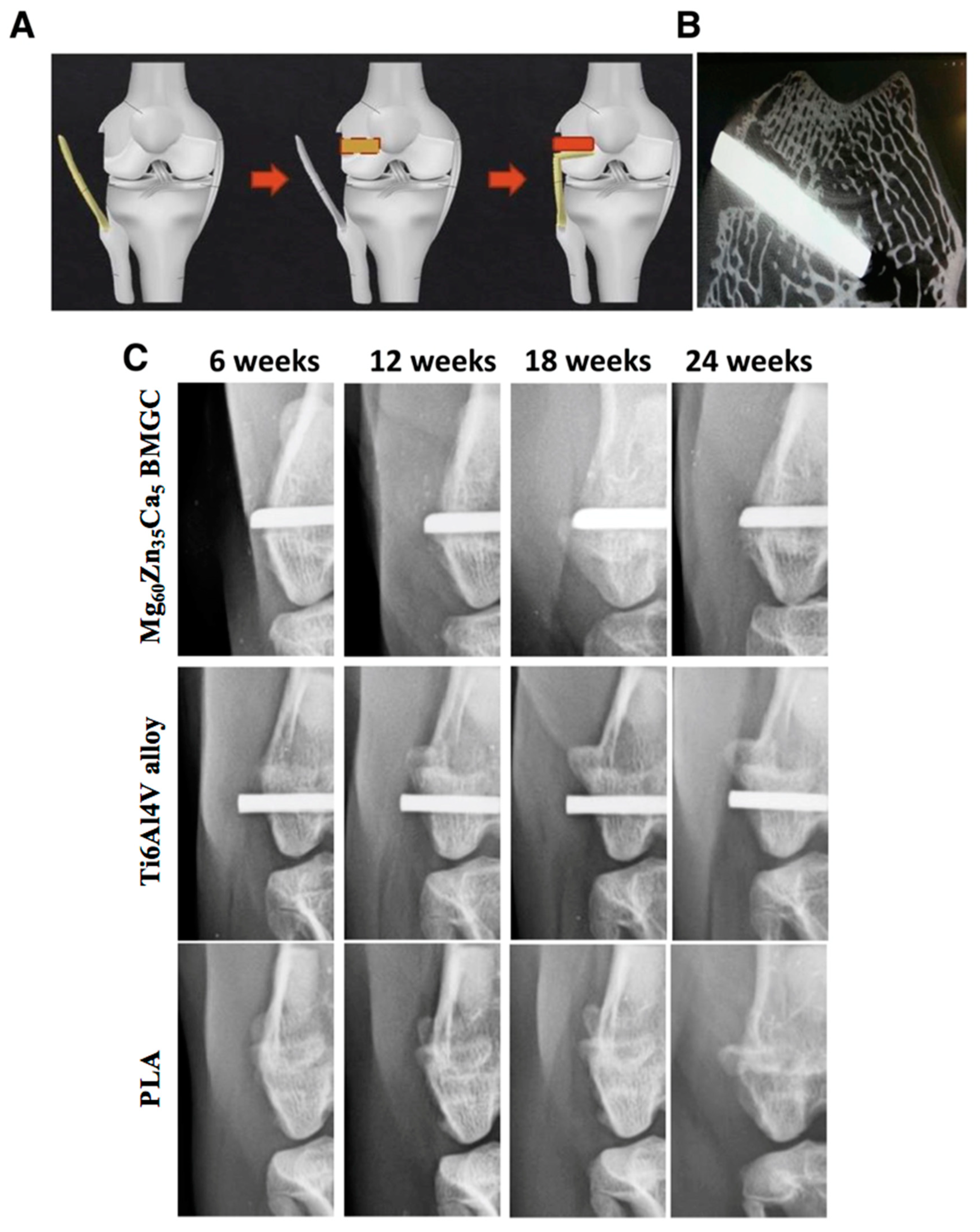
2.6. Radiological Evaluation
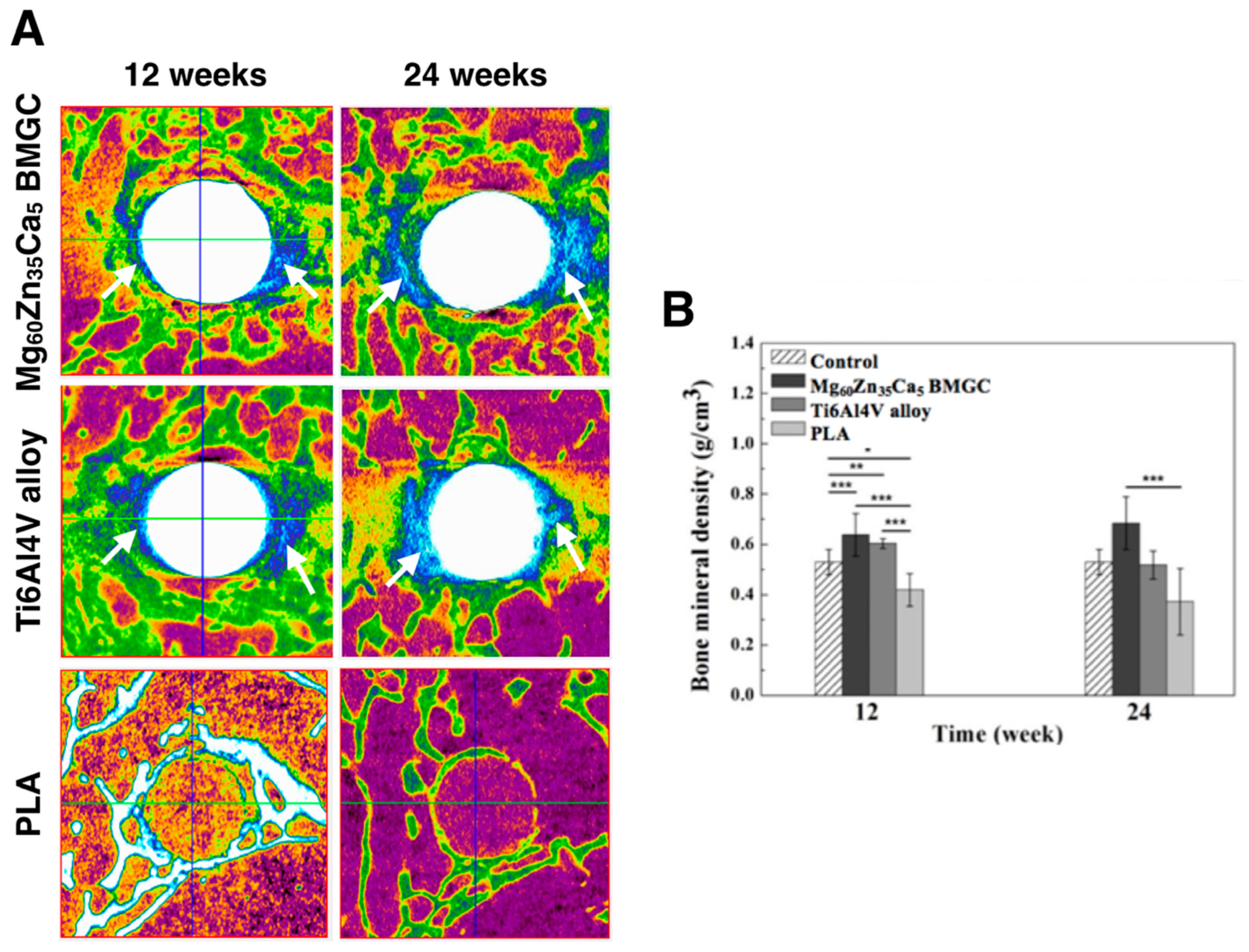
2.7. Micro-CT Image (Bone Mineral Density and 3D Image Reconstruction)
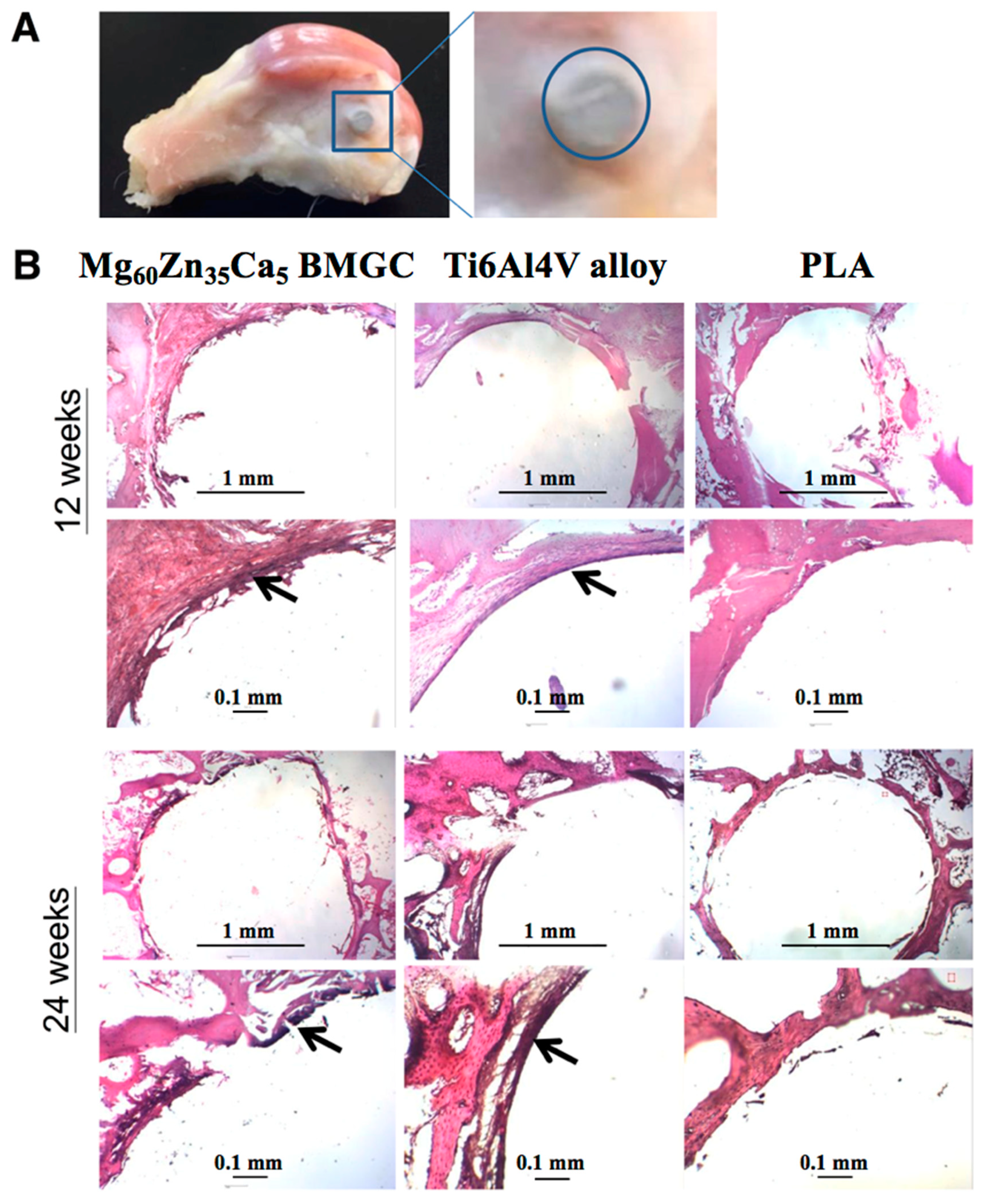
2.8. Histology Observation
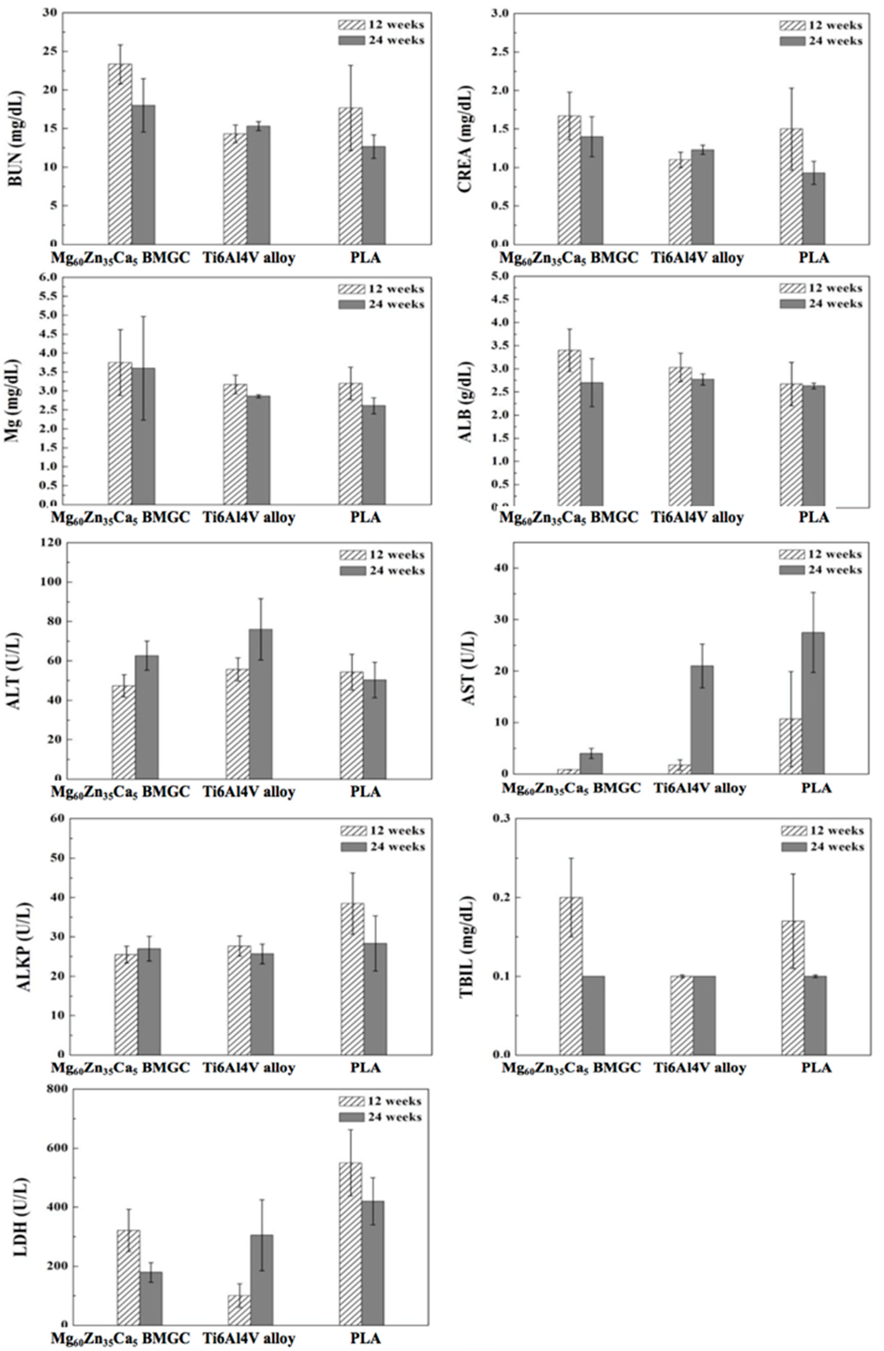
2.9. Hematology Analysis
3. Discussion
4. Materials and Methods
4.1. Sample Preparation
4.2. In Vitro Test
4.2.1. MTT Assay and Live/Dead Assay
4.2.2. ALP Staining
4.2.3. Alizarin Red S staining and Von Kossa Staining
4.2.4. Migration Test
4.2.5. Cell Morphology Observation
4.3. In Vivo Test
4.3.1. Experimental Design
4.3.2. Surgical Method
4.3.3. Radiological Observation
4.3.4. Micro-CT Scan and 3D Image Reconstruction
4.3.5. Hematology Analysis
4.3.6. Histological Observation
4.4. Statistical Analysis
5. Conclusions
- At low concentrations of extraction medium treatments, the cell survival rates of osteoblasts on Mg60Zn35Ca5 BMGC, Ti6Al4V alloy, and PLA can be higher than 80%. According to ISO-10993-5 [10], all samples could be classified as having first level cytotoxicity (slightly toxic).
- Mg60Zn35Ca5 BMGC demonstrated excellent in vivo biocompatibility, and the osteogenic and osteoconductive potentials of these implants were superior to the conventional Ti6Al4V alloy and PLA.
- With an improved biodegradation rate, excellent biocompatibility, and most importantly, osteogenic ability, Mg60Zn35Ca5 BMGC has great potential for future surgical implant development and application.
Author Contributions
Acknowledgments
Conflicts of Interest
References
- Huiskes, R.; Weinans, H.; van Rietbergen, B. The relationship between stress shielding and bone resorption around total hip stems and the effects of flexible materials. Clin. Orthop. Relat. Res. 1992, 274, 124–134. [Google Scholar] [CrossRef]
- Jiao, W.; Li, H.F.; Zhao, K.; Bai, H.Y.; Wang, Y.B.; Zheng, Y.F.; Wang, W.H. Development of CaZn based glassy alloys as potential biodegradable bone graft substitute. J. Non-Cryst. Solids 2011, 357, 3830–3840. [Google Scholar] [CrossRef]
- Witte, F.; Kaese, V.; Haferkamp, H.; Switzer, E.; Meyer-Lindenberg, A.; Wirth, C.J.; Windhagen, H. In vivo corrosion of four magnesium alloys and the associated bone response. Biomaterials 2005, 26, 3557–3563. [Google Scholar] [CrossRef] [PubMed]
- Wong, P.C.; Tsai, P.H.; Li, T.H.; Cheng, C.K.; Jang, J.S.C.; Huang, J.C. Degradation behavior and mechanical strength of Mg-Zn-Ca bulk metallic glass composites with Ti particles as biodegradable materials. J. Alloys Compounds 2017, 699, 914–920. [Google Scholar] [CrossRef]
- Wong, P.C.; Lee, T.H.; Tsai, P.H.; Cheng, C.K.; Li, C.; Jang, J.S.C.; Huang, J.C. Enhanced mechanical properties of MgZnCa bulk metallic glass composites with Ti-particle dispersion. Metals 2016, 6, 116. [Google Scholar] [CrossRef]
- He, L.Y.; Zhang, X.M.; Liu, B.; Tian, Y.; Ma, W.H. Effect of magnesium ion on human osteoblast activity. Braz. J. Med. Biol. Res. 2016, 49, e5257. [Google Scholar] [CrossRef] [PubMed]
- Yoshizawa, S.; Brown, A.; Barchowsky, A.; Sfeir, C. Magnesium ion stimulation of bone marrow stromal cells enhances osteogenic activity, simulating the effect of magnesium alloy degradation. Acta Biomater. 2014, 10, 2834–2842. [Google Scholar] [CrossRef] [PubMed]
- Abed, E.; Moreau, R. Importance of melastatin-like transient receptor potential 7 and magnesium in the stimulation of osteoblast proliferation and migration by platelet-derived growth factor. Am. J. Physiol. Cell Physiol. 2009, 297, C360–C368. [Google Scholar] [CrossRef] [PubMed] [Green Version]
- Yoshizawa, S.; Chaya, A.; Verdelis, K.; Bilodeau, E.A.; Sfeir, C. An in vivo model to assess magnesium alloys and their biological effect on human bone marrow stromal cells. Acta Biomater. 2015, 28, 234–239. [Google Scholar] [CrossRef] [PubMed] [Green Version]
- ISO-10993-5: Biological Evaluation of Medical Devices—Part 5: Test for Cytotoxicity: In Vitro Methods; ANSI/AAMI: Arlington, VA, USA, 1999.
- Jensen, E.D.; Gopalakrishnan, R.; Westendorf, J.J. Regulation of gene expression in osteoblasts. Biofactors 2010, 36, 25–32. [Google Scholar] [CrossRef] [PubMed]
- Att, W.; Hori, N.; Takeuchi, M.; Ouyang, J.; Yang, Y.; Anpo, M.; Ogawa, T. Time-dependent degradation of titanium osteoconductivity: an implication of biological aging of implant materials. Biomaterials 2009, 30, 5352–5363. [Google Scholar] [CrossRef] [PubMed]
- Zheng, Y.F.; Gu, X.N.; Witte, F. Biodegradable metals. Mater. Sci. Eng. R. Rep. 2014, 77, 1–34. [Google Scholar] [CrossRef]
- Janning, C.; Willbold, E.; Vogt, C.; Nellesen, J.; Meyer-Lindenberg, A.; Windhagen, H.; Thorey, F.; Witte, F. Magnesium hydroxide temporarily enhancing osteoblast activity and decreasing the osteoclast number in peri-implant bone remodelling. Acta Biomater. 2010, 6, 1861–1868. [Google Scholar] [CrossRef] [PubMed]
- Yang, J.X.; Cui, F.Z.; Lee, I.S.; Zhang, Y.; Yin, Q.S.; Xia, H.; Yang, S.X. In vivo biocompatibility and degradation behavior of Mg alloy coated by calcium phosphate in a rabbit model. Biomater. Appl. 2012, 27, 153–164. [Google Scholar] [CrossRef] [PubMed]
- Chaya, A.; Yoshizawa, S.; Verdelis, K.; Myers, N.; Costello, B.J.; Chou, D.T.; Pal, S.; Maiti, S.; Kumta, P.N.; Sfeir, C. In vivo study of magnesium plate and screw degradation and bone fracture healing. Acta Biomater. 2015, 18, 262–269. [Google Scholar] [CrossRef] [PubMed]


© 2019 by the authors. Licensee MDPI, Basel, Switzerland. This article is an open access article distributed under the terms and conditions of the Creative Commons Attribution (CC BY) license (http://creativecommons.org/licenses/by/4.0/).
Share and Cite
Wong, C.-C.; Wong, P.-C.; Tsai, P.-H.; Jang, J.S.-C.; Cheng, C.-K.; Chen, H.-H.; Chen, C.-H. Biocompatibility and Osteogenic Capacity of Mg-Zn-Ca Bulk Metallic Glass for Rabbit Tendon-Bone Interference Fixation. Int. J. Mol. Sci. 2019, 20, 2191. https://0-doi-org.brum.beds.ac.uk/10.3390/ijms20092191
Wong C-C, Wong P-C, Tsai P-H, Jang JS-C, Cheng C-K, Chen H-H, Chen C-H. Biocompatibility and Osteogenic Capacity of Mg-Zn-Ca Bulk Metallic Glass for Rabbit Tendon-Bone Interference Fixation. International Journal of Molecular Sciences. 2019; 20(9):2191. https://0-doi-org.brum.beds.ac.uk/10.3390/ijms20092191
Chicago/Turabian StyleWong, Chin-Chean, Pei-Chun Wong, Pei-Hua Tsai, Jason Shian-Ching Jang, Cheng-Kung Cheng, Hsiang-Ho Chen, and Chih-Hwa Chen. 2019. "Biocompatibility and Osteogenic Capacity of Mg-Zn-Ca Bulk Metallic Glass for Rabbit Tendon-Bone Interference Fixation" International Journal of Molecular Sciences 20, no. 9: 2191. https://0-doi-org.brum.beds.ac.uk/10.3390/ijms20092191