Review

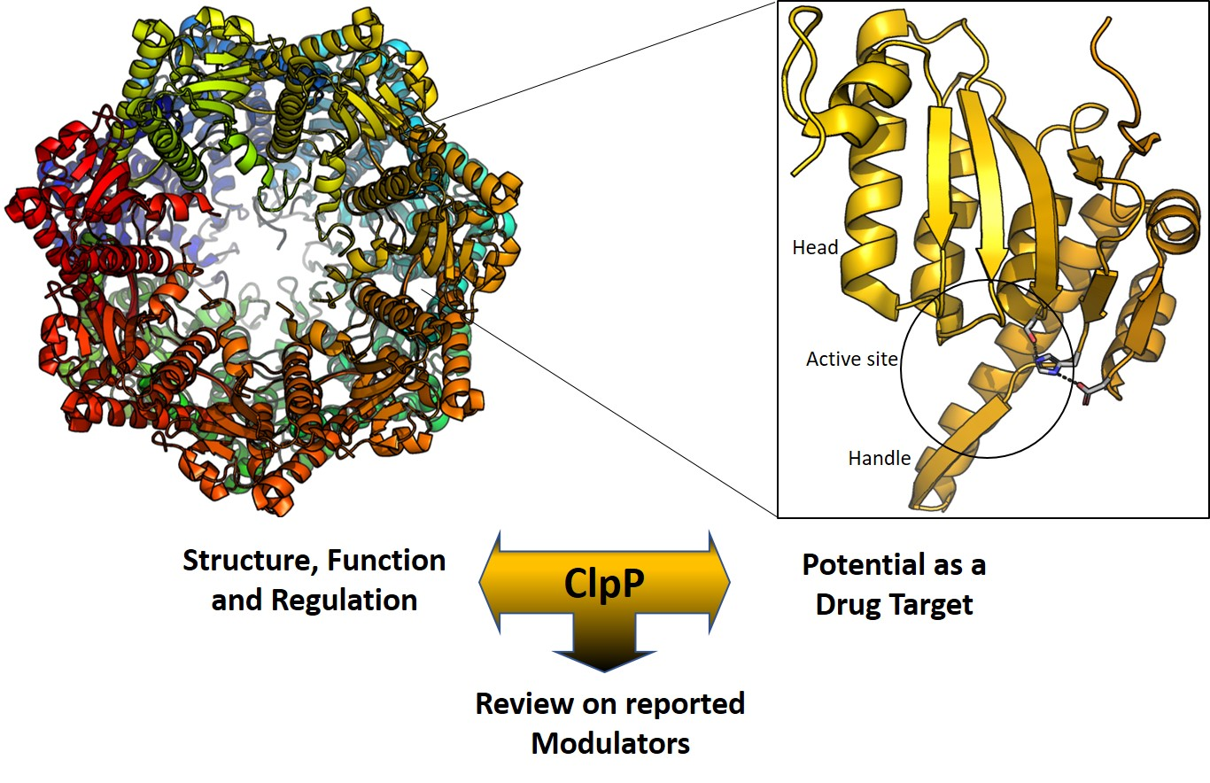
ClpP Protease, a Promising Antimicrobial Target

Laboratory of Medicinal Chemistry, University of Antwerp, Universiteitsplein 1, B-2610 Antwerp, Belgium
*
Author to whom correspondence should be addressed.
Int. J. Mol. Sci. 2019, 20(9), 2232; https://0-doi-org.brum.beds.ac.uk/10.3390/ijms20092232
Submission received: 25 March 2019 / Revised: 18 April 2019 / Accepted: 29 April 2019 / Published: 7 May 2019

Abstract

:
The caseinolytic protease proteolytic subunit (ClpP) is a serine protease playing an important role in proteostasis of eukaryotic organelles and prokaryotic cells. Alteration of ClpP function has been proved to affect the virulence and infectivity of a number of pathogens. Increased bacterial resistance to antibiotics has become a global problem and new classes of antibiotics with novel mechanisms of action are needed. In this regard, ClpP has emerged as an attractive and potentially viable option to tackle pathogen fitness without suffering cross-resistance to established antibiotic classes and, when not an essential target, without causing an evolutionary selection pressure. This opens a greater window of opportunity for the host immune system to clear the infection by itself or by co-administration with commonly prescribed antibiotics. A comprehensive overview of the function, regulation and structure of ClpP across the different organisms is given. Discussion about mechanism of action of this protease in bacterial pathogenesis and human diseases are outlined, focusing on the compounds developed in order to target the activation or inhibition of ClpP.

Graphical Abstract

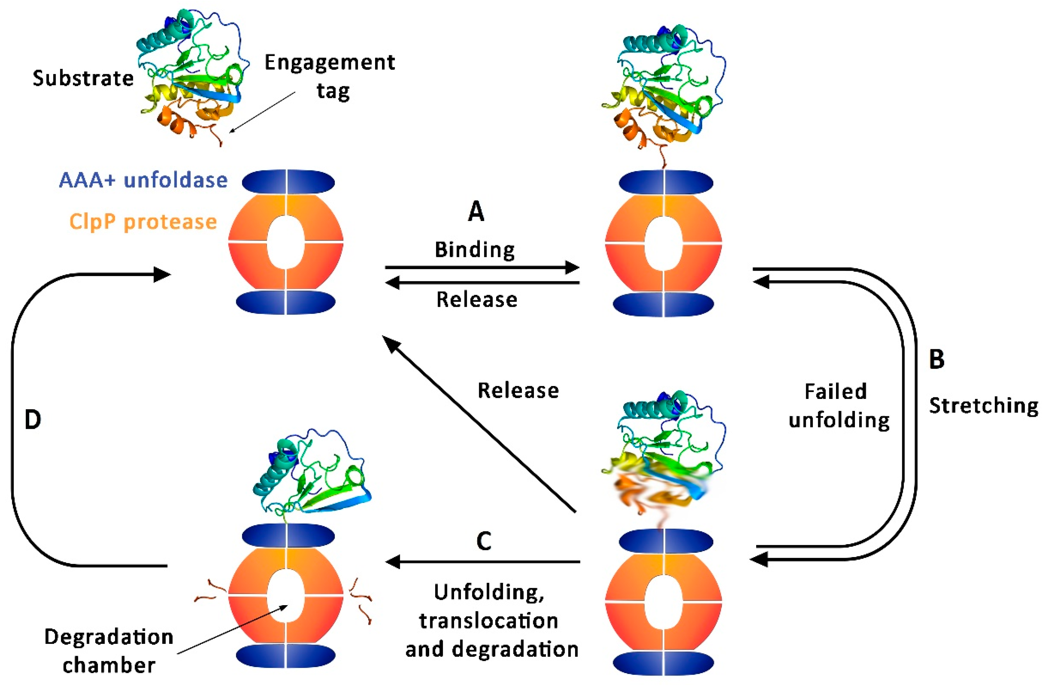
1. ClpP Function and Regulation

In every domain of life, cells and organelles are full of proteins, many of which are intrinsically disordered or in the process of folding [1]. Thus, proteolysis is an essential cellular activity, mediating protein turnover and the degradation of undesired or defective proteins from the intracellular environment. Since peptide bonds in an unstructured polypeptide are fairly sensitive to proteolysis, indiscriminate proteases are not present in the cytoplasm of archaea, bacteria and eukaryotic organelles. Instead, selected proteins in these intracellular environments are arrested, unfolded and degraded by proteolytic machines. These specific enzymes belong to the ATPases associated with diverse cellular activities (AAA+) superfamily, due to the presence of an AAA+ unfoldase partner in charge of the recognition of the desired substrates and the ATP-dependent mechanical unfolding of the targeted protein before its translocation to the proteolytic active site [2,3].
These proteolytic machines, enable the cell to keep proteostasis and to alter the protein levels to adapt to changing environments. The 26S proteasome present in eukaryotic cells is possibly the best known of these cellular machines [4]. AAA+ proteases present both in bacteria and eukaryotic organelles include FtsH, Lon, HsIUV, ClpPXP, ClpAP and ClpCP [2]. A protease found in all three domains of life is 20S peptidase, which associates with different AAA+ unfoldases such as Rpt1-Rpt6 ring of the 26S proteasome, proteasome-activating nucleotidase (Pan) and Mycobacterium proteasome-associated ATPase (Mpa) [4,5]. In bacteria, Lon and ClpP are estimated to carry out around the 80% of cellular proteolysis [6,7]. Coexistence of a number of AAA+ proteases is present in every species. Importance of their role and overlap of their functions determine the essentiality of each of them in every given organism.
ClpP is well characterized in many species, and it is always involved in the proteolysis of defective and misfolded proteins. More than 60 substrates of Escherichia coli ClpP were identified when using an inactive mutant as a trap [8]. These targeted proteins were involved in a variety of processes including transcription regulation, metabolism, damage repair and cell division. A sodium dodecyl sulfate polyacrylamide gel electrophoresis (SDS-PAGE) showed a similar selection in Bacillus subtilis, comparing wild-type (WT) and clpP-defective mutant (ΔclpP) strains [9]. In these and other bacteria, such as Staphylococcus aureus, [10] ClpP function affects greatly the degradation of proteins implicated in stationary phase adaptation, nutrient starvation, heat-stress response, biofilm formation, cell motility, cell-cycle progression or metabolism [11,12].
Although the targeted proteins are not the same in each of the organisms, the substrate identification in model bacteria shows the relevance of its function when regulating bacterial proteome. The important role that ClpP plays in the bacterial infectivity and virulence is explained by the range of processes in which its substrates are involved, pointing at ClpP as a prime target for antivirulence drug development [13,14,15,16,17].
This protease is also present in human mitochondria, where it is crucial for protein homeostasis and also plays a relevant role in the degradation and regulation of enzymes related with cellular metabolic pathways such as the electron transport chain [18,19,20]. ClpP overexpression has been associated with a broad range of carcinomas during the last years [21,22]. In contrast, clpP-recessive mutations were observed and identified as the cause of infertility and sensorineural deafness of Perrault syndrome [23,24].
As previously mentioned, presence of AAA+ unfoldases is required for the efficient action of this protease family. The Clp ATPases filter, unfold and introduce in the proteolytic chamber a range of proteins according to the situation following different regulatory processes (Figure 1) [25,26]. This target identification is often performed by inherent or added sequence motifs accessible either in the N or C terminus of these substrates [8].
The substrate selection will always rely on the specific unfoldase, and thus the common presence of diverse ATPases offers a tight regulation of the ClpP activity in cells [27]. Furthermore, the existence of adaptors binding to the Clp ATPases upon signals or stresses, influencing in this way the substrate choice, enlarges the control of this regulation. SspB, RssB and UmuD for E. coli caseinolytic protease subunit X (ClpX) [28,29,30], ClpS for E. coli caseinolytic protease subunit A (ClpA) or MecA for B. subtilis caseinolytic protease subunit C (ClpC) are some examples of these adaptors [31,32]. Recent identification of small anti-adaptor proteins, able to regulate the activity of the adaptors themselves, added an extra layer of complexity for control and fine-tuning of ClpP protease [33]. For further information on adaptor proteins regulating bacterial proteolysis, the excellent review by Battesti and Gottesman is recommended [34].

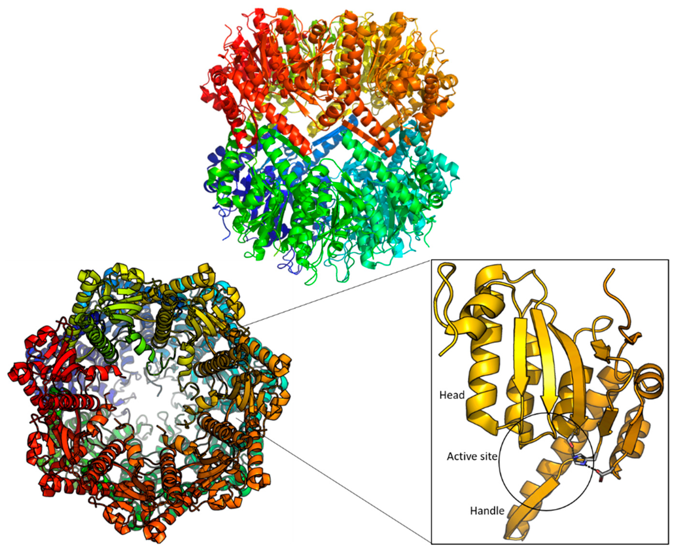
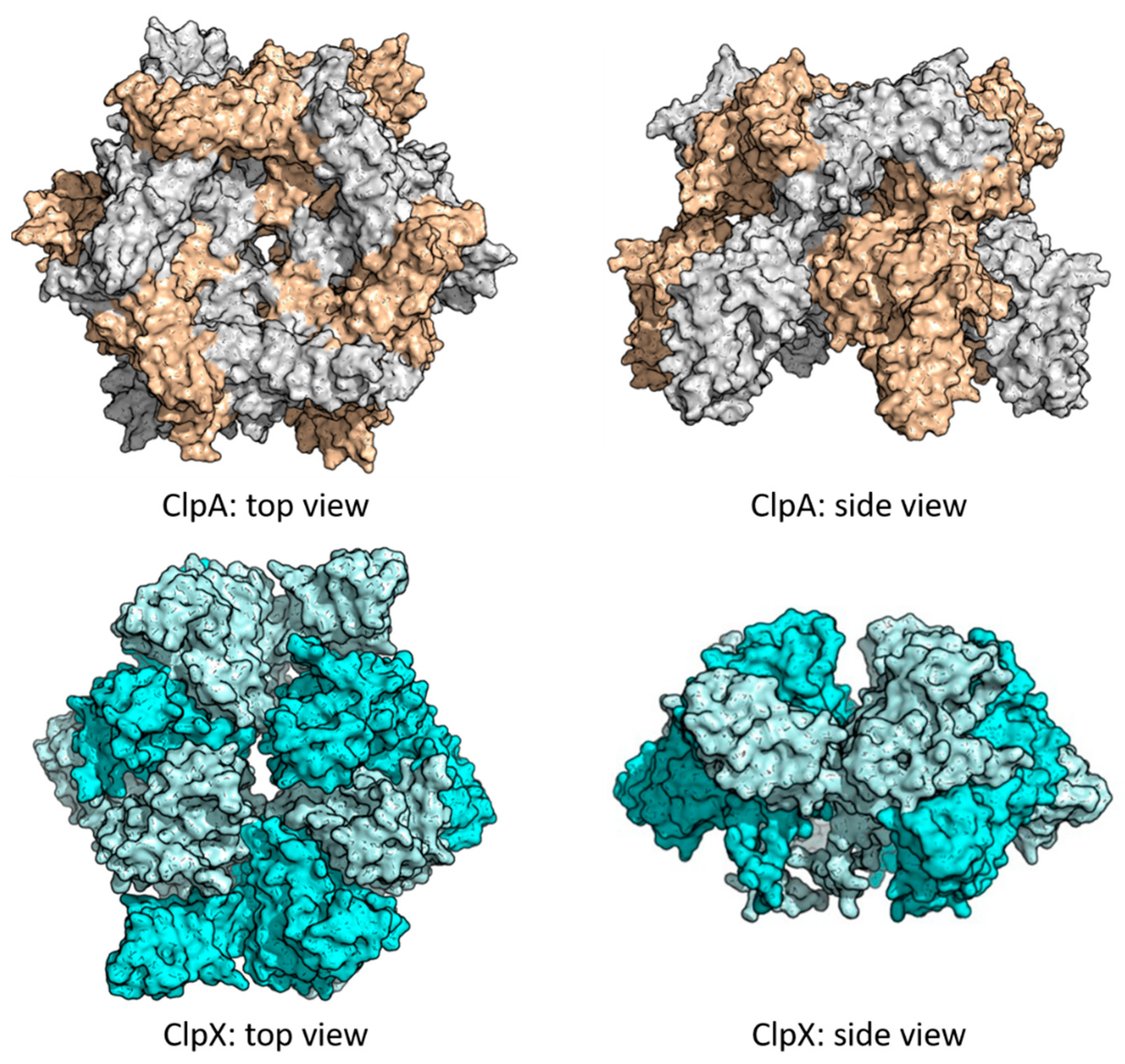
2. Structure of ClpP

The ClpP protease forms a tetradecameric cylinder with two heptameric rings longitudinally aligned (Figure 2). The axial pore of each ring (~10 Å diameter) acts as the entrance to the interior of the proteolytic chamber (~50 Å diameter) with the 14 active sites [35]. Each protomer, with a size ranging the 193-277 residues depending on the species, consists of a C terminus, an N terminal loop, a handle and a head domain (Figure 2). The handle domains of each heptameric ring are oriented to the same face, composing the interface that assembles the tetradecamer when the two handle faces bind to each other. When the tetradecamer is formed, the globular head domains comprise the main body of the ring, with most of their surface, including the active site, allocated in the interior of the barrel (Figure 2) [36]. Finally, the N-termini are highly flexible and are located in the axial region. Each of the active sites, contains the canonical Serine-Histidine-Aspartate catalytic triad [35].
ClpP tetradecamer in E. coli is formed by 14 identical protomers. Nevertheless, it can be found as a homomeric or heteromeric assembly in organisms were different isoforms of ClpP are present, as it is the case of ClpP1P2 in Mycobacterium tuberculosis, where each ring is formed with 7 protomers of a different paralog [37]. Other examples of heteromeric assembly include many cyanobacteria mixing ClpP and ClpR, an inactive variant of ClpP, leading to different types of tetradecameric complexes [38]. Pseudomonas aeruginosa also presents ClpP1 and ClpP2 isoforms, however in this case, they only form separate tetradecamers which perform distinct functions in the cell [39].
The peptidase activity of ClpP, characteristic of a chymotrypsin-like serine protease, results in peptides of 7-8 residues length, cutting after non-polar residues [27,40,41]. However, for efficient degradation of long peptides and proteins, as mentioned before, binding with an unfoldase partner such as ClpX or ClpA is needed. These chaperones belong to the HsP100 class of the AAA+ superfamily and act as a cap for the ClpP cylinder. Despite the existence of different Clp ATPases across different organisms, all share the structure of a hexameric complex with a central pore used as a channel to translocate the substrates into the proteolytic chamber (Figure 3).
They bind to seven grooves of about 10 Å of diameter, mainly composed of conserved hydrophobic residues and are compatible with the specific loop regions of the AAA+ chaperones as it is the case of IGF and IGL loop of ClpX and ClpA of E. coli respectively [44,45]. Thanks to these loop-groove interactions, the mismatch between the heptameric symmetry of the ClpP complex and the hexameric conformation of the chaperones is evaded. This mismatch has been suggested to be a result of evolution, which allows the chaperone to rotate around the ClpP complex, improving the efficiency of the unfolding and translocation [46]. It is also commonly accepted that the binding with the chaperone induces a conformational change in ClpP, turning a closed and inactive conformation into an open and proteolytically active conformer which accepts large peptide chains in the degradation chamber [45]. Further information about ClpP unfoldases structure and mechanistic can be found in a recent and comprehensive review by Olivares et al. [47].
With this information in mind, analogy with the well-known 20S peptidase could be easily done. ClpP and 20S peptidase share a self-compartmentalized nature, composed by aligned heptameric rings, with narrow axial pores leading to an inner proteolytic chamber with up to 14 catalytic sites. Need of association to ATP-unfoldase partners for an efficient proteolysis is also a common feature. However, they also display key differences. First, the size of proteasome is notably larger. Despite the residue count of its protomers and diameters of pores and inner chamber are comparable to the ones of ClpP, proteasome complex is formed by four heptameric rings, two of which have an exclusively structural nature. Furthermore, the active sites of 20S peptidase contain a catalytic threonine instead of the Ser-His-Asp of ClpP. For an in-depth comparison of ClpP and proteasome, readers are referred to the review published in by Liu et al. in 2014 [48].

3. ClpP as a Drug Target

3.1. ClpP in Bacteria and Parasites

ClpP started attracting attention due to its potential as an antibacterial target in the late 90s, when its direct relationship with bacterial virulence was proven for Gram-positive S. aureus and Listeria monocytogenes [49,50], together with Gram-negative Salmonella Typhimurium [51].
The ClpP of S. aureus is characterized most extensively. Direct relation between its virulence and ClpP function was demonstrated by Mei et al. when they performed a transposon-based mutagenesis screen, evaluating the virulence of the deletion strains in a murine model, identifying between others the ClpX ortholog [49]. Follow-up experiments enlarged the scope to ClpP itself, when clpP- and clpX-deficient mutants showed lack of virulence in a murine skin abscess model [10]. The same study established that the connection between ClpP function and the S. aureus virulence did not rely only on the regulation of stress responses, but also on the secretion of α-haemolysin (hla gene) and other effectors by the regulation of the accessory gene regulator (agr) locus [10]. Subsequent research related ClpP with the iron-regulated determinants (Isd) system regulation, which extracts heme-iron from the host hemoglobin and is fundamental for S. aureus pathogenesis [52].
In case of L. monocytogenes, Gaillot et al. established the essential ClpP role in the survival and growth of this bacteria under stress conditions [50]. Thus, the intracellular parasitism of L. monocytogenes is unfeasible, being susceptible to the bactericidal activity of the host [50]. From the two isoforms existing in this bacterial species, ClpP2 was found necessary for the Listeriolysin O factor expression [53]. The immunodominant virulence factor allows bacterial escape from the phagocytic vacuoles from the host and promotes intracellular growth [54].
Other Gram-positive pathogens where ClpP affects bacterial viability are Streptococcus pneumoniae and Enterococcus faecalis. In S. pneumoniae ΔclpP strains were unable to invade the lungs, lost the ability of colonizing the nasopharynx in mouse models and had lower survival rates in murine macrophages [55]. Furthermore, clpP-deficient mutant was found to be susceptible to nitric oxide and other oxidative stress [56]. In E. faecalis, deficiency of ClpP or Clp unfoldases led to loss of virulence in an invertebrate model [15].
The first Gram-negative ClpP studied was S. Typhimurium. Through a transposon-based mutational analysis, a ΔclpP strain unable to grow in a mouse model of typhoid fever was detected [51]. Subsequent studies explained how the survival of that strain is compromised in presence of peritoneal macrophages and therefore impedes virulence [57]. More recently, Knudsen et al. found, using a transcriptomic analysis, how ClpP regulation of RpoS (RNA polymerase, sigma S) and CsrA (Carbon storage regulator A) factors indirectly affects S. Typhimurium virulence [58].
More recent examples of Gram-negative bacteria where ClpP plays a central role in pathogenesis are P. aeruginosa and Legionella pneumophila [13,16]. For P. aeruginosa, ClpX and ClpP2 were identified as part of the proteolytic network of the exopolysaccharide alginate biosynthesis, a characteristic marker for the onset of chronic lung infection in cystic fibrosis [13]. Furthermore, Bishop et al. established the relevant role of ClpP in the Cell Surface Signaling (CSS) pathway, which enables iron uptake during P. aeruginosa infection [59]. Likewise L. monocytogenes, clpP-deficient mutants in L. pneumophila showed impaired virulence and inability to escape endosome-lysosome pathway in mammalian cells [16].
In E. coli, where ClpP is responsible for the cleavage of proteins involved in metabolism, transcription factors, as well as in oxidative stress response and starvation; [10] Robinson et al. showed significant differences between growth curves of wild type and ΔclpP E. coli under nitric oxide-stress [60].
Opposed to the described bacteria, where ClpP influences the growth in stress condition and virulence but not the viability of the species, M. tuberculosis ClpP1P2 was found to be an essential target [14,61]. As mentioned before, ClpP1 and ClpP2 isoforms ensemble a tetradecameric complex with one heptameric ring of each analogue [37]. These isoforms together with the chaperones ClpX and ClpC1 were identified as essential for the pathogen viability and infectivity in vitro [14].
ClpP has not only been studied in bacteria but also in protozoal parasites as it is Plasmodium falciparum. This unicellular eukaryotic parasite is the causative agent of malaria in humans. P. falciparum adopts several cellular morphologies and different reproductive stages when it moves from the mosquito carrier to human hosts. This parasite ClpP is localized in the apicoplast, a plastid bound by four membranes located near to a mitochondrion. This organelle is needed for isoprenoids, fatty acids, heme and iron-sulfur clusters biosynthesis [62]. An analysis of the ClpP expression through the P. falciparum life cycle demonstrated overexpression in the stages in which P. falciparum colony increases and infects red blood cells (late trophozoite an early schizont) [63]. Accordingly, growth decrease by inhibition of ClpP by a β-lactone was observed [63], interfering with the progression from early to late schizont stage.
In summary, the relevance of ClpP protease in bacterial pathogenesis offers an untapped target for new antivirulence and antimicrobial drugs [17].

3.2. ClpP in Human Mitochondria

Even though eukaryotic ClpPs have been less studied, human ClpP plays a key role in the quality control system of mitochondria. Human ClpP (hClpP) shares a high degree of sequence identity with its bacterial orthologs and the obtained X-rays suggest a close resemblance to their structure [48,64]. While human ClpP active form is the common tetradecameric complex with ATP unfoldases as human ClpX (hClpX) associated, ClpP exists as an inactive single heptameric ring, that only fuses into the tetradecamer by induction of hClpX binding [65]. Similarly to bacterial ClpP, hClpP degrades misfolded, mistranslated or aggregated proteins to keep mitochondrial respiratory efficiency, as hClpP target proteins are involved in a number of processes like fatty acid, amino acid and energy metabolism [19,20,21,66]. Given the relevant role of hClpP, it is not surprising to find human conditions in which this protease is involved.
In 2013, Gispert et al. reported that CLPP null mice showed infertility, reduced reproductive organs, severe growth retardation, diminished spontaneous motor activity and a strong decrease in survival [24]. Interestingly, these phenotypes correspond to Perrault syndrome symptoms in humans. This is a genetic disorder where also patients suffer sensorineural hearing loss and, in case of females, ovarian dysfunction leading to diminished fertility while male patients do not display sterility issues. Developmental delay or intellectual disability are some of the neurological defects present in some patients with this condition. During the same year, by a combination of homozygosity mapping, linkage analysis and exome sequencing in three families, CLPP recessive mutations were identified in a Pakistani family with Perrault syndrome. In this way, mutation in CLPP joined the already described mutations in HARS2, HSC17B4, LARS1 and C10orf2 [67]. Along the subsequent years new cases appeared [68,69]. It is suggested that hClpP structural integrity is compromised by these mutations, leading to lower ClpP levels in mitochondria, affecting mitochondrial respiratory function and leading to the symptoms of the disease.
Two years later, Cole et al. correlated hClpP overexpression with development of acute myeloid leukemia (AML) [21]. Shortly after, its complex with hClpX was identified as a key player of the bioenergetics stress control and found crucial for metastasis broadening the scope for a number of carcinomas [22]. Rapid and unregulated proliferation is a characteristic of cancer cells, it demands a significant alteration of the energy metabolism, increasing reactive oxygen species (ROS) and inducing oxidative stress [70]. This stress is balanced by upregulation of a number of mitochondrial proteases, such as Lon or ClpP, elevating its respiratory function to handle the oxidative damage to different biomolecules and other impairments in the cell [71]. Therefore, overexpression of ClpP and other mitochondrial proteolytic machines, such as heat shock protein-90 (Hsp90), in various types of cancer is consistent [22,72].
However, CLPP knockdown studies have revealed a variety of results across the different cell types. In experiments on AML cell lines, cell growth and viability were drastically reduced [21]. Prostate adenocarcinoma cell lines showed cell cycle arrest together with inability for colony formation [22]. However, barely any inhibition of cell proliferation was observed for the non-metastatic breast adenocarcinoma [22]. Potential co-administration of ClpP modulators with chemotherapy is also suggested by the correlation between ClpP expression and cisplatin resistance recently reported by Zhang et al. [73].

4. ClpP Modulation

Due to the complexity of these AAA+ proteolytic machines, many pathways to alter the normal function of the ClpP complex have been explored. While orthosteric inhibitors that bind and block the catalytic triad of the ClpP monomers seems like the most obvious choice, several alternatives impeding interaction of the tetradecamer with the ATP-unfoldases or upregulation of these chaperones have been investigated.

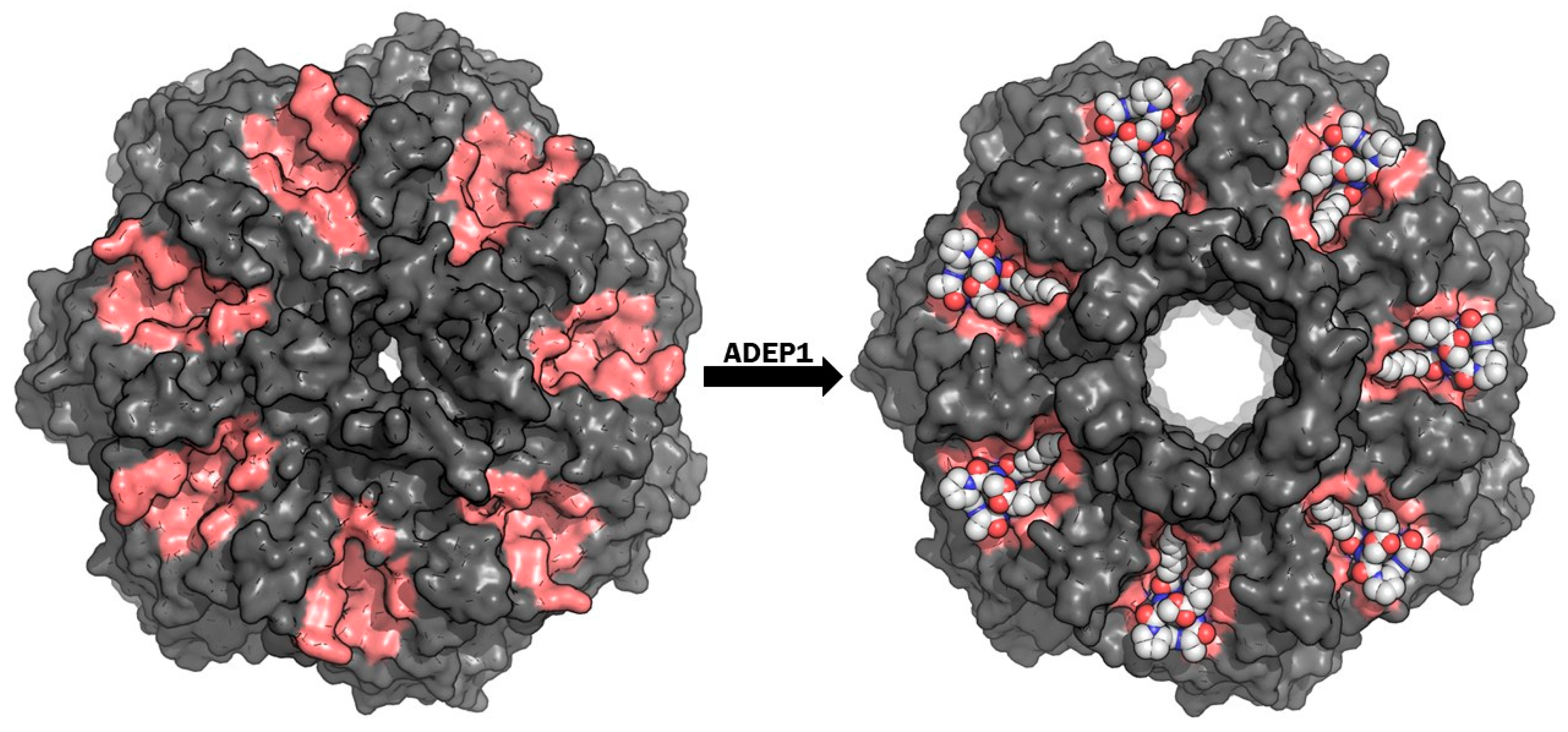
4.1. Targeting ClpP-ATPases Interaction

Interestingly, the most advanced drug-based studies for ClpP modulation do not proceed from a medicinal chemistry small molecule research focused on enzymatic inhibition, but from a factor isolated in the fermentation broth of Streptomyces hawaiiensis, which is able to dysregulate ClpP function [74]. The original group of eight factors was discovered by Eli Lilly and Company, who reported a notable in vitro antibacterial activity against Staphylococcus and Streptococcus strains [75]. Later, Osada et al. reported a depsinopeptide with similar antibacterial activity in E. faecalis and S. aureus called enopeptin A [76]. However, no suggested mechanism of action or activity in vivo was described until 2005, when Brötz-Oesterhelt et al. characterized the original cyclic acyldepsipeptide (ADEP1) structures from Eli Lilly and identified ClpP as their target [74]. ADEPs do not inhibit ClpP proteolytic activity nor interact with the catalytic triads, instead, these cyclopeptides compete with and displace the ATP-unfoldase ClpX specific loop regions (IGF and IGL) that bind into the ClpP grooves on the apical regions. By interaction with those grooves or pockets allocated between the adjacent monomers, ClpP tetradecameric complex adopts the active conformation of the ClpXP complex, broadening the diameter of the axial pores while lacking the selective and regulatory function of ClpX (Figure 4) [77,78]. In this way, ClpP performs uncontrolled proteolysis leading to bacterial death [74]. A comprehensive review about ClpP dysregulation by ADEPs was recently published by Malik et al. [79].
Improvement from the first ADEPs (ADEP1 and EnopeptinA, Figure 5), with poor ADME properties and antibacterial activity limited to Gram-positive pathogens [74,76], was progressively achieved by medicinal chemistry investigations. First, the inclusion of a pipecolate moiety to increase the rigidity of the depsipeptidic cycle, the replacement of a phenylalanine by its 3,5-difluorinated analogue and the partial saturation of the side chain led to a 160-fold increase of antibacterial activity and a boost of bioavailability and chemical stability (ADEP4, Figure 5) [74,80]. This compound was tested in mouse models proving its efficacy against S. aureus, E. faecalis and S. pneumoniae, proving as well desirable ADME properties with moderate to high clearance and distribution, with a half-life of 1–2 h in mice and dogs [74]. In contrast to other bacteria where these compounds activate an uncontrolled proteolysis leading to cell death, ADEP4 was able to eradicate M. tuberculosis by only preventing binding between ClpX and essential ClpP1P2 [81]. Furthermore, co-administration of ADEP4 and rifampicin in animal models eradicated S. aureus chronic biofilm infections [82]. Then, further constrains in the cycle were added by methylation of the piperidine and the cycled serine function were translated in improved antibacterial activity (ADEP10c and ADEP1g, Figure 5) [83,84]. Despite the success of the described compounds against Gram-positive bacteria, no ADEP with activity against Gram-negative was discovered until 2016 [85]. Goodreid et al. reported a set of compounds (ADEP26, Figure 5) which is not only able to kill S. aureus, E. faecalis, S. pneumoniae, B. subtilis and Listeria innocua; but also displayed antibacterial activity against Gram-negative Neisseria meningitidis and Neisseria gonorrhoeae [85]. With the aim of identifying new scaffolds with a similar mode of action to the ADEP family, a high-throughput screening (HTS) with E. coli ClpP was run [86]. Five clusters were identified, after lead optimization ACP1b (Figure 5) was the most developed structure, with a remarkable bactericidal activity against S. pneumoniae, S. aureus and Gram-negative N. gonorrhoeae, N. meningitidis, P. aeruginosa, L. monocytogenes and Haemophilus influenza [86]. Another scaffold of these activators was found by Lavey et al. in a fluorescence-based assay again with E. coli ClpP [87]. Despite sclerotiamide does not possess a comparable potency to its counterparts, the unique bicycle-[2.2.2]-diazoctane core opens a new and interesting spectrum for functionalization [87]. Finally, Stahl et al. discovered a species-selective activator for human ClpP (D9, Figure 5) [88]. This small molecule contains a halogenated benzyl moiety that binds to a unique aromatic amino acid network in the hClpP groove [88].
While ADEPs and the other new activators bind to the ClpP axial pockets of the tetradecamer, activating an uncontrolled proteolysis and impeding the connection with the AAA+ unfoldase, other families of compounds are known to target the unfoldases themselves.
This strategy has been broadly investigated in M. tuberculosis due to the essentiality of ClpP1P2 and its unfoldase ClpC1 in this pathogen. Large cyclic peptides such as cyclomarin A [89], lassomycin [90]. ecumicin and rufomycin (Figure 6) have shown their antibacterial action even with multidrug resistant Mtb strains [91,92]. Although their exact mode of action is not yet unraveled, they all target ClpC1, uncoupling its action from the ClpP1P2 proteolytic activity. Another recent example of ATP-unfoldase targeting is the dihydrothiazepine 334 (Figure 6) described by Fetzer et al. [93]. This compound was able to interact with S. aureus ClpX and provoke its deoligomerization. Toxin production levels of multidrug-resistant S. aureus were reduced and virulence attenuated.

4.2. ClpP Inhibition

A classical approach of designing a drug able to inhibit the catalytic triad of the serine protease has also been explored with uneven success. In 2008, a group of β-lactones were identified to inhibit S. aureus ClpP when Böttcher and Sieber used activity-based protein profiling to identify the targets involved in their antivirulence action [94]. From them, D3 (Figure 7) stood out as the most potent and selective irreversible inhibitor [95]. Optimization of this family led to synthesis of U1 (Figure 7), improving D3 potency between a 3 and a 5-fold [96]. Further characterization showed the ability of these compounds to also inhibit ClpP and therefore reducing the expression of extracellular virulence factors not only in S. aureus but also in L. monocytogenes [97]. β-Lactones have also displayed ClpP inhibition in other pathogens such as P. falciparum or M. tuberculosis [63,98]. Despite successful results in in vitro assays, the reduced plasma stability due to hydrolysis of the cyclic ester and their generally poor selectivity has limited their clinical development [99].
Thus, further investigations in search of a new class of ClpP inhibitors was carried out by Sieber et al., who conducted a HTS with more than 137.000 compounds [99]. The main result of this study was the identification of a phenyl ester AV170 (Figure 7), which triggered deoligomerization of the ClpP tetradecamer by covalent binding to the catalytic serine [99]. AV167 (Figure 7) also drew attention due to its inhibition of the hClpP [99]. A recent follow up study evolved this compound into a selective inhibitor of the human ClpP by addition of an alkyne in the naphtofurane moiety (TG42, Figure 7) [100]. However, their improved potency, pharmacokinetics and plasma stability were counteracted by their lower anti-virulence activity. In addition, no attempt of structure development was able to maintain the inhibitory activity [99]. A reinvestigation of the same HTS campaign with less strict selection criteria gave as a hit the first non-covalent binding inhibitor of S. aureus ClpP [101]. This compound AV145 (Figure 7) does not possess a substrate-like chemical structure and consequently does not bind to the active site but to the handle region of each monomer. This binding induces a novel and inactive conformation of the tetradecameric complex [101].
A new inactivation mechanism was discovered by Gersch et al. when they found β-sultams (RKS07, Figure 7) to selectively modify ClpP catalytic serine [102]. Nucleophilic attack of Ser98 to the cyclic sulfone, followed by elimination of the sulfonylated serine results in formation of dehydroalanine (Figure 8A). The alteration of the catalytic residue triggers disassembly of the active complex into two inactive heptameric rings [102].
Strong evidence of the potential of boron-derived compounds as ClpP inhibitors has been shown in M. tuberculosis. Bortezomib (Figure 7), a known 26S proteasome inhibitor studied in treatment for cancer, demonstrated to inhibit ClpP1P2 [103]. However, its high cost, poor pharmacokinetics, short half-life and proteasome inhibition limited the use of this drug at M. tuberculosis treatment [103]. Substrate-based boronates (N-(picolinoyl)-Trp-Lys-boroMet, Figure 7) by Akopian et al. also displayed selective targeting of ClpP1P2 inducing growth inhibition. New pyrrole cores (13i, Figure 7) were recently identified as ClpP1P2 inhibitors by Liu et al. after an in silico driven exploration, showing antibacterial properties and room for optimization [104]. Furthermore, a HTS setting for detection of Mtb ClpC1P1P2 complex inhibitors identified two hits [105]. Despite not showing antibacterial properties and having unclear mechanisms of action, both inhibitors showed a new chemical structure for ClpP inhibitors (GSK 17, Figure 7) and forthcoming use of broader libraries envisages a promising future for detection of new inhibitors for this pathogen [105]. Inhibition of P. falciparum ClpP has also been achieved with a novel class of ClpP inhibitors [106]. A family of pyrimidines (33, Figure 7) showed inhibition of growth and segregation of the apicoplast leading to parasite death [106].
Finally, although ClpPs of several organisms have been targeted, inhibition of Gram-negative bacteria ClpP remains barely untapped. The co-crystallized chloromethyl ketone (Z-LY-CMK, Figure 7) in E. coli ClpP was the only reported inhibitor for this pathogens until not long ago [107]. Recently, based on that co-crystallized structure, a new class of ClpP inhibitors with an α-amino diarylphosphonate warhead was reported by our group [108]. This family of compounds is a classic example of irreversible inhibitors of serine proteases [109,110,111,112,113,114]. Their mechanism of action consists of a nucleophilic attack of the catalytic serine to the phosphonate, which undergoes release of a phenolate group to recover the tetrahedral geometry from the pentacoordinate transition state (Figure 8B).
Nevertheless, from a screening of a focused library of about 150 compounds, 14 identified inhibitors displayed an unexpected reversible binding. This suggests that the phosphonate does not reach an accessible distance for the Ser98 to undergo nucleophilic attack, but still allows reversible blockage of the active site [108]. One of the reported hits (DPP 85, Figure 7), was also able to delay and decrease growth under nitric oxide-stress. This reinforced the results obtained by Robinson et al. [60], where the ΔclpP mutant strain displayed impaired growth under the nitric oxide conditions. Furthermore, administration of DPP 85 to the ΔclpP strain did not produce an additional effect, consistent with a ClpP mediated effect. We hope this study encourages further investigation in the ClpP of Gram-negative bacteria.

5. Remarks and Future Challenges

The bacterial ClpP protease is a promising novel drug target, not included in the large antibacterial screenings of pharmaceutical companies in the 1990s and 2000s because it is not essential in most of pathogens. The same lack of essentiality, poses ClpP as an adequate antivirulence target, as it has potential to disarm pathogens without killing them, which could avoid triggering of resistance development. In this way, modulation of ClpP joins a group of emerging antivirulence pathways such as disruption of quorum sensing and hindrance of biofilm formation [115,116,117].
Despite the amount of studies providing information about the mechanism and structure of ClpP, ClpX or ClpA, no high resolution structures of their complex, in the process of unfolding or translocating a protein, are available yet. In the same way, while the role of ClpP in pathogens has been broadly studied, understanding of hClpP influence in human mitochondria remains limited. Human ClpP connection with cancer and Perrault syndrome will be the focus of many studies in the coming years.
Although the number of reported molecules targeting the modulation of ClpP is considerable, no compound has reached the clinic yet, indicating the early stage of the research in this protease. Interestingly, from all the compounds modulating bacterial ClpP function, almost no studies include a description of their action in human ClpP. Given the high sequence conservation of ClpP and with the ultimate goal of developing an antimicrobial drug, comprehensive selectivity studies of ClpPs in different species should be considered.
Currently, the most successful and advanced strategy has been to target the protein-protein interaction between ClpP and its ATPases. Allosteric activation of ClpP, triggering uncontrolled proteolysis, presents an exceptional opportunity for drug development.
Finally, the majority of efforts in the ClpP research were focused on the Gram-positive bacteria and mycobacteria. Desperate need of new therapeutic options for multidrug resistant bacteria and the abundance of Gram-negative among the list of priority targets described by the World Health Organization: ESKAPE (acronym for Gram-positive E. faecalis and S. aureus and Gram-negative Klebsiella pneumoniae, Acinetobacter baumannii, P. aeruginosa and Enterobacter) point at the urgency of this matter [118]. Even though Gram-negative bacteria double membrane and resistance mechanisms pose the serious challenge, additional efforts in the Gram-negative ClpPs is encouraged.

Author Contributions

The manuscript was written through contributions of all authors. All authors have given approval to the final version of the manuscript.

Funding

This research is part of the MSCA-ITN-2014-ETN project INTEGRATE that has received funding from the European Union’s Horizon 2020 research and innovation programme under the Marie Sklodowska-Curie grant agreement No 642620.

Conflicts of Interest

The authors declare no conflict of interest.

Abbreviations

AAA+ATPases associated with diverse cellular activities
ADEPAcyldepsipeptide
agrAccessory gene regulator
AMLAcute myeloid leukemia
B. subtilisBacillus subtilis
ClpACaseinolytic protease subunit A
ClpCCaseinolytic protease subunit C
ClpPCaseinolytic protease proteolytic subunit
ClpSCaseinolytic protease subunit S
ClpXCaseinolytic protease subunit X
CSSCell surface signaling
E. coliEscherichia coli
E. faecalisEnterococcus faecalis
hClpPHuman ClpP
hClpXHuman ClpX
HTSHigh-throughput screening
Hsp90Heat shock protein-90
IsdIron-regulated determinants
L. monocytogenesListeria monocytogenes
L. pneumophilaLegionella pneumophila
M. tuberculosisMycobacterium tuberculosis
MtbMycobacterium tuberculosis
MpaMycobacterium proteasome-associated ATPase
N. gonorrhoeaeNeisseria gonorrhoeae
N. meningitidisNeisseria meningitidis
P. aeruginosaPseudomonas aeruginosa
P. falciparumPlasmodium falciparum
PanProteasome-activating nucleotidase
ROSReactive oxygen species
S. aureusStaphylococcus aureus
S. pneumoniaeStreptococcus pneumoniae
S. TyphimuriumSalmonella Typhimurium
SDS-PAGESodium dodecyl sulfate polyacrylamide gel electrophoresis
WTWild-type
Z-LY-CMKBenzyl ((S)-1-(((S)-4-chloro-1-(4-hydroxyphenyl)-3-oxobutan-2-yl)amino)-4-methyl-1-oxopentan-2-yl)carbamate
ΔclpPclpP-defective mutant

References

  1. Oldfield, C.J.; Dunker, A.K. Intrinsically disordered proteins and intrinsically disordered protein regions. Annu. Rev. Biochem. 2014, 83, 553–584. [Google Scholar] [CrossRef]
  2. Sauer, R.T.; Baker, T.A. AAA+ proteases: ATP-fueled machines of protein destruction. Annu. Rev. Biochem. 2011, 80, 587–612. [Google Scholar] [CrossRef]
  3. Matyskiela, M.E.; Martin, A. Design principles of a universal protein degradation machine. J. Mol. Biol. 2013, 425, 199–213. [Google Scholar] [CrossRef]
  4. Yedidi, R.S.; Wendler, P.; Enenkel, C. AAA-ATPases in protein degradation. Front. Mol. Biosci. 2017, 4, 42. [Google Scholar] [CrossRef] [PubMed]
  5. Barthelme, D.; Sauer, R.T. Identification of the Cdc48•20S proteasome as an ancient AAA+ proteolytic machine. Science 2012, 337, 843–846. [Google Scholar] [CrossRef]
  6. Maurizi, M.R.; Thompson, M.W.; Singh, S.K.; Kim, S.-H. Endopeptidase Clp: ATP-dependent Clp protease from Escherichia coli. Methods Enzymol. 1994, 244, 314–331. [Google Scholar] [CrossRef]
  7. Goldberg, A.L.; Moerschell, R.P.; Chung, C.H.; Maurizi, M.R. ATP-dependent protease La (Lon) from Escherichia Coli. Methods Enzymol. 1994, 244, 350–375. [Google Scholar] [CrossRef] [PubMed]
  8. Flynn, J.M.; Neher, S.B.; Kim, Y.-I.; Sauer, R.T.; Baker, T.A. Proteomic discovery of cellular substrates of the ClpXP protease reveals five classes of ClpX-recognition signals. Mol. Cell 2003, 11, 671–683. [Google Scholar] [CrossRef]
  9. Kock, H.; Gerth, U.; Hecker, M. The ClpP peptidase is the major determinant of bulk protein turnover in Bacillus Subtilis. J. Bacteriol. 2004, 186, 5856–5864. [Google Scholar] [CrossRef] [PubMed]
  10. Frees, D.; Qazi, S.N.; Hill, P.J.; Ingmer, H. Alternative roles of ClpX and ClpP in Staphylococcus aureus stress tolerance and virulence. Mol. Microbiol. 2003, 48, 1565–1578. [Google Scholar] [CrossRef]
  11. Frees, D.; Gerth, U.; Ingmer, H. Clp chaperones and proteases are central in stress survival, virulence and antibiotic resistance of Staphylococcus aureus. Int. J. Med. Microbiol. 2014, 304, 142–149. [Google Scholar] [CrossRef] [PubMed]
  12. Bhandari, V.; Wong, K.S.; Zhou, J.L.; Mabanglo, M.F.; Batey, R.A.; Houry, W.A. The role of ClpP protease in bacterial pathogenesis and human diseases. ACS Chem. Biol. 2018, 13, 1413–1425. [Google Scholar] [CrossRef]
  13. Qiu, D.; Eisinger, V.M.; Head, N.E.; Pier, G.B.; Yu, H.D. ClpXP proteases positively regulate alginate overexpression and mucoid conversion in Pseudomonas aeruginosa. Microbiology 2008, 154, 2119–2130. [Google Scholar] [CrossRef]
  14. Raju, R.M.; Unnikrishnan, M.; Rubin, D.H.; Krishnamoorthy, V.; Kandror, O.; Akopian, T.N.; Goldberg, A.L.; Rubin, E.J. Mycobacterium tuberculosis ClpP1 and ClpP2 function together in protein degradation and are required for viability in vitro and during infection. PLoS Pathog. 2012, 8, e1002511. [Google Scholar] [CrossRef]
  15. Cassenego, A.P.; de Oliveira, N.E.; Laport, M.S.; Abranches, J.; Lemos, J.A.; Giambiagi-deMarval, M. The CtsR regulator controls the expression of ClpC, ClpE and ClpP and is required for the virulence of Enterococcus faecalis in an invertebrate model. Antonie Van Leeuwenhoek 2016, 109, 1253–1259. [Google Scholar] [CrossRef]
  16. Zhao, B.B.; Li, X.H.; Zeng, Y.L.; Lu, Y.J. ClpP-deletion impairs the virulence of Legionella pneumophila and the optimal translocation of effector proteins. BMC Microbiol. 2016, 16, 174. [Google Scholar] [CrossRef] [PubMed]
  17. Culp, E.; Wright, G.D. Bacterial proteases, untapped antimicrobial drug targets. J. Antibiot. 2017, 70, 366–377. [Google Scholar] [CrossRef]
  18. Haynes, C.M.; Petrova, K.; Benedetti, C.; Yang, Y.; Ron, D. ClpP mediates activation of a mitochondrial unfolded protein response in C. elegans. Dev. Cell 2007, 13, 467–480. [Google Scholar] [CrossRef]
  19. Szczepanowska, K.; Maiti, P.; Kukat, A.; Hofsetz, E.; Nolte, H.; Senft, K.; Becker, C.; Ruzzenente, B.; Hornig-Do, H.T.; Wibom, R.; et al. ClpP coordinates mitoribosomal assembly through the regulation of ERAL1 levels. EMBO J. 2016, 35, 2566–2583. [Google Scholar] [CrossRef]
  20. Fischer, F.; Langer, J.D.; Osiewacz, H.D. Identification of potential mitochondrial CLPXP protease interactors and substrates suggests its central role in energy metabolism. Sci. Rep. 2015, 5, 18375. [Google Scholar] [CrossRef] [Green Version]
  21. Cole, A.; Wang, Z.; Coyaud, E.; Voisin, V.; Gronda, M.; Jitkova, Y.; Mattson, R.; Hurren, R.; Babovic, S.; Maclean, N.; et al. Inhibition of the mitochondrial protease ClpP as a therapeutic strategy for human acute myeloid leukemia. Cancer Cell 2015, 27, 864–876. [Google Scholar] [CrossRef]
  22. Seo, J.H.; Rivadeneira, D.B.; Caino, M.C.; Chae, Y.C.; Speicher, D.W.; Tang, H.Y.; Vaira, V.; Bosari, S.; Palleschi, A.; Rampini, P.; et al. The mitochondrial unfoldase-peptidase complex ClpXP controls bioenergetics stress and metastasis. PLoS Biol. 2016, 14, e1002507. [Google Scholar] [CrossRef]
  23. Hansen, J.; Corydon, T.J.; Palmfeldt, J.; Durr, A.; Fontaine, B.; Nielsen, M.N.; Christensen, J.H.; Gregersen, N.; Bross, P. Decreased expression of the mitochondrial matrix proteases Lon and ClpP in cells from a patient with hereditary spastic paraplegia (SPG13). Neuroscience 2008, 153, 474–482. [Google Scholar] [CrossRef]
  24. Gispert, S.; Parganlija, D.; Klinkenberg, M.; Drose, S.; Wittig, I.; Mittelbronn, M.; Grzmil, P.; Koob, S.; Hamann, A.; Walter, M.; et al. Loss of mitochondrial peptidase Clpp leads to infertility, hearing loss plus growth retardation via accumulation of CLPX, mtDNA and inflammatory factors. Hum. Mol. Genet. 2013, 22, 4871–4887. [Google Scholar] [CrossRef] [Green Version]
  25. Olivares, A.O.; Baker, T.A.; Sauer, R.T. Mechanistic insights into bacterial AAA+ proteases and protein-remodelling machines. Nat. Rev. Microbiol. 2016, 14, 33–44. [Google Scholar] [CrossRef]
  26. Gerth, U.; Kock, H.; Kusters, I.; Michalik, S.; Switzer, R.L.; Hecker, M. Clp-dependent proteolysis down-regulates central metabolic pathways in glucose-starved Bacillus subtilis. J. Bacteriol. 2008, 190, 321–331. [Google Scholar] [CrossRef]
  27. Gur, E.; Ottofueling, R.; Dougan, D.A. Regulated Proteolysis in Microrganisms, 1st ed.; Springer: Amsterdam, The Netherlands, 2013. [Google Scholar]
  28. Flynn, J.M.; Levchenko, I.; Sauer, R.T.; Baker, T.A. Modulating substrate choice: The SspB adaptor delivers a regulator of the extracytoplasmic-stress response to the AAA+ protease ClpXP for degradation. Genes Dev. 2004, 18, 2292–2301. [Google Scholar] [CrossRef]
  29. Neher, S.B.; Sauer, R.T.; Baker, T.A. Distinct peptide signals in the UmuD and UmuD’ subunits of UmuD/D’ mediate tethering and substrate processing by the ClpXP protease. Proc. Natl. Acad. Sci. USA 2003, 100, 13219–13224. [Google Scholar] [CrossRef]
  30. Peterson, C.N.; Ruiz, N.; Silhavy, T.J. RpoS proteolysis is regulated by a mechanism that does not require the SprE (RssB) response regulator phosphorylation site. J. Bacteriol. 2004, 186, 7403–7410. [Google Scholar] [CrossRef]
  31. Flynn, J.M.; Levchenko, I.; Seidel, M.; Wickner, S.H.; Sauer, R.T.; Baker, T.A. Overlapping recognition determinants within the ssrA degradation tag allow modulation of proteolysis. Proc. Natl. Acad. Sci. USA 2001, 98, 10584–10589. [Google Scholar] [CrossRef] [Green Version]
  32. Kirstein, J.; Schlothauer, T.; Dougan, D.A.; Lilie, H.; Tischendorf, G.; Mogk, A.; Bukau, B.; Turgay, K. Adaptor protein controlled oligomerization activates the AAA+ protein ClpC. EMBO J. 2006, 25, 1481–1491. [Google Scholar] [CrossRef] [Green Version]
  33. Battesti, A.; Hoskins, J.R.; Tong, S.; Milanesio, P.; Mann, J.M.; Kravats, A.; Tsegaye, Y.M.; Bougdour, A.; Wickner, S.; Gottesman, S. Anti-adaptors provide multiple modes for regulation of the RssB adaptor protein. Genes Dev. 2013, 27, 2722–2735. [Google Scholar] [CrossRef] [Green Version]
  34. Battesti, A.; Gottesman, S. Roles of adaptor proteins in regulation of bacterial proteolysis. Curr. Opin. Microbiol. 2013, 16, 140–147. [Google Scholar] [CrossRef]
  35. Wang, J.; Hartling, J.A.; Flanagan, J.M. The structure of ClpP at 2.3 A resolution suggests a model for ATP-dependent proteolysis. Cell 1997, 91, 447–456. [Google Scholar] [CrossRef]
  36. Alexopoulos, J.A.; Guarne, A.; Ortega, J. ClpP: A structurally dynamic protease regulated by AAA+ proteins. J. Struct. Biol. 2012, 179, 202–210. [Google Scholar] [CrossRef] [PubMed]
  37. Akopian, T.; Kandror, O.; Raju, R.M.; Unnikrishnan, M.; Rubin, E.J.; Goldberg, A.L. The active ClpP protease from M. tuberculosis is a complex composed of a heptameric ClpP1 and a ClpP2 ring. EMBO J. 2012, 31, 1529–1541. [Google Scholar] [CrossRef] [PubMed] [Green Version]
  38. Nishimura, K.; van Wijk, K.J. Organization, function and substrates of the essential Clp protease system in plastids. Biochim. Biophys. Acta 2015, 1847, 915–930. [Google Scholar] [CrossRef] [Green Version]
  39. Hall, B.M.; Breidenstein, E.B.M.; de la Fuente-Nunez, C.; Reffuveille, F.; Mawla, G.D.; Hancock RE, W.; Baker, T.A. Two isoforms of Clp peptidase in Pseudomonas aeruginosa control distinct aspects of cellular physiology. J. Bacteriol. 2017, 199, e00568-16. [Google Scholar] [CrossRef]
  40. Schelin, J.; Lindmark, F.; Clarke, A.K. The ClpP multigene family for the ATP-dependent Clp protease in the cyanobacterium Synechococcus. Microbiology 2002, 148, 2255–2265. [Google Scholar] [CrossRef]
  41. Ma, W.; Tang, C.; Lai, L. Specificity of trypsin and chymotrypsin: Loop-motion-controlled dynamic correlation as a determinant. Biophys. J. 2005, 89, 1183–1193. [Google Scholar] [CrossRef]
  42. Waterhouse, A.; Bertoni, M.; Bienert, S.; Studer, G.; Tauriello, G.; Gumienny, R.; Heer, F.T.; de Beer TA, P.; Rempfer, C.; Bordoli, L.; et al. SWISS-MODEL: Homology modelling of protein structures and complexes. Nucleic Acids Res. 2018, 46, W296–W303. [Google Scholar] [CrossRef] [PubMed]
  43. Webb, B.; Sali, A. Comparative protein structure modeling using MODELLER. Curr. Protoc. Bioinf. 2016, 54, 5.6.1–5.6.37. [Google Scholar] [CrossRef]
  44. Kim, D.Y.; Kim, K.K. Crystal structure of ClpX molecular chaperone from Helicobacter pylori. J. Biol. Chem. 2003, 278, 50664–50670. [Google Scholar] [CrossRef]
  45. Yu, A.Y.; Houry, W.A. ClpP: A distinctive family of cylindrical energy-dependent serine proteases. FEBS Lett. 2007, 581, 3749–3757. [Google Scholar] [CrossRef] [PubMed] [Green Version]
  46. Beuron, F.; Maurizi, M.R.; Belnap, D.M.; Kocsis, E.; Booy, F.P.; Kessel, M.; Steven, A.C. At sixes and sevens: Characterization of the symmetry mismatch of the ClpAP chaperone- assisted protease. J. Struct. Biol. 1998, 123, 248–259. [Google Scholar] [CrossRef]
  47. Olivares, A.O.; Baker, T.A.; Sauer, R.T. Mechanical Protein Unfolding and Degradation. Annu. Rev. Physiol. 2018, 80, 413–429. [Google Scholar] [CrossRef] [PubMed]
  48. Liu, K.; Ologbenla, A.; Houry, W.A. Dynamics of the ClpP serine protease: A model for self-compartmentalized proteases. Crit. Rev. Biochem. Mol. Biol. 2014, 49, 400–412. [Google Scholar] [CrossRef]
  49. Mei, J.M.; Nourbakhsh, F.; Ford, C.W.; Holden, D.W. Identification of Staphylococcus aureus virulence genes in a murine model of bacteraemia using signature-tagged mutagenesis. Mol. Microbiol. 1997, 26, 399–407. [Google Scholar] [CrossRef]
  50. Gaillot, O.; Pellegrini, E.; Bregenholt, S.; Nair, S.; Berche, P. The ClpP serine protease is essential for the intracellular parasitism and virulence of Listeria monocytogenes. Mol. Microbiol. 2000, 35, 1286–1294. [Google Scholar] [CrossRef]
  51. Hensel, M.; Shea, J.E.; Gleeson, C.; Jones, M.D.; Dalton, E.; Holden, D.W. Simultaneous identification of bacterial virulence genes by negative selection. Science 1995, 269, 400–403. [Google Scholar] [CrossRef]
  52. Farrand, A.J.; Reniere, M.L.; Ingmer, H.; Frees, D.; Skaar, E.P. Regulation of host hemoglobin binding by the Staphylococcus aureus Clp proteolytic system. J. Bacteriol. 2013, 195, 5041–5050. [Google Scholar] [CrossRef]
  53. Gaillot, O.; Bregenholt, S.; Jaubert, F.; Di Santo, J.P.; Berche, P. Stress-induced ClpP serine protease of Listeria monocytogenes is essential for induction of listeriolysin O-dependent protective immunity. Infect. Immun. 2001, 69, 4938–4943. [Google Scholar] [CrossRef] [PubMed]
  54. Beauregard, K.E.; Lee, K.-D.; Collier, R.J.; Swanson, J.A. pH-dependent perforation of macrophage phagosomes by Listeriolysin O from Listeria monocytogenes. J. Exp. Med. 1997, 186, 1159–1163. [Google Scholar] [CrossRef] [PubMed]
  55. Kwon, H.Y.; Ogunniyi, A.D.; Choi, M.H.; Pyo, S.N.; Rhee, D.K.; Paton, J.C. The ClpP protease of Streptococcus pneumoniae modulates virulence gene expression and protects against fatal pneumococcal challenge. Infect. Immun. 2004, 72, 5646–5653. [Google Scholar] [CrossRef]
  56. Park, C.Y.; Kim, E.H.; Choi, S.Y.; Tran, T.D.; Kim, I.H.; Kim, S.N.; Pyo, S.; Rhee, D.K. Virulence attenuation of Streptococcus pneumoniae clpP mutant by sensitivity to oxidative stress in macrophages via an NO-mediated pathway. J. Microbiol. 2010, 48, 229–235. [Google Scholar] [CrossRef] [PubMed]
  57. Yamamoto, T.; Sashinami, H.; Takaya, A.; Tomoyasu, T.; Matsui, H.; Kikuchi, Y.; Hanawa, T.; Kamiya, S.; Nakane, A. Disruption of the genes for ClpXP protease in Salmonella enterica serovar Typhimurium results in persistent infection in mice, and development of persistence requires endogenous gamma interferon and tumor necrosis factor alpha. Infect. Immun. 2001, 69, 3164–3174. [Google Scholar] [CrossRef]
  58. Knudsen, G.M.; Olsen, J.E.; Aabo, S.; Barrow, P.; Rychlik, I.; Thomsen, L.E. ClpP deletion causes attenuation of Salmonella Typhimurium virulence through mis-regulation of RpoS and indirect control of CsrA and the SPI genes. Microbiology 2013, 159, 1497–1509. [Google Scholar] [CrossRef] [PubMed]
  59. Bishop, T.F.; Martin, L.W.; Lamont, I.L. Activation of a cell surface signaling pathway in Pseudomonas aeruginosa requires ClpP protease and new sigma factor synthesis. Front. Microbiol. 2017, 8, 2442. [Google Scholar] [CrossRef]
  60. Robinson, J.L.; Brynildsen, M.P. An ensemble-guided approach identifies ClpP as a major regulator of transcript levels in nitric oxide-stressed Escherichia Coli. Metab. Eng. 2015, 31, 22–34. [Google Scholar] [CrossRef]
  61. Sassetti, C.M.; Boyd, D.H.; Rubin, E.J. Genes required for mycobacterial growth defined by high density mutagenesis. Mol. Microbiol. 2003, 48, 77–84. [Google Scholar] [CrossRef] [Green Version]
  62. Lim, L.; McFadden, G.I. The evolution, metabolism and functions of the apicoplast. Philos. Trans. R. Soc. B 2010, 365, 749–763. [Google Scholar] [CrossRef] [Green Version]
  63. Rathore, S.; Sinha, D.; Asad, M.; Bottcher, T.; Afrin, F.; Chauhan, V.S.; Gupta, D.; Sieber, S.A.; Mohmmed, A. A cyanobacterial serine protease of Plasmodium falciparum is targeted to the apicoplast and plays an important role in its growth and development. Mol. Microbiol. 2010, 77, 873–890. [Google Scholar] [CrossRef]
  64. Kang, S.G.; Maurizi, M.R.; Thompson, M.; Mueser, T.; Ahvazi, B. Crystallography and mutagenesis point to an essential role for the N-terminus of human mitochondrial ClpP. J. Struct. Biol. 2004, 148, 338–352. [Google Scholar] [CrossRef] [Green Version]
  65. Kang, S.G.; Dimitrova, M.N.; Ortega, J.; Ginsburg, A.; Maurizi, M.R. Human mitochondrial ClpP is a stable heptamer that assembles into a tetradecamer in the presence of ClpX. J. Biol. Chem. 2005, 280, 35424–35432. [Google Scholar] [CrossRef]
  66. Deepa, S.S.; Bhaskaran, S.; Ranjit, R.; Qaisar, R.; Nair, B.C.; Liu, Y.; Walsh, M.E.; Fok, W.C.; Van Remmen, H. Down-regulation of the mitochondrial matrix peptidase ClpP in muscle cells causes mitochondrial dysfunction and decreases cell proliferation. Free Radic. Biol. Med. 2016, 91, 281–292. [Google Scholar] [CrossRef]
  67. Jenkinson, E.M.; Rehman, A.U.; Walsh, T.; Clayton-Smith, J.; Lee, K.; Morell, R.J.; Drummond, M.C.; Khan, S.N.; Naeem, M.A.; Rauf, B.; et al. Perrault syndrome is caused by recessive mutations in CLPP, encoding a mitochondrial ATP-dependent chambered protease. Am. J. Hum. Genet. 2013, 92, 605–613. [Google Scholar] [CrossRef]
  68. Ahmed, S.; Jelani, M.; Alrayes, N.; Mohamoud, H.S.; Almramhi, M.M.; Anshasi, W.; Ahmed, N.A.; Wang, J.; Nasir, J.; Al-Aama, J.Y. Exome analysis identified a novel missense mutation in the CLPP gene in a consanguineous Saudi family expanding the clinical spectrum of Perrault Syndrome type-3. J. Neurol. Sci. 2015, 353, 149–154. [Google Scholar] [CrossRef]
  69. Dursun, F.; Mohamoud, H.S.; Karim, N.; Naeem, M.; Jelani, M.; Kirmizibekmez, H. A novel missense mutation in the CLPP gene causing Perrault Syndrome type 3 in a Turkish family. J. Clin. Res. Pediatr. Endocrinol. 2016, 8, 472–477. [Google Scholar] [CrossRef]
  70. Wallace, D.C. Mitochondria and cancer. Nat. Rev. Cancer 2012, 12, 685–698. [Google Scholar] [CrossRef] [Green Version]
  71. Pryde, K.R.; Taanman, J.W.; Schapira, A.H. A LON-ClpP proteolytic axis degrades complex I to extinguish ROS production in depolarized mitochondria. Cell. Rep. 2016, 17, 2522–2531. [Google Scholar] [CrossRef]
  72. Caino, M.C.; Chae, Y.C.; Vaira, V.; Ferrero, S.; Nosotti, M.; Martin, N.M.; Weeraratna, A.; O’Connell, M.; Jernigan, D.; Fatatis, A.; et al. Metabolic stress regulates cytoskeletal dynamics and metastasis of cancer cells. J. Clin. Investig. 2013, 123, 2907–2920. [Google Scholar] [CrossRef] [Green Version]
  73. Zhang, Y.; Maurizi, M.R. Mitochondrial ClpP activity is required for cisplatin resistance in human cells. Biochim. Biophys. Acta 2016, 1862, 252–264. [Google Scholar] [CrossRef]
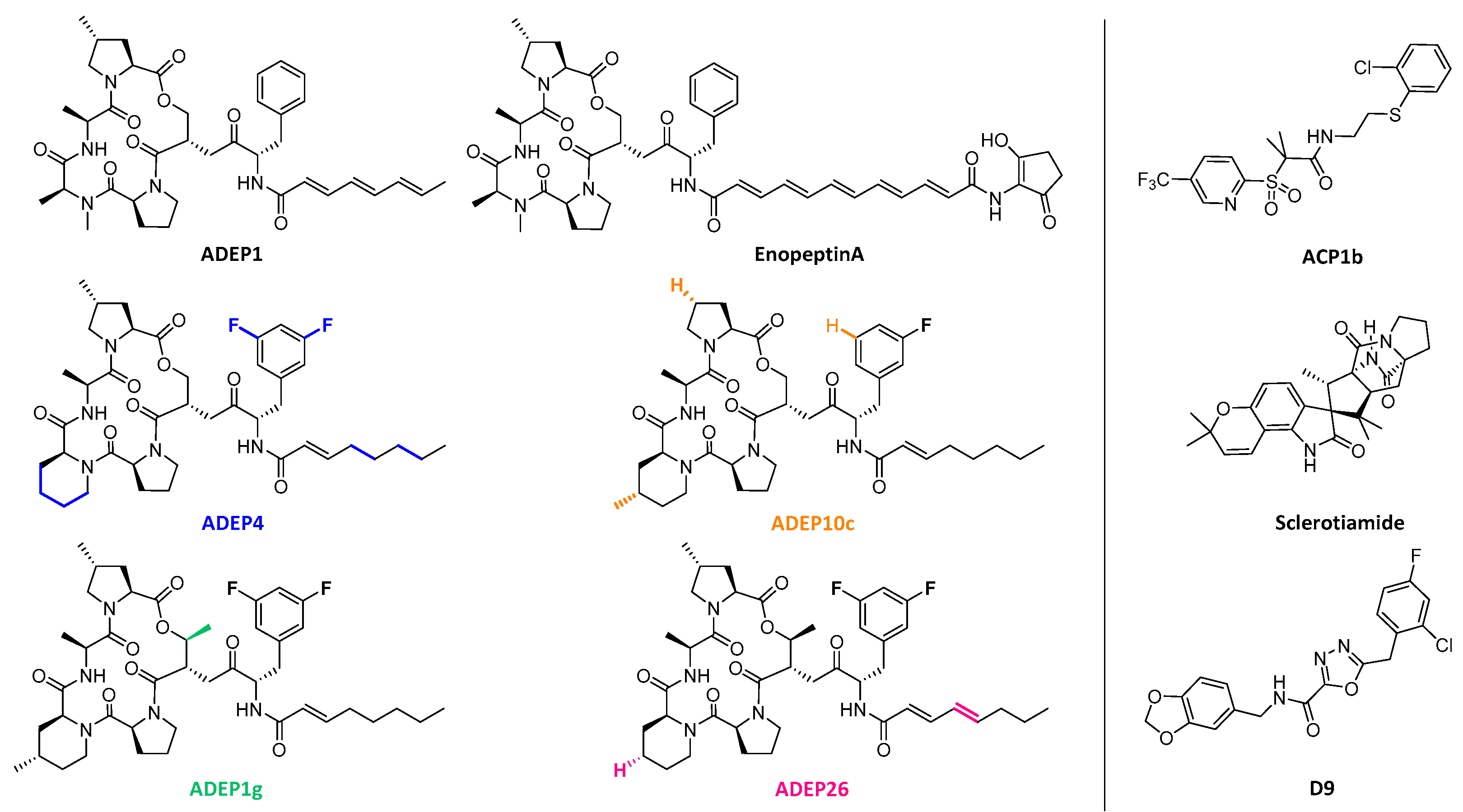
  74. Brotz-Oesterhelt, H.; Beyer, D.; Kroll, H.P.; Endermann, R.; Ladel, C.; Schroeder, W.; Hinzen, B.; Raddatz, S.; Paulsen, H.; Henninger, K.; et al. Dysregulation of bacterial proteolytic machinery by a new class of antibiotics. Nat. Med. 2005, 11, 1082–1087. [Google Scholar] [CrossRef]
  75. Michel, K.H.; Kastner, R.E. A54556 Antibiotics and Process for Production Thereof. U.S. Patent 4492650, 8 January 1985. [Google Scholar]
  76. Osada, H.; Yano, T.; Koshino, H.; Isono, K. Enopeptin A, a novel depsipeptide antibiotic with anti-bacteriophage activity. J. Antibiot. 1991, 44, 1463–1466. [Google Scholar] [CrossRef]
  77. Li, D.H.; Chung, Y.S.; Gloyd, M.; Joseph, E.; Ghirlando, R.; Wright, G.D.; Cheng, Y.Q.; Maurizi, M.R.; Guarne, A.; Ortega, J. Acyldepsipeptide antibiotics induce the formation of a structured axial channel in ClpP: A model for the ClpX/ClpA-bound state of ClpP. Chem. Biol. 2010, 17, 959–969. [Google Scholar] [CrossRef]
  78. Lee, B.G.; Park, E.Y.; Lee, K.E.; Jeon, H.; Sung, K.H.; Paulsen, H.; Rubsamen-Schaeff, H.; Brotz-Oesterhelt, H.; Song, H.K. Structures of ClpP in complex with acyldepsipeptide antibiotics reveal its activation mechanism. Nat. Struct. Mol. Biol. 2010, 17, 471–478. [Google Scholar] [CrossRef]
  79. Malik, I.T.; Brotz-Oesterhelt, H. Conformational control of the bacterial Clp protease by natural product antibiotics. Nat. Prod. Rep. 2017, 34, 815–831. [Google Scholar] [CrossRef] [Green Version]
  80. Hinzen, B.; Raddatz, S.; Paulsen, H.; Lampe, T.; Schumacher, A.; Habich, D.; Hellwig, V.; Benet-Buchholz, J.; Endermann, R.; Labischinski, H.; et al. Medicinal chemistry optimization of acyldepsipeptides of the enopeptin class antibiotics. Chemmedchem 2006, 1, 689–693. [Google Scholar] [CrossRef]
  81. Famulla, K.; Sass, P.; Malik, I.; Akopian, T.; Kandror, O.; Alber, M.; Hinzen, B.; Ruebsamen-Schaeff, H.; Kalscheuer, R.; Goldberg, A.L.; et al. Acyldepsipeptide antibiotics kill mycobacteria by preventing the physiological functions of the ClpP1P2 protease. Mol. Microbiol. 2016, 101, 194–209. [Google Scholar] [CrossRef] [Green Version]
  82. Conlon, B.P.; Nakayasu, E.S.; Fleck, L.E.; LaFleur, M.D.; Isabella, V.M.; Coleman, K.; Leonard, S.N.; Smith, R.D.; Adkins, J.N.; Lewis, K. Activated ClpP kills persisters and eradicates a chronic biofilm infection. Nature 2013, 503, 365–370. [Google Scholar] [CrossRef]
  83. Socha, A.M.; Tan, N.Y.; LaPlante, K.L.; Sello, J.K. Diversity-oriented synthesis of cyclic acyldepsipeptides leads to the discovery of a potent antibacterial agent. Bioorg. Med. Chem. 2010, 18, 7193–7202. [Google Scholar] [CrossRef]
  84. Carney, D.W.; Schmitz, K.R.; Truong, J.V.; Sauer, R.T.; Sello, J.K. Restriction of the conformational dynamics of the cyclic acyldepsipeptide antibiotics improves their antibacterial activity. J. Am. Chem. Soc. 2014, 136, 1922–1929. [Google Scholar] [CrossRef]
  85. Goodreid, J.D.; Janetzko, J.; Santa Maria, J.P., Jr.; Wong, K.S.; Leung, E.; Eger, B.T.; Bryson, S.; Pai, E.F.; Gray-Owen, S.D.; Walker, S.; et al. Development and characterization of potent cyclic acyldepsipeptide analogues with increased antimicrobial activity. J. Med. Chem. 2016, 59, 624–646. [Google Scholar] [CrossRef]
  86. Leung, E.; Datti, A.; Cossette, M.; Goodreid, J.; McCaw, S.E.; Mah, M.; Nakhamchik, A.; Ogata, K.; El Bakkouri, M.; Cheng, Y.Q.; et al. Activators of cylindrical proteases as antimicrobials: Identification and development of small molecule activators of ClpP protease. Chem. Biol. 2011, 18, 1167–1178. [Google Scholar] [CrossRef]
  87. Lavey, N.P.; Coker, J.A.; Ruben, E.A.; Duerfeldt, A.S. Sclerotiamide: The first non-peptide-based natural product activator of bacterial caseinolytic protease P. J. Nat. Prod. 2016, 79, 1193–1197. [Google Scholar] [CrossRef]
  88. Stahl, M.; Korotkov, V.S.; Balogh, D.; Kick, L.M.; Gersch, M.; Pahl, A.; Kielkowski, P.; Richter, K.; Schneider, S.; Sieber, S.A. Selective activation of human caseinolytic protease P (ClpP). Angew. Chem. Int. Ed. Engl. 2018, 57, 14602–14607. [Google Scholar] [CrossRef]
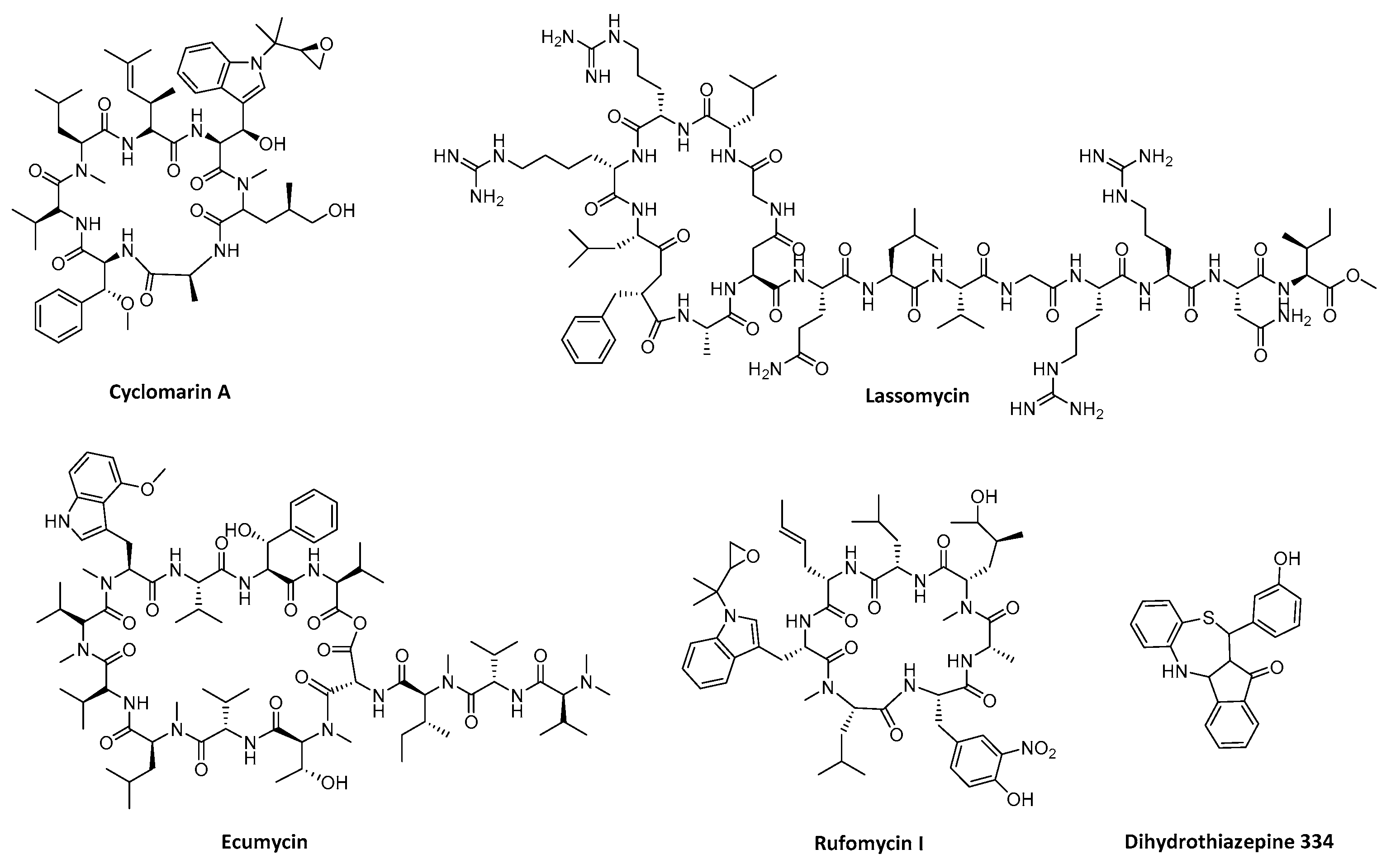
  89. Schmitt, E.K.; Riwanto, M.; Sambandamurthy, V.; Roggo, S.; Miault, C.; Zwingelstein, C.; Krastel, P.; Noble, C.; Beer, D.; Rao, S.P.; et al. The natural product cyclomarin kills Mycobacterium tuberculosis by targeting the ClpC1 subunit of the caseinolytic protease. Angew. Chem. Int. Ed. Engl. 2011, 50, 5889–5891. [Google Scholar] [CrossRef]
  90. Gavrish, E.; Sit, C.S.; Cao, S.; Kandror, O.; Spoering, A.; Peoples, A.; Ling, L.; Fetterman, A.; Hughes, D.; Bissell, A.; et al. Lassomycin, a ribosomally synthesized cyclic peptide, kills mycobacterium tuberculosis by targeting the ATP-dependent protease ClpC1P1P2. Chem. Biol. 2014, 21, 509–518. [Google Scholar] [CrossRef]
  91. Choules, M.P.; Wolf, N.M.; Lee, H.; Anderson, J.R.; Grzelak, E.M.; Wang, Y.; Ma, R.; Gao, W.; McAlpine, J.B.; Jin, Y.Y.; et al. Rufomycin targets ClpC1 proteolysis in Mycobacterium tuberculosis and M. abscessus. Antimicrob. Agents Chemother. 2019, 63, e02204-18. [Google Scholar] [CrossRef]
  92. Gao, W.; Kim, J.Y.; Anderson, J.R.; Akopian, T.; Hong, S.; Jin, Y.Y.; Kandror, O.; Kim, J.W.; Lee, I.A.; Lee, S.Y.; et al. The cyclic peptide ecumicin targeting ClpC1 is active against Mycobacterium tuberculosis in vivo. Antimicrob. Agents Chemother. 2015, 59, 880–889. [Google Scholar] [CrossRef]
  93. Fetzer, C.; Korotkov, V.S.; Thanert, R.; Lee, K.M.; Neuenschwander, M.; von Kries, J.P.; Medina, E.; Sieber, S.A. A chemical disruptor of the ClpX chaperone complex attenuates the virulence of multidrug-tesistant Staphylococcus aureus. Angew. Chem. Int. Ed. Engl. 2017, 56, 15746–15750. [Google Scholar] [CrossRef]
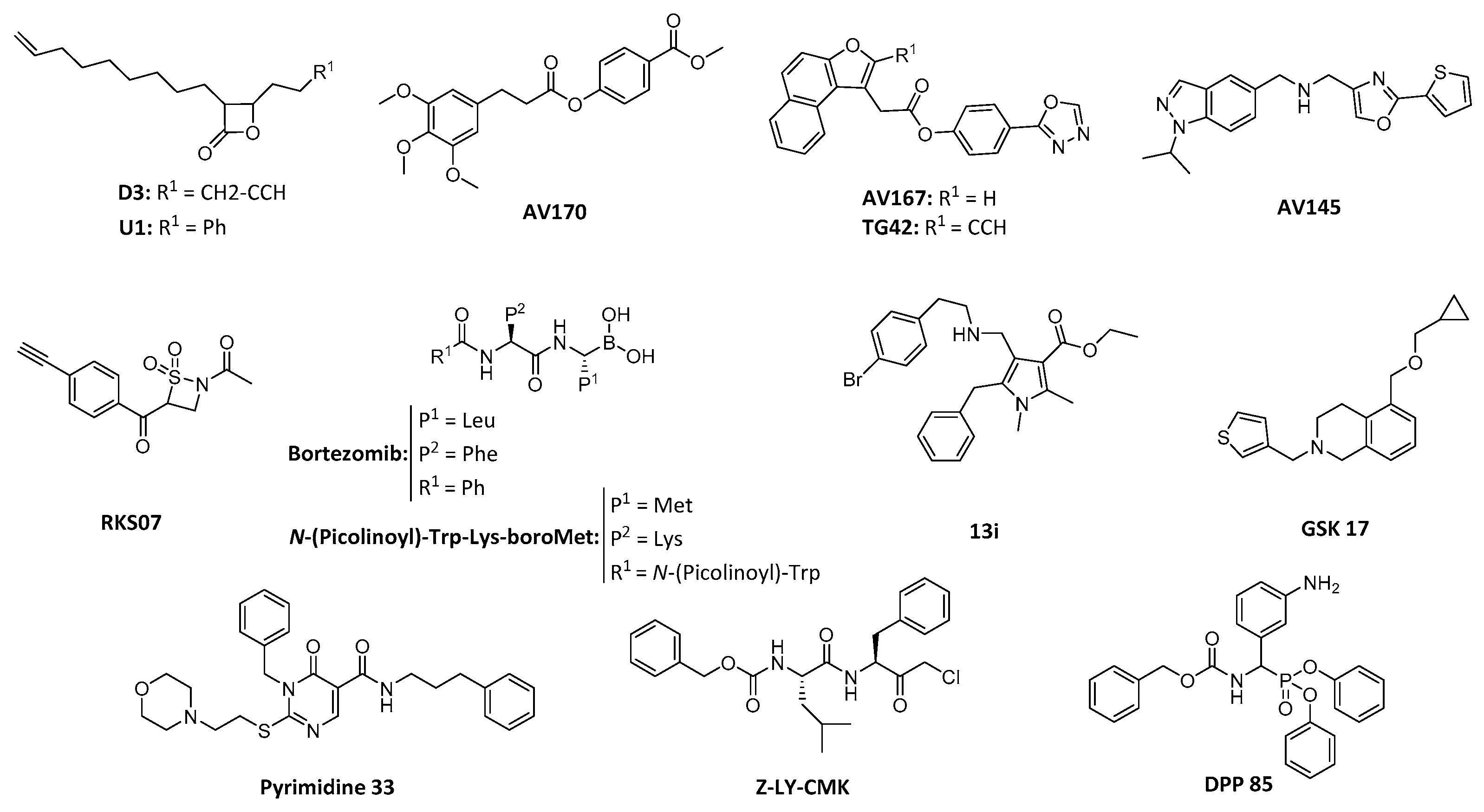
  94. Bottcher, T.; Sieber, S.A. beta-Lactones as privileged structures for the active-site labeling of versatile bacterial. Angew. Chem. Int. Ed. 2008, 47, 4600–4603. [Google Scholar] [CrossRef]
  95. Bottcher, T.; Sieber, S.A. beta-Lactones as specific inhibitors of CIpP attenuate the production of extracellular virulence factors of Staphylococcus aureus. J. Am. Chem. Soc. 2008, 130, 14400–14401. [Google Scholar] [CrossRef]
  96. Bottcher, T.; Sieber, S.A. Structurally refined beta-lactones as potent inhibitors of devastating bacterial virulence factors. Chembiochem 2009, 10, 663–666. [Google Scholar] [CrossRef]
  97. Bottcher, T.; Sieber, S.A. beta-Lactones decrease the Intracellular virulence of Listeria monocytogenes in macrophages. Chemmedchem 2009, 4, 1260–1263. [Google Scholar] [CrossRef]
  98. Compton, C.L.; Schmitz, K.R.; Sauer, R.T.; Sello, J.K. Antibacterial Activity of and Resistance to Small Molecule Inhibitors of the ClpP Peptidase. ACS Chem. Biol. 2013, 8, 2669–2677. [Google Scholar] [CrossRef] [Green Version]
  99. Hackl, M.W.; Lakemeyer, M.; Dahmen, M.; Glaser, M.; Pahl, A.; Lorenz-Baath, K.; Menzel, T.; Sievers, S.; Bottcher, T.; Antes, I.; et al. Phenyl esters are potent inhibitors of caseinolytic protease P and reveal a stereogenic switch for deoligomerization. J. Am. Chem. Soc. 2015, 137, 8475–8483. [Google Scholar] [CrossRef]
  100. Gronauer, T.F.; Mandl, M.M.; Lakemeyer, M.; Hackl, M.W.; Messner, M.; Korotkov, V.S.; Pachmayr, J.; Sieber, S.A. Design and synthesis of tailored human caseinolytic protease P inhibitors. Chem. Commun. 2018, 54, 9833–9836. [Google Scholar] [CrossRef]
  101. Pahl, A.; Lakemeyer, M.; Vielberg, M.T.; Hackl, M.W.; Vomacka, J.; Korotkov, V.S.; Stein, M.L.; Fetzer, C.; Lorenz-Baath, K.; Richter, K.; et al. Reversible Inhibitors Arrest ClpP in a defined conformational state that can be revoked by ClpX association. Angew. Chem. Int. Ed. 2015, 54, 15892–15899. [Google Scholar] [CrossRef]
  102. Gersch, M.; Kolb, R.; Alte, F.; Groll, M.; Sieber, S.A. Disruption of oligomerization and dehydroalanine formation as mechanisms for ClpP protease inhibition. J. Am. Chem. Soc. 2014, 136, 1360–1366. [Google Scholar] [CrossRef]
  103. Moreira, W.; Ngan, G.J.; Low, J.L.; Poulsen, A.; Chia, B.C.; Ang, M.J.; Yap, A.; Fulwood, J.; Lakshmanan, U.; Lim, J.; et al. Target mechanism-based whole-cell screening identifies bortezomib as an inhibitor of caseinolytic protease in mycobacteria. mBio 2015, 6, e00253-15. [Google Scholar] [CrossRef]
  104. Liu, P.; Yang, Y.; Ju, Y.; Tang, Y.; Sang, Z.; Chen, L.; Yang, T.; An, Q.; Zhang, T.; Luo, Y. Design, synthesis and biological evaluation of novel pyrrole derivatives as potential ClpP1P2 inhibitor against Mycobacterium tuberculosis. Bioorg. Chem. 2018, 80, 422–432. [Google Scholar] [CrossRef]
  105. Fraga, H.; Rodriguez, B.; Bardera, A.; Cid, C.; Akopian, T.; Kandror, O.; Park, A.; Colmenarejo, G.; Lelievre, J.; Goldberg, A. Development of high throughput screening methods for inhibitors of ClpC1P1P2 from Mycobacteria tuberculosis. Anal. Biochem. 2019, 567, 30–37. [Google Scholar] [CrossRef]
  106. Mundra, S.; Thakur, V.; Bello, A.M.; Rathore, S.; Asad, M.; Wei, L.; Yang, J.; Chakka, S.K.; Mahesh, R.; Malhotra, P.; et al. A novel class of Plasmodial ClpP protease inhibitors as potential antimalarial agents. Bioorg. Med. Chem. 2017, 25, 5662–5677. [Google Scholar] [CrossRef]
  107. Szyk, A.; Maurizi, M.R. Crystal structure at 1.9A of E. coli ClpP with a peptide covalently bound at the active site. J. Struct. Biol. 2006, 156, 165–174. [Google Scholar] [CrossRef]
  108. Moreno-Cinos, C.; Sassetti, E.; Salado, I.G.; Witt, G.; Benramdane, S.; Reinhardt, L.; DCruz, C.; Joossens, J.; Van der Veken, P.; Brötz-Oesterhelt, H.; et al. alpha-Amino diphenyl phosphonates as novel inhibitors of Escherichia coli ClpP protease. J. Med. Chem. 2019, 62, 774–797. [Google Scholar] [CrossRef]
  109. Lamden, L.A.; Bartlett, F.A. Aminoalkylphosphonofluoridate derivatives: Rapid and potentially selective inactivators of serine peptidases. Biochem. Biophys. Res. Commun. 1983, 112, 1085–1090. [Google Scholar] [CrossRef]
  110. Joossens, J.; Van der Veken, P.; Surpateanu, G.; Lambeir, A.M.; El-Sayed, I.; Ali, O.M.; Augustyns, K.; Haemers, A. Diphenyl phosphonate inhibitors for the urokinase-type plasminogen activator: Optimization of the P4 position. J. Med. Chem. 2006, 49, 5785–5793. [Google Scholar] [CrossRef]
  111. Van der Veken, P.; Soroka, A.; Brandt, I.; Chen, Y.S.; Maes, M.B.; Lambeir, A.M.; Chen, X.; Haemers, A.; Scharpe, S.; Augustyns, K.; et al. Irreversible inhibition of dipeptidyl peptidase 8 by dipeptide-derived diaryl phosphonates. J. Med. Chem. 2007, 50, 5568–5570. [Google Scholar] [CrossRef]
  112. Winiarski, L.; Oleksyszyn, J.; Sienczyk, M. Human neutrophil elastase phosphonic inhibitors with improved potency of action. J. Med. Chem. 2012, 55, 6541–6553. [Google Scholar] [CrossRef]
  113. Pietrusewicz, E.; Sienczyk, M.; Oleksyszyn, J. Novel diphenyl esters of peptidyl alpha-aminoalkylphosphonates as inhibitors of chymotrypsin and subtilisin. J. Enzyme Inhib. Med. Chem. 2009, 24, 1229–1236. [Google Scholar] [CrossRef]
  114. Burchacka, E.; Zdzalik, M.; Niemczyk, J.S.; Pustelny, K.; Popowicz, G.; Wladyka, B.; Dubin, A.; Potempa, J.; Sienczyk, M.; Dubin, G.; et al. Development and binding characteristics of phosphonate inhibitors of SplA protease from Staphylococcus aureus. Protein Sci. 2014, 23, 179–189. [Google Scholar] [CrossRef]
  115. Dickey, S.W.; Cheung GY, C.; Otto, M. Different drugs for bad bugs: Antivirulence strategies in the age of antibiotic resistance. Nat. Rev. Drug Discov. 2017, 16, 457–471. [Google Scholar] [CrossRef]
  116. Defoirdt, T. Quorum-sensing systems as targets for antivirulence therapy. Trends Microbiol. 2018, 26, 313–328. [Google Scholar] [CrossRef]
  117. Koo, H.; Allan, R.N.; Howlin, R.P.; Stoodley, P.; Hall-Stoodley, L. Targeting microbial biofilms: Current and prospective therapeutic strategies. Nat. Rev. Microbiol. 2017, 15, 740–755. [Google Scholar] [CrossRef]
  118. Rice, L.B. Federal funding for the study of antimicrobial resistance in nosocomial pathogens: No ESKAPE. J. Infect. Dis. 2008, 197, 1079–1081. [Google Scholar] [CrossRef]
Figure 1. Cycle of ATPases associated with diverse cellular activities (AAA+) caseinolytic protease proteolytic subunit (ClpP) complex. (A) Substrate selective binding by the AAA+ unfoldase. (B) Sequence of ATP-driven stretching, leading to a strained structure or alternatively to a substrate release. (C) Successful unfolding leading to translocations and degradation. (D) Initiation of a new cycle.
Ijms 20 02232 g001
Figure 2. Structure of the full E. coli ClpP tetradecamer (top: side view, bottom left: top view) and the ClpP monomer (bottom right). Every monomer is shown in a different color in cartoon representation. The monomer is shown to emphasize the active site and the catalytic triad (S172, H122, D171) is shown in grey stick representation. (PDB ID: 1YG6).
Ijms 20 02232 g002
Figure 3. Top and side view of E. coli caseinolytic protease subunit A (ClpA) (top) and E. coli ClpX (bottom) hexamers. The ClpA hexamer is shown in grey and wheat surface representation. The structure was obtained through homology modeling, using the B. subtilis caseinolytic protease subunit C (ClpC) monomer as template (PDB ID: 3PXI) in SWISS-MODEL [42]. The subunits in the ClpC hexamer (PDB ID: 3PXG) where then replaced with the ClpA monomers, and the structure was then relaxed through energy minimization. The caseinolytic protease subunit X (ClpX) hexamer is shown in cyan and light cyan surface representation. Missing loops were modeled using Modeller 9.17 [43].
Ijms 20 02232 g003
Figure 4. Top view of E. coli ClpP in unbound state (left) (PDB ID: 1YG6) and bound with acyldepsipeptide (ADEP)1 (right) (PDB ID: 3MT6). When bound, an increase in pore size can be observed which defines an active, non-selective conformation. The protein is shown in grey surface representation, with the ADEP1 binding pockets shown in pink. ADEP1 molecules are represented by white spheres.
Ijms 20 02232 g004
Figure 5. Chemical structures of ClpP activators. On the left, chemical structures of the mentioned ADEPs are shown. In the color matching their name, consecutive modifications respect to the previous ADEP are highlighted: the original ADEP1 and EnopeptinA (black), ADEP4 (blue), ADEP10c (orange), ADEP1g (green) and ADEP26 (pink). On the right side, alternative cores extracted from screening including ACP1b, Sclerotiamide and D9.
Ijms 20 02232 g005
Figure 6. Chemical structures of compounds targeting ATP-unfoldases.
Ijms 20 02232 g006
Figure 7. Chemical structure of reported ClpP inhibitors.
Ijms 20 02232 g007
Figure 8. (A) Mechanism of action of RKS07 [102], a β-sultam undergoing sulfonylation and subsequent elimination to transform the catalytic serine into an inactive dehydroalanine. (B) Inhibitory mechanism of a diphenyl phosphonate inhibitor with a serine protease. (I) The unreacted inhibitor enters the active site, with the R1 moiety filling the S1 (recognition) pocket while the phosphonate stays at a reachable distance from the catalytic serine and the oxyanion hole. (II) Nucleophilic attack of the serine to the phosphonate induced by the interaction with the oxyanion hole residues, forming the pentacoordinate transition state. (III) Release of the phenolate group due to the recovery of tetrahedral geometry. (IV) Final configuration leading to irreversible inhibition of the serine protease.
Ijms 20 02232 g008

