Low Intensity Pulsed Ultrasound Prevents Recurrent Ischemic Stroke in a Cerebral Ischemia/Reperfusion Injury Mouse Model via Brain-derived Neurotrophic Factor Induction

 , and
, and 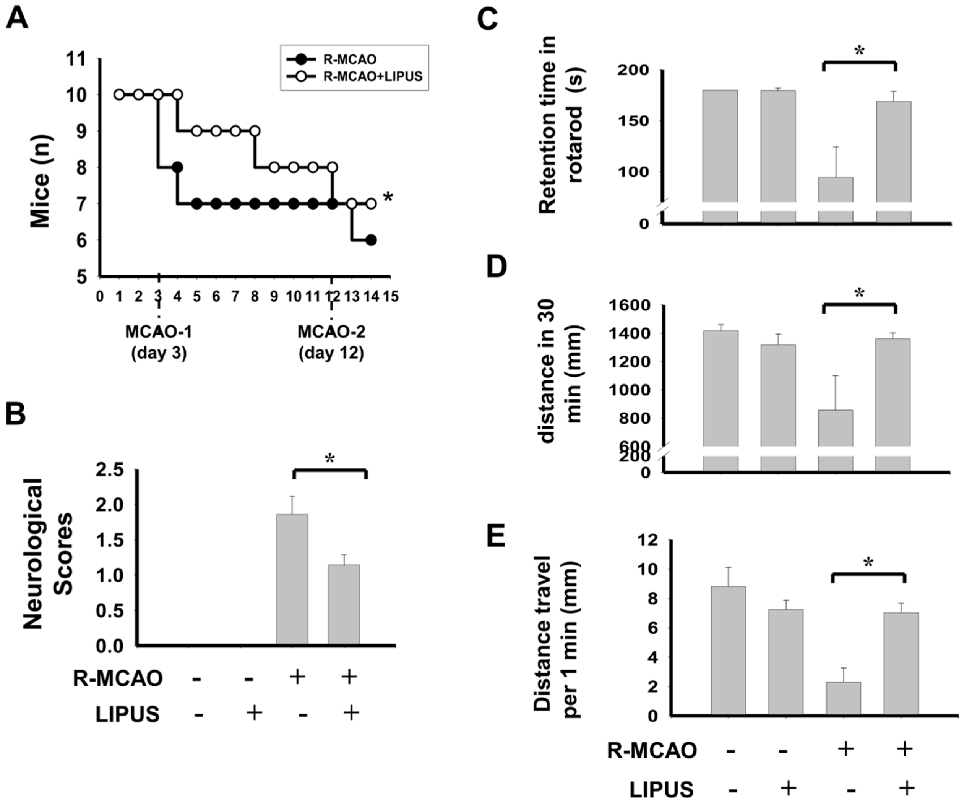
Abstract
:1. Introduction
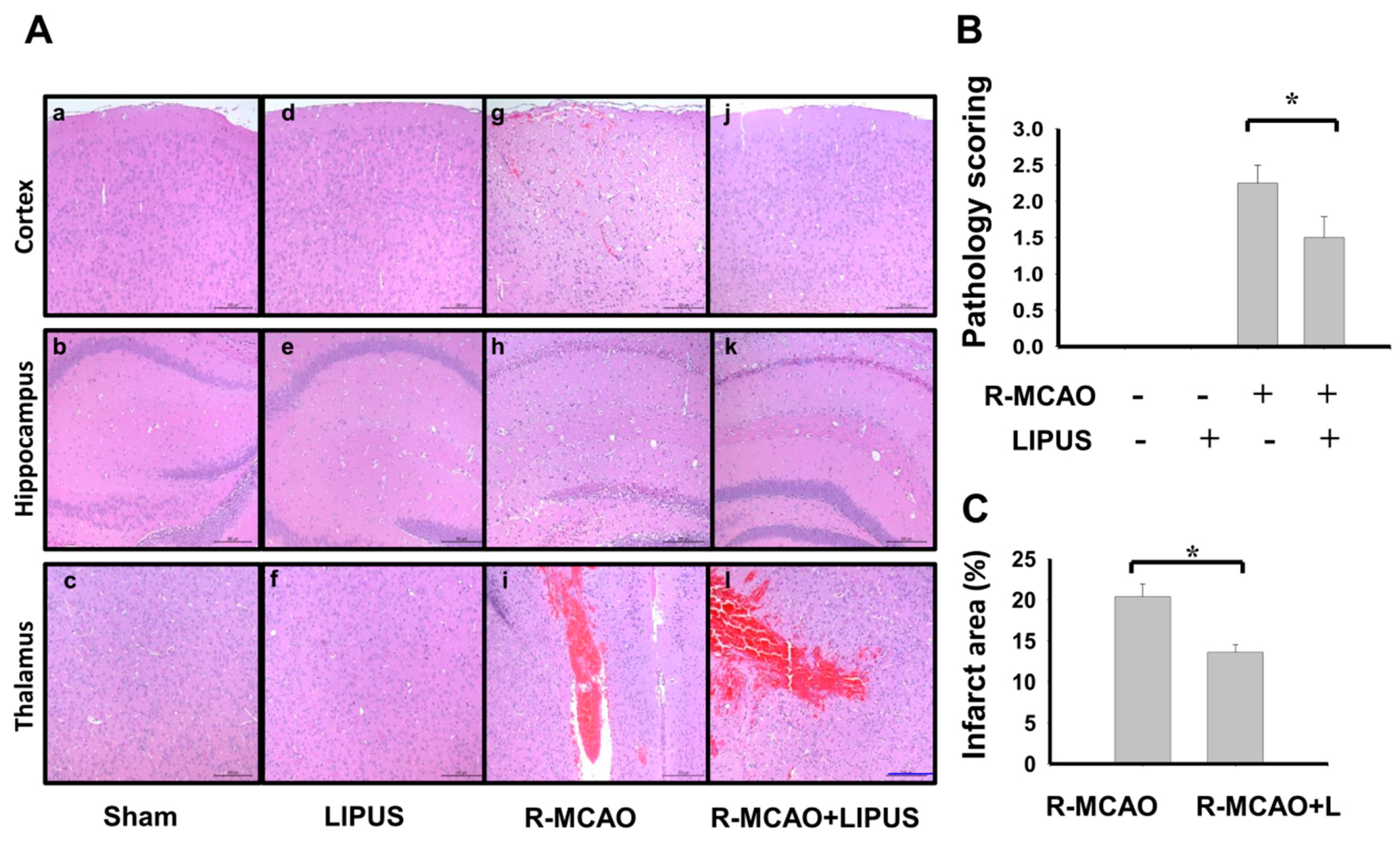
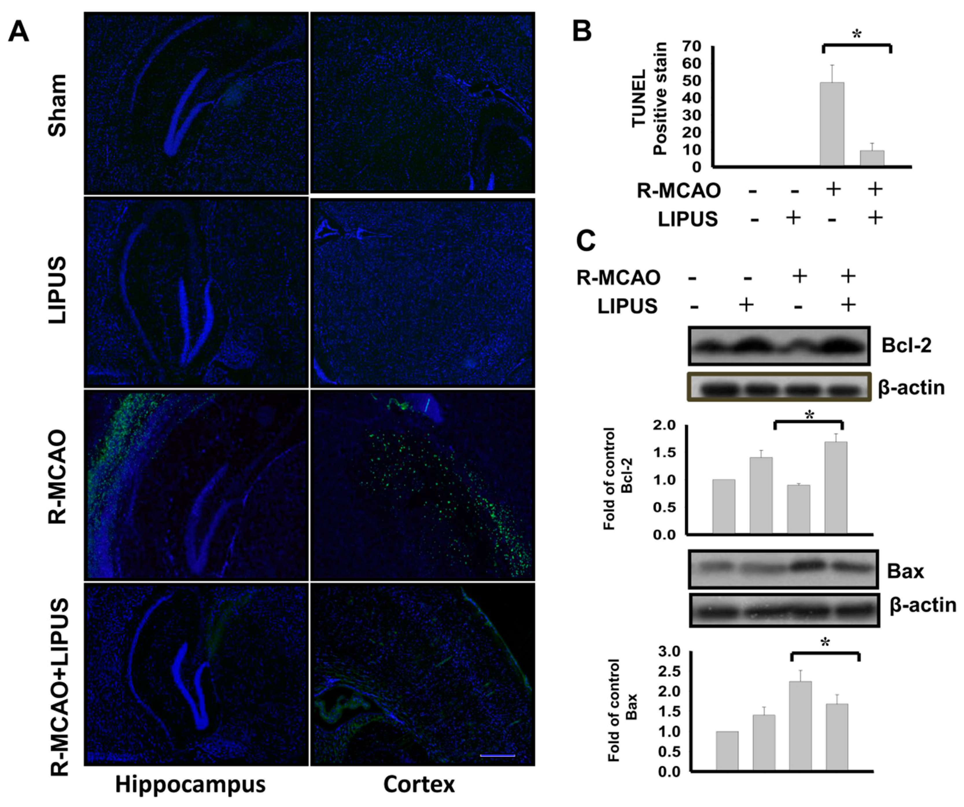
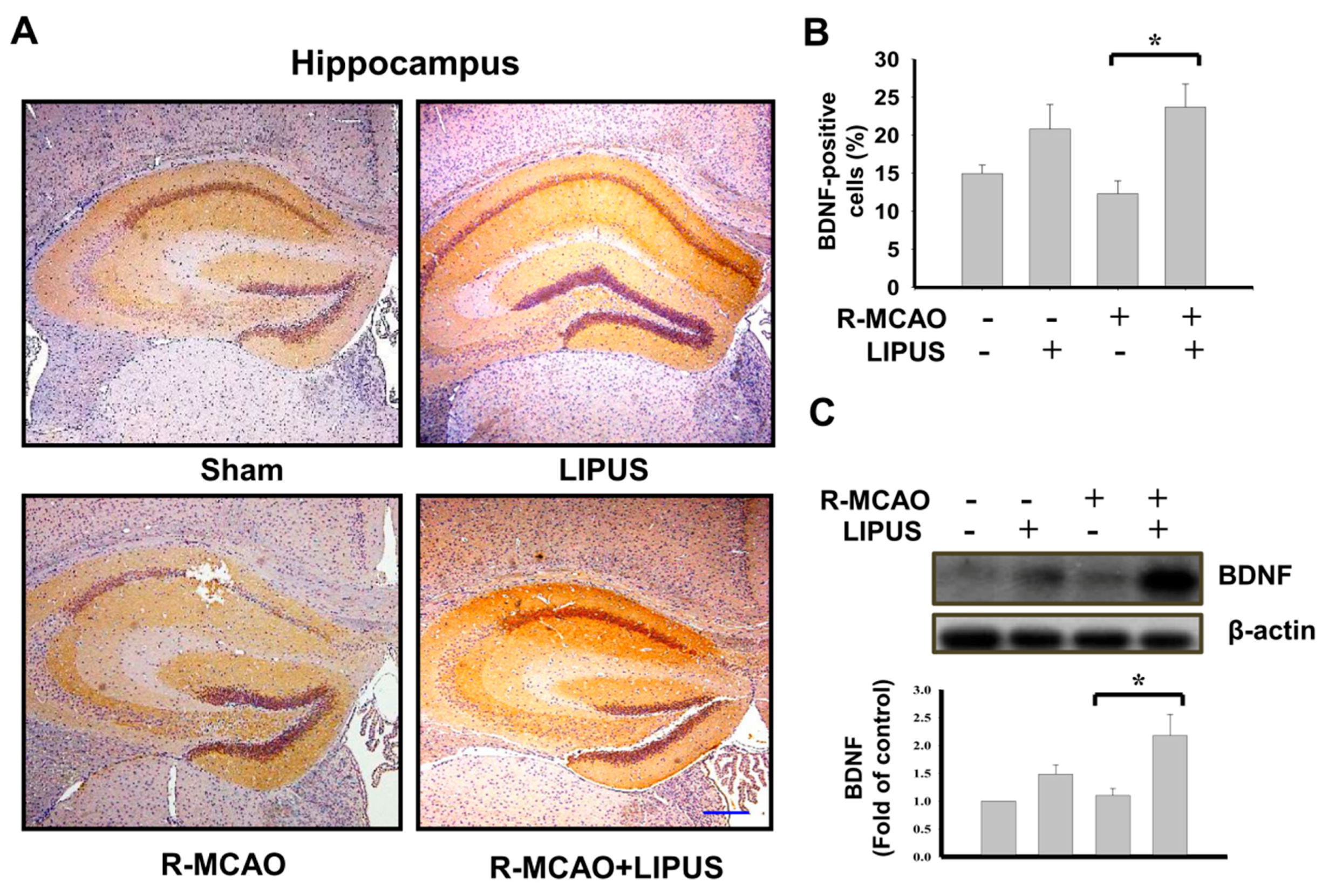
2. Results
3. Discussion
4. Materials and Methods
4.1. Animals and Middle Cerebral Artery Occlusion (MCAO) Recurrent Surgical Procedure
4.2. LIPUS Treatment
4.3. Neurological Function Assessment
4.4. Locomotor Activity Evaluation
4.5. Assessment of Motor Equilibrium Performance on Rotarod
4.6. Infarct Volume Detection
4.7. Histopathological and Immunohistochemical Analysis
4.8. Western Blotting
4.9. Fluorescent Terminal Deoxynucleotidyl Transferase (TdT) dUTP Nick End Labeling (TUNEL) Staining
4.10. Statistics
5. Conclusions
Author Contributions
Funding
Conflicts of Interest
Abbreviations
| LIPUS | Low intensity pulsed ultrasound |
| BDNF | Brain-derived neurotrophic factor |
| MCAO | Middle cerebral artery occlusion |
| TUNEL | Terminal deoxynucleotidyl transferase (TdT) dUTP nick end labeling |
References
- WHO. The Top 10 Causes of Death. Available online: https://www.who.int/news-room/fact-sheets/detail/the-top-10-causes-of-death (accessed on 1 August 2018).
- Johnson, W.; Onuma, O.; Owolabi, M.; Sachdev, S. Stroke: A global response is needed. Bull. World Health Organ. 2016, 94, 634–634A. [Google Scholar] [CrossRef]
- Bergstrom, L.; Irewall, A.L.; Soderstrom, L.; Ogren, J.; Laurell, K.; Mooe, T. One-year incidence, time trends, and predictors of recurrent ischemic stroke in sweden From 1998 to 2010: An observational study. Stroke 2017, 48, 2046–2051. [Google Scholar] [CrossRef]
- Feigin, V.L.; Mensah, G.A.; Norrving, B.; Murray, C.J.; Roth, G.A. Atlas of the global burden of stroke (1990–2013): The GBD 2013 Study. Neuroepidemiology 2015, 45, 230–236. [Google Scholar] [CrossRef]
- Siragusa, S.; Malato, A.; Saccullo, G.; Iorio, A.; Di Ianni, M.; Caracciolo, C.; Coco, L.L.; Raso, S.; Santoro, M.; Guarneri, F.P.; et al. Residual vein thrombosis for assessing duration of anticoagulation after unprovoked deep vein thrombosis of the lower limbs: The extended DACUS study. Am. J. Hematol. 2011, 86, 914–917. [Google Scholar] [CrossRef] [Green Version]
- Conte, G.; Rigon, N.; Perrone, A.; Lauro, S. Acute cardiovascular diseases and respiratory sleep disorders. Minerva Cardioangiol. 1999, 47, 195–202. [Google Scholar]
- Tuttolomondo, A.; Maida, C.; Pinto, A. Diabetic foot syndrome as a possible cardiovascular marker in diabetic patients. J. Diabetes Res. 2015, 2015, 268390. [Google Scholar] [CrossRef]
- Oza, R.; Rundell, K.; Garcellano, M. Recurrent ischemic stroke: Strategies for prevention. Am. Fam. Physician 2017, 96, 436–440. [Google Scholar]
- Yamamoto, H.; Bogousslavsky, J. Mechanisms of second and further strokes. J. Neurol. Neurosurg. Psychiatry 1998, 64, 771–776. [Google Scholar] [CrossRef]
- Ward, R.S.; Hayes-Lundy, C.; Reddy, R.; Brockway, C.; Mills, P.; Saffle, J.R. Evaluation of topical therapeutic ultrasound to improve response to physical therapy and lessen scar contracture after burn injury. J. Burn Care Rehabil. 1994, 15, 74–79. [Google Scholar] [CrossRef]
- Miller, D.L.; Smith, N.B.; Bailey, M.R.; Czarnota, G.J.; Hynynen, K.; Makin, I.R. Overview of therapeutic ultrasound applications and safety considerations. J. Ultrasound Med. 2012, 31, 623–634. [Google Scholar] [CrossRef]
- Piper, R.J.; Hughes, M.A.; Moran, C.M.; Kandasamy, J. Focused ultrasound as a non-invasive intervention for neurological disease: A review. Br. J. Neurosurg. 2016, 30, 286–293. [Google Scholar] [CrossRef] [PubMed]
- Lou, S.; Lv, H.; Li, Z.; Tang, P.; Wang, Y. Effect of low-intensity pulsed ultrasound on distraction osteogenesis: A systematic review and meta-analysis of randomized controlled trials. J. Orthop. Surg. Res. 2018, 13, 205. [Google Scholar] [CrossRef] [PubMed]
- Zhao, L.; Feng, Y.; Shi, A.; Zhang, L.; Guo, S.; Wan, M. Neuroprotective effect of low-intensity pulsed ultrasound against MPP(+)-induced neurotoxicity in PC12 cells: Involvement of K2P channels and stretch-activated ion channels. Ultrasound Med. Biol. 2017, 43, 1986–1999. [Google Scholar] [CrossRef] [PubMed]
- Su, W.S.; Wu, C.H.; Chen, S.F.; Yang, F.Y. Low-intensity pulsed ultrasound improves behavioral and histological outcomes after experimental traumatic brain injury. Sci. Rep. 2017, 7, 15524. [Google Scholar] [CrossRef]
- Rodriguez-Frutos, B.; Otero-Ortega, L.; Ramos-Cejudo, J.; Martinez-Sanchez, P.; Barahona-Sanz, I.; Navarro-Hernanz, T.; Gomez-de Frutos Mdel, C.; Diez-Tejedor, E.; Gutierrez-Fernandez, M. Enhanced brain-derived neurotrophic factor delivery by ultrasound and microbubbles promotes white matter repair after stroke. Biomaterials 2016, 100, 41–52. [Google Scholar] [CrossRef] [Green Version]
- Lin, W.T.; Chen, R.C.; Lu, W.W.; Liu, S.H.; Yang, F.Y. Protective effects of low-intensity pulsed ultrasound on aluminum-induced cerebral damage in Alzheimer’s disease rat model. Sci. Rep. 2015, 5, 9671. [Google Scholar] [CrossRef]
- Liu, S.H.; Lai, Y.L.; Chen, B.L.; Yang, F.Y. Ultrasound enhances the expression of brain-derived neurotrophic factor in astrocyte through activation of TrkB-Akt and calcium-CaMK signaling pathways. Cereb. Cortex 2017, 27, 3152–3160. [Google Scholar] [CrossRef]
- Chen, T.T.; Lan, T.H.; Yang, F.Y. Low-intensity pulsed ultrasound attenuates LPS-induced neuroinflammation and memory impairment by modulation of TLR4/NF-κB signaling and CREB/BDNF expression. Cereb. Cortex 2019, 29, 1430–1438. [Google Scholar] [CrossRef]
- Li, J.; Zhang, D.D.; Wang, C.Q.; Shi, M.; Wang, L.L. Protective effects of low-intensity pulsed ultrasound on aluminum overload-induced cerebral damage through epigenetic regulation of brain-derived neurotrophic factor expression. Biosci. Rep. 2019, 39, BSR20181185. [Google Scholar] [CrossRef]
- Chen, C.M.; Wu, C.T.; Yang, T.H.; Liu, S.H.; Yang, F.Y. Preventive effect of low intensity pulsed ultrasound against experimental cerebral ischemia/reperfusion injury via apoptosis reduction and brain-derived neurotrophic factor induction. Sci. Rep. 2018, 8, 5568. [Google Scholar] [CrossRef]
- Liu, L.; Du, J.; Zheng, T.; Hu, S.; Zhao, M.; Wang, X.; Wu, S.; Shi, Q. Readout-segmented echo-planar diffusion-weighted MR at 3.0T for the evaluation the effect of low-intensity transcranial ultrasound on stroke in a rat model. Magn. Reson. Imaging 2019. [Google Scholar] [CrossRef] [PubMed]
- Adams, H.P.; Bendixen, B.H.; Kappelle, L.J.; Biller, J.; Love, B.B.; Gordon, D.L.; Marsh, E.E. Classification of subtype of acute ischemic stroke. Definitions for use in a multicenter clinical trial. TOAST. Trial of ORG 10172 in Acute Stroke Treatment. Stroke 1993, 24, 35–41. [Google Scholar] [CrossRef] [PubMed]
- Chung, J.W.; Park, S.H.; Kim, N.; Kim, W.J.; Park, J.H.; Ko, Y.; Yang, M.H.; Jang, M.S.; Han, M.K.; Jung, C.; et al. Trial of ORG 10172 in Acute Stroke Treatment (TOAST) classification and vascular territory of ischemic stroke lesions diagnosed by diffusion-weighted imaging. J. Am. Heart Assoc. 2014, 3, e001119. [Google Scholar] [CrossRef] [PubMed]
- Kocaman, G.; Dürüyen, H.; Koçer, A.; Asil, T. Recurrent ischemic stroke characteristics and assessment of sufficiency of secondary stroke prevention. Noro Psikiyatr Ars 2015, 52, 139–144. [Google Scholar] [CrossRef]
- Yang, X.; Liu, M.; Zhu, Y.; Zhang, X.; Gao, S.; Ni, J. An overlooked cause in a patient with recurrent ischemic stroke: A case report. Med. (Baltim.) 2018, 97, e0146. [Google Scholar] [CrossRef]
- Qiao, M.; Zhao, Z.; Barber, P.A.; Foniok, T.; Sun, S.; Tuor, U.I. Development of a model of recurrent stroke consisting of a mild transient stroke followed by a second moderate stroke in rats. J. Neurosci. Methods 2009, 184, 244–250. [Google Scholar] [CrossRef]
- Hill, M.D.; Yiannakoulias, N.; Jeerakathil, T.; Tu, J.V.; Svenson, L.W.; Schopflocher, D.P. The high risk of stroke immediately after transient ischemic attack: A population-based study. Neurology 2004, 62, 2015–2020. [Google Scholar] [CrossRef]
- Johnston, D.C.; Hill, M.D. The patient with transient cerebral ischemia: A golden opportunity for stroke prevention. CMAJ 2004, 170, 1134–1137. [Google Scholar] [CrossRef]
- Nguyen-Huynh, M.N.; Johnston, S.C. Evaluation and management of transient ischemic attack: An important component of stroke prevention. Nat. Clin. Pr. Cardiovasc Med. 2007, 4, 310–318. [Google Scholar] [CrossRef]
- Chen, J.; Zhang, X.; Zhang, C.; Wang, W.; Chen, R.; Jiao, H.; Li, L.; Zhang, L.; Cui, L. Anti-inflammation of natural components from medicinal plants at low concentrations in brain via inhibiting Neutrophil Infiltration after Stroke. Mediat. Inflamm. 2016, 2016, 9537901. [Google Scholar] [CrossRef]
- Guo, T.; Li, H.; Lv, Y.; Lu, H.; Niu, J.; Sun, J.; Yang, G.Y.; Ren, C.; Tong, S. Pulsed transcranial ultrasound stimulation immediately after the ischemic brain injury is neuroprotective. IEEE Trans. Biomed. Eng. 2015, 62, 2352–2357. [Google Scholar] [CrossRef] [PubMed]
- Radak, D.; Katsiki, N.; Resanovic, I.; Jovanovic, A.; Sudar-Milovanovic, E.; Zafirovic, S.; Mousad, S.A.; Isenovic, E.R. Apoptosis and acute brain ischemia in ischemic stroke. Curr. Vasc. Pharm. 2017, 15, 115–122. [Google Scholar] [CrossRef] [PubMed]
- Korsmeyer, S.J.; Shutter, J.R.; Veis, D.J.; Merry, D.E.; Oltvai, Z.N. Bcl-2/Bax: A rheostat that regulates an anti-oxidant pathway and cell death. Semin. Cancer Biol. 1993, 4, 327–332. [Google Scholar] [PubMed]
- Li, J.; Zhang, S.; Li, C.; Li, M.; Ma, L. Sitagliptin rescues memory deficits in Parkinsonian rats via upregulating BDNF to prevent neuron and dendritic spine loss. Neurol. Res. 2018, 40, 736–743. [Google Scholar] [CrossRef]
- Bomba, M.; Granzotto, A.; Castelli, V.; Onofrj, M.; Lattanzio, R.; Cimini, A.; Sensi, S.L. Exenatide reverts the high-fat-diet-induced impairment of BDNF signaling and inflammatory response in an animal model of Alzheimer’s disease. J. Alzheimers Dis. 2019, 70, 793–810. [Google Scholar] [CrossRef]
- Luo, L.; Li, C.; Du, X.; Shi, Q.; Huang, Q.; Xu, X.; Wang, Q. Effect of aerobic exercise on BDNF/proBDNF expression in the ischemic hippocampus and depression recovery of rats after stroke. Behav. Brain Res. 2019, 362, 323–331. [Google Scholar] [CrossRef]
- Eleftheriou, D.; Ganesan, V.; Hong, Y.; Klein, N.J.; Brogan, P.A. Endothelial repair in childhood arterial ischaemic stroke with cerebral arteriopathy. Cereb. Dis. Extra 2015, 5, 68–74. [Google Scholar] [CrossRef]
- Mang, C.S.; Campbell, K.L.; Ross, C.J.; Boyd, L.A. Promoting neuroplasticity for motor rehabilitation after stroke: Considering the effects of aerobic exercise and genetic variation on brain-derived neurotrophic factor. Phys. Ther. 2013, 93, 1707–1716. [Google Scholar] [CrossRef]
- Su, Q.; Cheng, Y.; Jin, K.; Cheng, J.; Lin, Y.; Lin, Z.; Wang, L.; Shao, B. Estrogen therapy increases BDNF expression and improves post-stroke depression in ovariectomy-treated rats. Exp. Med. 2016, 12, 1843–1848. [Google Scholar] [CrossRef]
- Schabitz, W.R.; Steigleder, T.; Cooper-Kuhn, C.M.; Schwab, S.; Sommer, C.; Schneider, A.; Kuhn, H.G. Intravenous brain-derived neurotrophic factor enhances poststroke sensorimotor recovery and stimulates neurogenesis. Stroke 2007, 38, 2165–2172. [Google Scholar] [CrossRef]
- Kermani, P.; Hempstead, B. Brain-derived neurotrophic factor: A newly described mediator of angiogenesis. Trends Cardiovasc Med. 2007, 17, 140–143. [Google Scholar] [CrossRef] [PubMed]
- Ko, I.G.; Shin, M.S.; Kim, B.K.; Kim, S.E.; Sung, Y.H.; Kim, T.S.; Shin, M.C.; Cho, H.J.; Kim, S.C.; Kim, S.H.; et al. Tadalafil improves short-term memory by suppressing ischemia-induced apoptosis of hippocampal neuronal cells in gerbils. Pharm. Biochem. Behav. 2009, 91, 629–635. [Google Scholar] [CrossRef] [PubMed]
- Liu, J.; Solway, K.; Messing, R.O.; Sharp, F.R. Increased neurogenesis in the dentate gyrus after transient global ischemia in gerbils. J. Neurosci. 1998, 18, 7768–7778. [Google Scholar] [CrossRef] [PubMed]
- Yagita, Y.; Kitagawa, K.; Ohtsuki, T.; Takasawa, K.; Miyata, T.; Okano, H.; Hori, M.; Matsumoto, M. Neurogenesis by progenitor cells in the ischemic adult rat hippocampus. Stroke 2001, 32, 1890–1896. [Google Scholar] [CrossRef] [PubMed]
- Tsukahara, T.; Iihara, K.; Hashimoto, N.; Nishijima, T.; Taniguchi, T. Increases in levels of brain-derived neurotrophic factor mRNA and its promoters after transient forebrain ischemia in the rat brain. Neurochem. Int. 1998, 33, 201–207. [Google Scholar] [CrossRef]
- Hara, H.; Huang, P.L.; Panahian, N.; Fishman, M.C.; Moskowitz, M.A. Reduced brain edema and infarction volume in mice lacking the neuronal isoform of nitric oxide synthase after transient MCA occlusion. J. Cereb Blood Flow Metab. 1996, 16, 605–611. [Google Scholar] [CrossRef] [PubMed]
- Ansari, S.; Azari, H.; McConnell, D.J.; Afzal, A.; Mocco, J. Intraluminal middle cerebral artery occlusion (MCAO) model for ischemic stroke with laser doppler flowmetry guidance in mice. J. Vis. Exp. 2011, 51, 2879. [Google Scholar] [CrossRef]
- Yang, F.Y.; Lu, W.W.; Lin, W.T.; Chang, C.W.; Huang, S.L. Enhancement of neurotrophic factors in astrocyte for neuroprotective effects in brain disorders using low-intensity pulsed ultrasound stimulation. Brain Stimul. 2015, 8, 465–473. [Google Scholar] [CrossRef]
- Bederson, J.B.; Pitts, L.H.; Tsuji, M.; Nishimura, M.C.; Davis, R.L.; Bartkowski, H. Rat middle cerebral artery occlusion: Evaluation of the model and development of a neurologic examination. Stroke 1986, 17, 472–476. [Google Scholar] [CrossRef]
- Shackelford, C.; Long, G.; Wolf, J.; Okerberg, C.; Herbert, R. Qualitative and quantitative analysis of nonneoplastic lesions in toxicology studies. Toxicol. Pathol. 2002, 30, 93–96. [Google Scholar] [CrossRef]
- Xiong, J.Y.; Li, S.C.; Sun, Y.X.; Zhang, X.S.; Dong, Z.Z.; Zhong, P.; Sun, X.R. Long-term treadmill exercise improves spatial memory of male APPswe/PS1dE9 mice by regulation of BDNF expression and microglia activation. Biol. Sport 2015, 32, 295–300. [Google Scholar] [CrossRef]
- Chen, C.M.; Wu, C.T.; Chiang, C.K.; Liao, B.W.; Liu, S.H. C/EBP homologous protein (CHOP) deficiency aggravates hippocampal cell apoptosis and impairs memory performance. Plos ONE 2012, 7, e40801. [Google Scholar] [CrossRef]


© 2019 by the authors. Licensee MDPI, Basel, Switzerland. This article is an open access article distributed under the terms and conditions of the Creative Commons Attribution (CC BY) license (http://creativecommons.org/licenses/by/4.0/).
Share and Cite
Wu, C.-T.; Yang, T.-H.; Chen, M.-C.; Chung, Y.-P.; Guan, S.-S.; Long, L.-H.; Liu, S.-H.; Chen, C.-M. Low Intensity Pulsed Ultrasound Prevents Recurrent Ischemic Stroke in a Cerebral Ischemia/Reperfusion Injury Mouse Model via Brain-derived Neurotrophic Factor Induction. Int. J. Mol. Sci. 2019, 20, 5169. https://0-doi-org.brum.beds.ac.uk/10.3390/ijms20205169
Wu C-T, Yang T-H, Chen M-C, Chung Y-P, Guan S-S, Long L-H, Liu S-H, Chen C-M. Low Intensity Pulsed Ultrasound Prevents Recurrent Ischemic Stroke in a Cerebral Ischemia/Reperfusion Injury Mouse Model via Brain-derived Neurotrophic Factor Induction. International Journal of Molecular Sciences. 2019; 20(20):5169. https://0-doi-org.brum.beds.ac.uk/10.3390/ijms20205169
Chicago/Turabian StyleWu, Cheng-Tien, Ting-Hua Yang, Man-Chih Chen, Yao-Pang Chung, Siao-Syun Guan, Lin-Hwa Long, Shing-Hwa Liu, and Chang-Mu Chen. 2019. "Low Intensity Pulsed Ultrasound Prevents Recurrent Ischemic Stroke in a Cerebral Ischemia/Reperfusion Injury Mouse Model via Brain-derived Neurotrophic Factor Induction" International Journal of Molecular Sciences 20, no. 20: 5169. https://0-doi-org.brum.beds.ac.uk/10.3390/ijms20205169