Figure 2.
PIP-2 inhibits the increased lung aiPLA2 activity and increased ROS generation after LPS administration. LPS (5 µg/g body weight) was administered by intratracheal (IT) instillation along with liposomes alone (labeled as LPS) or with PIP-2 in liposomes (labeled as +PIP-2). Control was liposomes alone without LPS (labeled as control). Mice were sacrificed at 6, 12, or 24 h after LPS and lungs were perfused in situ for 15 min with saline solution containing the fluorophore difluorofluoroscein diacetate (DFF-DA). Lungs were then homogenized and assayed for (A) aiPLA2 activity; and (B) fluorescence of the lung homogenate as an index of ROS production. Results are mean ± SE for n = 3 for (A) and n = 4 for (B). * p < 0.05 vs. corresponding control and corresponding +PIP-2 values at the same time point; Δ p < 0.05 vs. the corresponding value at 6 h.
Figure 2.
PIP-2 inhibits the increased lung aiPLA2 activity and increased ROS generation that follows LPS administration. LPS (5 µg/g body weight) was administered by intratracheal (IT) instillation along with liposomes alone (labeled as LPS) or with PIP-2 in liposomes (labeled as +PIP-2). Control was liposomes alone without LPS (labeled as control). Mice were sacrificed at 6, 12, or 24 h after LPS and some lungs were perfused in situ for 15 min with saline solution (A) while others were perfused in situ for 15 min with saline solution containing the fluorophore difluorofluoroscein diacetate (DFF-DA) (B). Lungs were then homogenized and assayed for aiPLA2 activity (A), or fluorescence of the lung homogenate as an index of ROS production (B). Results are mean ± SE for n = 3 for (A) and n = 4 for (B). * p < 0.05 vs. both the corresponding control and the corresponding LPS+PIP-2 values at the same time point; § p < 0.05 vs. 12 h and 24 h LPS values; Δ p < 0.05 vs. corresponding control value.
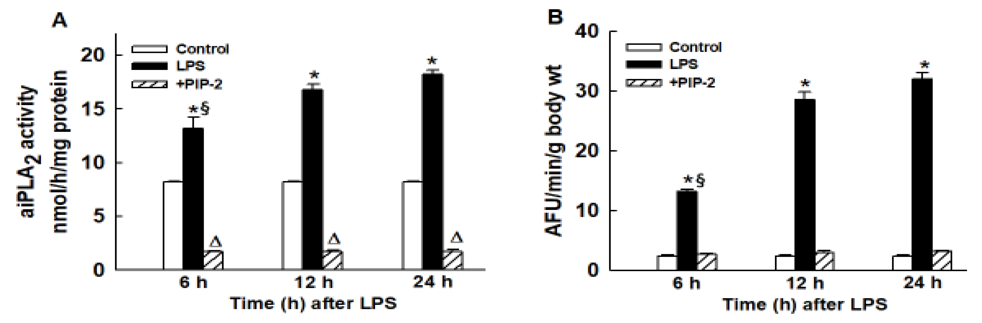
![Ijms 21 00398 g002]()
Figure 5.
PIP-2 prevents mouse mortality with high dose LPS. Mice were administered LPS (15 µg/g body weight) by intratracheal instillation and divided into two groups. At 12 h after LPS, one group was given PIP-2 in liposomes by intravenous injection (IV) while the other group (placebo) was given liposomes alone. The time of the treatment initiation (12 h after LPS) is plotted as zero time. Treatment was repeated at 12, 36, 60, and 84 h after the initial dose of PIP-2,co-incident with the plotted points. Surviving mice were sacrificed at 108 h. n = 12 for placebo and n = 11 for PIP-2.
Figure 5.
PIP-2 prevents mouse mortality with high dose LPS. Mice were administered LPS (15 µg/g body weight) by intratracheal instillation and divided into two groups. At 12 h after LPS, one group was given PIP-2 (2 µg/g body weight) in liposomes by intravenous injection (IV) while the other group (placebo) was given liposomes alone. The time of treatment initiation (12 h after LPS) is plotted as zero time. Treatment was repeated at 12, 36, 60, and 84 h after the initial dose of PIP-2, as indicated by the arrows. Surviving mice were sacrificed at 108 h after start of PIP-2 (120 h after LPS). n = 12 for placebo and n = 11 for PIP-2.


