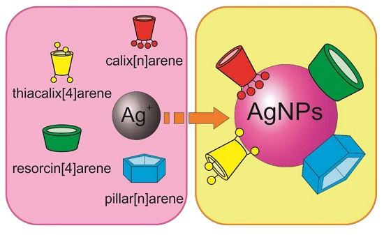
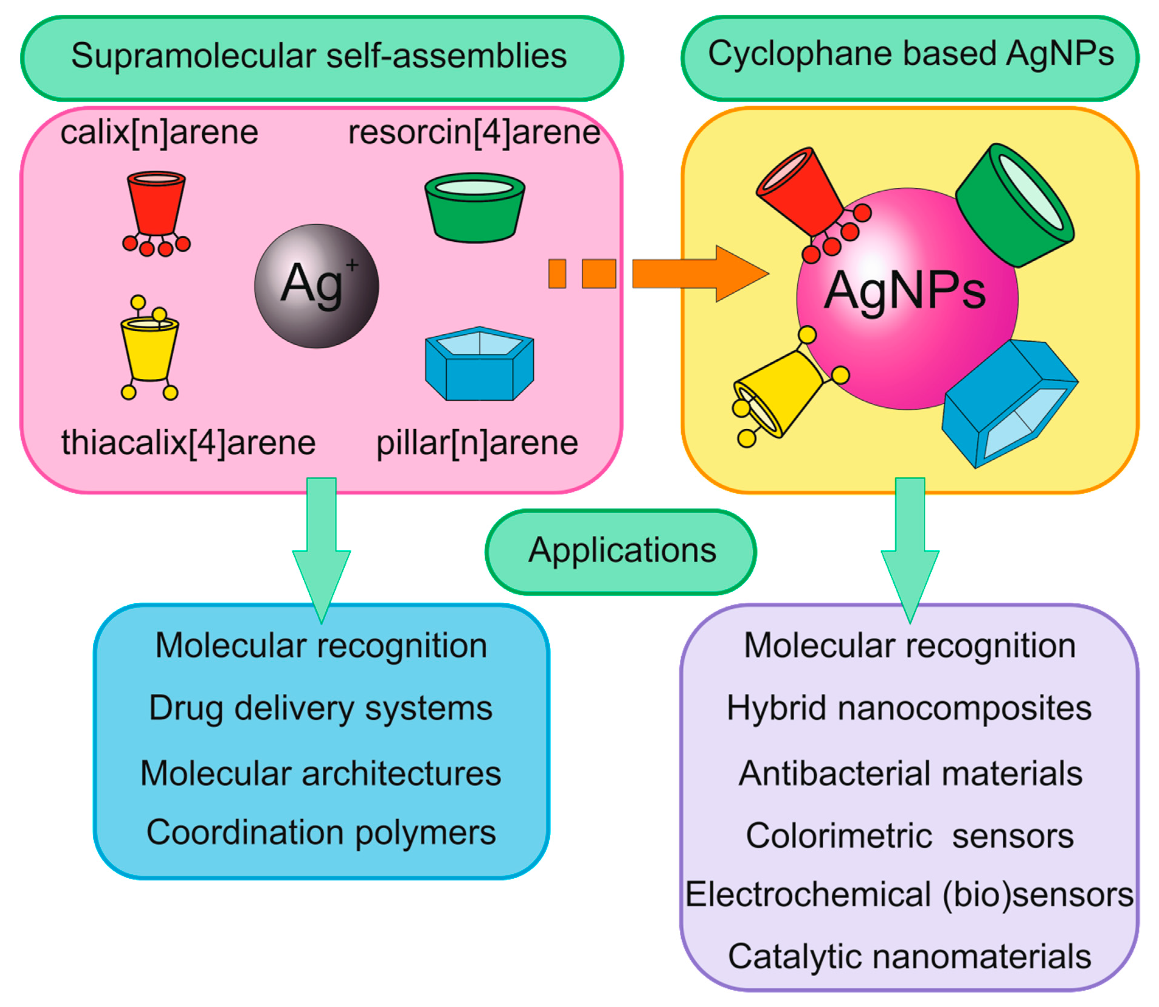
The Role of Calix[n]arenes and Pillar[n]arenes in the Design of Silver Nanoparticles: Self-Assembly and Application


Abstract
:
1. Introduction
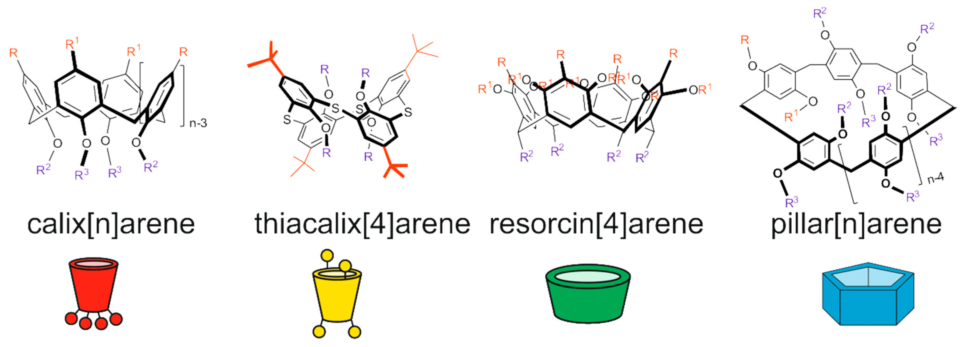
2. Supramolecular Self-Assembly of Calix[n]arenes with Ag(I) Ions and Calix[n]arene-Based AgNPs
2.1. Self-Assembly of Calix[n]arenes with Ag(I) Ions
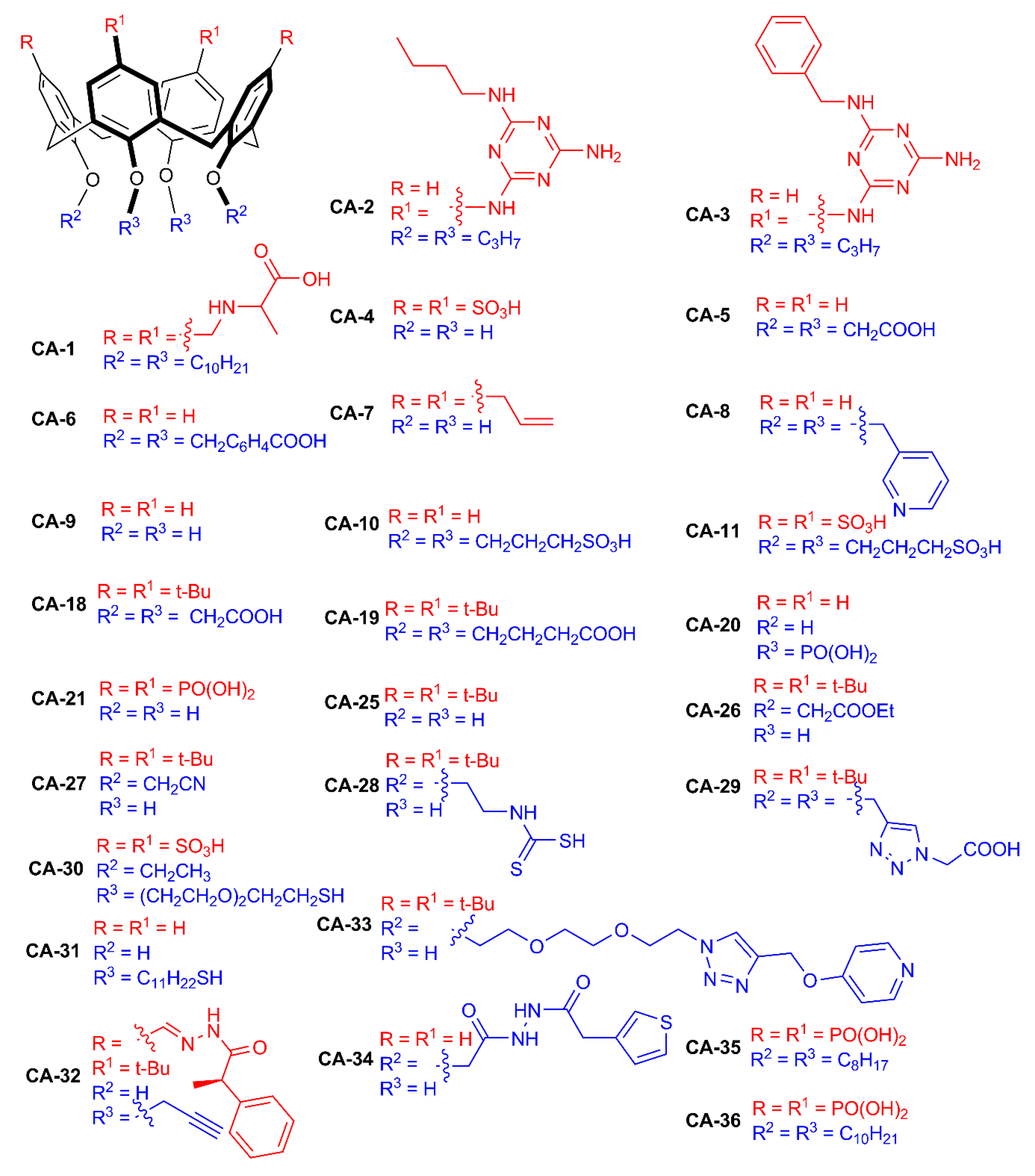
2.2. Synthesis and Application of Calix[n]arene-Based AgNPs
2.2.1. Calix[n]arene-Based AgNPs Obtained Using Chemical Reduction
2.2.2. Calix[n]arene-Based AgNPs Obtained Using Photochemical Reduction
2.2.3. Calix[n]arene-Based AgNPs Obtained Using Electrochemical Reduction
3. Supramolecular Self-Assembly of Thiacalix[4]arenes with Ag(I) Ions and Thiacalix[4]arene-Based AgNPs
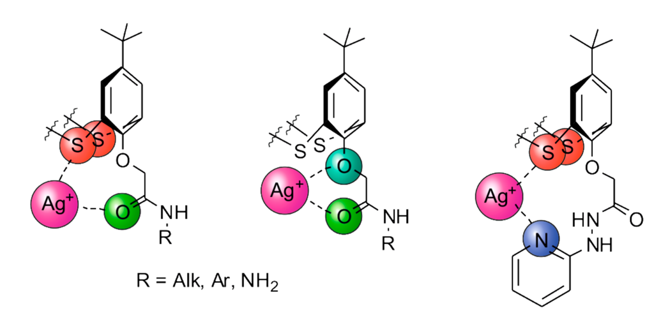
3.1. Self-Assembly of Thiacalix[4]arenes with Ag(I) Ions
3.2. Synthesis and Application of Thiacalix[4]arene-Based AgNPs
4. Synthesis and Application of Resorcin[4]arene-Based AgNPs
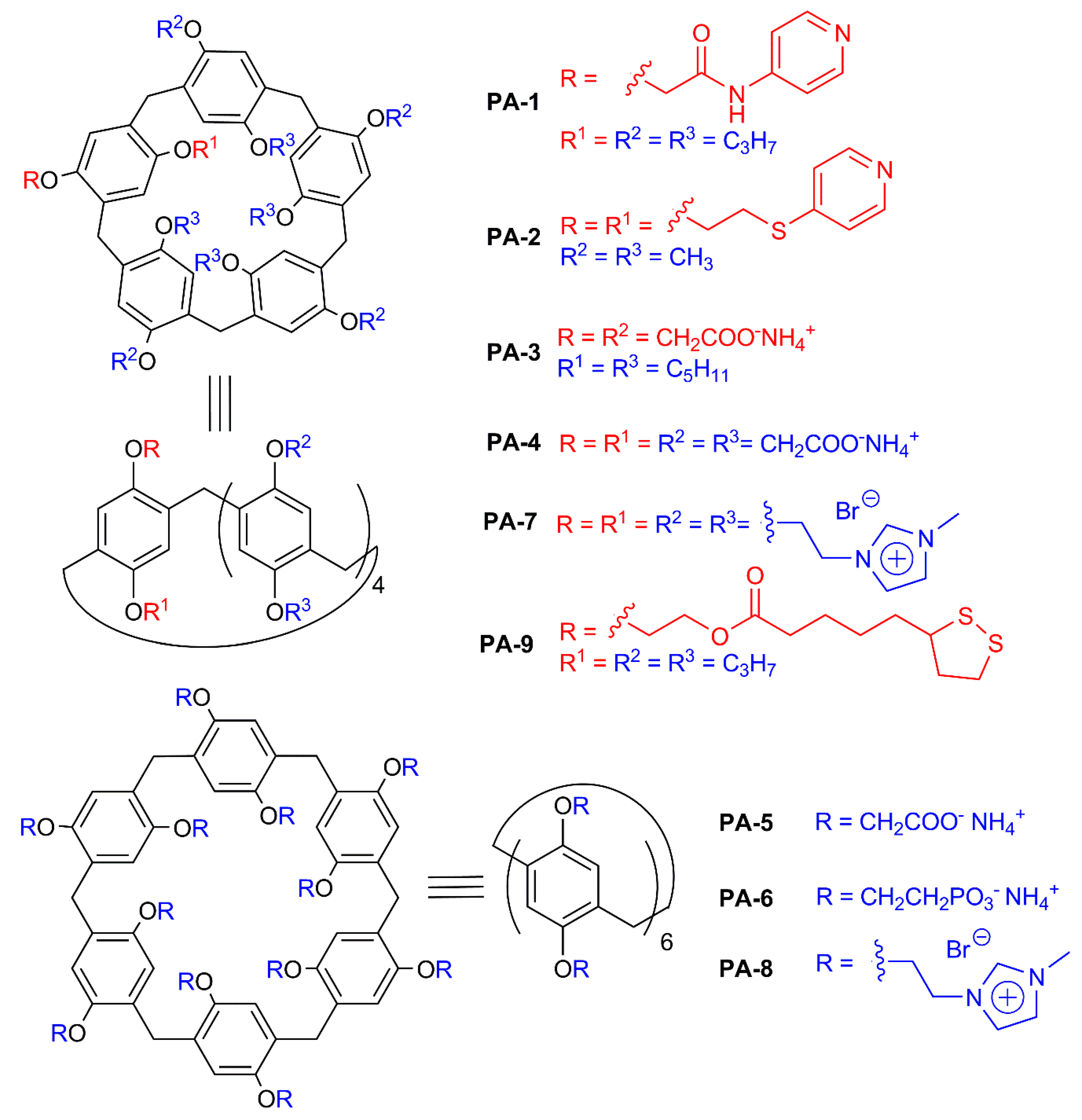
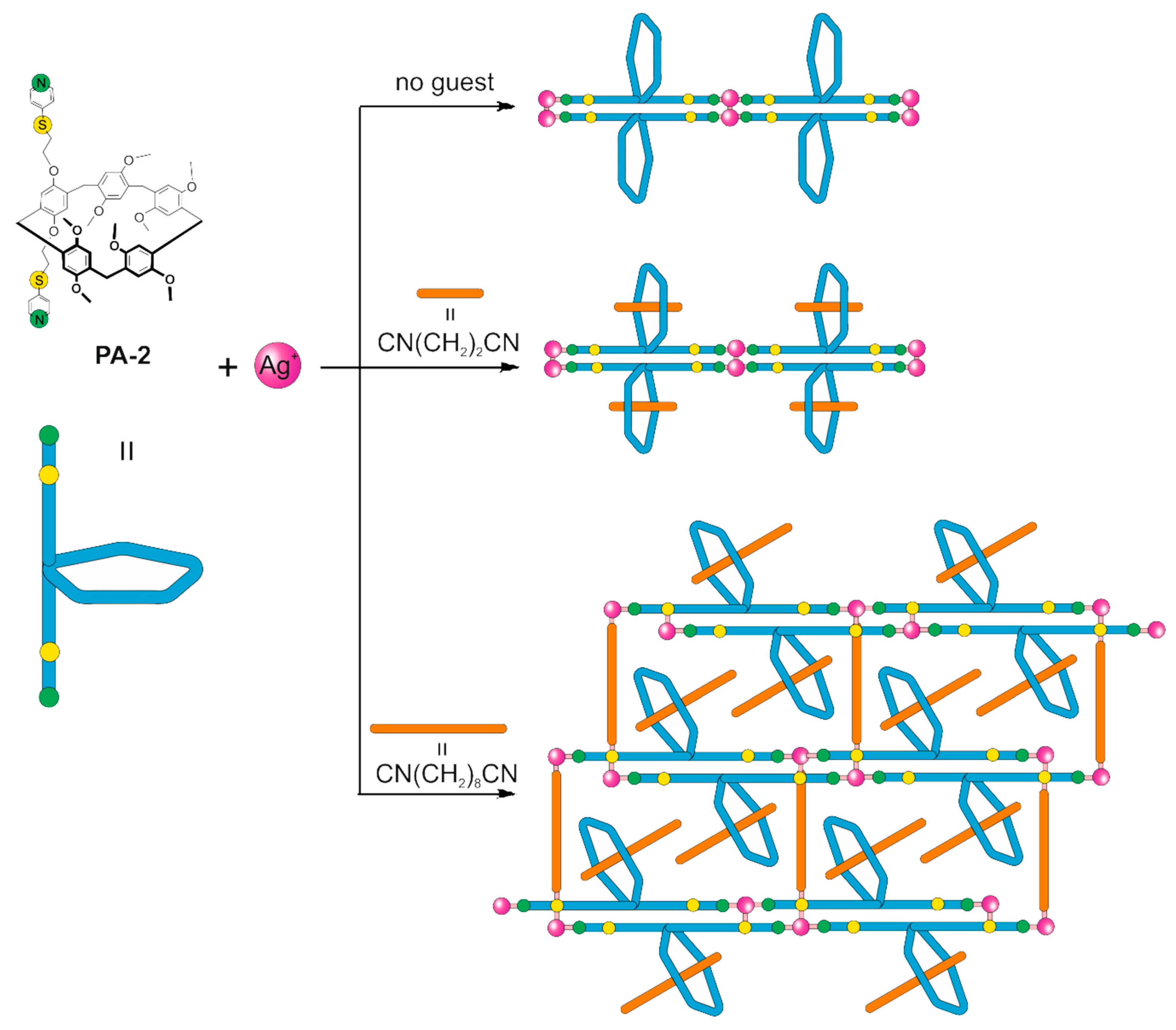
5. Supramolecular Self-Assembly of Pillar[n]arenes with Ag(I) Ions and Pillar[n]arene-Based AgNPs
5.1. Self-Assembly of Pillar[n]arenes with Ag(I) Ions
5.2. Synthesis and Application of Pillar[n]arene-Based AgNPs
6. Conclusions
Author Contributions
Funding
Conflicts of Interest
Abbreviations
| AgNPs | Silver nanoparticles |
| CA | Calixarene |
| TCA | Thiacalixarene |
| PA | Pillararene |
| RA | Resorcinarene |
| TEM | Transmission electron microscopy |
| API | Active pharmaceutical ingredient |
| PAHs | Polycyclic aromatic hydrocarbons |
| SERS | Surface-enhanced Raman scattering |
| DLS | Dynamic light scattering |
| D-FAA | N-Fmoc-d-aspartic acid |
| L-FAA | N-Fmoc-l-aspartic acid |
| ppb | Parts per billion |
| AFM | Atomic force microscopy |
| BSA | Bovine serum albumin |
| DMF | Dimethylformamide |
| DNA | Deoxyribonucleic acid |
| SEM | Scanning electron microscopy |
| PQ | Paraquat |
| SWCNT | Single-walled carbon nanotube |
| COF | Covalent organic framework |
| MOF | Metal–organic framework |
References
- Sabela, M.; Balme, S.; Bechelany, M.; Janot, J.M.; Bisetty, K. A review of gold and silver nanoparticle-based colorimetric sensing assays. Adv. Eng. Mater. 2017, 19, 1700270. [Google Scholar] [CrossRef]
- Chandraker, K.; Nagwanshi, R.; Jadhav, S.K.; Ghosh, K.K.; Satnami, M.L. Antibacterial properties of amino acid functionalized silver nanoparticles decorated on graphene oxide sheets. Spectrochim. Acta Part A Mol. Biomol. Spectrosc. 2017, 181, 47–54. [Google Scholar] [CrossRef] [PubMed]
- Malekzad, H.; Zangabad, P.S.; Mirshekari, H.; Karimi, M.; Hamblin, M.R. Noble metal nanoparticles in biosensors: Recent studies and applications. Nanotechnol. Rev. 2017, 6, 301–329. [Google Scholar] [CrossRef] [PubMed]
- Montes-García, V.; Pérez-Juste, J.; Pastoriza-Santos, I.; Liz-Marzán, L.M. Metal nanoparticles and supramolecular macrocycles: A tale of synergy. Chem. A Eur. J. 2014, 20, 10874–10883. [Google Scholar] [CrossRef] [PubMed]
- Wong, K.K.; Liu, X. Silver nanoparticles—The real “silver bullet” in clinical medicine? MedChemComm 2010, 1, 125–131. [Google Scholar] [CrossRef]
- Burdușel, A.C.; Gherasim, O.; Grumezescu, A.M.; Mogoantă, L.; Ficai, A.; Andronescu, E. Biomedical applications of silver nanoparticles: An up-to-date overview. Nanomaterials 2018, 8, 681. [Google Scholar] [CrossRef] [Green Version]
- Hamouda, R.A.; Hussein, M.H.; Abo-elmagd, R.A.; Bawazir, S.S. Synthesis and biological characterization of silver nanoparticles derived from the cyanobacterium Oscillatoria limnetica. Sci. Rep. 2019, 9, 1–17. [Google Scholar] [CrossRef]
- Lee, S.H.; Jun, B.H. Silver Nanoparticles: Synthesis and application for nanomedicine. Int. J. Mol. Sci. 2019, 20, 865. [Google Scholar] [CrossRef] [Green Version]
- Rasheed, T.; Bilal, M.; Li, C.; Nabeel, F.; Khalid, M.; Iqbal, H.M. Catalytic potential of bio-synthesized silver nanoparticles using Convolvulus arvensis extract for the degradation of environmental pollutants. J. Photochem. Photobiol. B Biol. 2018, 181, 44–52. [Google Scholar] [CrossRef]
- Zhang, X.F.; Liu, Z.G.; Shen, W.; Gurunathan, S. Silver nanoparticles: Synthesis, characterization, properties, applications, and therapeutic approaches. Int. J. Mol. Sci. 2016, 17, 1534. [Google Scholar] [CrossRef]
- Hussain, M.; Nafady, A.; Avcı, A.; Pehlivan, E.; Nisar, J.; Sherazi, S.T.H.; Balouch, A.; Shah, M.R.; Almaghrabi, O.A.; Ul-Haq, M.A. Biogenic silver nanoparticles for trace colorimetric sensing of enzyme disrupter fungicide vinclozolin. Nanomaterials 2019, 9, 1604. [Google Scholar] [CrossRef] [PubMed] [Green Version]
- Kailasa, S.K.; Singhal, R.K.; Basu, H.; Park, T.J. Surface-modified metal nanoparticles for recognition of toxic organic molecules. In Handbook of Nanomaterials in Analytical Chemistry; Elsevier: London, UK, 2020; pp. 415–432. [Google Scholar] [CrossRef]
- Wu, Z.; Song, N.; Menz, R.; Pingali, B.; Yang, Y.W.; Zheng, Y. Nanoparticles functionalized with supramolecular host–guest systems for nanomedicine and healthcare. Nanomedicine 2015, 10, 1493–1514. [Google Scholar] [CrossRef] [Green Version]
- Akiba, U.; Minaki, D.; Anzai, J.I. Host-guest chemistry in layer-by-layer assemblies containing calix[n]arenes and cucurbit[n]urils: A review. Polymers 2018, 10, 130. [Google Scholar] [CrossRef] [PubMed] [Green Version]
- Kongor, A.R.; Mehta, V.A.; Modi, K.M.; Panchal, M.K.; Dey, S.A.; Panchal, U.S.; Jain, V.K. Calix-based nanoparticles: A review. Top. Curr. Chem. 2016, 374, 28. [Google Scholar] [CrossRef] [PubMed]
- Sanabria Español, E.; Maldonado, M. Host–guest recognition of pesticides by calixarenes. Crit. Rev. Anal. Chem. 2019, 49, 383–394. [Google Scholar] [CrossRef] [PubMed]
- Padnya, P.L.; Andreyko, E.A.; Mostovaya, O.A.; Rizvanov, I.K.; Stoikov, I.I. The synthesis of new amphiphilic p-tert-butylthiacalix[4]arenes containing peptide fragments and their interaction with DNA. Organ. Biomol. Chem. 2015, 13, 5894–5904. [Google Scholar] [CrossRef] [PubMed]
- Yakimova, L.; Padnya, P.; Tereshina, D.; Kunafina, A.; Nugmanova, A.; Osin, Y.; Evtugyn, V.; Stoikov, I. Interpolyelectrolyte mixed nanoparticles from anionic and cationic thiacalix[4]arenes for selective recognition of model biopolymers. J. Mol. Liq. 2019, 279, 9–17. [Google Scholar] [CrossRef]
- Mostovaya, O.A.; Gorbachuk, V.V.; Bazanova, O.B.; Gerasimov, A.V.; Evtugyn, V.G.; Osin, Y.N.; Myakushev, V.D.; Rizvanov IKh Stoikov, I.I. Thiacalixarene “knot” effect on protein binding by oligolactic acid particles. Mater. Chem. Front. 2019, 3, 292–300. [Google Scholar] [CrossRef]
- Shu, X.; Xu, K.; Hou, D.; Li, C. Molecular Recognition of Water-soluble pillar[n]arenes towards biomolecules and drugs. Isr. J. Chem. 2018, 58, 1230–1240. [Google Scholar] [CrossRef]
- Español, E.S.; Villamil, M.M. Calixarenes: Generalities and their role in improving the solubility, biocompatibility, stability, bioavailability, detection, and transport of biomolecules. Biomolecules 2019, 9, 90. [Google Scholar] [CrossRef] [Green Version]
- Späth, A.; König, B. Molecular recognition of organic ammonium ions in solution using synthetic receptors. Beilstein J. Org. Chem. 2010, 6, 32. [Google Scholar] [CrossRef] [PubMed]
- Li, Z.; Li, X.; Yang, Y.W. Photoactive nanoparticles capped with macrocycles as platforms and hosts. In Photoactive Inorganic Nanoparticles; Elsevier: London, UK, 2019; pp. 139–167. [Google Scholar] [CrossRef]
- Kim, H.J.; Lee, M.H.; Mutihac, L.; Vicens, J.; Kim, J.S. Host–guest sensing by calixarenes on the surfaces. Chem. Soc. Rev. 2012, 41, 1173–1190. [Google Scholar] [CrossRef]
- Cho, E.J.; Kang, J.K.; Han, W.S.; Jung, J.H. Stimuli-responsive supramolecular nanostructure from amphiphilic calix[4]arene and its three-dimensional dendritic silver nanostructure. Langmuir 2008, 24, 5229–5232. [Google Scholar] [CrossRef] [PubMed]
- Kinge, S.; Crego-Calama, M.; Reinhoudt, D. Silver nanoparticles from hydrogen-bonded supramolecular scaffolds. New J. Chem. 2008, 32, 2071–2073. [Google Scholar] [CrossRef]
- Houmadi, S.; Coquiere, D.; Legrand, L.; Faure, M.C.; Goldmann, M.; Reinaud, O.; Remita, S. Architecture-controlled “SMART” calix[6]arene self-assemblies in aqueous solution. Langmuir 2007, 23, 4849–4855. [Google Scholar] [CrossRef] [PubMed]
- Li, J.; Zhang, S.; Chen, Y.G.; Du, X.; Yu, H.; Yu, J. Supramolecular compounds with coordination polymeric chains of Ag ions, p-sulfonatocalix[4]arene and ethylenediamine. J. Incl. Phenom. Macrocycl. Chem. 2015, 81, 485–491. [Google Scholar] [CrossRef]
- Park, K.M.; Lee, E.; Park, C.S.; Lee, S.S. Tube-type coordination polymers: Two-and four-silver (i)-mediated linear networking of calix[4]arene tetracarboxylates. Inorg. Chem. 2011, 50, 12085–12090. [Google Scholar] [CrossRef]
- Shi, Q.; Luo, W.Z.; Li, B.; Xie, Y.P.; Zhang, T. Versatile architectures of silver (i) organometallic polymers with tetra-allyl functionalized calix[4]arene fine-tuned by distinct Anions. Cryst. Growth Des. 2015, 16, 493–498. [Google Scholar] [CrossRef]
- Liu, L.L.; Chen, J.; Yu, C.X.; Lv, W.X.; Yu, H.Y.; Cui, X.Q.; Liu, L. A novel Ag (I)-calix[4]arene coordination polymer for the sensitive detection and efficient photodegradation of nitrobenzene in aqueous solution. Dalton Trans. 2017, 46, 178–185. [Google Scholar] [CrossRef]
- Zheng, G.L.; Li, Y.Y.; Deng, R.P.; Song, S.Y.; Zhang, H.J. Self-assembly of guest-induced calix[4]arene nanocapsules into three-dimensional molecular architecture. CrystEngComm 2008, 10, 658–660. [Google Scholar] [CrossRef]
- Tauran, Y.; Kim, B.; Coleman, A.W. Bio-applications of calix[n]arene capped silver nanoparticles. J. Nanosci. Nanotechnol. 2015, 15, 6308–6326. [Google Scholar] [CrossRef]
- Tauran, Y.; Brioude, A.; Shahgaldian, P.; Cumbo, A.; Kim, B.; Perret, F.; Coleman, A.W.; Montasser, I. Calix-arene silver nanoparticles interactions with surfactants are charge, size and critical micellar concentration dependent. Chem. Commun. 2012, 48, 9483–9485. [Google Scholar] [CrossRef] [PubMed]
- Stephens, E.K.; Tauran, Y.; Coleman, A.W.; Fitzgerald, M. Structural requirements for anti-oxidant activity of calix [n] arenes and their associated anti-bacterial activity. Chem. Commun. 2015, 51, 851–854. [Google Scholar] [CrossRef] [PubMed] [Green Version]
- Boudebbouze, S.; Coleman, A.W.; Tauran, Y.; Mkaouar, H.; Perret, F.; Garnier, A.; Brioude, A.; Kim, B.; Maguin, E.; Rhimi, M. Discriminatory antibacterial effects of calix[n]arene capped silver nanoparticles with regard to Gram positive and Gram negative bacteria. Chem. Commun. 2013, 49, 7150–7152. [Google Scholar] [CrossRef] [PubMed]
- Tauran, Y.; Grosso, M.; Brioude, A.; Kassab, R.; Coleman, A.W. Colourimetric and spectroscopic discrimination between nucleotides and nucleosides using para-sulfonato-calix[4]arene capped silver nanoparticles. Chem. Commun. 2011, 47, 10013–10015. [Google Scholar] [CrossRef] [Green Version]
- Tauran, Y.; Rhimi, M.; Ueno, R.; Grosso, M.; Brioude, A.; Janneau, E.; Suwinska, K.; Kassab, R.; Shahgaldian, P.; Cumbo, A.; et al. Cytosine: Para-sulphonato-calix[4]arene assemblies: In solution, in the solid-state and on the surface of hybrid silver nanoparticles. J. Incl. Phenom. Macrocycl. Chem. 2013, 77, 213–221. [Google Scholar] [CrossRef]
- Chen, M.; Ding, W.; Kong, Y.; Diao, G. Conversion of the surface property of oleic acid stabilized silver nanoparticles from hydrophobic to hydrophilic based on host−guest binding interaction. Langmuir 2008, 24, 3471–3478. [Google Scholar] [CrossRef]
- Xiong, D.; Chen, M.; Li, H. Synthesis of para-sulfonatocalix[4]arene-modified silver nanoparticles as colorimetric histidine probes. Chem. Commun. 2008, 7, 880–882. [Google Scholar] [CrossRef]
- Abe, N.; Iki, N. Multi-coloration of calixarene-coated silver nanoparticles for the visual discrimination of metal elements. Anal. Sci. 2017, 33, 1141–1145. [Google Scholar] [CrossRef] [Green Version]
- Xiong, D.; Li, H. Colorimetric detection of pesticides based on calixarene modified silver nanoparticles in water. Nanotechnology 2008, 19, 465502. [Google Scholar] [CrossRef]
- Hu, R.; Long, G.; Chen, J.; Yin, Y.; Liu, Y.; Zhu, F.; Feng, J.; Mei, Y.; Wang, R.; Xue, H.; et al. Highly sensitive colorimetric sensor for the detection of H2PO4− based on self-assembly of p-sulfonatocalix[6]arene modified silver nanoparticles. Sens. Actuators B Chem. 2015, 218, 191–195. [Google Scholar] [CrossRef]
- Kellici, S.; Acord, J.; Vaughn, A.; Power, N.P.; Morgan, D.J.; Heil, T.; Facq, S.P.; Lampronti, G.I. Calixarene assisted rapid synthesis of silver-graphene nanocomposites with enhanced antibacterial activity. ACS Appl. Mater. Interfaces 2016, 8, 19038–19046. [Google Scholar] [CrossRef] [PubMed]
- Akermi, N.; Mkaouar, H.; Kriaa, A.; Jablaoui, A.; Soussou, S.; Gargouri, A.; Coleman, A.W.; Perret, F.; Maguin, E.; Rhimi, M. para-Sulphonato-calix[n]arene capped silver nanoparticles challenge the catalytic efficiency and the stability of a novel human gut serine protease inhibitor. Chem. Commun. 2019, 55, 8935–8938. [Google Scholar] [CrossRef] [PubMed]
- Perret, F.; Tauran, Y.; Suwinska, K.; Kim, B.; Chassain-Nely, C.; Boulet, M.; Coleman, A.W. Molecular recognition and transport of active pharmaceutical ingredients on anionic calix[4]arene-capped silver nanoparticles. J. Chem. 2012, 2013, 1–9. [Google Scholar] [CrossRef]
- Tauran, Y.; Brioude, A.; Kim, B.; Perret, F.; Coleman, A. Anionic calixarene-capped silver nanoparticles show species-dependent binding to serum albumins. Molecules 2013, 18, 5993–6007. [Google Scholar] [CrossRef]
- Hartlieb, K.J.; Saunders, M.; Raston, C.L. Templating silver nanoparticle growth using phosphonated calixarenes. Chem. Commun. 2009, 21, 3074–3076. [Google Scholar] [CrossRef]
- Tauran, Y.; Brioude, A.; Coleman, A.W.; Rhimi, M.; Kim, B. Molecular recognition by gold, silver and copper nanoparticles. World J. Biol. Chem. 2013, 4, 35. [Google Scholar] [CrossRef] [Green Version]
- Leyton, P.; Sanchez-Cortes, S.; Garcia-Ramos, J.V.; Domingo, C.; Campos-Vallette, M.; Saitz, C.; Clavijo, R.E. Selective molecular recognition of polycyclic aromatic hydrocarbons (PAHs) on calix [4] arene-functionalized Ag nanoparticles by surface-enhanced Raman scattering. J. Phys. Chem. B 2004, 108, 17484–17490. [Google Scholar] [CrossRef] [Green Version]
- Guerrini, L.; Garcia-Ramos, J.V.; Domingo, C.; Sanchez-Cortes, S. Functionalization of Ag nanoparticles with dithiocarbamate calix [4] arene as an effective supramolecular host for the surface-enhanced Raman scattering detection of polycyclic aromatic hydrocarbons. Langmuir 2006, 22, 10924–10926. [Google Scholar] [CrossRef]
- Guerrini, L.; Garcia-Ramos, J.V.; Domingo, C.; Sanchez-Cortes, S. Self-assembly of a dithiocarbamate calix[4]arene on Ag nanoparticles and its application in the fabrication of surface-enhanced Raman scattering based nanosensors. Phys. Chem. Chem. Phys. 2009, 11, 1787–1793. [Google Scholar] [CrossRef]
- Guerrini, L.; Garcia-Ramos, J.V.; Domingo, C.; Sanchez-Cortes, S. Sensing polycyclic aromatic hydrocarbons with dithiocarbamate-functionalized Ag nanoparticles by surface-enhanced Raman scattering. Anal. Chem. 2009, 81, 953–960. [Google Scholar] [CrossRef] [PubMed]
- Brown, P.O.; Enright, G.D.; Ripmeester, J.A. Nanocrystalline Ag from supramolecular stabilization of metals in 4-tert-butylcalix[4]arene lattices. Chem. Asian J. 2006, 1, 529–535. [Google Scholar] [CrossRef] [PubMed]
- Mehra, C.; Gala, R.; Kakatkar, A.; Kumar, V.; Khurana, R.; Chatterjee, S.; Kumar, N.N.; Barooah, N.; Bhasikuttan, A.C.; Mohanty, J. Cooperative enhancement of antibacterial activity of sanguinarine drug through p-sulfonatocalix[6]arene functionalized silver nanoparticles. Chem. Commun. 2019, 55, 14275–14278. [Google Scholar] [CrossRef] [PubMed]
- Nsengiyuma, G.; Hu, R.; Li, J.; Li, H.; Tian, D. Self-assembly of 1,3-alternate calix[4]arene carboxyl acids-modified silver nanoparticles for colorimetric Cu2+ sensing. Sens. Actuators B Chem. 2016, 236, 675–681. [Google Scholar] [CrossRef]
- Pandya, A.; Sutariya, P.G.; Lodha, A.; Menon, S.K. A novel calix[4]arene thiol functionalized silver nanoprobe for selective recognition of ferric ion with nanomolar sensitivity via DLS selectivity in human biological fluid. Nanoscale 2013, 5, 2364–2371. [Google Scholar] [CrossRef] [PubMed]
- Vita, F.; Boccia, A.; Marrani, A.G.; Zanoni, R.; Rossi, F.; Arduini, A.; Secchi, A. Calix[4]arene-functionalised silver nanoparticles as hosts for pyridinium-loaded gold nanoparticles as guests. Chem. A Eur. J. 2015, 21, 15428–15438. [Google Scholar] [CrossRef]
- Sun, Y.; Zhao, H.; Boussouar, I.; Zhang, F.; Tian, D.; Li, H. Highly sensitive chiral sensing by calix[4]arene-modified silver nanoparticles via dynamic light scattering. Sens. Actuators B Chem. 2015, 216, 235–239. [Google Scholar] [CrossRef]
- Zhan, J.; Wen, L.; Miao, F.; Tian, D.; Zhu, X.; Li, H. Synthesis of a pyridyl-appended calix[4]arene and its application to the modification of silver nanoparticles as an Fe3+ colorimetric sensor. New J. Chem. 2012, 36, 656–661. [Google Scholar] [CrossRef]
- Vyas, G.; Bhatt, S.; Paul, P. Synthesis of calixarene-capped silver nanoparticles for colorimetric and amperometric detection of mercury (HgII, Hg0). ACS Omega 2019, 4, 3860–3870. [Google Scholar] [CrossRef] [Green Version]
- Hartlieb, K.J.; Martin, A.D.; Saunders, M.; Raston, C.L. Photochemical generation of small silver nanoparticles involving multi-functional phosphonated calixarenes. New J. Chem. 2010, 34, 1834–1837. [Google Scholar] [CrossRef]
- Ray, P.; Clément, M.; Martini, C.; Abdellah, I.; Beaunier, P.; Rodriguez-Lopez, J.L.; Huc, V.; Remita, H.; Lampre, I. Stabilisation of small mono-and bimetallic gold–silver nanoparticles using calix[8]arene derivatives. New J. Chem. 2018, 42, 14128–14137. [Google Scholar] [CrossRef]
- Bian, Y.; Li, C.; Li, H. para-Sulfonatocalix[6]arene-modified silver nanoparticles electrodeposited on glassy carbon electrode: Preparation and electrochemical sensing of methyl parathion. Talanta 2010, 81, 1028–1033. [Google Scholar] [CrossRef] [PubMed]
- Baghayeri, M.; Namadchian, M.; Karimi-Maleh, H.; Beitollahi, H. Determination of nifedipine using nanostructured electrochemical sensor based on simple synthesis of Ag nanoparticles at the surface of glassy carbon electrode: Application to the analysis of some real samples. J. Electroanal. Chem. 2013, 697, 53–59. [Google Scholar] [CrossRef]
- Ahmadi, F.; Raoof, J.B.; Ojani, R.; Baghayeri, M.; Lakouraj, M.M.; Tashakkorian, H. Synthesis of Ag nanoparticles for the electrochemical detection of anticancer drug flutamide. Chin. J. Catal. 2015, 36, 439–445. [Google Scholar] [CrossRef]
- Raoof, J.B.; Ojani, R.; Hasheminejad, E.; Rashid-Nadimi, S. Electrochemical synthesis of Ag nanoparticles supported on glassy carbon electrode by means of p-isopropyl calix[6]arene matrix and its application for electrocatalytic reduction of H2O2. Appl. Surf. Sci. 2012, 258, 2788–2795. [Google Scholar] [CrossRef]
- Zhou, R.; Teo, S.; Srinivasan, M.P. In situ formation of silver nanoparticle layer by supramolecule-directed assembly. Thin Solid Films 2014, 550, 210–219. [Google Scholar] [CrossRef]
- Zhou, R.; Srinivasan, M.P. Photocatalysis in a packed bed: Degradation of organic dyes by immobilized silver nanoparticles. J. Environ. Chem. Eng. 2015, 3, 609–616. [Google Scholar] [CrossRef]
- Tian, H.W.; Liu, Y.C.; Guo, D.S. Assembling features of calixarene-based amphiphiles and supra-amphiphiles. Mater. Chem. Front. 2020, 4, 46–98. [Google Scholar] [CrossRef]
- Mostovaya, O.A.; Gorbachuk, V.V.; Padnya, P.L.; Vavilova, A.A.; Evtugyn, G.A.; Stoikov, I.I. Modification of Oligo-and Polylactides With Macrocyclic Fragments: Synthesis and Properties. Front. Chem. 2019, 7, 554. [Google Scholar] [CrossRef]
- Bi, Y.; Du, S.; Liao, W. Thiacalixarene-based nanoscale polyhedral coordination cages. Coord. Chem. Rev. 2014, 276, 61–72. [Google Scholar] [CrossRef]
- Kozlova, M.N.; Ferlay, S.; Solovieva, S.E.; Antipin, I.S.; Konovalov, A.I.; Kyritsakas, N.; Hosseini, M.W. Molecular tectonics: On the formation of 1-D silver coordination networks by thiacalixarenes bearing nitrile groups. Dalton Trans. 2007, 44, 5126–5131. [Google Scholar] [CrossRef] [PubMed]
- Bourlier, J.; Hosseini, M.W.; Planeix, J.M.; Kyritsakas, N. Molecular tectonics: Generation of 1-D interdigitated and 2-D interwoven helical silver coordination networks by oligoethylene glycol based tectons bearing two benzonitrile moieties. New J. Chem. 2007, 31, 25–32. [Google Scholar] [CrossRef]
- Kozlova, M.N.; Ferlay, S.; Kyritsakas, N.; Hosseini, M.W.; Solovieva, S.E.; Antipin, I.S.; Konovalov, A.I. Molecular tectonics: 3-D organisation of decanuclear silver nanoclusters. Chem. Commun. 2009, 18, 2514–2516. [Google Scholar] [CrossRef] [PubMed]
- Ovsyannikov, A.; Lang, M.N.; Ferlay, S.; Solovieva, S.E.; Antipin, I.S.; Konovalov, A.I.; Kyritsakas, N.; Hosseini, M.W. Molecular tectonics: Pyridyl containing thiacalix[4]arene based tectons for the generation of 2-and 3-D silver coordination networks. Dalton Trans. 2013, 42, 116–126. [Google Scholar] [CrossRef]
- Ovsyannikov, A.; Ferlay, S.; Solovieva, S.E.; Antipin, I.S.; Konovalov, A.I.; Kyritsakas, N.; Hosseini, M.W. Molecular tectonics: Anion control of dimensionality and connectivity in meta-pyridyl appended tetramercaptotetrathiacalix[4]arene based silver coordination networks. Dalton Trans. 2014, 43, 158–165. [Google Scholar] [CrossRef]
- Hosseini, M.W. Molecular tectonics an approach to crystal enginee. In Applications of Supramolecular Chemistry; Taylor & Francis Group: London, UK, 2012; pp. 231–254. [Google Scholar] [CrossRef]
- Ovsyannikov, A.S.; Epifanova, N.A.; Popova, E.V.; Kyritsakas, N.; Ferlay, S.; Hosseini, M.W.; Latypov, S.h.K.; Solovieva, S.E.; Konovalov, A.I. Template synthesis of tetrakis-triazolylthiacalix[4]arene in the cone conformation and supramolecular structure of its hexanuclear complex with Ag (I). Macroheterocycles 2014, 7, 189–195. [Google Scholar] [CrossRef] [Green Version]
- Ovsyannikov, A.S.; Ferlay, S.; Solovieva, S.E.; Antipin, I.S.; Konovalov, A.I.; Kyritsakas, N.; Hosseini, M.W. Molecular tectonics: Silver coordination networks based on tetramercaptothiacalix[4]arene in 1,3-alternate conformation bearing four nitrile groups. Russ. Chem. Bull. 2015, 64, 1955–1962. [Google Scholar] [CrossRef]
- Ovsyannikov, A.S.; Noamane, M.H.; Abidi, R.; Ferlay, S.; Solovieva, S.E.; Antipin, I.S.; Konovalov, A.I.; Kyritsakas Hosseini, M.W. Molecular tectonics: Dimensionality and geometry control of silver coordination networks based on pyrazolyl appended thiacalixarenes. CrystEngComm 2016, 18, 691–703. [Google Scholar] [CrossRef]
- Noamane, M.H.; Ferlay, S.; Abidi, R.; Kyritsakas, N.; Hosseini, M.W. Discrete di-and tetranuclear silver complexes based on ortho-imino-or ortho-amino-methylpyridyl-appended p-tert-butylcalix [4] arene or p-tert-butylthiacalix[4]arene in 1,3-alternate conformation. Eur. J. Inorg. Chem. 2017, 2017, 3327–3336. [Google Scholar] [CrossRef]
- Sýkora, J.; Himl, M.; Stibor, I.; Císařová, I.; Lhoták, P. Unique self-assembly patterns based on thiacalix[4]arene–silver interactions. Tetrahedron 2007, 63, 2244–2248. [Google Scholar] [CrossRef]
- Evtugyn, G.A.; Stoikov, I.I.; Beljyakova, S.V.; Shamagsumova, R.V.; Stoikova, E.E.; Zhukov, A.Y.; Antipin, I.S.; Budnikov, H.C. Ag selective electrode based on glassy carbon electrode covered with polyaniline and thiacalix[4]arene as neutral carrier. Talanta 2007, 71, 1720–1727. [Google Scholar] [CrossRef] [PubMed]
- Galitskaya, P.; Fomin, V.; Stoikov, I.; Andreyko, E.; Selivanovskaya, S. Antimicrobial activity of nanoparticles fromsolid phase supramolecular assembliesbased on stereoisomers of p-tert-butylthiacalix[4]arene with silver cations. Int. J. Pharm. Technol. 2016, 8, 15048–15053. [Google Scholar]
- Yakimova, L.S.; Gilmanova, L.H.; Evtugyn, V.G.; Osin, Y.N.; Stoikov, I.I. Self-assembled fractal hybrid dendrites from water-soluble anionic (thia)calix[4]arenes and Ag+. J. Nanoparticle Res. 2017, 19, 173. [Google Scholar] [CrossRef]
- Stoikov, I.I.; Yushkova, E.A.; Bukharaev, A.A.; Biziaev, D.A.; Selivanovskaya, S.Y.; Chursina, M.A.; Antipin, I.S.; Konovalov, A.I.; Zharov, I. Self-assembly of p-tert-butylthiacalix[4]arenes and metal cations into nanoscale three-dimensional particles. J. Phys. Org. Chem. 2012, 25, 1177–1185. [Google Scholar] [CrossRef]
- Yushkova, E.A.; Stoikov, I.I.; Zhukov, A.Y.; Puplampu, J.B.; Rizvanov, I.K.; Antipin, I.S.; Konovalov, A. Heteroditopic p-tert-butylthiacalix[4]arenes for creating supramolecular self-assembles by cascade or commutative mechanisms. RSC Adv. 2012, 2, 3906–3919. [Google Scholar] [CrossRef]
- Andreyko, E.A.; Puplampu, J.B.; Ignacio-De Leon, P.A.; Zharov, I.; Stoikov, I.I. p-tert-Butylthiacalix[4]arenes containing guanidinium groups: Synthesis and self-assembly into nanoscale aggregates. Supramol. Chem. 2019, 31, 473–483. [Google Scholar] [CrossRef]
- Yakimova, L.S.; Padnya, P.L.; Kunafina, A.F.; Nugmanova, A.R.; Stoikov, I.I. Sulfobetaine derivatives of thiacalix[4]arene: Synthesis and supramolecular self-assembly of submicron aggregates with AgI cations. Mendeleev Commun. 2019, 29, 86–88. [Google Scholar] [CrossRef]
- Andreyko, E.A.; Padnya, P.L.; Stoikov, I.I. Supramolecular self-assembly of water-soluble nanoparticles based on amphiphilic p-tert-butylthiacalix[4]arenes with silver nitrate and fluorescein. Colloids Surf. A Physicochem. Eng. Asp. 2014, 454, 74–83. [Google Scholar] [CrossRef]
- Stoikov, I.I.; Yushkova, E.A.; Antipin, I.S.; Konovalov, A.I. Synthesis of silver and lithium sub-micro-and nanoparticles coated with derivatives of p-tert-butylthiacalix[4]arenes. J. Nanoparticle Res. 2011, 13, 6603–6611. [Google Scholar] [CrossRef]
- Darjee, S.M.; Bhatt, K.D.; Panchal, U.S.; Jain, V.K. Scrupulous recognition of biologically important acids by fluorescent “turn off-on” mechanism of thaicalix reduced silver nanoparticles. Chin. Chem. Lett. 2017, 28, 312–318. [Google Scholar] [CrossRef]
- Guan, Z.J.; Zeng, J.L.; Nan, Z.A.; Wan, X.K.; Lin, Y.M.; Wang, Q.M. Thiacalix[4]arene: New protection for metal nanoclusters. Sci. Adv. 2016, 2, e1600323. [Google Scholar] [CrossRef] [PubMed] [Green Version]
- Evtugyn, G.A.; Shamagsumova, R.V.; Sitdikov, R.R.; Stoikov, I.I.; Antipin, I.S.; Ageeva, M.V.; Hianik, T. Dopamine sensor based on a composite of silver nanoparticles implemented in the electroactive matrix of calixarenes. Electroanalysis 2011, 23, 2281–2289. [Google Scholar] [CrossRef]
- Evtugyn, G.; Porfireva, A.; Sitdikov, R.; Evtugyn, V.; Stoikov, I.; Antipin, I.; Hianik, T. Electrochemical aptasensor for the determination of ochratoxin A at the Au electrode modified with Ag nanoparticles decorated with macrocyclic ligand. Electroanalysis 2013, 25, 1847–1854. [Google Scholar] [CrossRef]
- Evtugyn, G.A.; Shamagsumova, R.V.; Padnya, P.V.; Stoikov, I.I.; Antipin, I.S. Cholinesterase sensor based on glassy carbon electrode modified with Ag nanoparticles decorated with macrocyclic ligands. Talanta 2014, 127, 9–17. [Google Scholar] [CrossRef] [PubMed]
- Kuzin, Y.; Porfireva, A.; Stepanova, V.; Evtugyn, V.; Stoikov, I.; Evtugyn, G.; Hianik, T. Impedimetric detection of DNA damage with the sensor based on silver nanoparticles and neutral red. Electroanalysis 2015, 27, 2800–2808. [Google Scholar] [CrossRef]
- Gorbatchuk, V.V.; Porfireva, A.V.; Stepanova, V.B.; Kuzin, Y.I.; Evtugyn, V.G.; Shamagsumova, R.V.; Stoikov, I.I.; Evtugyn, G.A. Co-polymers of oligolactic acid and tetrasubstituted thiacalix[4]arenes as a new material for electrochemical sensor development. Sens. Actuators B Chem. 2017, 246, 136–145. [Google Scholar] [CrossRef]
- Porifreva, A.V.; Gorbatchuk, V.V.; Evtugyn, V.G.; Stoikov, I.I.; Evtugyn, G.A. Glassy carbon electrode modified with silver nanodendrites implemented in polylactide-thiacalix[4]arene copolymer for the electrochemical determination of tryptophan. Electroanalysis 2018, 30, 641–649. [Google Scholar] [CrossRef]
- Kobayashi, K.; Yamanaka, M. Self-assembled capsules based on tetrafunctionalized calix[4]resorcinarene cavitands. Chem. Soc. Rev. 2015, 44, 449–466. [Google Scholar] [CrossRef]
- Wei, H.; Abtahi, S.M.H.; Vikesland, P.J. Plasmonic colorimetric and SERS sensors for environmental analysis. Environ. Sci. Nano 2015, 2, 120–135. [Google Scholar] [CrossRef] [Green Version]
- Salorinne, K.; Lopez-Acevedo, O.; Nauha, E.; Häkkinen, H.; Nissinen, M. Solvent driven formation of silver embedded resorcinarene nanorods. CrystEngComm 2012, 14, 347–350. [Google Scholar] [CrossRef]
- Menon, S.K.; Modi, N.R.; Pandya, A.; Lodha, A. Ultrasensitive and specific detection of dimethoate using ap-sulphonato-calix [4] resorcinarene functionalized silver nanoprobe in aqueous solution. RSC Adv. 2013, 3, 10623–10627. [Google Scholar] [CrossRef]
- Sun, Y.; Yao, Y.; Yan, C.G.; Han, Y.; Shen, M. Selective decoration of metal nanoparticles inside or outside of organic microstructures via self-assembly of resorcinarene. ACS Nano 2010, 4, 2129–2141. [Google Scholar] [CrossRef] [PubMed]
- Ermakova, A.M.; Morozova, J.E.; Shalaeva, Y.V.; Syakaev, V.V.; Nizameev, I.R.; Kadirov, M.K.; Antipin, I.S.; Konovalov, A.I. Calixresorcinarene-capped silver nanoparticles as new supramolecular hybrid nanocontainers. Mendeleev Commun. 2017, 27, 335–337. [Google Scholar] [CrossRef]
- Ermakova, A.M.; Morozova, J.E.; Shalaeva, Y.V.; Syakaev, V.V.; Nizameev, I.R.; Kadirov, M.K.; Antipin, I.S.; Konovalov, A.I. The supramolecular approach to the phase transfer of carboxylic calixresorcinarene-capped silver nanoparticles. Colloids Surf. A Physicochem. Eng. Asp. 2017, 524, 127–134. [Google Scholar] [CrossRef]
- Makwana, B.A.; Vyas, D.J.; Bhatt, K.D.; Jain, V.K.; Agrawal, Y.K. Highly stable antibacterial silver nanoparticles as selective fluorescent sensor for Fe3+ ions. Spectrochim. Acta Part A Mol. Biomol. Spectrosc. 2015, 134, 73–80. [Google Scholar] [CrossRef] [PubMed]
- Makwana, B.A.; Vyas, D.J.; Bhatt, K.D.; Darji, S.; Jain, V.K. Novel fluorescent silver nanoparticles: Sensitive and selective turn off sensor for cadmium ions. Appl. Nanosci. 2016, 6, 555–566. [Google Scholar] [CrossRef] [Green Version]
- Mishra, D.; Kongor, A.; Panchal, M.; Modi, K.; Jain, V. Resorcinarene-embedded stable silver nanoparticles: A fluorescent nanoprobe for Pb (II) in water. Int. J. Res. Appl. Sci. Eng. Technol. 2018, 6, 1360–1370. [Google Scholar] [CrossRef]
- Makwana, B.A.; Darjee, S.; Jain, V.K.; Kongor, A.; Sindhav, G.; Rao, M.V. A comparative study: Metal nanoparticles as fluorescent sensors for biomolecules and their biomedical application. Sens. Actuators B Chem. 2017, 246, 686–695. [Google Scholar] [CrossRef]
- Sergeeva, T.Y.; Samigullina, A.I.; Gubaidullin, A.T.; Nizameev, I.R.; Kadirov, M.K.; Mukhitova, R.K.; Ziganshina, A.Y.; Konovalov, A.I. Application of ferrocene-resorcinarene in silver nanoparticle synthesis. RSC Adv. 2016, 6, 87128–87133. [Google Scholar] [CrossRef] [Green Version]
- Ogoshi, T.; Kanai, S.; Fujinami, S.; Yamagishi, T.A.; Nakamoto, Y. para-Bridged symmetrical pillar[5]arenes: Their Lewis acid catalyzed synthesis and host–guest property. J. Am. Chem. Soc. 2008, 130, 5022–5023. [Google Scholar] [CrossRef]
- Tan, L.L.; Yang, Y.W. Molecular recognition and self-assembly of pillarenes. J. Incl. Phenom. Macrocycl. Chem. 2015, 81, 13–33. [Google Scholar] [CrossRef]
- Song, N.; Yang, Y.W. Hybrid Materials Based on Pillararenes. In Pillararenes; Royal Society of Chemistry: London, UK, 2015; pp. 229–262. [Google Scholar] [CrossRef]
- Yang, K.; Chao, S.; Zhang, F.; Pei, Y.; Pei, Z. Recent advances in the development of rotaxanes and pseudorotaxanes based on pillar[n]arenes: From construction to application. Chem. Commun. 2019, 55, 13198–13210. [Google Scholar] [CrossRef] [PubMed]
- Nazarova, A.A.; Padnya, P.L.; Gilyazeva, A.I.; Khannanov, A.A.; Evtugyn, V.G.; Kutyreva, M.P.; Klochkov, V.V.; Stoikov, I.I. Supramolecular motifs for the self-assembly of monosubstituted pillar[5]arenes with an amide fragment: From nanoparticles to supramolecular polymers. New J. Chem. 2018, 42, 19853–19863. [Google Scholar] [CrossRef]
- Zhang, C.W.; Chen, L.J.; Yang, H.B. Pillarene-involved metallic supramolecular nanostructures. Chin. J. Chem. 2015, 33, 319–328. [Google Scholar] [CrossRef]
- Hua, B.; Shao, L.; Zhang, Z.; Liu, J.; Huang, F. Cooperative silver ion-pair recognition by peralkylated pillar[5]arenes. J. Am. Chem. Soc. 2019, 141, 15008–15012. [Google Scholar] [CrossRef] [PubMed]
- Wang, P.; Ma, J.; Xia, D. AH2S and I− dual-responsive supramolecular polymer constructed via pillar[5]arene-based host–guest interactions and metal coordination. Org. Chem. Front. 2018, 5, 1297–1302. [Google Scholar] [CrossRef]
- Lee, E.; Park, I.H.; Ju, H.; Kim, S.; Jung, J.H.; Habata, Y.; Lee, S.S. Formation of a pillar[5]arene-based two-dimensional poly-pseudo-rotaxane: Threading and crosslinking by the same guest molecules. Angew. Chem. Int. Ed. 2019, 58, 11296–11300. [Google Scholar] [CrossRef]
- Yao, Y.; Wei, P.; Yue, S.; Li, J.; Xue, M. Amphiphilic pillar[5]arenes: Influence of chemical structure on self-assembly morphology and application in gas response and λ-DNA condensation. RSC Adv. 2014, 4, 6042–6047. [Google Scholar] [CrossRef]
- Yao, Y.; Zhou, Y.; Dai, J.; Yue, S.; Xue, M. Host–guest recognition-induced color change of water-soluble pillar[5]arene modified silver nanoparticles for visual detection of spermine analogues. Chem. Commun. 2014, 50, 869–871. [Google Scholar] [CrossRef]
- Yao, Y.; Jie, K.; Zhou, Y.; Xue, M. Reversible assembly of silver nanoparticles driven by host–guest interactions based on water-soluble pillar[n]arenes. Chem. Commun. 2014, 50, 5072–5074. [Google Scholar] [CrossRef] [Green Version]
- Sun, J.; Guo, F.; Shi, Q.; Wu, H.; Sun, Y.; Chen, M.; Diao, G. Electrochemical detection of paraquat based on silver nanoparticles/water-soluble pillar[5]arene functionalized graphene oxide modified glassy carbon electrode. J. Electroanal. Chem. 2019, 847, 113221. [Google Scholar] [CrossRef]
- Zhao, G.; Gao, Z.; Li, H.; Liu, S.; Chen, L.; Zhang, R.; Guo, H. Controlled assembly of Ag nanoparticles on the surface of phosphate pillar[6]arene functionalized single-walled carbon nanotube for enhanced catalysis and sensing performance. Electrochim. Acta 2019, 318, 711–719. [Google Scholar] [CrossRef]
- Tan, X.; Zhang, Z.; Cao, T.; Zeng, W.; Huang, T.; Zhao, G. Control assembly of pillar[6]arene-modified Ag nanoparticles on covalent organic framework surface for enhanced sensing performance toward paraquat. ACS Sustain. Chem. Eng. 2019, 7, 20051–20059. [Google Scholar] [CrossRef]
- Yao, Y.; Jie, K.; Zhou, Y.; Xue, M. Water-soluble pillar[6]arene stabilized silver nanoparticles: Preparation and application in amino acid detection. Tetrahedron Lett. 2014, 55, 3195–3199. [Google Scholar] [CrossRef]
- Muhammed, M.A.H.; Cruz, L.K.; Emwas, A.H.; El-Zohry, A.M.; Moosa, B.; Mohammed, O.F.; Khashab, N.M. Pillar[5]arene-stabilized silver nanoclusters: Extraordinary stability and luminescence enhancement induced by host–guest interactions. Angew. Chem. 2019, 131, 15812–15817. [Google Scholar] [CrossRef]


| Cyclophanes Studied | AgNPs Synthesis Method 1 | AgNPs Sizes (Method) | Surface Plasmon Resonance λ | Application, Target Substrate, SPR λ, nm in the Presence of Substrate 2 | Comparison Substrates | Lit. |
|---|---|---|---|---|---|---|
| CA-4, CA-10–CA-17 | B (NaBH4) | - | 390 nm | CMC determination of cationic surfactants (CPB, CTAB) intensity of 390 nm band CA-10—decrease CA-4, CA-11—increase CA-12–CA-17—no changes | cetyl pyridium bromide, cetyl trimethyl ammonium bromide, N-octyl glucopyranoside. | [34] |
| CA-4, CA-10–CA-17 | B (NaBH4) | - | - | Structure-dependent inhibition of Gram+ and Gram− bacteria growth, anti-oxidant capacity, antibacterial effect against E. coli, B. subtilis | - | [35,36] |
| CA-4 | B (NaBH4) | ~20 nm (TEM) | ~380 nm | Multicolor response to nucleotides and desoxynucleotides | cytosine, guanine—540 nm, uracil—580 nm, thymine—590 nm, adenine—no changes, deoxy-adenosine, deoxy-guanosine—460 nm, deoxy-thymidine—520 nm, deoxy-cytidine—decoloration (grey colored solution) | [37] |
| CA-2, CA-3 | B (electron beam) | CA-2: 2.3 ± 0.3 nm (TEM) CA-3: 2.1 ± 0.1 nm (TEM) | - | - | - | [26] |
| CA-4 | B (NaBH4) | ~30 nm (TEM) | 390 nm | 540 nm (cytosine) | - | [38] |
| CA-4 | A (NaBH4) | 11.6 ± 3 nm (TEM) | 407 nm (hexane) oleic acid stabilized | 413 nm (water) oleic acid stabilized+calixarene; Oleic-stabilized particles become hydrophilic | - | [39] |
| CA-4 | B (NaBH4) | 8.0 ± 1.0 nm (TEM) | 394 nm | His ~ 500 nm | Alanine, valine, leucine, methionine, phenylalanine, histidine tyrosine, threonine, serine, proline, glutamic acid, aspartic acid | [40] |
| CA-4 | B (NaBH4) | ~6–12 nm (TEM) | 391 nm | 450–600 nm (transition metal hydroxide particles at pH = 10) | Effectivity is: Ni2 + <Tb3 + ~Zn2 + <Cu2 + <Co2+~Cd2+ << Pb2+ | [41] |
| CA-4, CA-15 | A (NaBH4) | CA-4: 8 nm (TEM) CA-15: 4 nm (TEM) | CA-4: 393 nm CA-15: 391 nm | CA-4 Optunal ~ 500 nm CA-15 - | iprodione, pyrimethanil, thiabendazole, optunal, parathion-methyl, methomyl and acetamiprid | [42] |
| CA-12 | A (NaBH4) | - | - | Three-component supramolecular system with dipyrene, discriminates; H2PO4- | NaF, NaCl, NaBr, NaI, NaH2PO4, NaHSO3, Na2SO4, NaNO3, NaNO2 and NaHCO3 | [43] |
| CA-12 | B (KOH,400º C, H2) | 14.9 ±6.7 nm – AgNPs (TEM) 15.6 ± 9.1 nm – graphene nanocomposites (TEM) | - | Antibacterial activity against S. aureus, E. Coli | - | [44] |
| CA-4, CA-12, CA-15 | B (NaBH4) | CA-4: 4.5 nm (TEM) CA-12: 5 nm (TEM) CA-15: 8 nm (TEM) | 400 nm 400 nm 400 nm | 415 nm 410, 465 nm 410, 450, 490 nm (Saburopin) | increased Saburopin efficiency and stability. | [45] |
| CA-4, CA-10, CA-11, CA-18, CA-19, CA-20 | B(NaBH4) | - | CA-4: 390 nm CA-10: 390 nm CA-11: 380 nm CA-18: 420 nm CA-19: 400 nm CA-20: 400 nm | Chlorohexidine, gentamycine | chlorohexidine, chloramphenicol, gentamycine sulfate, | [46] |
| CA-4, CA-20 | B(NaBH4) | - | CA-4: 390 nm CA-20: 398 nm | CA-4: 398 nm (BSA), CA-20: 404 nm (BSA) | - | [47] |
| CA-21 CA-22 CA-23 CA-24 | B (H2) (pH = 9) 70 °C | CA-21: 2.1± 0.8 nm (TEM) CA-22: 2.9 ±1.3 nm (TEM) CA-23: 5 ± 3.6 nm (TEM) CA-24: 5.3± 2.4 nm (TEM) | ~ 350, 390 nm | - | - | [48] |
| CA-25, CA-26, CA-27 | A (NH2OH·HCl) | ~23 nm (TEM) | 420 nm | 436 nm (pyrene) | pyrene, triphenylene, benzo[c]phenantrene, anthracene, coronene, chrysene, dibenzoanthracene, rubicene | [50] |
| CA-28 | A (sodium citrate or NH2OH) | - | - | SERS (coronene) | pyrene, triphenylene, benzo[c]phenantrene, coronene | [51] |
| CA-25 | B (ethylene-diamine) | 9.4 nm (nanocrystalline) (TEM) | - | - | - | [54] |
| CA-12 | B (NaBH4) | 7 nm (TEM) | 393 nm | 406 nm (Sanguinarine) | Enhancement of antibacterial activity | [55] |
| CA-29 | A (NaBH4, C18H37NH2) | 46.7 nm (DLS) | 430 nm | 430 nm (intensity increase) | CoCl2, NiCl2, MnCl2, Cd(NO3)2, AgNO3, Cu(NO3)2), Pb(NO3)2, Hg(NO3)2, BaCl2 | [56] |
| CA-30 | A (NaBH4) | 52 nm (TEM) | 422 nm | 554 nm (Fe3+) | Zn Fe3+, Cu2+, Ca2+, Co2+, Mg2+, Cd2+, Ba2+, Na+, K+, Mn2+, Fe2+, Pb2+, Ni2+, Pd2+, Hg2+, pepsin, cytochrome c, BSA, myoglobin | [57] |
| CA-31 | A (interphase NaBH4, C12H25SH) | 3 ± 1 nm (TEM) | 450 nm | 580 nm (association with AuNPs) | AuNPs functionalized with pyridinium fragments | [58] |
| CA-33 | B (NaBH4) | 9 nm (TEM) | ~ 415 nm | ~ 525 nm (N-Fmoc-L-aspartic acid) | N-Fmoc-L-aspartic acid, N-Fmoc-D-aspartic acid | [59] |
| CA-32 | B (photo-reduction, 365 nm) | 10 ± 1.0 nm (TEM) | 364 nm | Fe3+ (414 nm) | Li+, Na+, K+, Cs+, Mg2+, Ca2+, Sr2+, Ba2+, Cr3+, Fe3+, Fe2+, Cu2+, Pb2+, Ag+, Ni2+, Mn2+, Co2+, Cd2+, Zn2+. | [60] |
| CA-34 | B (photo-reduction with sunlight) | 3-5 nm (TEM) | 432 nm | 432 nm, lower intensity (Hg2+) | Li+, Na+, K+, Cs+, Ca2+, Mg2+, Ba2+, Cr3+, Sr2+, Co2+, Ni2+, Cd2+, Zn2+, Rb+, Hg2+, Pb2+, Cu2+ | [61] |
| CA-21, CA-23, CA-24, CA-35, CA-36 | B (photo-reduction, pH = 9, 365 nm) | CA-21: 3.6 ± 1.2 nm CA-23: 2.6 ± 0.8 nm CA-24: 2.6 ± 0.8 nm CA-35: 3.6 ± 1.2 nm CA-36: 3.6 ± 1.2 nm | CA-21: 400 nm CA-23: 400,485 nm CA-24: 400,485 nm CA-35: 400 nm CA-36: 400 nm | - | - | [62] |
| CA-37 | B (gamma-irradiation) | 1.55–0.5 nm in order AgPF6 < AgClO4 < AgOTf < AgBF4 Bimetallic: 1 nm and 3 nm (HAADF-EDX-STEM) | 410 nm | nitrothiophenol reduction | nitrothiophenol, nitrophenol | [63] |
| CA-12 | B (electrochemical reduction) | ~ 100 nm (TEM) | - | methyl parathion | PO43−, SO42−, CO32−, NO3−, p-nitrophenol, nitrobenzene | [64] |
| CA-25 | B (electrochemical reduction) | 40–70 nm (TEM) | - | nifedipine | dopamine, ascorbic acid, L-Dopa, epinephrine, tryptophan, L-cysteine, uric acid | [65] |
| CA-25, CA-38 | B (electrochemical reduction) | ~ 100–2000 nm (TEM) | - | flutamide | dopamine, ascorbic acid, L-Dopa, epinephrine, tryptophan, L-cysteine, uric acid | [66] |
| CA-39 | B (electrochemical reduction) | 70 nm (TEM) | - | electrochemical reduction of H2O2 | - | [67] |
| CA-40, CA-41 | Combination of C and B (electrochemical reduction with participation of cyclophane as reducing agent) | 10 nm, height = 2 nm (AFM) | - | Photocatalytic degradation of methyl orange, methylene blue, rhodamine 6G chloride | - | [68,69] |
| TCA-3, TCA-8 | B (reduction with DMF) | three-dimensional self-assembled monolayer (DMF) (1–13 nm; 46–622 nm | - | - | - | [92] |
| TCA-5 | C | 20 nm | ~425 nm | ~ 452 nm (histidine) ~414 nm (tryptophan) | valine, proline, arginine, cysteine, aspartic acid, glutamic acid, glutamine, leucine, methionine, phenylalanine, tryptophan, isoleucine, histidine | [93] |
| TCA-1 | A | Two nanoclusters with 35 and 34 atoms | Ag36: 501, 336, 300 (shoulder) nm Ag35: 495, 336, 300 (shoulder) nm Ag34: 482, 336, 300 (shoulder) nm | - | - | [94] |
| TCA-27 | C | 4–6 nm (TEM) | - | Substrate depending on electrode composition: A: dopamine B: ochratoxin C: cholinesterase D: DNA damage | - | [95,96,97,98] |
| TCA-28 | B (electrochemical reduction) | Micro-sized, contacted granules (TEM) | - | A: factors affecting DNA charge and structure (thermal denaturing, methylene blue intercalation, oxidative damage), thiocholline B: tryptophan | B: phenylalanine, histidine, cysteine, tyrosine | [99,100] |
| RA-1 | A | 45 ± 10 nm (DLS) | 420 nm | 519 nm (dimethoate) | dichlorvos, parathion, 2,4-D, dimethoate, hexaconazole, imidacloprid and monocrotophos | [104] |
| RA-2 | A | 20.9 ± 16.4 nm (TEM) | 416 nm | reversible association with resorcinarene self-assembled microtubes | - | [105] |
| RA-3 | B (NaBH4) | 2–3 nm (TEM) | - | doxorubicin | - | [106] |
| RA-4 | B (NaBH4) | 4–6 nm (TEM) | 429 nm | Cetyltrimethylammonium (phase transfer of AgNPs from water to chloroform upon interaction with surfactant) | - | [107] |
| RA-5 | C | 7 ± 1 nm (TEM) | 406 nm | Fe3+ (fluorescence quenching at 560 nm), antibacterial activity against E. coli, B. subtilis, S. aureus, B. megaterium | Zn2+, Cd2+, Hg2+, Co2+, Cu2+, Pb2+, Cr3+, V3+ | [108] |
| RA-6 | C | 5 ± 2 nm (TEM) | 415 nm | Cd2+ (fluorescence quenching at 458 nm) | Zn2+, Pb2+, Co2+, Cu2+, Ba2+, Cd2+, Mn2+, Hg2+, Ca2+, Mg2+, Sr2+, Ni2+ | [109] |
| RA7 | C | 15 ± 5 nm (TEM) | 426 nm | Pb2+ (fluorescence quenching at 580 nm) | Cr3+, Mn2+, Fe3+, Co2+, Ni2+, Cu2+, Zn2+, Cd2+, Hg2+ | [110] |
| RA-8 | C | 7 ± 5 nm (TEM) | 408 nm | 420 nm (CT-DNA, S-DNA) Histidine (fluorescence quenching at 540 nm) | arginine, cysteine, aspartic acid, glutamic acid, glutamine, leucine, methionine, threonine, histidine, L-Dopa, tryptophan | [111] |
| RA-9 | C | 30 nm (TEM) 60 nm (AFM, DLS) | 440 nm | Catalysis of nitrophenol reduction | - | [112] |
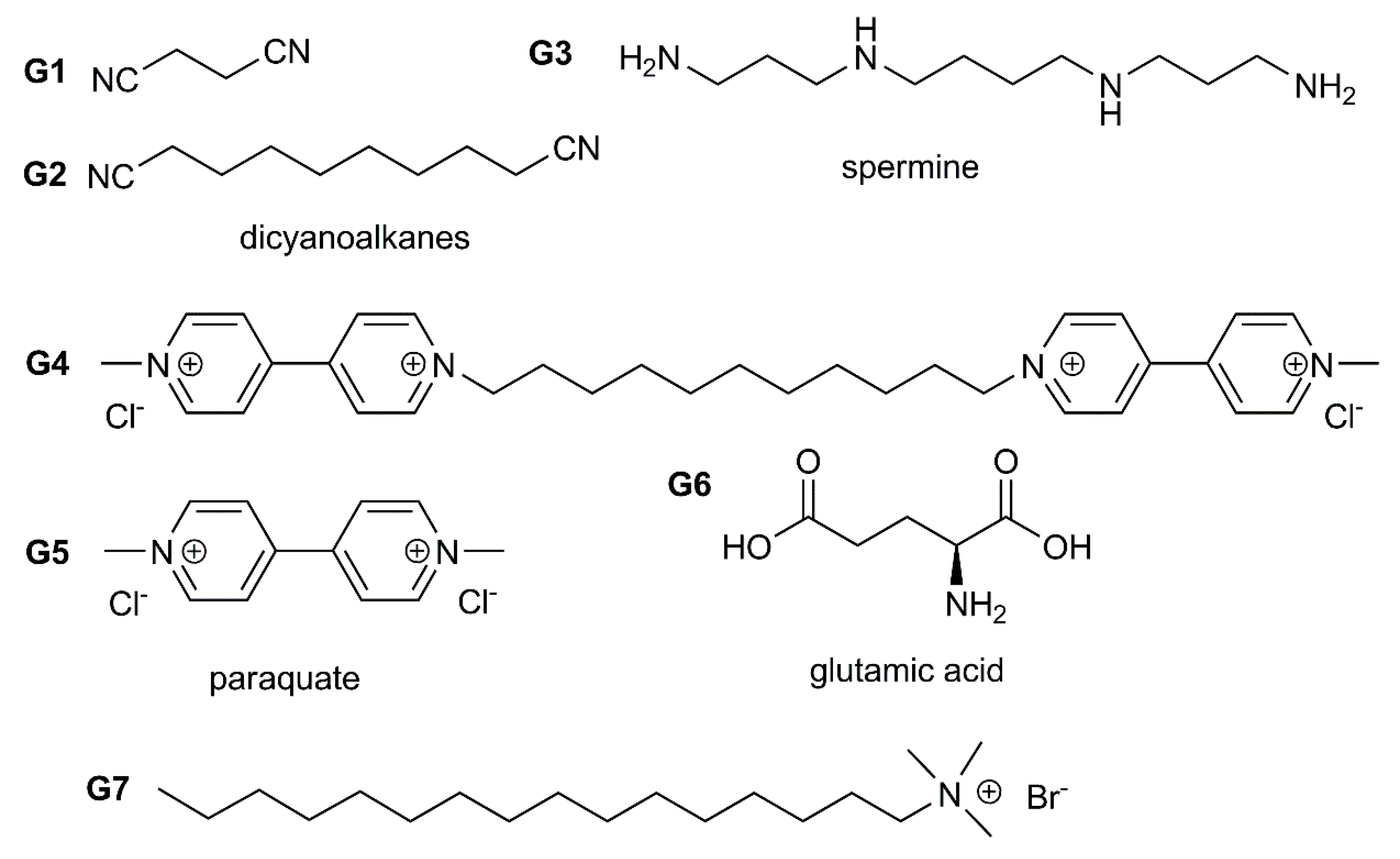
| PA-5 | B (NaBH4) | 18.7 ± 2.18 nm (TEM) | 400 nm | 400 nm intensity decrease (Structural analogues of spermine G3) | spermine, ursol, tetraethylenepentamine, triethylenetetramine, ethanediamine, 1,12-dodecylamine and 1,6-hexamethylenediamine | [123] |
| PA-4 | B (NaBH4) | 10 nm (TEM) | 404 nm | 456 nm (series of alkylidene- linked two paraquat units) binding is reversible upon addition of PA-5 | - | [124] |
| PA-4 | B (NaBH4) | ~10 nm (TEM) | 394 nm | Electrochemical detection of paraquate | - | [125] |
| PA-6 | B (NaBH4 in the presence of SWCNT) | 3–4 nm (TEM) | 410 nm | Catalytic reduction of nitrophenol, catalytic degradation of methylene blue | - | [126] |
| PA-5 | B (NaBH4) | 6.01 ± 0.94 nm (TEM) | 400 nm | Electrochemical detection of paraquat | - | [127] |
| PA-7, PA-8 | B (NaBH4) | 13.57 ± 2.18 nm (TEM) | 400 nm | 500 nm glutamic acid | lysine, arginine, histidine, glycine, glutamic acid, tyrosine, aspartic acid, threonine | [128] |
| PA-9 | A (ligand-exchange), B (synthesis in the presence of cyclophane) | Ag29 nanoclusters For case B, formation of small nanoparticles is possible | 330, 455, 513, 623, 700 nm | Photoluminiscence enhancement (810 nm, Neutral alkylamines; 650 nm, quaternary alkylammonium salts) | hexylamine, dodecaneamine, oleylamine, 1,8-diaminooctane trimethyloctadecylammonium bromide, | [129] |
© 2020 by the authors. Licensee MDPI, Basel, Switzerland. This article is an open access article distributed under the terms and conditions of the Creative Commons Attribution (CC BY) license (http://creativecommons.org/licenses/by/4.0/).
Share and Cite
Padnya, P.; Gorbachuk, V.; Stoikov, I. The Role of Calix[n]arenes and Pillar[n]arenes in the Design of Silver Nanoparticles: Self-Assembly and Application. Int. J. Mol. Sci. 2020, 21, 1425. https://0-doi-org.brum.beds.ac.uk/10.3390/ijms21041425
Padnya P, Gorbachuk V, Stoikov I. The Role of Calix[n]arenes and Pillar[n]arenes in the Design of Silver Nanoparticles: Self-Assembly and Application. International Journal of Molecular Sciences. 2020; 21(4):1425. https://0-doi-org.brum.beds.ac.uk/10.3390/ijms21041425
Chicago/Turabian StylePadnya, Pavel, Vladimir Gorbachuk, and Ivan Stoikov. 2020. "The Role of Calix[n]arenes and Pillar[n]arenes in the Design of Silver Nanoparticles: Self-Assembly and Application" International Journal of Molecular Sciences 21, no. 4: 1425. https://0-doi-org.brum.beds.ac.uk/10.3390/ijms21041425