Article

Role of Chitin Deacetylase 1 in the Molting and Metamorphosis of the Cigarette Beetle Lasioderma serricorne

1
Provincial Key Laboratory for Agricultural Pest Management of Mountainous Regions, Institute of Entomology, Guizhou University, Guiyang 550025, China
2
Guizhou Provincial Key Laboratory for Rare Animal and Economic Insect of the Mountainous Region, College of Biology and Environmental Engineering, Guiyang University, Guiyang 550005, China
*
Authors to whom correspondence should be addressed.
Int. J. Mol. Sci. 2020, 21(7), 2449; https://0-doi-org.brum.beds.ac.uk/10.3390/ijms21072449
Submission received: 13 February 2020 / Revised: 24 March 2020 / Accepted: 30 March 2020 / Published: 1 April 2020
(This article belongs to the Special Issue Molecular Ecology, Physiology and Biochemistry of Insects 2.0)

Abstract

:
Chitin deacetylases (CDAs) are chitin-modifying enzymes known to play vital roles in insect metamorphosis and development. In this study, we identified and characterized a chitin deacetylase 1 gene (LsCDA1) from the cigarette beetle Lasioderma serricorne. LsCDA1 contains a 1614 bp open reading frame encoding a protein of 537 amino acids that includes domain structures typical of CDAs. LsCDA1 was mainly expressed in the late larval and late pupal stages. In larval tissues, the highest level of LsCDA1 was detected in the integument. The expression of LsCDA1 was induced by 20-hydroxyecdysone (20E) in vivo, and it was significantly suppressed by knocking down the expression of ecdysteroidogenesis genes and 20E signaling genes. RNA interference (RNAi)-aided silencing of LsCDA1 in fifth-instar larvae prevented the larval–pupal molt and caused 75% larval mortality. In the late pupal stage, depletion of LsCDA1 resulted in the inhibition of pupal growth and wing abnormalities, and the expression levels of four wing development-related genes (LsDY, LsWG, LsVG, and LsAP) were dramatically decreased. Meanwhile, the chitin contents of LsCDA1 RNAi beetles were significantly reduced, and expressions of three chitin synthesis pathway genes (LsTRE1, LsUAP1, and LsCHS1) were greatly decreased. The results suggest that LsCDA1 is indispensable for larval–pupal and pupal–adult molts, and that it is a potential target for the RNAi-based control of L. serricorne.

Graphical Abstract

1. Introduction

Molting and metamorphosis are important aspects of insect growth, as the insect periodically sheds and replaces the rigid exoskeleton. The insect cuticle is repeatedly renewed while the old cuticle is digested, and a series of enzymes and cofactors function in the process of molting and development [1,2]. Chitin is a linear polymer of N-acetylglucosamine and is a major structural component of the insect cuticle [3]. Chitin synthesis and degradation are tightly coupled with the chitin content balance, which is key for the molting cycle and metamorphosis of insects [4,5]. Chitin degradation is accomplished by three types of chitinolytic enzyme: chitin deacetylases (EC 3.5.1.41, CDAs), chitinase (EC 3.2.1.14, CHTs), and β-N-acetylglucosaminidases (EC3.2.1.30, NAGs). CDAs enzymatically alter chitin by a deacetylating process, whereas CHTs and NAGs degrade chitin by hydrolyzation [6,7]. These three enzymes mainly function in insect growth and development. Studies in Nilaparvata lugens revealed that one NAG, five CHTs, and three CDAs participate in chitin degradation and are critical for nymph molting [8,9,10].
CDAs are extracellular chitin-modifying enzymes and belong to the carbohydrate esterase family 4 [11]. The first cDNA encoding chitin deacetylase-like protein was cloned from the cabbage looper Trichopusia ni [12]. Since then, hundreds of CDA genes have been identified in numerous insect species, including Coleoptera [13,14,15,16], Hemiptera [10], Hymenoptera [17], Orthoptera [18,19], Diptera [20,21], Lepidoptera [7,22,23,24,25], and Isoptera [26]. Based on sequence similarity and domain organization, insect CDAs have been divided into five groups (group I−V) [17]. Both group I and II CDAs contain a chitin-binding (ChtB) domain, a low-density lipoprotein receptor class A (LDLa) domain, and a polysaccharide deacetylase-like (CDA) catalytic domain, but with substantial differences in amino acid sequences. Group III and IV CDAs contain ChtB and CDA catalytic domains, whereas group V CDA has only the CDA catalytic domain [17]. Different groups of CDA have various functions during insect development. For example, studies have been conducted in the model species Tribolium castaneum, in which all nine CDA genes have been identified and characterized. The RNA interference (RNAi)-mediated knockdown of TcCDA1 or TcCDA2 resulted in severe molt blocks at the larval–larval, larval–pupal, and pupal–adult transitions; however, no developmental abnormalities were observed after the RNAi knockdown of TcCDA3 to TcCDA9 [13]. Based on the complete genome sequencing of N. lugens, four CDA-like genes were also identified. The silencing of NlCDA1, NlCDA2, and NlCDA4 caused molting failure and high mortality, while nonviable phenotypes were found after injection with double-stranded RNA (dsRNA) for NlCDA3 [10]. Similar molting defects were observed after RNAi of CDA genes in many insect species, including Stegobium paniceum [14], Leptinotarsa decemlineata [15,16], Locusta migratoria [19], Choristoneura fumiferana [22], Hyphantria cunea [24], Bombyx mori [25], and Heortia vitessoides [27]. In Drosophila melanogaster, mutations of DmCDA1 (Serpentine) and DmCDA2 (vermiform) led to limited tracheal elongation and affected the formation of the larval cuticle [20,28,29]. Subsequently, RNAi experiments confirmed that DmCDA1 and DmCDA2 have distinct roles in Drosophila adult wing development [30]. Some CDAs in Bombyx mori and Mamestra configurata were found to be expressed mainly in the peritrophic membrane (PM) and to play important roles in modifying the physical properties of the PM [31,32].
The cigarette beetle Lasioderma serricorne (Fabricius) (Coleoptera: Anobiidae) is a destructive and widespread storage pest that feeds on postharvest agricultural products, such as grains, tobacco leaves, spices, dry foods, and Chinese medicinal materials [33,34]. Adult beetles do not feed; they simply locate suitable products, mate, and lay eggs [35]. Immediately after egg hatching, L. serricorne larvae directly feed on the stored products and cause serious economic damage [36]. Many methods have been employed for L. serricorne management, including using extreme temperatures [37,38], a low-oxygen modified atmosphere [39], entomopathogenic fungi [40], and ultraviolet light [41]. The most effective control methods rely heavily on the use of insecticides and repellents, such as phosphine [42,43], pyrethrin [44], and essential oils [45]. Unfortunately, overreliance on chemical controls has led to several problems such as insecticide resistance, environmental pollution, and pest resurgence [42,43]. There is an urgent need to find novel and sustainable control strategies. Due to chitin being absent in plants and vertebrates, chitin metabolic enzymes have been considered as potential targets for eco-friendly insecticide development [46]. Recently, we identified two chitin-degrading enzyme genes (LsNAG1 and LsNAG2), and demonstrated their crucial roles in L. serricorne molting and wing development [47,48]. In this study, we report: (1) the full-length open reading frame (ORF) of the chitin deacetylase 1 gene (LsCDA1) in L. serricorne, (2) the expression profiles of LsCDA1 in different developmental stages and larval tissues, (3) the effects of 20-hydroxyecdysone (20E) and the 20E signaling pathway on the expression of LsCDA1 in the fourth-instar larvae of L. serricorne, and (4) the functional analysis of LsCDA1 by RNAi in the larval–pupal and pupal–adult transitions. These analyses will facilitate our understanding of the potential functions of LsCDA1 in L. serricorne development.

2. Results

2.1. Identification and Characterization of LsCDA1

Based on the L. serricorne transcriptome, the putative cDNA sequence of LsCDA1 (GenBank accession number AXU05961.1) was obtained and verified by reverse transcription PCR (RT-PCR). The ORF of LsCDA1 consisted of 1614 bp, encoding 537 amino acid residues with a calculated molecular weight of approximately 61.1 kDa and a theoretical isoelectric point of 4.99. Signal peptides with 21 amino acids (from M1 to A21) were found at the N-terminal ends of LsCDA1. The deduced amino acid sequence of LsCDA1 includes a putative ChtB domain (residues 59–94), a LdLa domain (residues 120–154), and a CDA catalytic domain (residues 195–471). In the CDA catalytic region of the LsCDA1 protein, five signature motifs (TFDD, HSITH, RAPY, FLYDS, and MYFRMP) characteristic of the deacetylase proteins were also identified (Figure 1).
Sequence alignment showed that LsCDA1 shared 96.5%, 93.4%, 91.3%, and 88.5% identity with the CDA1 of S. paniceum (AYA83838.1), T. castaneum (NP_001095946.1), L. migratoria (ANA57443.1), and Ostrinia furnacalis (AKJ26157.1), respectively. Phylogenetic analysis was performed to display the relationships among these insect CDAs. The tree showed that these proteins were divided into five major groups (I–V); LsCDA1 was most closely related to SpCDA1 of S. paniceum and belonged to group Ib CDAs (Figure 2).

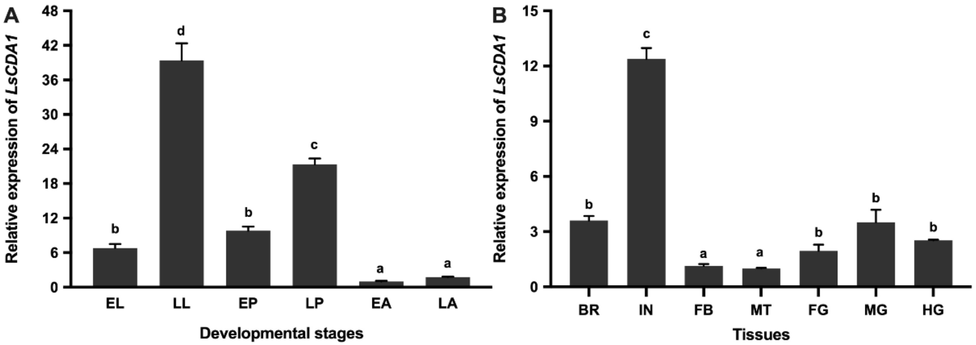
2.2. Developmental and Tissue-Specific Expression of LsCDA1

Quantitative real-time PCR (qPCR) was used to analyze the expression of LsCDA1 in different developmental stages and different tissues of L. serricorne. Among the six assessed stages, LsCDA1 was stably expressed at all of the stages; however, it was highly expressed in the late larval and late pupal stages (Figure 3a). The expression of LsCDA1 varied among the seven tissues of late larvae, and the highest expression level of LsCDA1 was detected in the integument (Figure 3b).

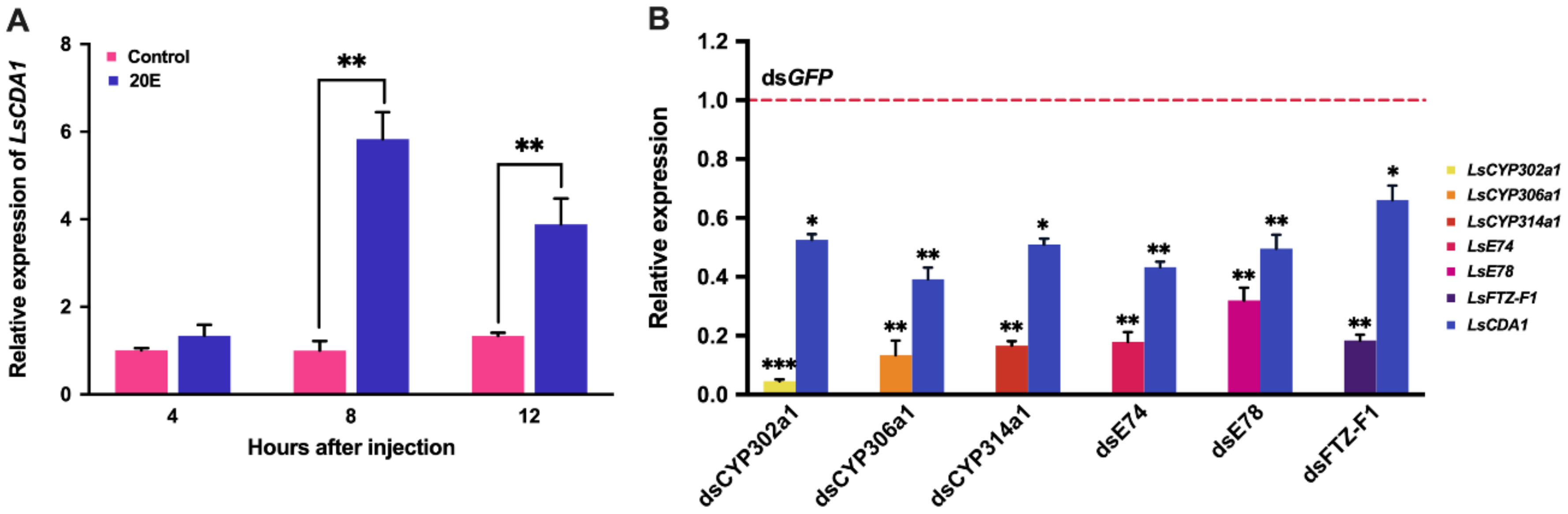
2.3. Expression of LsCDA1 in Response to 20E Signaling

To test whether LsCDA1 is induced by 20E, the fourth-instar larvae were injected with 20E for 4, 8 and 12 h. The expression of LsCDA1 in the 20E treatment group was significantly increased, and the ratios of inducible expression were 5.8- and 3.9-fold at 8 and 12 h post-injection, respectively, compared to the control group (Figure 4a). To further validate the regulation of LsCDA1 in response to 20E signaling, we individually silenced genes encoding ecdysteroidogenesis enzymes (CYP302a1, CYP306a1, and CYP314a1) and 20E signaling components (E74, E78, and FTZ-F1) by the injection of dsRNAs. The mRNA level of LsCDA1 was significantly decreased in the CYP302a1, CYP306a1, and CYP314a1 RNAi larvae compared to that in controls. We also found that the inhibition of the 20E signal in the E74, E78, and FTZ-F1 RNAi larvae significantly downregulated the expression of LsCDA1 (Figure 4b).

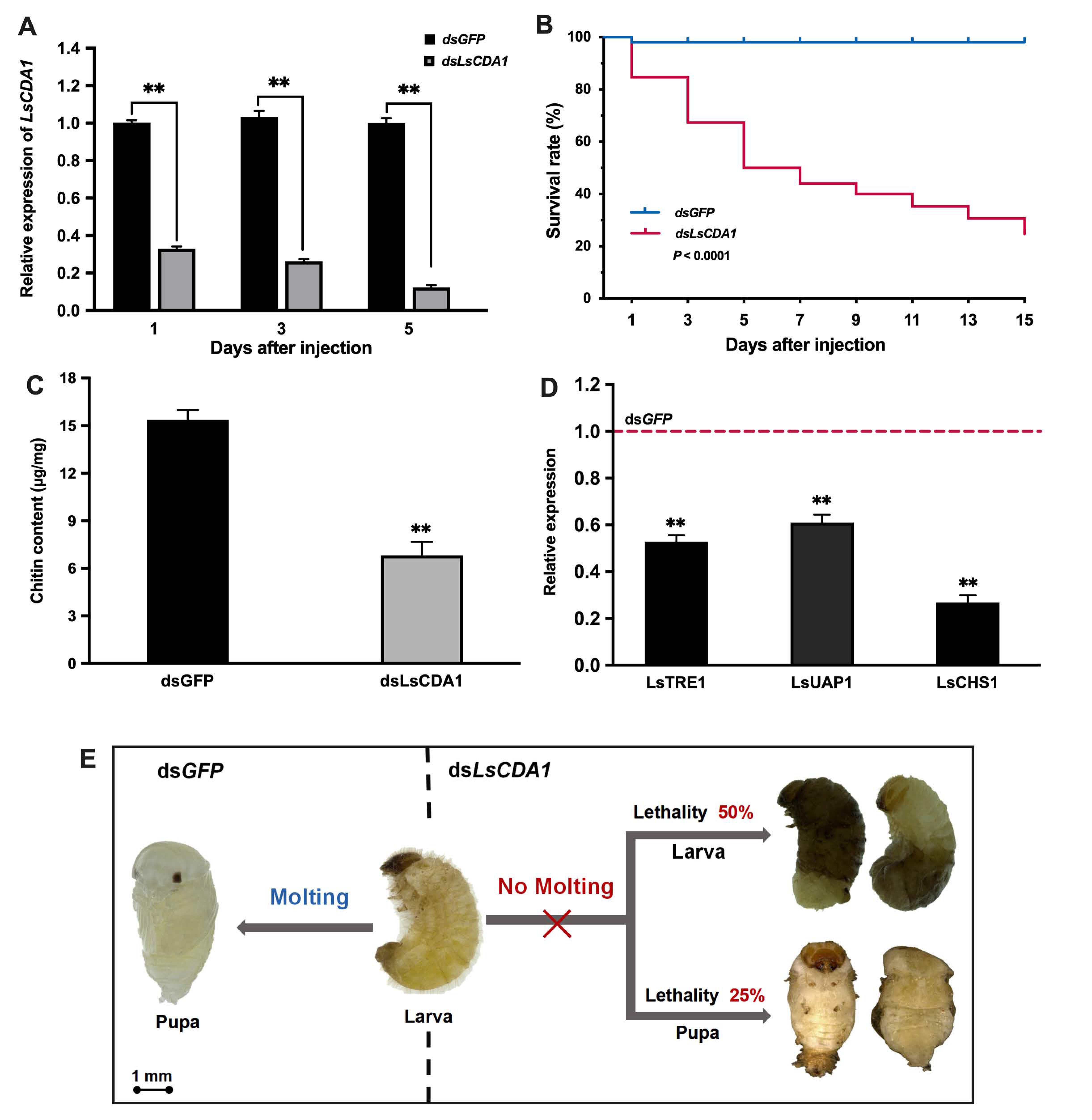
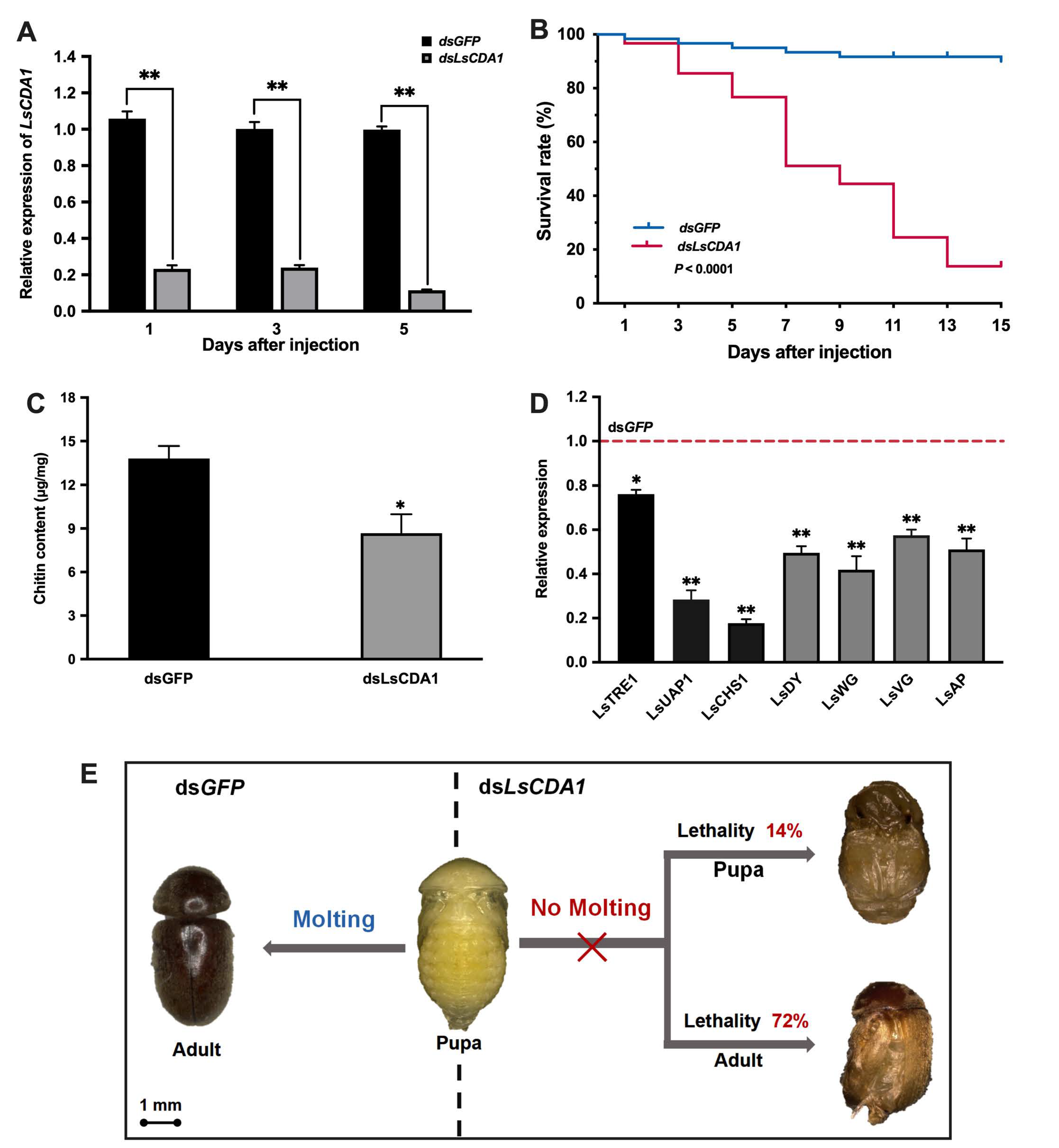
2.4. Knockdown of LsCDA1 Impairs Larval–Pupal and Pupal–Adult Transitions

To investigate the role of LsCDA1 in the molting process of L. serricorne, dsRNAs of LsCDA1 and green fluorescent protein (GFP) were synthesized in vitro and injected into fifth-instar larvae and late pupae, respectively. Compared to the control, the expression of LsCDA1 was significantly reduced after injection with dsLsCDA1 in the fifth-instar larvae (Figure 5a). The survival rate of L. serricorne was reduced to 25% in the dsLsCDA1 group, whereas 98% of the individuals in the control group could molt normally to pupae in the dsGFP group (Figure 5b). After the silencing of LsCDA1, the chitin contents were greatly decreased in the LsCDA1 RNAi larvae compared to those in the control samples (Figure 5c). The expression levels of chitin synthesis pathway genes, trehalase 1 (LsTRE1), UDP-N-acetylglucosamine pyrophosphorylase 1 (LsUAP1), and chitin synthase 1 (LsCHS1) were significantly lowered in the LsCDA1-depleted larvae (Figure 5d). Following the injection of dsLsCDA1, 50% of larvae were unable to shed their old cuticles and failed to complete the larval–pupal molting, and they eventually died; approximately 25% of the individuals successfully molted but died later (Figure 5e).
For the late pupae treated with dsLsCDA1, the transcript level of LsCDA1 was significantly decreased compared with the dsGFP group (Figure 6a). The survival rate of pupae injected with dsLsCDA1 was significantly lower compared to the controls, and 86% of individuals exhibited abnormal phenotypes and eventually died during the pupal–adult transition (Figure 6b). The chitin contents of the pupae were also reduced after the injection of dsLsCDA1 compared to those of control insects (Figure 6c), and the expression levels of three chitin synthesis pathway genes were significantly reduced in LsCDA1 RNAi pupae (Figure 6d). Furthermore, we examined the expression of four genes related to wing development in pupae after LsCDA1 silencing. The results showed that the expression levels of four genes, wingless (LsWG), vestigial (LsVG), dusky (LsDY), and apterous (LsAP), were significantly decreased compared to those in control insects (Figure 6d). Specifically, 14% of pupae failed to shed puparia and died without completing adult eclosion, whereas 72% of the pupae molted to deformed adults and emerged with wrinkled wings (Figure 6e). No visible phenotypic changes were observed in the dsGFP-injected group.

3. Discussion

3.1. Characteristics and Expression Profiles of LsCDA1

Chitin deacetylases play essential roles in chitin degradation during insect growth and development, and these enzymes have been identified in several insect species [10,13,14,15,16]. In this study, we report the complete ORF sequence encoding chitin deacetylase 1, LsCDA1, from the cigarette beetle. Bioinformatic analysis showed that LsCDA1 contained a signal peptide, a ChtB domain, an LDLa binding domain, and a CDA catalytic domain, indicating that it belonged to the carbohydrate esterase family 4 [11]. These structural domains are also considered as expression sequence tags of group I and II chitin deacetylases [7,17]. Five well-conserved motifs of deacetylase proteins [49], TFDD, HSITH, RAPY, FLYDS, and MYFRMP, were present in the CDA catalytic region of LsCDA1. Homology alignment and phylogenetic analysis revealed that LsCDA1 belonged to subgroup Ib CDAs. LsCDA1 shares a closer relationship with SpCDA1 in S. paniceum and TcCDA1 in T. castaneum, suggesting that they may have similar functions during insect molting [13,14].
Developmental expression analyses indicated that LsCDA1 was highly expressed in late larval and late pupal stages, which are specific periods prior to each molt. Similar results have been confirmed for many CDA1 genes of other insect species, such as N. lugens [10], T. castaneum [13], S. paniceum [14], and H. vitessoides [27]. The dramatically increased expression of CDA1 was probably related to the requirement for chitin deposition during molt periods. Our previous studies have demonstrated a significant increase in the expression levels of two chitin-degrading enzyme genes (LsNAG1 and LsNAG2) during the larval–pupal and pupal–adult molts of L. serricorne [47,48]. Collectively, these results suggest that LsCDA1 and two LsNAGs signified their important roles in the molting of L. serricorne. In Helicoverpa armigera, large amounts of HaCDA1 transcripts were observed in the fat body [23]. In D. melanogatser, DmCDA1 was highly expressed in the embryonic trachea [28]. In L. decemlineata, LdCDA1 was copiously expressed in the larval ileum, rectum, and epidermis [15]. In L. migratoria, LmCDA1 was highly expressed in the foregut, and lower transcripts were also detected in the integument and hindgut, but were not detected in other examined tissues [19]. The rice leaf folder CmCDA1 was expressed primarily in the head and midgut [50]. In the pharate appendages of T. castaneum adults, TcCDA1-specific in situ hybridization showed a strong signal in the epidermal cells [13]. In the present study, LsCDA1 was predominantly expressed in the integument at the larval stages, which is consistent with the expression of NlCDA1, NlCDA2, and NlCDA4 from N. lugens [10], HcCDA2 from H. cunea [24], and BmCDA1 and BmCDA2 from B. mori [25]. We speculated that these CDAs are responsible for chitin metabolism in the integument and are crucial for insect molting. Actually, the results were not always in accordance with those of previous studies. For example, the injection of dsRNAs of BmCDA1 and BmCDA2 in the larvae of B. mori did not reveal any morphological abnormalities, but rather led to delayed pupation times [25]. Therefore, we proposed that the high expression of CDAs in the integument might be associated with additional physiological functions.

3.2. Transcriptional Regulation of LsCDA1 by 20E

There is a functional relationship between 20E and the chitin metabolism system. It has been documented that 20E and its signal pathway play vital roles in regulating insect molting and metamorphosis [51]. Cholesterol or other sterols are precursors and are converted to ecdysone and its derivative 20E by a series of Halloween genes, such as Spook (CYP307a1), Phantom (CYP306a1), Disembodied (CYP302a1), Shadow (CYP315a1), and Shade (CYP314a1) [52]. Then, 20E binds its complex nuclear receptors, and transduces its signal through a subset of primary response genes (E74, E75, Broad, HR3, HR4, E78, FTZ-F1, and others) [53]. Previous studies in Spodotera exigua showed that 20E and its receptors could induce the expression of five chitin synthesis pathway genes [54]. In L. migratoria, the injection of 20E and the downregulation of ecdysone receptor genes increased and decreased the expression of two variants of the chitinase 5 gene (LmCht5) [55]. The injection of 20E could upregulate the gene expression of NAG genes in L. serricorne [47] and L. migratoria [56]. Interestingly, 20E also stimulated the expression of the chitin synthase 1 gene in B. dorsalis [57], B. mori [58], and L. decemlineate [59]. In this study, we found that LsCDA1 expression was induced by 20E in vivo, and the expression was suppressed by the silencing of three ecdysteroidogenesis genes (CYP302a1, CYP306a1, and CYP314a1) and three 20E signaling genes (E74, E78, and FTZ-F1). The results indicated that LsCDA1 expression could be regulated by 20E synthesis and the 20E signaling pathway. We confirmed that LsCDA1 was affected by a 20E pulse. Similar findings have been reported for LdCDA1 and LdCDA2 in L. decemlineata; however, the expressions levels of two LdCDAs were inhibited after the application of juvenile hormone (JH) and its analogs, and were stimulated by the silencing of JH synthesis and signaling genes [15,16]. In S. paniceum [14] and H. vitessoides [27], exposure to 20E could activate the expression of CDA genes at various times. Substantial upregulation of six BmCDAs (BmCDA1BmCDA6) by 20E was observed not only in the larval cuticles and midguts, but also in embryo cell lines of B. mori [25]. These results strongly suggest that CDAs are important 20E late-responsive genes.

3.3. Knockdown of LsCDA1 Affects Chitin Metabolism and Impairs Molting

It has been reported that CDA1 plays roles in the formation of the cuticle during insect molting. In this study, RNAi was used to evaluate the functions of LsCDA1, and specific dsLsCDA1 was injected into the fifth-instar larvae and late pupae of L. serricorne. Our results revealed that the silencing of LsCDA1 caused developmental disturbances and lethal morphological phenotypes, suggesting that LsCDA1 is critical for successful larval–pupal and pupal–adult molts. This phenomenon is consistent with previous reports. In T. castaneum and N. lugens, the knockdown of CDA1 disrupted the metamorphic transitions, and the treated insects failed to shed their old cuticles and eventually died [10,13]. In Oxya chinensis, nymphs injected with dsCDA1 displayed arrested development, and more than 95% died during the molting process [18]. The function of CDA1 in the molting was also studied in S. paniceum [14], L. migratoria [19], and H. vitessoides [27]. Given that CDA1 is essential for insect metamorphosis, it could be a promising target for RNAi-based insecticide development. Meanwhile, we found the chitin content was significantly reduced in the LsCDA1 RNAi beetles, and the expression levels of LsTRE1, LsUAP1, and LsCHS1 were dramatically decreased. Similar results have been observed in L. decemlineata larvae, where RNAi of LdCDA1 decreased the chitin amounts and the mRNA levels of five chitin synthesis pathway genes [15]. This indicates that repression of CDA1 may simultaneously affect chitin synthesis during the molting process. Taken together, the results suggest that LsCDA1 is responsible for chitin degradation and synthesis in the cuticle, and is critical for L. serricorne development. However, the expression levels of chitin synthesis pathway genes were not affected after the silencing of HvNAG1 in the larvae of H. vitessoides [60]. Therefore, the underlying mechanism of how chitin-degrading enzymes assist chitin synthesis is complicated and needs further investigation.

3.4. The Function of LsCDA1 Involved in Wing Development During Pupal–Adult Metamorphosis

Functional studies in D. melanogaster demonstrated that Group I CDAs play distinct roles in wing development. Specifically, DmCDA1 is the main enzyme responsible for chitin deacetylation in wing cuticle formation, whereas DmCDA2 is required for the laminar arrangement of chitin. Additionally, both enzymes contribute to the establishment of the cuticular inward barrier against the penetration of xenobiotics [30]. In the current study, the silencing of LsCDA1 in the late pupae of L. serricorne resulted in wing deformities. In this group, 72% of the individuals molted to adults but died with wrinkled wings. Similar phenotypes have also been observed after RNAi of CDA1 in L. decemlineata [15] and H. vitessoides [27]. Interestingly, other chitin-degrading enzymes, such as NAG and chitinase, have been reported to be crucial for normal wing formation. In H. vitessoides and L. serricorne, the suppression of NAG expression led to abnormal wing phenotypes [47,60]. In Sogatella furcifera, the injection of SfCHT7 dsRNA caused individuals to emerge with deformed wings [61]. It seems that chitin deposition is indispensable for adult wing development. However, the mechanism by which chitin-degrading genes control the development of wings remains unclear. Previous studies demonstrated that these genes, such as dusky (DY), wingless (WG), vestigial (VG), and apterous (AP), have essential roles during the wing morphogenesis of T. castaneum [62,63]. Here, we found that the expression levels of LsDY, LsWG, LsVG, and LsAP were downregulated after the knockdown of LsCDA1 in L. serricorne. This suggests that LsCDA1 controls beetle molting by regulating the expression of wing development genes during the pupal–adult transition.

4. Materials and Methods

4.1. Insects

The cigarette beetle L. serricorne was originally collected from a tobacco warehouse (N26°30′, E106°40′) in Guizhou Province, China. All of the insects were incubated in our laboratory at 28 °C under a constant 24-h darkness with 40% relative humidity. The larvae were reared on the dried roots of Angelica sinensis as described previously [47].

4.2. Molecular Cloning and Bioinformatic Analysis

Total RNA was extracted from L. serricorne larvae using a MiniBEST Universal Extraction Kit (TaKaRa, Dalian, China), following the manufacturer’s instructions. The integrity and quantity of RNA were evaluated using 1% agarose gel electrophoresis and a NanoDrop 2000C spectrophotometer (Thermo Fisher Scientific, Waltham, MA, USA). One microgram of total RNA was used to synthesize first-strand cDNA with the TransScript Synthesis Supermix (TransGen, Beijing, China). The cDNA library of L. serricorne integument was constructed using an mRNA sequencing assay on an Illumina HisSeq2000 platform by Biomarker Technologies (Beijing, China). The putative CDA1 sequence was obtained from this transcriptome database. The full-length ORF sequence of CDA1 was acquired by using the ORF Finder software (https://0-www-ncbi-nlm-nih-gov.brum.beds.ac.uk/orffinder). The correctness of the sequence was substantiated by RT-PCR using specific primers (Supplementary Table S2). PCR was conducted as follows: 95 °C for 5 min; 34 cycles of 95 °C for 30 s, 57 °C for 30 s, and 72 °C for 2 min; and then an extension at 72 °C for 10 min. The PCR product was cloned into the pMD19-T vector (TaKaRa, Dalian, China) and then sequenced (Tsingke Bio, Chengdu, China).
Sequence similarities were determined using the Basic Local Alignment Search Tool program of the NCBI website (https://blast.ncbi.nlm.nih.gov/Blast.cgi). DNAMAN 6.0 (LynnonBiosoft, Vaudreuil, Quebec, Canada) was used to edit the sequence. The signal peptide, molecular weight, and isoelectric point were predicted by the ExPASy Proteomics Server (http://www.expasy.ch). Domain predictions were produced by the Simple Modular Architecture Research Tool (http://smart.embl.de/). Amino acid sequences were aligned with Clustal W [64]. Phylogenetic tree construction was performed using MEGA7 [65] with the Maximum Likelihood method and General Reversible Chloroplast model with 1000 bootstrap replicates.

4.3. Developmental and Tissue-Specific Expression Analysis of LsCDA1

Developmental samples were collected from early larvae (<24 h after hatching), late larvae (older than fourth-instar, including prepupae), early pupae (>48 h after pupation), late pupae (>120 h after pupation, before emergence), early adults (<24 h after emergence), and late adults (one week old). For tissue distribution, seven tissues (integument, brain, foregut, midgut, hindgut, fat body, and Malpighian tubules) were dissected from the late larvae. Each sample included 30–50 individuals, and three biological replications were prepared. Total RNA extraction and first-strand cDNA synthesis were performed as described above. A qPCR was used to detect the expression profiles of LsCDA1. The qPCR was conducted on a CFX-96 real-time PCR Detection system (Bio-Rad, Hercules, CA, USA) with the GoTaq qPCR Master Mix (Promega, Madison, WI, USA) under the following conditions: initial denaturation at 95 °C for 2 min, followed by 40 cycles 95 °C for 30 s and 60 °C for 30 s. A melting curve analysis was performed to ensure the specificity of the qPCR. The fivefold dilution series of cDNA from the late larvae was used to construct a relative standard curve to determine the PCR efficiency of each primer for each gene. L. serricorne elongation factor 1-alpha (EF1α) and 18S ribosomal RNA (18S) were used as internal reference genes according to our published results [48,66]. Data were analyzed by the 2−∆∆Ct method [67], using the geometric mean of the two internal reference genes for normalization. All methods and data were confirmed to follow the Minimum Information for Publication of Quantitative Real-Time PCR Experiments (MIQE) guidelines [68].

4.4. Expression of LsCDA1 after 20E Treatment and RNAi of Ecdysone Synthesis and Signaling Genes

The newly ecdysed fourth-instar larvae were used for 20E treatment. According to our previous study [47], each larva in the treatment group was injected with 120 ng of 20E (Sigma-Aldrich, St. Louis, MO, USA) dissolved in 0.1% ethanol, to a final concentration of 1 μg/μL, while the control group was injected with an equal volume of 0.1% ethanol. Each larva was injected at the junction between the second and third abdominal segments using a Nanoliter 2010 injector (World Precision Instruments, Sarasota, FL, USA). For transcriptional analysis of LsCDA1, thirty individuals were randomly collected from each group at 4, 8, and 12 h post-injection. To further confirm whether LsCDA1 was regulated by 20E synthesis and signaling, we performed RNAi experiments using dsRNAs of ecdysteroidogenesis genes (CYP302a1, CYP306a1, and CYP314a1) and 20E signal pathway genes (E74, E78, and FTZ-F1). These dsRNAs were prepared using a TranscriptAid T7 High Yield Transcription Kit (Thermo Scientific, Wilmington, DE, USA), and dsGFP was used as the negative control. The dsRNAs of each ecdysone-related gene were injected into fifth-instar larvae (300 ng/larva) using a microinjector. The larval samples were collected from each group at 3 days post-injection, and total RNA was extracted to analyze the RNAi efficiency and expression of LsCDA1 by using qPCR as described above.

4.5. Function Analysis of LsCDA1 by RNAi

To evaluate the roles of LsCDA1 in L. serricorne development, dsRNAs (300 ng) of LsCDA1 and GFP were synthesized in vitro as described above and were injected into the fifth-instar larvae and the late pupae, respectively. The insects treated with dsRNA were reared under the same conditions as mentioned above. Insect survival and abnormal phenotypes were observed and photographed using a stereomicroscope (Keyence VHX-6000, Keyence Corporation, Osaka, Japan). To determine RNAi efficiency, the relative expression levels of LsCDA1 in both the dsLsCDA1 and dsGFP groups at 1, 3, and 5 days post-injection were detected by qPCR, as described above. To explore the effects of LsCDA1 RNAi on chitin metabolism, a chitin content assay was performed according to the previously described method [69]. In brief, the chitin content of whole insect bodies was measured by the SpectraMax M2 microplate reader (Molecular Devices, Sunnyvale, CA, USA) at 3 days after dsRNA injection. The chitin contents were given as micrograms per milligram of insect body. After LsCDA1 was knocked down in L. serricorne, the transcript levels of three chitin synthesis pathway genes (LsTRE1, LsUAP1, and LsCHS1) were detected by qPCR at 3 days after injection. To further explore the effects of LsCDA1 RNAi on wing development, samples were collected from the late pupae injected with dsLsCDA1 and dsGFP for 3 days. The relative expression levels of four wing development-related genes (LsDY, LsWG, LsVG, and LsAP) were determined by qPCR.

4.6. Statistical Analysis

Data were presented as mean ± standard error (SE) and were analyzed using SPSS 21.0 software (SPSS Inc., Chicago, IL, USA). Survival curves were analyzed using the Kaplan–Meier method, and the Log-Rank test was used to evaluate the significance of differences between the RNAi-treatment and control groups. Differences between two groups were compared using Student’s t-test (* p < 0.05, ** p < 0.01, *** p < 0.001). A one-way analysis of variance (ANOVA) followed by a least significant difference (LSD) test was applied for comparing the differences among more than two samples.

5. Conclusions

In summary, we identified a chitin deacetylase gene (LsCDA1) from L. serricorne. LsCDA1 was highly expressed in the integument and showed periodic expression during each molting. The expression of LsCDA1 could be regulated by a 20E pulse. The RNAi results reveal that LsCDA1 plays vital roles in molting transitions by blocking cuticle chitin synthesis and deposition. The knockdown of LsCDA1 inhibited the expression of wing development-related genes, resulting in wing deformities. These findings indicated LsCDA1 as a potential target for the control of L. serricorne.

Supplementary Materials

Supplementary materials can be found at https://0-www-mdpi-com.brum.beds.ac.uk/1422-0067/21/7/2449/s1, Table S1: Details of chitin deacetylase sequences used for phylogenetic analysis, Table S2: Primers used in this study.

Author Contributions

Conceived and designed this study: W.-J.Y. and D.-C.J. Collected the samples: K.-K.X. and Y.Y. Performed experiments: W.-J.Y., K.-K.X., and Y.Y. Analyzed the data: W.-J.Y and K.-K.X. Wrote the manuscript: W.-J.Y., C.L., and D.-C.J. All authors have read and agreed to the published version of the manuscript.

Funding

This study was supported in part by the National Natural Science Foundation of China (31501649), the Program for Science and Technology Outstanding talents in Higher Learning Institution of Guizhou Province (2017083), the Program for Science and Technology Youth Talents in Department of Education in Guizhou Province (2018298), the Program for Academician Workstation in Guiyang University (20195605), and the Program for First-class Discipline Construction in Guizhou Province (201785).

Conflicts of Interest

The authors declare no conflict of interest. The funders had no role in the design of the study; in the collection, analyses, or interpretation of data; in the writing of the manuscript, or in the decision to publish the results.

References

  1. Choi, H.K.; Choi, K.H.; Kramer, K.J.; Muthukrishnan, S. Isolation and characterization of a genomic clone for the gene of an insect molting enzyme, chitinase. Insect Biochem. Mol. Biol. 1997, 27, 37–47. [Google Scholar] [CrossRef]
  2. Merzendorfer, H.; Zimoch, L. Chitin metabolism in insects: Structure, function and regulation of chitin synthases and chitinases. J. Exp. Biol. 2003, 206, 4393–4412. [Google Scholar] [CrossRef] [PubMed] [Green Version]
  3. Moussian, B.; Schwarz, H.; Bartoszewski, S.; Nusslein-Volhard, C. Involvement of chitin in exoskeleton morphogenesis in Drosophila melanogaster. J. Morphol. 2005, 264, 117–130. [Google Scholar] [CrossRef] [PubMed]
  4. Nation, J.L. Insect Physiology and Biochemistry, 2nd ed.; CRC Press: Boca Raton, FL, USA, 2008. [Google Scholar]
  5. Cohen, E. Chitin synthesis and inhibition: A revisit. Pest Manag. Sci. 2001, 57, 946–950. [Google Scholar] [CrossRef]
  6. Kramer, K.; Muthukrishnan, S. Chitin metabolism in insects. Compr. Mol. Insect Sci. 2005, 4, 111–144. [Google Scholar]
  7. Tetreau, G.; Cao, X.L.; Chen, Y.R.; Muthukrishnan, S.; Jiang, H.B.; Blissard, G.W.; Kanost, M.R.; Wang, P. Overview of chitin metabolism enzymes in Manduca sexta: Identification, domain organization, phylogenetic analysis and gene expression. Insect Biochem. Mol. Biol. 2015, 62, 114–126. [Google Scholar] [CrossRef] [Green Version]
  8. Xi, Y.; Pan, P.L.; Ye, Y.X.; Yu, B.; Xu, H.J.; Zhang, C.X. Chitinase-like gene family in the brown planthopper, Nilaparvata lugens. Insect Mol. Biol. 2015, 24, 29–40. [Google Scholar] [CrossRef]
  9. Xi, Y.; Pan, P.L.; Zhang, C.X. The β-N-acetylhexosaminidase gene family in the brown planthopper, Nilaparvata lugens. Insect Mol. Biol. 2015, 24, 601–610. [Google Scholar] [CrossRef]
  10. Xi, Y.; Pan, P.L.; Ye, Y.X.; Yu, B.; Zhang, C.X. Chitin deacetylase family genes in the brown planthopper, Nilaparvata lugens (Hemiptera: Delphacidae). Insect Mol. Biol. 2014, 23, 695–705. [Google Scholar] [CrossRef]
  11. Tsigos, I.; Martinou, A.; Kafetzopoulos, D.; Bouriotis, V. Chitin deacetylases: New, versatile tools in biotechnology. Trends Biotechnol. 2000, 18, 305–312. [Google Scholar] [CrossRef]
  12. Guo, W.; Li, G.; Pang, Y.; Wang, P. A novel chitin binding protein identified from the peritrophic membrane of the cabbage looper, Trichoplusia ni. Insect Biochem. Mol. Biol. 2005, 35, 1224–1234. [Google Scholar] [CrossRef] [PubMed]
  13. Arakane, Y.; Dixit, R.; Begum, K.; Park, Y.; Specht, C.A.; Merzendorfer, H.; Kramer, K.J.; Muthukrishnan, S.; Beeman, R.W. Analysis of functions of the chitin deacetylase gene family in Tribolium castaneum. Insect Biochem. Mol. Biol. 2009, 39, 355–365. [Google Scholar] [CrossRef] [PubMed]
  14. Yang, W.J.; Xu, K.K.; Yan, X.; Chen, C.X.; Cao, Y.; Meng, Y.L.; Li, C. Functional characterization of chitin deacetylase 1 gene disrupting larval–pupal transition in the drugstore beetle using RNA interference. Comp. Biochem. Physiol. B 2018, 219–220, 10–16. [Google Scholar] [CrossRef] [PubMed]
  15. Wu, J.J.; Mu, L.L.; Chen, Z.C.; Fu, K.Y.; Guo, W.C.; Li, C.; Li, G.Q. Disruption of ecdysis in Leptinotarsa decemlineata by knockdown of chitin deacetylase 1. J. Asia Pac. Entomol. 2019, 22, 443–452. [Google Scholar] [CrossRef]
  16. Wu, J.J.; Chen, Z.C.; Wang, Y.W.; Fu, K.Y.; Guo, W.C.; Li, G.Q. Silencing chitin deacetylase 2 impairs larval–pupal and pupal-adult molts in Leptinotarsa decemlineata. Insect Mol. Biol. 2019, 28, 52–64. [Google Scholar] [CrossRef] [Green Version]
  17. Dixit, R.; Arakane, Y.; Specht, C.A.; Richard, C.; Kramer, K.J.; Beeman, R.W.; Muthukrishnan, S. Domain organization and phylogenetic analysis of proteins from the chitin deacetylase gene family of Tribolium castaneum and three other species of insects. Insect Biochem. Mol. Biol. 2008, 38, 440–451. [Google Scholar] [CrossRef] [Green Version]
  18. Ding, G.W.; Yu, R.R.; Yang, M.L.; Ma, E.B.; Yang, J.; Zhang, J.Z. Molecular characterization and functional analysis of chitin deacetylase 1 gene in Oxya chinensis (Orthoptera: Acrididae). Acta Entomol. Sin. 2014, 57, 1265–1271. [Google Scholar]
  19. Yu, R.R.; Liu, W.M.; Zhao, X.M.; Zhang, M.; Li, D.Q.; Zuber, R.; Ma, E.B.; Zhu, K.Y.; Moussian, B.; Zhang, J.Z. LmCDA1 organizes the cuticle by chitin deacetylation in Locusta migratoria. Insect Mol. Biol. 2019, 28, 301–312. [Google Scholar] [CrossRef] [Green Version]
  20. Luschnig, S.; Batz, T.; Armbruster, K.; Krasnow, M.A. Serpentine and vermiform encode matrix proteins with chitin binding and deacetylation domains that limit tracheal tube length in Drosophila. Curr. Biol. 2006, 16, 186–194. [Google Scholar] [CrossRef] [Green Version]
  21. Liu, S.H.; Li, H.F.; Yang, Y.; Yang, R.L.; Yang, W.J.; Jiang, H.B.; Dou, W.; Smagghe, G.; Wang, J.J. Genome-wide identification of chitinase and chitin deacetylase gene families in the oriental fruit fly, Bactrocera dorsalis (Hendel). Comp. Biochem. Physiol. D 2018, 27, 13–22. [Google Scholar] [CrossRef]
  22. Quan, G.; Ladd, T.; Duan, J.; Wen, F.; Doucet, D.; Cusson, M.; Krell, P.J. Characterization of a spruce budworm chitin deacetylase gene: Stage- and tissue-specific expression, and inhibition using RNA interference. Insect Biochem. Mol. Biol. 2013, 43, 683–691. [Google Scholar] [CrossRef] [PubMed]
  23. Han, G.; Li, X.; Zhang, T.; Zhu, X.; Li, J. Cloning and tissue-specific expression of a chitin deacetylase gene from Helicoverpa armigera (Lepidoptera: Noctuidae) and its response to Bacillus thuringiensis. J. Insect Sci. 2015, 15, 95. [Google Scholar] [CrossRef] [PubMed]
  24. Yan, X.P.; Zhao, D.; Zhang, Y.K.; Guo, W.; Wang, W.; Zhao, K.L.; Gao, Y.J.; Wang, X.Y. Identification and characterization of chitin deacetylase 2 from the American white moth, Hyphantria cunea (Drury). Gene 2018, 670, 98–105. [Google Scholar] [CrossRef] [PubMed]
  25. Zhang, Z.Y.; Yan, J.M.; Liu, Q.; Zhang, Y.H.; Gong, J.; Hou, Y. Genome-wide analysis and hormone regulation of chitin deacetylases in silkworm. Int. J. Mol. Sci. 2019, 20, 1679. [Google Scholar] [CrossRef] [Green Version]
  26. Sandoval-Mojica, A.F.; Scharf, M.E. Gut genes associated with the peritrophic matrix in Reticulitermes flavipes (Blattodea: Rhinotermitidae): Identification and characterization. Arch. Insect Biochem. Physiol. 2016, 92, 127–142. [Google Scholar] [CrossRef] [PubMed]
  27. Wang, C.Y.; Cheng, J.; Lyu, Z.H.; Li, Z.X.; Chen, J.X.; Lin, T. Chitin deacetylase 1 and 2 are indispensable for larval–pupal and pupal–adult molts in Heortia vitessoides (Lepidoptera: Crambidae). Comp. Biochem. Physiol. B 2019, 237, 110325. [Google Scholar] [CrossRef]
  28. Wang, S.; Jayaram, S.J.; Senti, K.; Tsarouhas, V.; Jin, H.; Samakovlis, C. Septate-junction-dependent luminal deposition of chitin deacetylases restricts tube elongation in the Drosophila trachea. Curr. Biol. 2006, 16, 180–185. [Google Scholar] [CrossRef] [Green Version]
  29. Gangishetti, U.; Veerkamp, J.; Bezdan, D.; Schwarz, H.; Lohmann, I.; Moussian, B. The transcription factor grainy head and the steroid hormone ecdysone cooperate during differentiation of the skin of Drosophila melanogaster. Insect Mol. Biol. 2012, 21, 283–295. [Google Scholar] [CrossRef]
  30. Zhang, M.; Ji, Y.N.; Zhang, X.B.; Ma, P.J.; Wang, Y.W.; Moussian, B.; Zhang, J.Z. The putative chitin deacetylases Serpentine and Vermiform have non-redundant functions during Drosophila wing development. Insect Biochem. Mol. Biol. 2019, 110, 128–135. [Google Scholar] [CrossRef]
  31. Toprak, U.; Baldwin, D.; Erlandson, M.; Gillott, C.; Hou, X.; Coutu, C.; Hegedus, D.D. A chitin deacetylase and putative insect intestinal lipases are components of the Mamestra configurata (Lepidoptera: Noctuidae) peritrophic matrix. Insect Mol. Biol. 2010, 17, 573–585. [Google Scholar] [CrossRef]
  32. Zhong, X.W.; Wang, X.H.; Tan, X.; Xia, Q.Y.; Xiang, Z.H.; Zhao, P. Identification and molecular characterization of a chitin deacetylase from Bombyx mori peritrophic membrane. Int. J. Mol. Sci. 2014, 15, 1946–1961. [Google Scholar] [CrossRef] [PubMed] [Green Version]
  33. Mahroof, R.M.; Phillips, T.W. Life history parameters of Lasioderma serricorne (F.) as influenced by food sources. J. Stored Prod. Res. 2008, 44, 219–226. [Google Scholar] [CrossRef]
  34. Li, C.; Li, Z.Z.; Cao, Y.; Zhou, B.; Zheng, X.W. Partial characterization of stress-induced carboxylesterase from adults of Stegobium paniceum and Lasioderma serricorne (Coleoptera: Anobiidae) subjected to CO2-enriched atmosphere. J. Pest Sci. 2009, 82, 7–11. [Google Scholar] [CrossRef]
  35. Minor, M.F. Do adult cigarette beetle feed? Tob. Sci. 1979, 23, 61–64. [Google Scholar]
  36. Riudavets, J.; Salas, I.; Pons, M.J. Damage characteristics produced by insect pests in packaging film. J. Stored Prod. Res. 2007, 43, 564–570. [Google Scholar] [CrossRef]
  37. Collins, D.; Conyers, S. The effect of sub-zero temperatures on different lifestages of Lasioderma serricorne (F.) and Ephestia elutella (Hübner). J. Stored Prod. Res. 2010, 46, 234–241. [Google Scholar] [CrossRef]
  38. Yu, C.; Subramanyam, B.; Flinn, P.W.; Gwirtz, J.A. Susceptibility of Lasioderma serricorne (Coleoptera: Anobiidae) life stages to elevated temperatures used during structural heat treatments. J. Econ. Entomol. 2011, 104, 317–324. [Google Scholar] [CrossRef]
  39. Imai, T. The additive effect of carbon dioxide on mortality of the cigarette beetle Lasioderma serricorne (Coleoptera: Anobiidae) in low-oxygen atmospheres. Appl. Entomol. Zool. 2015, 50, 11–15. [Google Scholar] [CrossRef]
  40. Saeed, M.B.E.E.E.M.; Laing, M.D.; Miller, R.M.; Bancole, B. Ovicidal, larvicidal and insecticidal activity of strains of Beauveria bassiana (Balsamo) Vuillemin against the cigarette beetle, Lasioderma serricorne Fabricius (Coleoptera: Anobiidae), on rice grain. J. Stored Prod. Res. 2017, 74, 78–86. [Google Scholar] [CrossRef]
  41. Hironaka, M.; Kamura, T.; Osada, M.; Sasaki, R.; Shinoda, K.; Hariyama, T.; Miyatake, T. Adults of Lasioderma serricorne and Stegobium paniceum (Anobiidae: Coleoptera) are attracted to ultraviolet (UV) over blue light LEDs. J. Econ. Entomol. 2017, 110, 1911–1915. [Google Scholar] [CrossRef]
  42. Rajendran, S.; Narasimhan, K.S. Phosphine resistance in the cigarette beetle Lasioderma serricorne (Coleoptera: Anobiidae) and overcoming control failures during fumigation of stored tobacco. Int. J. Pest Manag. 1994, 40, 207–210. [Google Scholar] [CrossRef]
  43. Sağlam, Ö.; Edde, P.A.; Phillips, T.W. Resistance of Lasioderma serricorne (Coleoptera: Anobiidae) to fumigation with phosphine. J. Econ. Entomol. 2015, 108, 2489–2495. [Google Scholar] [CrossRef] [PubMed]
  44. Abdelghany, A.Y.; Awadalla, S.S.; Abdel-Baky, N.F.; EL-Syrafi, H.A.; Fields, P.G. Efficacy of reduced risk insecticides on penetration into jute and polyethylene bags by Lasioderma serricorne (F.) (Coleoptera: Anobiidae). J. Stored Prod. Res. 2016, 69, 190–194. [Google Scholar] [CrossRef]
  45. Wu, Y.; Zhang, W.J.; Huang, D.Y.; Wang, Y.; Wei, J.Y.; Li, Z.H.; Sun, J.S.; Bai, J.F.; Tian, Z.F.; Wang, P.J.; et al. Chemical compositions and insecticidal activities of Alpinia kwangsiensis essential oil against Lasioderma serricorne. Molecules 2015, 20, 21939–21945. [Google Scholar] [CrossRef]
  46. Cohen, E. Chitin synthesis and degradation as targets for pesticide action. Arch. Insect Biochem. Physiol. 1993, 22, 245–261. [Google Scholar] [CrossRef]
  47. Chen, X.Y.L.; Xu, K.K.; Yan, X.; Chen, C.X.; Cao, Y.; Wang, Y.W.; Li, C.; Yang, W.J. Characterization of a β-N-acetylglucosaminidase gene and its involvement in the development of Lasioderma serricorne (Fabricius). J. Stored Prod. Res. 2018, 77, 156–165. [Google Scholar] [CrossRef]
  48. Yang, W.J.; Xu, K.K.; Yan, Y.; Li, C. Knockdown of β-N-acetylglucosaminidase 2 impairs molting and wing development in Lasioderma serricorne (Fabricius). Insects 2019, 10, 396. [Google Scholar] [CrossRef] [Green Version]
  49. Blair, D.E.; Schuttelkopf, A.W.; MacRae, J.I.; vanAalten, D.M. Structure and metal-dependent mechanism of peptidoglycan deacetylase, a streptococcal virulence factor. Proc. Natl. Acad. Sci. USA 2005, 102, 15429–15434. [Google Scholar] [CrossRef] [Green Version]
  50. Yu, H.Z.; Liu, M.H.; Wang, X.Y.; Yang, X.; Wang, W.L.; Geng, L.; Yu, D.; Liu, X.L.; Xu, J.P. Identification and expression profiles of chitin deacetylase genes in the rice leaf folder, Cnaphalocrocis medinalis. J. Asia Pac. Entomol. 2016, 19, 691–696. [Google Scholar] [CrossRef]
  51. Luan, J.B.; Ghanim, M.; Liu, S.S.; Czosnek, H. Silencing the ecdysone synthesis and signaling pathway genes disrupts nymphal development in the whitefly. Insect Biochem. Mol. Biol. 2013, 43, 740–746. [Google Scholar] [CrossRef]
  52. Niwa, R.; Niwa, Y.S. Enzymes for ecdysteroid biosynthesis: Their biological functions in insects and beyond. Biosci. Biotechnol. Biochem. 2014, 78, 1283–1292. [Google Scholar] [CrossRef] [PubMed] [Green Version]
  53. Hill, R.J.; Billas, I.M.; Bonneton, F.; Graham, L.D.; Lawrence, M.C. Ecdysone receptors: From the Ashburner model to structural biology. Annu. Rev. Entomol. 2013, 58, 251–271. [Google Scholar] [CrossRef] [PubMed]
  54. Yao, Q.; Zhang, D.W.; Tang, B.; Chen, J.; Chen, J.; Lu, L.; Zhang, W.Q. Identification of 20-hydroxyecdysone late-response genes in the chitin biosynthesis pathway. PLoS ONE 2010, 5, e14058. [Google Scholar] [CrossRef] [PubMed]
  55. Li, D.Q.; Zhang, J.Q.; Wang, Y.; Liu, X.J.; Ma, E.B.; Sun, Y.; Li, S.; Zhu, K.Y.; Zhang, J.Z. Two chitinase 5 genes from Locusta migratoria: Molecular characteristics and functional differentiation. Insect Biochem. Mol. Biol. 2015, 58, 46–54. [Google Scholar] [CrossRef] [PubMed]
  56. Rong, S.; Li, D.Q.; Zhang, X.Y.; Li, S.; Zhu, K.Y.; Guo, Y.P.; Ma, E.B.; Zhang, J.Z. RNA interference to reveal roles of β-N-acetylglucosaminidase gene during molting process in Locusta migratoria. Insect Sci. 2013, 20, 109–119. [Google Scholar] [CrossRef] [PubMed] [Green Version]
  57. Yang, W.J.; Xu, K.K.; Cong, L.; Wang, J.J. Identification, mRNA expression, and functional analysis of chitin synthase 1 gene and its two alternative splicing variants in oriental fruit fly, Bactrocera dorsalis. Int. J. Biol. Sci. 2013, 9, 331–342. [Google Scholar] [CrossRef] [Green Version]
  58. Zhuo, W.W.; Fang, Y.; Kong, L.F.; Li, X.; Sima, Y.H.; Xu, S.Q. Chitin synthase A: A novel epidermal development regulation gene in the larvae of Bombyx mori. Mol. Biol. Rep. 2014, 41, 4177–4186. [Google Scholar] [CrossRef]
  59. Shi, J.F.; Mu, L.L.; Guo, W.C.; Li, G.Q. Identification and hormone induction of putative chitin synthase genes and splice variants in Leptinotarsa decemlineata (SAY). Arch. Insect Biochem. Physiol. 2016, 92, 242–258. [Google Scholar] [CrossRef]
  60. Lyu, Z.H.; Chen, J.X.; Li, Z.X.; Cheng, J.; Wang, C.Y.; Lin, T. Knockdown of β-N-acetylglucosaminidase gene disrupts molting process in Heortia vitessoides Moore. Arch. Insect Biochem. Physiol. 2019, 101, e21561. [Google Scholar] [CrossRef]
  61. Chen, C.; Yang, H.; Tang, B.; Yang, W.J.; Jin, D.C. Identification and functional analysis of chitinase 7 gene in white-backed planthopper, Sogatella furcifera. Comp. Biochem. Physiol. B 2017, 208–209, 19–28. [Google Scholar] [CrossRef]
  62. Clark-Hachtel, C.M.; Linz, D.M.; Tomoyasu, Y. Insights into insect wing origin provided by functional analysis of vestigial in the red flour beetle, Tribolium castaneum. Proc. Natl. Acad. Sci. USA 2013, 110, 16951–16956. [Google Scholar] [CrossRef] [PubMed] [Green Version]
  63. Li, C.; Li, B.; Ma, S.; Lü, P.; Chen, K. Dusky works upstream of Four-jointed and Forked in wing morphogenesis in Tribolium castaneum. Insect Mol. Biol. 2017, 26, 677–686. [Google Scholar] [CrossRef] [PubMed]
  64. Larkin, M.A.; Blackshields, G.; Brown, N.P.; Chenna, R.; McGettigan, P.A.; McWilliam, H.; Valentin, F.; Wallace, I.M.; Wilm, A.; Lopez, R.; et al. Clustal W and Clustal X version 2.0. Bioinformatics 2007, 23, 2947–2948. [Google Scholar] [CrossRef] [PubMed] [Green Version]
  65. Kumar, S.; Stecher, G.; Tamura, K. MEGA7: Molecular evolutionary genetics analysis version 7.0 for bigger datasets. Mol. Biol. Evol. 2016, 33, 1870–1874. [Google Scholar] [CrossRef] [PubMed] [Green Version]
  66. Yang, W.J.; Xu, K.K.; Cao, Y.; Meng, Y.L.; Liu, Y.; Li, C. Identification and expression analysis of four small heat shock protein genes in cigarette beetle, Lasioderma serricorne (Fabricius). Insects 2019, 10, 139. [Google Scholar] [CrossRef] [Green Version]
  67. Livak, K.J.; Schmittgen, T.D. Analysis of relative gene expression data using real-time quantitative PCR and the 2-ΔΔCT method. Methods 2001, 25, 402–408. [Google Scholar] [CrossRef]
  68. Bustin, S.A.; Benes, V.; Garson, J.A.; Hellemas, J.; Huggett, J.; Kubista, M.; Mueller, R.; Nolan, T.; Pfaffl, M.W.; Shipley, G.L.; et al. The MIQE guidelines: Minimum information for publication of quantitative real-time PCR experiments. Clin Chem. 2009, 55, 611–622. [Google Scholar] [CrossRef] [Green Version]
  69. Yang, W.J.; Chen, C.X.; Yan, Y.; Xu, K.K.; Li, C. Clip-domain serine protease gene (LsCLIP3) is essential for larval–pupal molting and immunity in Lasioderma serricorne. Front. Physiol. 2020, 10, 1631. [Google Scholar] [CrossRef]
Figure 1. The nucleotide and deduced amino acid sequences of LsCDA1 cDNA. The start codon is indicated in bold and the stop codon in bold with an asterisk. The predicted signal peptide, chitin-binding domain, low-density lipoprotein receptor class A domain, and polysaccharide deacetylase-like catalytic domain are underlined. Five signature motifs (motifs 1–5) of the sequence catalytic domain are boxed. The primers for LsCDA1 dsRNA synthesis are shaded.
Ijms 21 02449 g001
Figure 2. Phylogenetic analysis of insect chitin deacetylases (CDAs). The phylogenetic tree was constructed using the MEGA7 software based on the Maximum Likelihood method. A bootstrap analysis of 1000 replications was used, and bootstrap values (only above 50%) are shown in the cladogram. Am, Apis mellifera; Bd, Bactrocera dorsalis; Bm, Bombyx mori; Dm, Drosophila melanogaster; Ha, Helicoverpa armigera; Hv, Heortia vitessoides; Ld, Leptinotarsa decemlineata; Lm, Locusta migratoria; Ls, Lasioderma serricorne; Mb, Mamestra brassicae; Nv, Nasonia vitripennis; Sp, Stegobium paniceum; Tc, Tribolium castaneum. The L. serricorne LsCDA1 protein is marked with a red triangle. The sequences presented in the phylogenetic tree are listed in Supplementary Table S1.
Ijms 21 02449 g002
Figure 3. Expression profiles of LsCDA1 in the different developmental stages (A) and different larval tissues (B) of L. serricorne. Various stages are collected. EL, early larvae; LL, late larvae; EP, early pupae; LP, late pupae; EA, early adults; LA, late adults. Different tissues are listed. BA, brain; IN, integument; FB, fat body; MT, Malpighian tubules; FG, foregut; MG, midgut; HG, hindgut. Data were normalized to the geometric mean of the expression of EF1α and 18S. Different letters above bars indicate significant differences based on one-way ANOVA followed by a least significance difference test (p < 0.05).
Ijms 21 02449 g003
Figure 4. 20-Hydroxyecdysone-responsiveness of LsCDA1 in L. serricorne. (A) The in vivo effects of 20E on the expression of LsCDA1. The larvae were collected for qPCR analysis at 4, 8 and 12 h after injection with 20E. Control: insects injected with distilled water containing 0.1% ethanol; 20E: insects injected with 20E (120 ng/larva). (B) The relative expression levels of LsCDA1, LsCYP302a1, LsCYP306a1, LsCYP314a1, LsE74, LsE78, and LsFTZ-F1 at 3 days after gene-specific dsRNA injection. The gene expression in the dsGFP group was set as 1 and is shown by the dotted line. Data were normalized to the geometric mean of the expression of EF1α and 18S. Significant differences between the treatment group and control group were determined using Student’s t-test (* p < 0.05, ** p < 0.01, *** p < 0.001).
Ijms 21 02449 g004
Figure 5. The effect of LsCDA1 knockdown on larval–pupal molting in L. serricorne. (A) The relative expression levels of LsCDA1 at 1, 3, and 5 days after LsCDA1 or GFP dsRNA injection in the fifth-instar larvae. (B) Kaplan-Meier survival curves of L. serricorne larvae after LsCDA1 or GFP dsRNA injection. The effects of LsCDA1 knockdown on chitin contents (C) and the expressions of three chitin synthesis pathway genes (D). The expression values of chitin synthesis pathway genes were calculated by comparison to the dsGFP group, which was normalized at 1. (E) Representative phenotypes of larvae after LsCDA1 or GFP dsRNA injection. Significant differences between the RNAi group and the control group were determined using Student’s t-test (** p < 0.01).
Ijms 21 02449 g005
Figure 6. The effect of LsCDA1 knockdown on pupal–adult metamorphosis in L. serricorne. (A) The relative expression levels of LsCDA1 at 1, 3, and 5 days after LsCDA1 or GFP dsRNA injection at the late pupal stage. (B) Kaplan-Meier survival curves of L. serricorne pupae after LsCDA1 or GFP dsRNA injection. The effects of LsCDA1 knockdown on chitin contents (C) and the expressions of three chitin synthesis pathway genes and four genes involved in wing development (D). The expression values were calculated by comparison to the dsGFP group, which was normalized at 1. (E) Representative phenotypes of pupae after LsCDA1 or GFP dsRNA injection. Significant differences between the RNAi group and the control group were determined using by Student’s t-test (* p < 0.05, ** p < 0.01).
Ijms 21 02449 g006

