Recent Advances in Electrospun Sustainable Composites for Biomedical, Environmental, Energy, and Packaging Applications


 ,
, 
Abstract
:1. Introduction
2. Electrospinning Technology
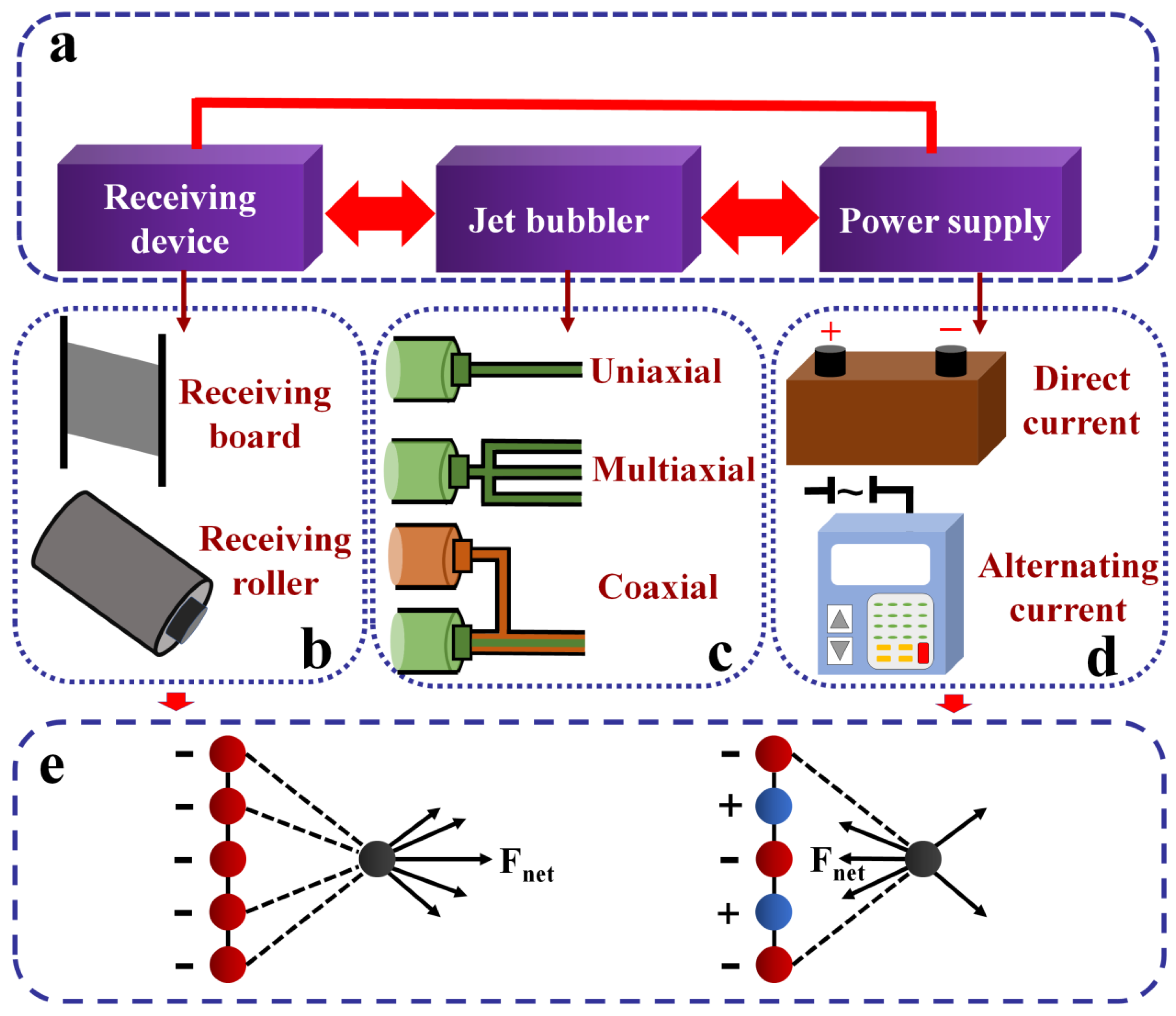
2.1. Electrospinning Devices
2.1.1. Direct Current (DC) Electrospinning
2.1.2. Alternating Current (AC) Electrospinning
2.2. Factors Influencing the Electrospinning Process
2.2.1. The Effect of Electrospinning Fluid Properties
2.2.2. The Effect of Operating Conditions
2.2.3. The Effect of Environment Conditions

| Factor | Parameter | Point of Action | Reference |
|---|---|---|---|
| Properties of electrospinning fluid | Liquid viscosity | Fiber diameter and uniformity | [2,28,64,68,70,74,80,81,82,83,84,85,86,87] |
| Electrical conductivity | Fiber diameter and distribution | [2,28,66,70,71,81,84,87,88,89,90] | |
| Surface tension | Fiber formation | [64,67,68,70,91,92] | |
| Operating conditions | Voltage | Fiber diameter | [68,72,76,83] |
| Needle size | Fiber diameter | [65,69,76] | |
| Receiving distance | Solvent volatilization and fiber diameter | [69,74] | |
| Spinning solution flow rate | Fiber diameter | [74,75,93] | |
| Environment conditions | Temperature | Solvent volatilization and liquid viscosity | [28,77] |
| Humidity | Solvent volatilization | [78,79] |
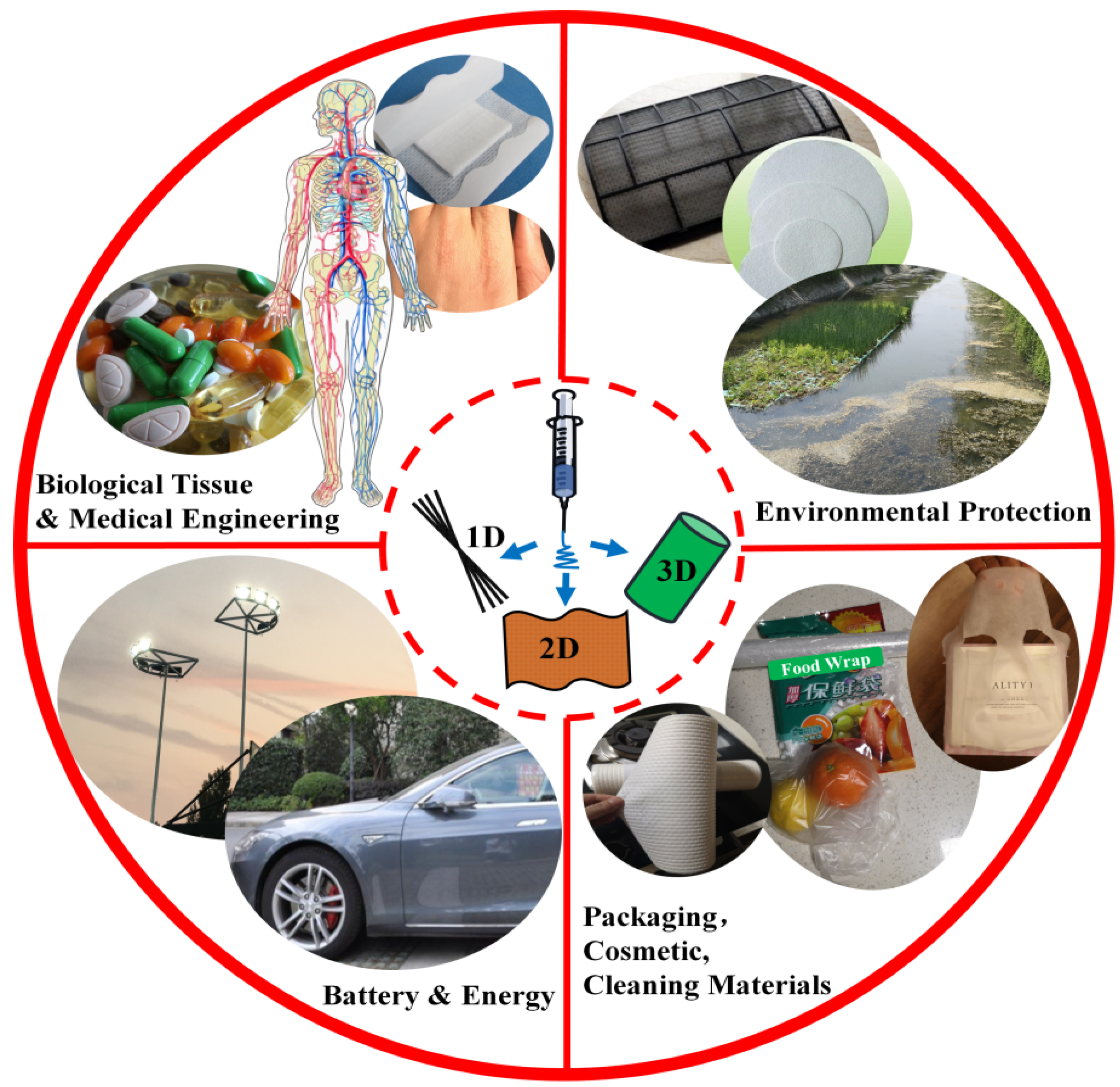
3. Application of Electrospun Sustainable Composite Materials
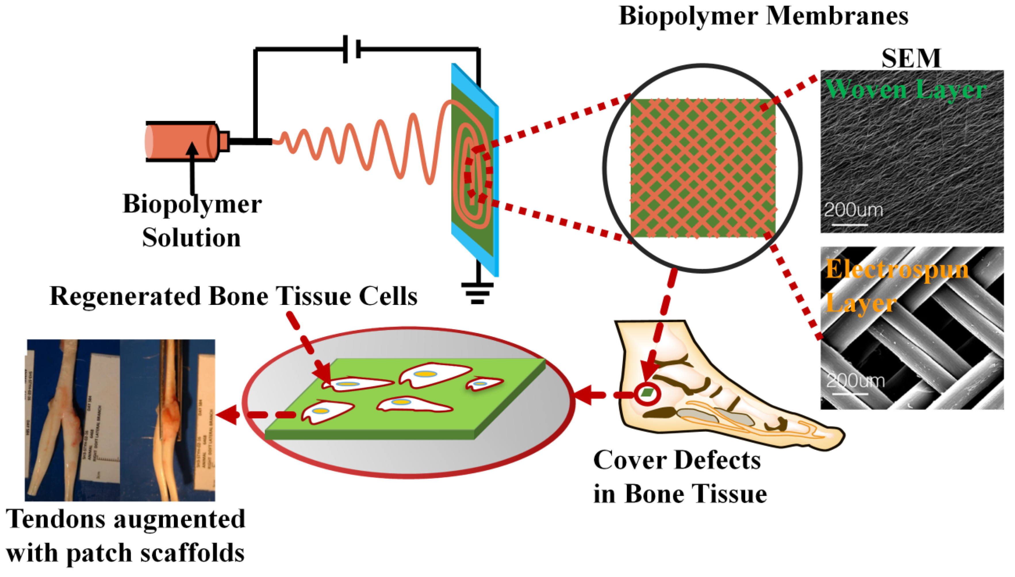
3.1. Recent Biological and Medical Engineering Applications
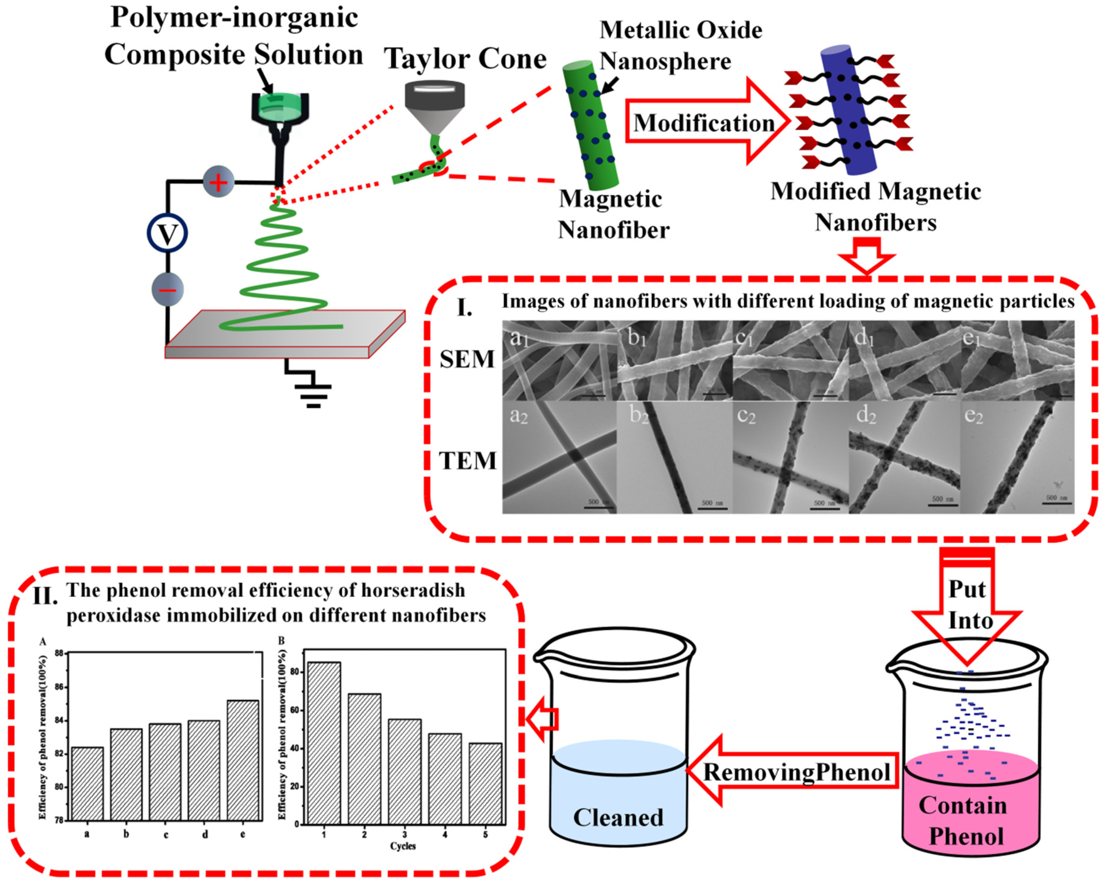
3.2. Recent Environmental Engineering Applications
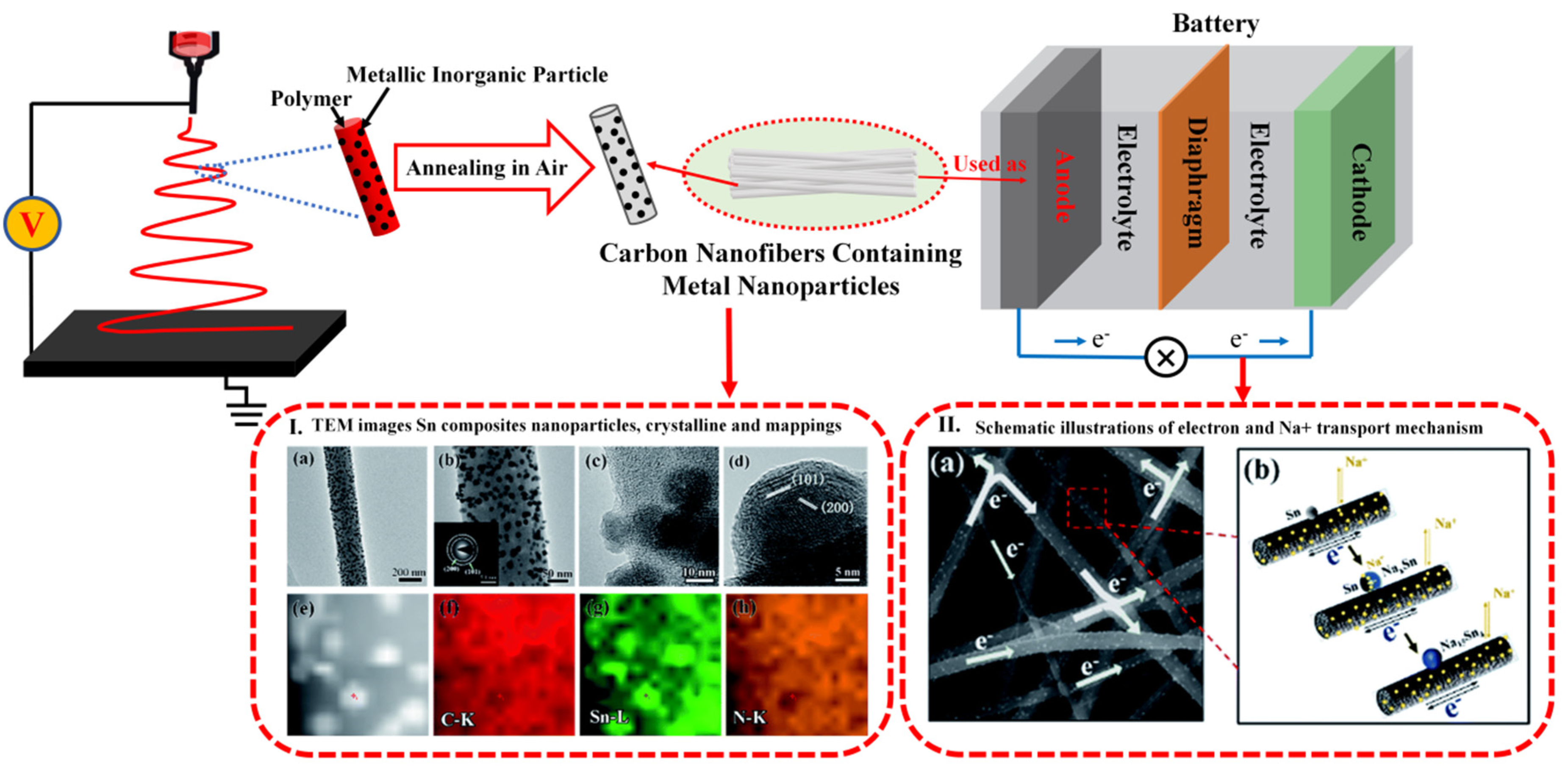
3.3. Recent Energy Material Applications
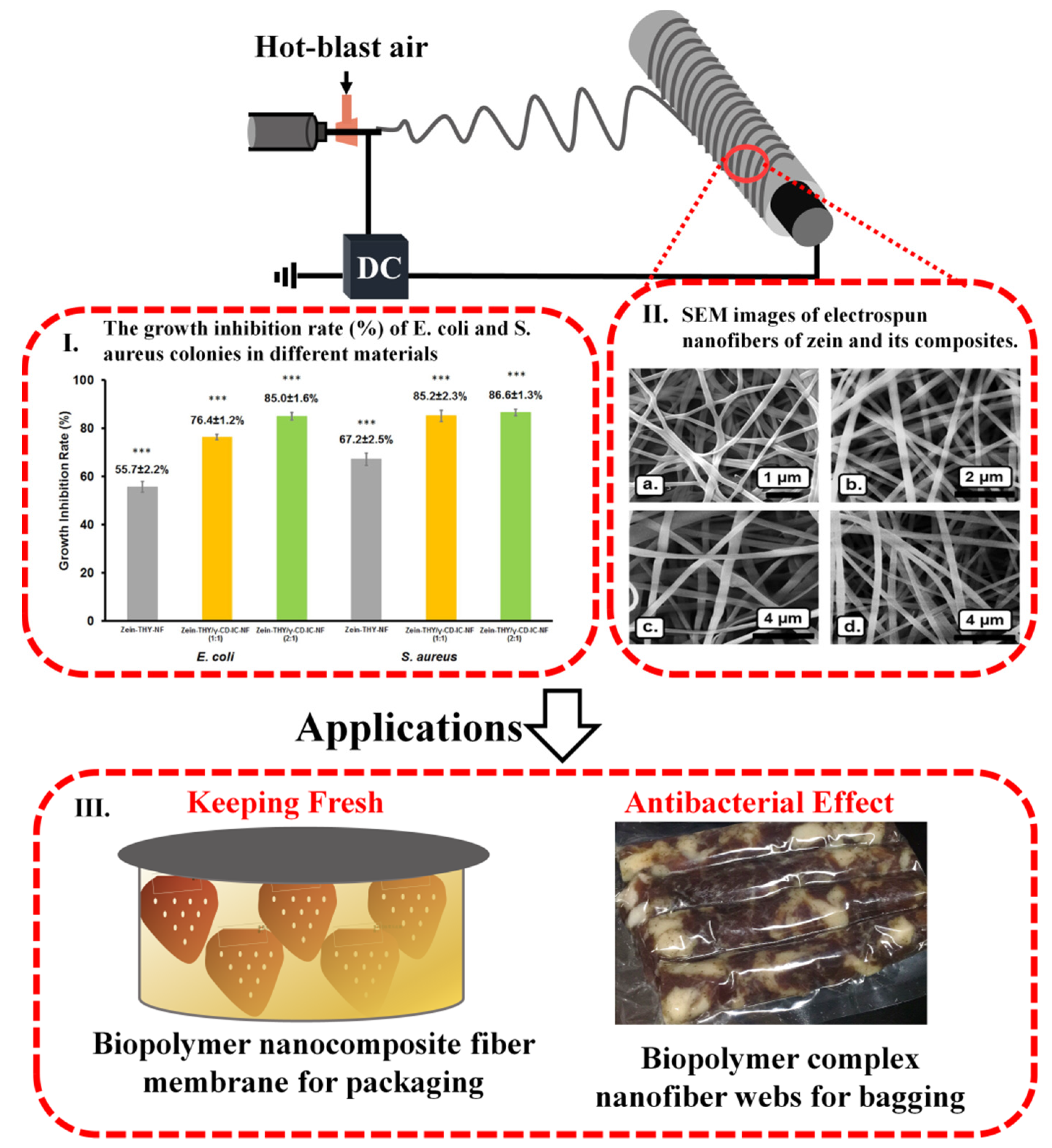
3.4. Recent Packaging Material Applications
| Application Direction | Materials | Solvent | Operating Parameters | References | ||
|---|---|---|---|---|---|---|
| Voltage (kV) | Distance (cm) | Flow Rate (mL/h) | ||||
| Biology | PLLA, Pluronic | Chloroform, DMF | 18 | 14 | 0.5 | [3] |
| SF, PEO | HFIP | 20 | 18 | 0.7–2.5 | [4] | |
| PCL, PLGA | HFP | 7.5–37.5 | 10–25 | 0.75 | [5] | |
| PS | DMF | 18 | 20 | 0.1 | [7] | |
| PCL | DCM, TEF | 15 | 20 | 0.6, 0.8, 1.6 | [9] | |
| PEO | FA | 21 | 10 | 0.8–1.2 | [17] | |
| Gliadin, IBU | HFIP, TFA | 15 | - | 0.2, 0.3 | [18] | |
| Chitosan, Zein, PVP, PVA | Ethanol, Acetic acid | 22 | 8 | 0.7 | [19] | |
| KGM, PDA | Ethanol, Distillated water | 16 | 13 | 0.03 | [20] | |
| Zein, Quercetin | Ethanol | 20 | 15 | 0.6 | [21] | |
| PVP, PVB, PVPI | Ethanol | 10 | 8 | - | [22] | |
| PLA | HFIP | 16, 12.5 | 23, 20 | 1.5, 2.5 | [23,30] | |
| Fibrin | HFIP, Distilled water | 22 | 10 | 0.5 | [24] | |
| PCL | HFIP | 16 | 10 | 2 | [25] | |
| Poly(pro-17β- estradiol-alt-oEG) | DCM | 12.5 | 5 | 0.75 | [26] | |
| PCL, COL | HFIP | 15 | 15 | 1 | [27] | |
| PHBV, MCC | Chloroform, DMF | 15 | 18 | 1 | [28] | |
| Tecoflex EG-80A | DMF | 10.5 | 20 | 0.5 | [29] | |
| PVA, PE | Water, IPA | 28 | 15 | 2 | [35] | |
| Glycerol sebacic acid, PLLA | DCM, DMF, trichloromathane | 20 | - | - | [50] | |
| Zn(CH3COO)2·4H2O, Co(CH3COO)2·4H2O PVP | DMF | 12 | 14 | - | [51] | |
| PLG, PLA | DCM, DMF | - | - | - | [52] | |
| SPIR, HPMC | Ethanol, DCM | 25 | 20 | 10, 30 | [56] | |
| PCL | AA | 15–38 | [60] | |||
| Al(NO3)3·9H2O, PVP | DI water, ethanol | 15–40 | 30–35 | 15–40 | [61] | |
| gelatin | acetic acid, distilled water, ethanol | 34 | - | - | [62] | |
| PEO, PIB, PS | Toluene, Ethanol | 4–5.5 | 7 | 0.036–0.072 | [63] | |
| PS | DMF | 10–20 | 5–20 | 0.5–2 | [66] | |
| PVB, PA6, PES | Ethanol, FA, Acetic acid | 32 | - | - | [94] | |
| PCL, Ch, Ferulic acid | DMF, THF | 13 | 12 | 0.7 | [80] | |
| COL, PVA, SA | Acetic acid | 18 | 15 | 0.4 | [100] | |
| PVP, Loratadine | Ethanol | 10, 20 | 1, 6 | 5 | [75] | |
| PU, Eudragit | DMF, THF | 10, 15, 18 | 15 | 1, 1.5 | [81] | |
| PLCL, Gelatin, NaHCO3 | HFIP | 15 | 23 | 0.8 | [88] | |
| PCL, Gelatin | AA, FA | 15 | 11 | 0.4 | [106] | |
| PDO, PCL | HFIP | 8.2–8.4 | 20 | - | [107] | |
| PLCL, Gelatin | DMF, TFA | 12 | 15 | 1 | [109] | |
| ECM, PCL | HFIP | 20 | 21 | 3 | [111] | |
| PLCL, PLLA, SF | Chloroform, FA | 22–23, 17 | 10–12, 7 | 0.24–0.36, 1 | [112,115] | |
| Collagen, Ch | HFP | 15 | 20 | 0.2 | [110] | |
| SF, PLLACL | HFIP | 12, 15 | 12, 15 | 0.1, 0.6 | [114] | |
| PVA | TFA, THMs, Deionize water | 10 | 15 | - | [116] | |
| TSF | Deionize water | 20 | 18 | 0.1–0.3 | [117] | |
| PAN, Fe3O4 magnetic nanofiber | DMF | 10, 15 | 10, 15 | 0.72, 1 | [119,121] | |
| Environment | PVDF, GPS | DMF | 30 | 20 | 0.5 | [2] |
| Soy flour, PA-6 | FA, Acetic acid | 12–18 | 5–11, 20 | 0.2–0.3, 3 | [64,129,130] | |
| PAN | DMF | 20 | 10, 15, 20 | 0.15 | [69,133] | |
| LPI | DMAc | 7–20 | 12–35 | 0.0025–0.1 | [74] | |
| PAN, PVP | DMAc | 10, 14 | 15 | 1, 2 | [77] | |
| PAN, PMMA | DMF | 14 | 15 | 1.6 | [95] | |
| PVDF, PTFE | DMF | 30 | 15 | 0.5 | [71] | |
| PVA, Gelatin | Ultrapure water, Glacial acetic acid | 20 | - | 0.3 | [98] | |
| PAN | DMF | 15 | 10 | - | [118] | |
| PET | TFA, DCM | 5–25 | 8-21 | 1 | [122] | |
| Nylon 6,6 | DMF, FA, Chloroform | 22 | 12 | 1 | [126] | |
| PMDA, ODA | DMAc | 11–14 | 21 | 0.2 | [127] | |
| PCL, PEO | Chloroform, Acetone | 25, 15 | 25, 15 | 0.3, 1 | [128] | |
| PAN, PA-66, PES | DMF | 20, 12, 75 | 20, 11 | 0.8 | [79,82,83] | |
| PA-6 | Acetic acid, FA | 27–28 | 15 | - | [131] | |
| SPAC, PS | DMF | 40 | 15 | 1 | [84] | |
| PVA | Deionize water | 20 | 15 | 0.5 | [132] | |
| PVA, Tetraetho ysilane, Zirconium oxychloride | DMF, Deionize water | 15, 20 | 15, 20 | 0.4 | [93,134] | |
| PVA, PVP, PAN | NaOH, Distilled water | 10–25 | 10–20 | 0.6, 0.9 | [135,136] | |
| Energy | TPP, PVDF-HFP | DMAc, BLA | 13 | - | - | [11] |
| CA | Acetone, DMAc | 20 | - | 0.2 | [16] | |
| PMMA, PAN | DMF | 18 | - | 1.5 | [138,139] | |
| PAN | DMF | 13 | 16 | - | [91] | |
| PAN | DMF | 15, 25 | 15 | 0.05, 0.5, 1 | [140,141] | |
| PVP, SiO2 | Ethanol, DMF | 16 | 18 | 1 | [142] | |
| PAN | DMF, Acetone | 18 | 15 | 1 | [143] | |
| PVDF, PU | DMAc, EMC, Acetone, THF | 30 | 20 | 0.6, 1 | [145] | |
| PPESK | NMP, THF | 13 | 20 | - | [147] | |
| PVDF-HFP, PVDF | DMF, NMP | 20 | 30 | - | [148] | |
| PU, GO | DMF, THF | 9–10 | 13 | - | [90] | |
| PVDF | DMF | 12 | 18 | 1 | [149] | |
| PEO, CMCS | Distilled water | 20 | 20 | - | [153] | |
| Package | PVA | Deionize water | 30 | 12 | - | [65] |
| BSA, Ascorbic acid | MilliQ water | 12.5 | 15 | 1 | [108] | |
| Zein | Ethanol | 11 | 10 | 0.15 | [92] | |
| Gliadin | Acetic acid | 18 | 10 | 1 | [152] | |
| Zein | Ethanol | 15 | 10 | 1 | [85] | |
| PEO, Lentil powder | Chloroform, Ethanol | 20 | 8 | 0.5 | [70] | |
| PEO, | Chloroform | 15 | 30 | 0.6 | [86] | |
| Zein, Thymol | DMF | 17 | 17 | 0.5 | [87] | |
| Zein, Gelatin | Acetic acid, Ethanol | 14–16 | 12 | 0.1, 0.3–0.7 | [154,155] | |
| PLA, CEO, Ch, Glucose oxidase | DCM, DMF | 12–16 | 10–14 | 2–2.4 | [156] | |
4. Conclusions
Author Contributions
Funding
Conflicts of Interest
References
- Lv, D.; Zhu, M.M.; Jiang, Z.C.; Jiang, S.H.; Zhang, Q.L.; Xiong, R.H.; Huang, C.B. Green Electrospun Nanofibers and Their Application in Air Filtration. Macromol. Mater. Eng. 2018, 303, 1800336. [Google Scholar] [CrossRef]
- Ding, X.X.; Li, Y.Y.; Si, Y.; Yin, X.; Yu, J.Y.; Ding, B. Electrospun polyvinylidene fluoride/SiO2 nanofibrous membranes with enhanced electret property for efficient air filtration. Chem. Commun. 2019, 13, 57–62. [Google Scholar] [CrossRef]
- Birhanu, G.; Tanha, S.; Javar, H.A.; Seyedjafari, E.; Zandi-Karimi, A.; Dehkordi, B.K. Dexamethasone Loaded Multi-Layer Poly-L-Lactic Acid/Pluronic P123 Composite Electrospun Nanofiber Scaffolds for Bone Tissue Engineering and Drug Delivery. Pharm. Dev. Technol. 2019, 24, 338–347. [Google Scholar] [CrossRef] [PubMed]
- Chan, A.H.P.; Filipe, E.C.; Tan, R.P.; Miguel, S.; Yang, N.J.; Hung, J.; Feng, J.Y.; Nazir, S.; Benn, J.A.; Ng, C.K.M.; et al. Altered processing enhances the efficacy of small-diameter silk fibroin vascular grafts. Sci. Rep. 2019, 9, 1–14. [Google Scholar] [CrossRef] [Green Version]
- Nagiah, N.; Murdock, C.J.; Bhattacharjee, M.; Nair, L.; Laurencin, C.T. Development of Tripolymeric Triaxial Electrospun Fibrous Matrices for Dual Drug Delivery Applications. Sci. Rep. 2020, 10, 1–11. [Google Scholar] [CrossRef] [Green Version]
- Ghosal, K.; Chandra, A.; Praveen, G.; Snigdha, S.; Roy, S.; Agatemor, C.; Thomas, S.; Provaznik, I. Electrospinning over solvent casting: Tuning of mechanical properties of membranes. Sci. Rep. 2018, 8, 1–9. [Google Scholar] [CrossRef] [Green Version]
- Kim, D.; Kim, S.M.; Lee, S.; Yoon, M.H. Investigation of neuronal pathfinding and construction of artificial neuronal networks on 3D-arranged porous fibrillar scaffolds with controlled geometry. Sci. Rep. 2017, 7, 1–10. [Google Scholar] [CrossRef]
- Petropoulou, A.; Kralj, S.; Karagiorgis, X.; Savva, I.; Loizides, E.; Panagi, M.; Krasia, C.T.; Riziotis, C. Multifunctional Gas and pH Fluorescent Sensors Based on Cellulose Acetate Electrospun Fibers Decorated with Rhodamine B-Functionalised Core-Shell Ferrous Nanoparticles. Sci. Rep. 2020, 10, 1–14. [Google Scholar] [CrossRef]
- Han, D.; Serra, R.; Gorelick, N.; Fatima, U.; Eberhart, G.C.; Brem, H.; Tyler, B.; Steckl, J.A. Multi-layered core-sheath fiber membranes for controlled drug release in the local treatment of brain tumor. Sci. Rep. 2019, 9, 1–12. [Google Scholar] [CrossRef]
- Radacsi, N.; Campos, D.F.; Chisholm, I.R.C.; Giapis, P.K. Spontaneous formation of nanoparticles on electrospun nanofibers. Nat. Commun. 2018, 9, 1–8. [Google Scholar] [CrossRef] [Green Version]
- Liu, K.; Liu, W.; Qiu, Y.C.; Kong, B.; Sun, Y.M.; Chen, Z.; Zhuo, D.; Lin, D.C.; Cui, Y. Electrospun core-shell microfiber separator with thermal-triggered flame-retardant properties for lithium-ion batteries. Sci. Adv. 2017, 3, e1601978. [Google Scholar] [CrossRef] [PubMed] [Green Version]
- Wang, Z.; Lee, W.J.; Koh, B.T.H.; Hong, M.; Wang, W.; Lim, P.N.; Feng, J.; Park, L.S.; Kim, M.; Thian, E.S. Functional regeneration of tendons using scaffolds with physical anisotropy engineered via microarchitectural manipulation. Sci. Adv. 2018, 4, eaat4537. [Google Scholar] [CrossRef] [Green Version]
- Liao, X.J.; Dulle, M.; Silva, J.M.D.S.E.; Wehrspohn, R.B.; Agarwal, S.; Förster, S.; Hou, H.Q.; Smith, P.; Greiner, A. High strength in combination with high toughness in robust and sustainable polymeric materials. Science 2019, 366, 1376–1379. [Google Scholar] [CrossRef]
- Subbiah, T.; Bhat, G.S.; Tock, R.W.; Parameswaran, S.; Ramkumar, S.S. Electrospinning of nanofibers. Appl. Polym. Sci. 2005, 96, 557–569. [Google Scholar] [CrossRef]
- Mirjalili, M.; Zohoori, S. Review for application of electrospinning and electrospun nanofibers technology in textile industry. J. Nanostruct. Chem. 2016, 6, 207–213. [Google Scholar] [CrossRef] [Green Version]
- Lima, N.; Baptista, A.C.; Faustino, B.M.M.; Taborda, S.; Marques, A.; Ferreira, I. Carbon threads sweat-based supercapacitors for electronic textiles. Sci. Rep. 2020, 10, 7703. [Google Scholar] [CrossRef]
- Jung, H.S.; Kim, M.H.; Shin, J.Y.; Park, S.R.; Jung, J.Y.; Park, W.H. Electrospinning and wound healing activity of β-chitin extracted from cuttlefish bone. Carbohydr. Polym. 2018, 193, 205–211. [Google Scholar] [CrossRef] [PubMed]
- Yang, Y.Y.; Li, W.B.; Yu, D.G.; Wang, G.H.; Williams, G.R.; Zhang, Z. Tunable drug release from nanofibers coated with blank cellulose acetate layers fabricated using tri-axial electrospinning. Carbohydr. Polym. 2019, 203, 228–237. [Google Scholar] [CrossRef] [PubMed]
- Mirzaeei, S.; Berenjian, K.; Khazaei, R. Preparation of the potential ocular inserts by electrospinning method to achieve the prolong release profile of triamcinolone acetonide. Adv. Pharm. Bull. 2018, 8, 21–27. [Google Scholar] [CrossRef] [PubMed]
- Wang, L.; Mu, R.J.; Yuan, Y.; Gong, J.N.; Ni, Y.S.; Wang, W.H.; Pang, J. Novel nanofiber membrane fabrication from konjac glucomannan and polydopamine via electrospinning method. J. Sol-Gel Sci. Technol. 2018, 85, 253–258. [Google Scholar] [CrossRef]
- Moradkhannejhad, L.; Abdouss, M.; Nikfarjam, N.; Mazinani, S.; Heydari, V. Electrospinning of zein/propolis nanofibers; antimicrobial properties and morphology investigation. J. Mater. Sci. Mater. Med. 2018, 29, 165. [Google Scholar] [CrossRef] [PubMed]
- Liu, G.S.; Yan, X.; Yan, F.F.; Chen, F.X.; Hao, L.Y.; Chen, S.J.; Lou, T.; Ning, X.; Long, Y. In situ electrospinning iodine-based fibrous meshes for antibacterial wound dressing. Nanoscale Res. Lett. 2018, 13, 309. [Google Scholar] [CrossRef]
- Grant, R.; Hallett, J.; Forbes, S.; Hay, D.; Callanan, A. Blended electrospinning with human liver extracellular matrix for engineering new hepatic microenvironments. Sci. Rep. 2019, 9, 6293. [Google Scholar] [CrossRef] [PubMed]
- Kayal, T.A.; Losi, P.; Pierozzi, S.; Soldani, G. A new method for fibrin-based electrospun/sprayed scaffold fabrication. Sci. Rep. 2020, 10, 1–4. [Google Scholar] [CrossRef] [PubMed] [Green Version]
- Horejs, C.M.; St-Pierre, J.P.; Ojala, J.R.M.; Steele, J.A.M.; da Silva, P.B.; Rynne-Vidal, A.; Maynard, S.A.; Hansel, C.S.; Rodríguez-Fernández, C.; Mazo, M.M.; et al. Preventing tissue fibrosis by local biomaterials interfacing of specific cryptic extracellular matrix information. Nat. Commun. 2017, 8, 15509. [Google Scholar] [CrossRef] [PubMed]
- D’Amato, A.R.; Puhl, D.L.; Ellman, S.A.T.; Balouch, B.; Gilbert, R.J.; Palermo, E.F. Vastly extended drug release from poly(pro-17β-estradiol) materials facilitates in vitro neurotrophism and neuroprotection. Nat. Commun. 2019, 10, 4830. [Google Scholar] [PubMed] [Green Version]
- Kim, J.I.; Kim, Y.J.; Park, C.H. Fabrication of transparent hemispherical 3D nanofibrous scaffolds with radially aligned patterns via a novel electrospinning method. Sci. Rep. 2018, 8, 3424. [Google Scholar] [CrossRef]
- Cheng, M.; Qin, Z.Y.; Hu, S.; Dong, S.; Ren, Z.C.; Yu, H.Y. Achieving Long-Term Sustained Drug Delivery for Electrospun Biopolyester Nanofibrous Membranes by Introducing Cellulose Nanocrystals. ACS Biomater. Sci. Eng. 2017, 3, 1666–1676. [Google Scholar] [CrossRef]
- Li, G.B.; Li, P.Q.; Chen, Q.A.; Mani, M.P.; Jaganathan, S.K. Enhanced mechanical, thermal and biocompatible nature of dual component electrospun nanocomposite for bone tissue engineering. Peer J. 2019, 7, e6986. [Google Scholar] [CrossRef]
- Apalangya, V.A.; Rangari, V.K.; Tiimob, B.J.; Jeelani, S.; Samue, T. Eggshell Based Nano-Engineered Hydroxyapatite and Poly (lactic) Acid Electrospun Fibers as Potential Tissue Scaffold. Int. J. Polym. Mater. 2019, 11, 6762575. [Google Scholar] [CrossRef]
- Marchesan, S.; Ballerini, L.; Prato, M. Nanomaterials for stimulating nerve growth. Science 2017, 356, 1010–1011. [Google Scholar] [CrossRef] [PubMed] [Green Version]
- DeFrates, K.G.; Moore, R.; Borgesi, J.; Lin, G.; Mulderig, T.; Beachley, V.; Hu, X. Protein-Based Fiber Materials in Medicine: A Review. Nanomaterials 2018, 8, 457. [Google Scholar] [CrossRef] [PubMed] [Green Version]
- Xu, C.; Yang, Q.; Wang, F.X.; Fang, X.M.; Zhang, Z.G. Research progress on novel solar steam generation system based on black nanomaterials. Can. J. Chem. Eng. 2018, 96, 2086–2099. [Google Scholar] [CrossRef]
- Xin, Q.; Shah, H.; Nawaz, A.; Xie, W.J.; Akram, M.Z.; Batool, A.; Tian, L.Q.; Jan, S.U.; Boddula, R.; Guo, B.D.; et al. Antibacterial carbon-based nanomaterials. Adv. Mater. 2019, 31, 1804838. [Google Scholar] [CrossRef] [PubMed]
- Si, Y.; Zhang, Z.; Wu, W.R.; Fu, Q.X.; Huang, K.; Nitin, N. Daylight-driven rechargeable antibacterial and antiviral nanofibrous membranes for bioprotective applications. Sci. Adv. 2018, 4, eaar5931. [Google Scholar] [CrossRef] [Green Version]
- Park, J.; Kim, J.; Kim, S.Y.; Cheong, W.H.; Jang, J.; Park, Y.G.; Na, K.; Kim, Y.T.; Heo, J.H.; Lee, C.Y.; et al. Soft, smart contact lenses with integrations of wireless circuits, glucose sensors, and displays. Sci. Adv. 2018, 4, eaap9841. [Google Scholar] [CrossRef] [Green Version]
- Xue, Y.L.; Huang, J.; Lau, C.H.; Cao, B.; Li, P. Tailoring the molecular structure of crosslinked polymers for pervaporation desalination. Nat. Commun. 2020, 11, 1–9. [Google Scholar] [CrossRef] [Green Version]
- Kim, Y.; Wu, X.W.; Oh, J.H. Fabrication of triboelectric nanogenerators based on electrospun polyimide nanofibers membrane. Sci. Rep. 2020, 10, 2742. [Google Scholar] [CrossRef]
- Yu, Z.M.; Shen, L.F.; Li, D.H.; Pun, E.Y.B.; Zhao, X.; Lin, H. Fluctuation of photon-releasing with ligand coordination in polyacrylonitrile-based electrospun nanofibers. Sci. Rep. 2020, 10, 926. [Google Scholar] [CrossRef]
- Cho, S.K.; Cho, W.J. Performance improvement in electrospun InGaZnO nanofibres field-effect-transistors using low thermal budget microwave calcination and Ar/O2 mixed-plasma surface treatment. Sci. Rep. 2020, 10, 3645. [Google Scholar] [CrossRef]
- Kweon, O.Y.; Lee, S.J.; Oh, J.H. Wearable high-performance pressure sensors based on three-dimensional electrospun conductive nanofibers. NPG Asia Mater. 2018, 10, 540–551. [Google Scholar] [CrossRef]
- Mohammed, N.; Grishkewich, N.; Tam, K.C. Cellulose nanomaterials: Promising sustainable nanomaterials for application in water/wastewater treatment processes. Environ. Sci. Nano 2018, 5, 623–658. [Google Scholar] [CrossRef]
- Chen, R.X.; Wan, Y.Q.; Wu, W.W.; Yang, C.; He, J.H.; Cheng, J.H.; Jetter, R.; Ko, F.K.; Chen, Y.C. A lotus effect-inspired flexible and breathable membrane with hierarchical electrospinning micro/nanofibers and ZnO nanowires. Mater. Des. 2019, 162, 246–248. [Google Scholar] [CrossRef]
- Fang, J.; Wang, X.G.; Lin, T. Electrical power generator from randomly oriented electrospun poly(vinylidene fluoride) nanofibre membranes. J. Mater. Chem. 2011, 21, 11088–11091. [Google Scholar] [CrossRef] [Green Version]
- Li, X.H.; Song, T.D.; Chen, Z.Y.; Liu, Y.X.; He, H.; Zhang, Y.C.; Liu, Y. Modes of electrospinning. J. Text. Res. 2014, 35, 163–168. [Google Scholar]
- Zhang, B.; Yan, X.; He, H.W.; Yu, M.; Ning, X.; Long, Y.Z. Solvent-free Electrospinning: Opportunities and Challenges. Polym. Chem. 2016, 8, 333–352. [Google Scholar] [CrossRef]
- Garg, K.; Bowlin, G.L. Electrospinning jets and nanofibrous structures. Biomicrofluidics 2011, 5, 013403. [Google Scholar] [CrossRef] [Green Version]
- Reneker, D.H.; Yarin, A.L. Electrospinning jets and polymer nanofibers. Polymer 2008, 49, 2387–2425. [Google Scholar] [CrossRef] [Green Version]
- Yarin, A.L.; Koombhongse, S.; Reneker, D.H. Bending instability in electrospinning of nanofibers. J. Appl. Phys. 2001, 89, 3018. [Google Scholar] [CrossRef] [Green Version]
- Yang, X.P.; Li, L.F.; Yang, D.Z.; Nie, J.; Ma, G.P. Electrospun Core–Shell Fibrous 2D Scaffold with Biocompatible Poly(Glycerol Sebacate) and Poly-L-Lactic Acid for Wound Healing. Adv. Fiber Mater. 2020. [Google Scholar] [CrossRef] [Green Version]
- Yu, H.Q.; Zhao, H.Y.; Wu, Y.B.; Chen, B.J.; Sun, J.S. Electrospun ZnCo2O4/C composite nanofibers with superior electrochemical performance for supercapacitor. J. Phys. Chem. Solids 2020, 140, 109385. [Google Scholar] [CrossRef]
- Liu, S.H.; Zhang, H.G.; Hu, Q.X.; Wang, B.; Li, S.; Zhang, C. Development and Evaluation of Biomimetic 3D Coated Composite Scaffold for Application as Skin Substitutes. Macromol. Mater. Eng. 2020, 305, 1900848. [Google Scholar] [CrossRef]
- Park, Y.S.; Kim, J.; Oh, J.M.; Park, S.; Cho, S.; Ko, H.; Cho, Y.K. Near-Field Electrospinning for Three-Dimensional Stacked Nanoarchitectures with High Aspect Ratios. Nano Lett. 2020, 20, 441–448. [Google Scholar] [CrossRef]
- Feltz, K.P.; Kalaf, E.A.G.; Chen, C.P.; Martin, R.S.; Sell, S.A. A review of electrospinning manipulation techniques to direct fiber deposition and maximize pore size. Electrospinning 2017, 1, 46–61. [Google Scholar] [CrossRef]
- Sarkar, S.; Deevi, S.; Tepper, G. Biased AC Electrospinning of Aligned Polymer Nanofibers. Macromol. Rapid Commun. 2007, 28, 1034–1039. [Google Scholar] [CrossRef]
- Balogh, A.; Farkas, B.; Verreck, G.; Mensch, J.; Borbás, E.; Nagy, B.; Marosi, G.; Nagy, Z.K. AC and DC electrospinning of hydroxypropylmethylcellulose with polyethylene oxides as secondary polymer for improved drug dissolution. Int. J. Pharm. 2016, 505, 159–166. [Google Scholar] [CrossRef] [PubMed] [Green Version]
- Liu, W.Y.; Thomopoulos, S.; Xia, Y.N. Electrospun Nanofibers for Regenerative Medicine. Adv. Healthc. Mater. 2012, 1, 10–25. [Google Scholar] [CrossRef] [PubMed]
- Maheshwari, S.; Chang, H.C. Assembly of multi-stranded nanofiber threads through AC electrospinning. Adv. Mater. 2010, 21, 349–354. [Google Scholar] [CrossRef]
- Kalayci, V.E.; Patra, P.K.; Kim, Y.K.; Ugbolue, S.C.; Warner, S.B. Charge consequences in Electrospun polyacrylonitrile (PAN) nanofibers. Polymer 2005, 46, 7191–7200. [Google Scholar] [CrossRef]
- Lawson, C.; Stanishevsky, A.; Sivan, M.; Pokorny, P.; Lukas, D. Rapid fabrication of poly(ε-caprolactone) nanofibers using needleless alternating current electrospinning. J. Appl. Polym. Sci. 2016, 133, 43232. [Google Scholar] [CrossRef]
- Stanishevsky, A.; Brayer, W.A.; Pokorny, P.; Kalous, T.; Lukas, D. Nanofibrous alumina structures fabricated using high-yield alternating current electrospinning. Ceram. Int. 2016, 42, 17154–17161. [Google Scholar] [CrossRef] [Green Version]
- Jirkovec, R.; Kalous, T.; Brayer, W.A.; Stanishevky, A.V.; Chvojka, J. Production of gelatin nanofibrous layers via alternating current electrospinning. Mater. Lett. 2019, 252, 186–190. [Google Scholar] [CrossRef]
- Xue, J.J.; Wu, T.; Dai, Y.; Xia, Y.N. Electrospinning and electrospun nanofibers: Methods, materials, and applications. Chem. Rev. 2019, 119, 5298–5415. [Google Scholar] [CrossRef] [PubMed]
- Chowdhury, M.; Stylios, G. Effect of experimental parameters on the morphology of electrospun Nylon 6 fibres. Int. J. Basic Appl. Sci. 2010, 10, 70–78. [Google Scholar]
- Prabu, G.T.V.; Dhurai, B. A Novel Profiled Multi-Pin Electrospinning System for Nanofiber Production and Encapsulation of Nanoparticles into Nanofibers. Sci. Rep. 2020, 10, 4302. [Google Scholar] [CrossRef] [PubMed] [Green Version]
- Uyar, T.; Besenbacher, F. Electrospinning of uniform polystyrene fibers: The effect of solvent conductivity. Polymer 2008, 49, 5336–5343. [Google Scholar] [CrossRef]
- Jiang, S.H.; Chen, Y.M.; Duan, G.G.; Mei, C.T.; Greiner, A.; Agarwal, S. Electrospun nanofiber reinforced composites: A review. Polym. Chem. 2018, 9, 2685–2720. [Google Scholar] [CrossRef]
- Bazrafshan, Z.; Stylios, G.K. Spinnability of collagen as a biomimetic material: A review. Int. J. Biol. Macromol. 2019, 129, 693–705. [Google Scholar] [CrossRef]
- Jin, S.X.; Yu, J.L.; Zheng, Y.S.; Wang, W.Y.; Xin, B.J.; Kan, C.W. Preparation and characterization of electrospun PAN/PSA carbonized nanofibers: Experiment and simulation study. Nanomaterials 2018, 8, 821. [Google Scholar] [CrossRef] [Green Version]
- Kara, H.H.; Xiao, F.G.; Sarker, M.; Jin, T.Z.; Sousa, A.M.M.; Liu, C.K.; Tomasula, P.M.; Liu, L.S. Antibacterial poly (lactic acid) (PLA) films grafted with electrospun PLA/allyl isothiocyanate fibers for food packaging. J. Appl. Polym. Sci. 2016, 133, 42475. [Google Scholar] [CrossRef]
- Wang, S.; Zhao, X.L.; Yin, X.; Yu, J.Y.; Ding, B. Electret polyvinylidene fluoride nanofibers hybridized by polytetrafluoroethylene nanoparticles for high-efficiency air filtration. ACS Appl. Mater. Interfaces 2016, 8, 23985–23994. [Google Scholar] [CrossRef] [PubMed]
- Guo, Y.H.; He, W.D.; Liu, J.X. Electrospinning polyethylene terephthalate/SiO2 nanofiber composite needle felt for enhanced filtration performance. J. Appl. Polym. Sci. 2020, 137, 48282. [Google Scholar] [CrossRef]
- Agarwal, S.; Greiner, A.; Wendorff, J.H. Functional materials by electrospinning of polymers. Prog. Polym. Sci. 2013, 38, 963–991. [Google Scholar] [CrossRef]
- Megelski, S.; Stephens, S.J.; Chase, D.B.; Rabolt, F.J. Micro-and nanostructured surface morphology on electrospun polymer fibers. Macromolecules 2002, 35, 8456–8466. [Google Scholar] [CrossRef]
- Akhgari, A.; Ghalambor, D.A.; Rezaei, M.; Kiarsi, M.; Abbaspour, M. The design and evaluation of a fast-dissolving drug delivery system for loratadine using the electrospinning method. Jundishapur J. Nat. Pharm. Prod. 2016, 11, e33613. [Google Scholar] [CrossRef]
- Katti, D.S.; Robinson, K.W.; Ko, F.K.; Laurencin, C.T. Bioresorbable nanofiber-based systems for wound healing and drug delivery: Optimization of fabrication parameters. J. Biomed. Mater. Res. Part B 2004, 70B, 286–296. [Google Scholar] [CrossRef]
- Yang, G.Z.; Li, H.P.; Yang, J.; Wan, J.; Yu, D.G. Influence of working temperature on the formation of electrospun polymer nanofibers. Nanoscale Res. Lett. 2017, 12, 55. [Google Scholar] [CrossRef] [Green Version]
- Jan, P.; Julijana, K.; Biljana, J.; Saša, B.; Petra, K. The impact of relative humidity during electrospinning on the morphology and mechanical properties of nanofibers. Int. J. Pharm. 2013, 456, 125–134. [Google Scholar]
- Yan, S.L.; Yu, Y.X.; Ma, R.; Fang, J.Y. The formation of ultrafine polyamide 6 nanofiber membranes with needleless electrospinning for air filtration. Polym. Adv. Technol. 2019, 30, 1635–1643. [Google Scholar] [CrossRef]
- Yakub, G.; Ignatova, M.; Manolova, N.; Rashkov, I.; Toshkova, R.; Georgieva, A.; Markova, N. Chitosan/ferulic acid-coated poly(ε-caprolactone) electrospun materials with antioxidant, antibacterial and antitumor properties. Int. J. Biol. Macromol. 2018, 107, 689–702. [Google Scholar] [CrossRef]
- Aguilar, L.E.; Unnithan, A.R.; Amarjargal, A.; Tiwari, A.P.; Hong, S.T.; Park, C.H.; Kim, C.S. Electrospun polyurethane/Eudragit® L100-55 composite mats for the pH dependent release of paclitaxel on duodenal stent cover application. Int. J. Pharm. 2015, 478, 1–8. [Google Scholar] [CrossRef] [PubMed]
- Al-Attabi, R.; Dumée, L.F.; Kong, L.; Schütz, J.A.; Morsi, Y. High efficiency poly (acrylonitrile) electrospun nanofiber membranes for airborne nanomaterials filtration. Adv. Eng. Mater. 2018, 20, 1700572. [Google Scholar] [CrossRef]
- Li, L.; Shang, L.M.; Li, Y.X.; Yang, C.F. Three-layer composite filter media containing electrospun polyimide nanofibers for the removal of fine particles. Fibers Polym. 2017, 18, 749–757. [Google Scholar] [CrossRef]
- Apul, O.G.; Reitzenstein, N.H.V.; Schoepf, J.; Ladner, D.; Hristovski, K.D.; Westerhoff, P. Superfine powdered activated carbon incorporated into electrospun polystyrene fibers preserve adsorption capacity. Sci. Total Environ. 2017, 592, 458–464. [Google Scholar] [CrossRef]
- Amjadi, S.; Almasi, H.; Ghorbani, M.; Ramazani, S. Reinforced ZnONPs/rosemary essential oil-incorporated zein electrospun nanofibers by κ-carrageenan. Carbohydr. Polym. 2020, 232, 115800. [Google Scholar] [CrossRef]
- Aydogdu, A.; Yildiz, E.; Aydogdu, Y.; Sumnu, G.; Sahin, S.; Ayhan, Z. Enhancing oxidative stability of walnuts by using gallic acid loaded lentil flour based electrospun nanofibers as active packaging material. Food Hydrocoll. 2019, 95, 245–255. [Google Scholar] [CrossRef]
- Aytac, Z.; Ipek, S.; Durgun, E.; Tekinay, T.; Uyar, T. Antibacterial electrospun zein nanofibrous web encapsulating thymol/cyclodextrin-inclusion complex for food packaging. Food Chem. 2017, 233, 117–124. [Google Scholar] [CrossRef]
- Sang, Q.Q.; Williams, R.G.; Wu, H.L.; Liu, K.; Li, H.Y.; Zhu, L.M. Electrospun gelatin/sodium bicarbonate and poly (lactide-co-ε-caprolactone)/sodium bicarbonate nanofibers as drug delivery systems. Mater. Sci. Eng. C 2017, 81, 359–365. [Google Scholar] [CrossRef]
- Reich, S.; Burgard, M.; Langner, M.; Jiang, S.H.; Wang, X.Q.; Agarwal, S.; Ding, B.; Yu, J.Y.; Greiner, A. Polymer nanofibre composite nonwovens with metal-like electrical conductivity. NPJ Flex. Electron. 2018, 2, 5. [Google Scholar] [CrossRef]
- Liu, X.; Song, K.D.; Lu, C.; Huang, Y.T.; Duan, X.L.; Li, S.; Ding, Y.H. Electrospun PU@GO separators for advanced lithium ion batteries. J. Membr. Sci. 2018, 555, 1–6. [Google Scholar] [CrossRef]
- Chen, Y.M.; Yu, X.Y.; Li, Z.; Paik, U.; Lou, X.W. Hierarchical MoS2 tubular structures internally wired by carbon nanotubes as a highly stable anode material for lithium-ion batteries. Sci. Adv. 2016, 2, e1600021. [Google Scholar] [CrossRef] [PubMed] [Green Version]
- Moreno, M.A.; Orqueda, M.E.; Gómez-Mascaraque, L.G.; Isla, M.I.; López-Rubio, A. Crosslinked electrospun zein-based food packaging coatings containing bioactive chilto fruit extracts. Food Hydrocoll. 2019, 95, 496–505. [Google Scholar] [CrossRef]
- Wen, H.F.; Yang, C.; Yu, D.G.; Li, X.Y.; Zhang, D.F. Electrospun zein nanoribbons for treatment of lead-contained wastewater. Chem. Eng. J. 2016, 290, 263–272. [Google Scholar] [CrossRef] [Green Version]
- Valtera, J.; Kalous, T.; Pokorny, P.; Batka, O.; Bilek, M.; Chvojka, J.; Mikes, P.; Kostakova, E.K.; Zabka, P.; Ornstova, J.; et al. Fabrication of dual-functional composite yarns with a nanofibrous envelope using high throughput AC needleless and collectorless electrospinning. Sci. Rep. 2019, 9, 1801. [Google Scholar] [CrossRef] [PubMed]
- Xu, W.C.; Hu, X.Z.; Zhuang, S.D.; Wang, Y.X.; Li, X.Q.; Zhou, L.; Zhu, S.N.; Zhu, J. Flexible and salt resistant Janus absorbers by electrospinning for stable and efficient solar desalination. Adv. Energy Mater. 2018, 8, 1702884. [Google Scholar] [CrossRef]
- Chu, L.L.; Kang, X.J. Adsorption/Desorption Performance of Electrospun Nanofibers on Volatile Sulfur Compounds from Onion Juice. Nanosci. Nanotechnol. Lett. 2019, 11, 776–783. [Google Scholar] [CrossRef]
- Guo, H.W.; Tan, S.J.; Gao, J.; Wang, L. Sequential release of drugs form a dual-delivery system based on pH-responsive nanofibrous mats towards wound care. J. Mater. Chem. B 2020, 8, 1759–1770. [Google Scholar] [CrossRef]
- Meng, J.; Lin, X.Y.; Li, H.N.; Zhang, Y.D.; Zhou, J.; Chen, Y.; Shang, R.; Luo, X.G. Adsorption capacity of kelp-like electrospun nanofibers immobilized with bayberry tannin for uranium (vi) extraction from seawater. RSC Adv. 2019, 9, 8091–8103. [Google Scholar] [CrossRef] [Green Version]
- Ghosal, K.; Agatemor, C.; Špitálsky, Z.; Thomas, S.; Kny, E. Electrospinning tissue engineering and wound dressing scaffolds from polymer–titanium dioxide nanocomposites. Chem. Eng. J. 2018, 358, 1262–1278. [Google Scholar] [CrossRef]
- Zhang, X.L.; Tang, K.Y.; Zheng, X.J. Electrospinning and crosslinking of COL/PVA nanofiber-microsphere containing salicylic acid for drug delivery. J. Bionic Eng. 2016, 13, 143–149. [Google Scholar] [CrossRef]
- You, X.L.; He, J.X.; Nan, N.; Sun, X.Q.; Qi, K.; Zhou, Y.M.; Shao, W.L.; Liu, F.; Cui, S.Z. Stretchable capacitive fabric electronic skin woven by electrospun nanofiber coated yarns for detecting tactile and multimodal mechanical stimuli. J. Mater. Chem. C 2018, 6, 12981–12991. [Google Scholar] [CrossRef]
- Sheng, S.J.; Wang, F.; Ma, Q.Y.; Hu, X. Impact of foaming air on melting and crystallization behaviors of microporous PLA scaffolds. J. Therm. Anal. Calorim. 2015, 122, 1077–1088. [Google Scholar] [CrossRef]
- Sheng, S.J.; Hu, X.; Wang, F.; Ma, Q.Y.; Gu, M.F. Mechanical and thermal property characterization of poly-L-lactide (PLLA) scaffold developed using pressure-controllable green foaming technology. Mater. Sci. Eng. C 2015, 49, 612–622. [Google Scholar] [CrossRef]
- Yu, H.Y.; Wang, F.; Liu, Q.C.; Ma, Q.Y.; Gu, Z.G. Structure and kinetics of thermal decomposition mechanism of novel silk fibroin films. Acta Phys. Chim. Sin. 2017, 33, 344–355. [Google Scholar] [CrossRef]
- Wang, F.; Yu, H.Y.; Gu, Z.G.; Si, L.; Liu, Q.C.; Xiao, H. Impact of calcium chloride concentration on structure and thermal property of Thai silk fibroin films. J. Therm. Anal. Calorim. 2017, 130, 851–859. [Google Scholar] [CrossRef]
- Liverani, L.; Raffel, N.; Fattahi, A.; Preis, A.; Hoffmann, I.; Boccaccini, A.R.; Beckmann, M.W.; Dittrich, R. Electrospun patterned porous scaffolds for the support of ovarian follicles growth: A feasibility study. Sci. Rep. 2019, 9, 1150. [Google Scholar] [CrossRef] [Green Version]
- Rashid, M.; Dudhia, J.; Dakin, S.G.; Snelling, S.J.B.; Godoy, R.D.; Mouthuy, P.A.; Smith, R.K.W.; Morrey, M.; Carr, A.J. Histopathological and immunohistochemical evaluation of cellular response to a woven and electrospun polydioxanone (pDo) and polycaprolactone (pcL) patch for tendon repair. Sci. Rep. 2020, 10, 4754. [Google Scholar] [CrossRef] [Green Version]
- Evrova, O.; Kellenberger, D.; Scalera, C.; Calcagni, M.; Giovanoli, P.; Vogel, V.; Buschmann, J. Impact of UV sterilization and short term storage on the in vitro release kinetics and bioactivity of biomolecules from electrospun scaffolds. Sci. Rep. 2019, 9, 15117. [Google Scholar] [CrossRef] [PubMed] [Green Version]
- Ran, X.L.; Ye, Z.Y.; Fu, M.L.; Wang, Q.L.; Wu, H.D.; Lin, S.; Yin, T.Y.; Hu, T.Z.; Wang, G.X. Design, Preparation, and Performance of a Novel Bilayer Tissue-Engineered Small-Diameter Vascular Graft. Macromol. Biosci. 2019, 19, 1800189. [Google Scholar] [CrossRef] [PubMed]
- Sankar, S.; Sharma, C.S.; Rath, S.N.; Ramakrishna, S. Electrospun nanofibres to mimic natural hierarchical structure of tissues: Application in musculoskeletal regeneration. J. Tissue Eng. Regen. Med. 2018, 12, e604–e619. [Google Scholar] [CrossRef]
- Carvalho, M.S.; Silva, J.C.; Udangawa, R.N.; Cabral, J.M.S.; Ferreira, F.C.; Silva, C.L.D.; Linhardt, R.J.; Vashishth, D. Co-culture cell-derived extracellular matrix loaded electrospun microfibrous scaffolds for bone tissue engineering. Mater. Sci. Eng. C 2019, 99, 479–490. [Google Scholar] [CrossRef] [PubMed]
- Roy, T.; Maity, P.P.; Rameshbabu, A.P.; Das, B.; John, A.; Dutta, A.; Ghorai, S.K.; Chattopadhyay, S.; Dhara, S. Core-Shell Nanofibrous Scaffold Based on Polycaprolactone-Silk Fibroin Emulsion Electrospinning for Tissue Engineering Applications. Bioengineering 2018, 5, 68. [Google Scholar] [CrossRef] [PubMed] [Green Version]
- Lotfi, G.; Shokrgozar, M.A.; Mofid, R.; Abbas, F.M.; Ghanavati, F.; Baghban, A.A.; Yavari, S.K.; Pajoumshariati, S. Biological evaluation (in vitro and in vivo) of bilayered collagenous coated (nano electrospun and solid wall) chitosan membrane for periodontal guided bone regeneration. Ann. Biomed. Eng. 2016, 44, 2132–2144. [Google Scholar] [CrossRef] [PubMed]
- Yin, L.H.; Yang, S.H.; He, M.M.; Chang, Y.C.; Wang, K.J.; Zhu, Y.D.; Liu, Y.H.; Chang, Y.R.; Yu, Z.H. Physicochemical and biological characteristics of BMP-2/IGF-1-loaded three-dimensional coaxial electrospun fibrous membranes for bone defect repair. J. Mater. Sci. Mater. Med. 2017, 28, 94. [Google Scholar]
- Oyama, H.T.T.; Cortella, L.R.X.; Rosa, I.N.S.; Filho, L.E.R.; Hui, W.S.; Cestari, I.N.; Cestari, I.A. Assessment of the Biocompatibility of the PLLA-PLCL Scaffold Obtained by Electrospinning. Procedia Eng. 2015, 110, 135–142. [Google Scholar] [CrossRef] [Green Version]
- Khan, M.Q.; Kharaghani, D.; Nishat, N.; Shahzad, A.; Yamamoto, T.; Inoue, Y.; Kim, I.S. In vitro assessment of dual-network electrospun tubes from poly (1, 4 cyclohexane dimethylene isosorbide terephthalate)/PVA hydrogel for blood vessel application. J. Appl. Polym. Sci. 2019, 136, 47222. [Google Scholar] [CrossRef]
- Shao, W.L.; He, J.X.; Sang, F.; Ding, B.; Chen, L.; Cui, S.Z.; Li, K.J.; Han, Q.M.; Tan, W.L. Coaxial electrospun aligned tussah silk fibroin nanostructured fiber scaffolds embedded with hydroxyapatite–tussah silk fibroin nanoparticles for bone tissue engineering. Mater. Sci. Eng. C 2016, 58, 342–351. [Google Scholar] [CrossRef]
- Liu, Y.K.; Huang, Q.; Jiang, G.H.; Liu, D.P.; Yu, W.J. Cu2O nanoparticles supported on carbon nanofibers as a cost-effective and efficient catalyst for RhB and phenol degradation. J. Mater. Res. 2017, 32, 3605–3615. [Google Scholar] [CrossRef]
- Dai, Y.R.; Yao, J.; Song, Y.H.; Wang, S.Y.; Yuan, Y. Enhanced adsorption and degradation of phenolic pollutants in water by carbon nanotube modified laccase-carrying electrospun fibrous membranes. Environ. Sci. Nano 2016, 3, 857–868. [Google Scholar] [CrossRef]
- DelRe, C.; Huang, C.; Li, T.; Dennis, P.; Drockenmuller, E.; Xu, T. Reusable Enzymatic Fiber Mats for Neurotoxin Remediation in Water. ACS Appl. Mater. Interfaces 2018, 10, 44216–44220. [Google Scholar] [CrossRef]
- Jia, Y.Y.; Yue, X.Y.; Wang, Y.L.; Yan, C.; Zheng, G.Q.; Dai, K.; Liu, C.T.; Shen, C.Y. Multifunctional stretchable strain sensor based on polydopamine/ reduced graphene oxide/ electrospun thermoplastic polyurethane fibrous mats for human motion detection and environment monitoring. Compos. Pt. B Eng. 2020, 183, 107696. [Google Scholar] [CrossRef]
- Ma, W.J.; Zhao, J.T.; Oderinde, O.; Han, J.Q.; Liu, Z.C.; Gao, B.H.; Xiong, R.H.; Zhang, Q.L.; Jiang, S.H.; Huang, C.B. Durable superhydrophobic and superoleophilic electrospun nanofibrous membrane for oil-water emulsion separation. J. Colloid Interface Sci. 2018, 532, 12–23. [Google Scholar] [CrossRef]
- Zhu, M.M.; Xiong, R.H.; Huang, C.B. Bio-based and photocrosslinked electrospun antibacterial nanofibrous membranes for air filtration. Carbohydr. Polym. 2019, 205, 55–62. [Google Scholar] [CrossRef]
- Wang, B.; Sun, Z.M.; Sun, Q.; Wang, J.; Du, Z.X.; Li, C.J.; Li, X.Y. The preparation of bifunctional electrospun air filtration membranes by introducing attapulgite for the efficient capturing of ultrafine PMs and hazardous heavy metal ions. Environ. Pollut. 2019, 249, 851–859. [Google Scholar] [CrossRef]
- Li, J.L.; Chen, X.Y.; Xu, D.F.; Pan, K. Immobilization of horseradish peroxidase on electrospun magnetic nanofibers for phenol removal. Ecotox. Environ. Safe 2019, 170, 716–721. [Google Scholar] [CrossRef]
- Parlayıcı, Ş.; Avcı, A.; Pehlivan, E. Electrospinning of polymeric nanofiber (nylon 6, 6/graphene oxide) for removal of Cr (VI): Synthesis and adsorption studies. J. Anal. Sci. Technol. 2019, 10, 13. [Google Scholar] [CrossRef] [Green Version]
- Wang, Q.N.; Bai, Y.Y.; Xie, J.F.; Jiang, Q.R.; Qiu, Y.P. Synthesis and filtration properties of polyimide nanofiber membrane/carbon woven fabric sandwiched hot gas filters for removal of PM 2.5 particles. Powder Technol. 2016, 292, 54–63. [Google Scholar] [CrossRef]
- Huang, X.X.; Jiao, T.F.; Liu, Q.Q.; Zhang, L.X.; Zhou, J.X.; Li, B.B.; Peng, Q.M. Hierarchical electrospun nanofibers treated by solvent vapor annealing as air filtration mat for high-efficiency PM2. 5 capture. Sci. China Mater. 2019, 62, 423–436. [Google Scholar] [CrossRef] [Green Version]
- Yu, Y.X.; Ma, R.; Yan, S.L.; Fang, J.Y. Preparation of multi-layer nylon-6 nanofibrous membranes by electrospinning and hot pressing methods for dye filtration. RSC Adv. 2018, 8, 12173–12178. [Google Scholar] [CrossRef] [Green Version]
- Wang, H.L.; Lin, S.; Yang, S.; Yang, X.D.; Song, J.N.; Wang, D.; Wang, H.Y.; Liu, Z.L.; Li, B.; Fang, M.H.; et al. High-temperature particulate matter filtration with resilient yttria-stabilized ZrO2 nanofiber sponge. Small 2018, 14, 1800258. [Google Scholar] [CrossRef] [PubMed]
- Jiang, Z.C.; Zhang, H.Y.; Zhu, M.M.; Lv, D.; Yao, J.F.; Xiong, R.H.; Huang, C.B. Electrospun soy-protein-based nanofibrous membranes for effective antimicrobial air filtration. Appl. Polym. Sci. 2018, 135, 45766. [Google Scholar] [CrossRef]
- Liu, Y.M.; Hou, C.L.; Jiao, T.F.; Song, J.W.; Zhang, X.; Xing, R.R.; Zhou, J.X.; Zhang, L.X.; Peng, Q.M. Self-assembled AgNP-containing nanocomposites constructed by electrospinning as efficient dye photocatalyst materials for wastewater treatment. Nanomaterials 2018, 8, 35. [Google Scholar] [CrossRef] [PubMed] [Green Version]
- Kahraman, H.T.; Avcı, A.; Pehlivan, E. Preparation and Adsorption Behavior of Novel Sandwiched Composite Electro-Spun Aminated Membrane for Hexavalent Chromium Removal. Int. J. Chem. Eng. Appl. 2018, 9, 180–183. [Google Scholar] [CrossRef]
- Tang, Y.F.; Liu, Z.W.; Zhao, K.; Fu, S. Positively charged and flexible SiO2@ZrO2 nanofibrous membranes and their application in adsorption and separation. RSC Adv. 2018, 8, 13018–13025. [Google Scholar] [CrossRef] [Green Version]
- Camiré, A.; Espinasse, J.; Chabot, B.; Lajeunesse, A. Development of electrospun lignin nanofibers for the adsorption of pharmaceutical contaminants in wastewater. Environ. Sci. Pollut. Res. 2018, 27, 1–14. [Google Scholar] [CrossRef] [PubMed]
- Xiao, F.F.; Guo, X.; Li, J.; Sun, H.B.; Zhang, H.; Wang, W.W. Electrospinning preparation and dye adsorption capacity of TiO2@ Carbon flexible fiber. Ceram. Int. 2019, 45, 11856–11860. [Google Scholar] [CrossRef]
- Jin, T.; Wang, X.J.; Jiao, L.F. Recent progress in electrospinning method for secondary ion batteries and electrocatalysis. Sci. China-Chem. 2019, 49, 692–703. [Google Scholar] [CrossRef]
- Zhou, Z.P.; Liu, T.Y.; Khan, A.U.; Liu, G.L. Block copolymer–based porous carbon fibers. Sci. Adv. 2019, 5, eaau6852. [Google Scholar] [CrossRef] [Green Version]
- Liu, T.Y.; Serrano, J.; Elliott, J.; Elliott, J.; Yang, X.Z.; Cathcart, W.; Wang, Z.X.; He, Z.; Liu, G.L. Exceptional capacitive deionization rate and capacity by block copolymer–based porous carbon fibers. Sci. Adv. 2020, 6, eaaz0906. [Google Scholar] [CrossRef] [Green Version]
- Sha, M.; Zhang, H.; Nie, Y.T.; Nie, K.Q.; Lv, X.X.; Sun, N.; Xie, X.K.; Ma, Y.Y.; Sun, X.H. Sn nanoparticles@nitrogen-doped carbon nanofiber composites as high-performance anodes for sodium-ion batteries. J. Mater. Chem. A 2017, 5, 6277–6283. [Google Scholar] [CrossRef]
- Wang, J.K.; Xie, S.M.; Li, L.; Li, Z.H.; Asiri, A.M.; Marwani, H.M.; Han, X.G.; Wang, H.K. Electrospinning Synthesis of Porous NiCoO2 Nanofibers as High-Performance Anode for Lithium-Ion Batteries. Part. Part. Syst. Charact. 2019, 36, 1900109. [Google Scholar] [CrossRef]
- Ren, Y.R.; Yang, B.; Wei, H.M.; Ding, J.N. Electrospun SiO2/C composite fibers as durable anode materials for lithium ion batteries. Solid State Ion. 2016, 292, 27–31. [Google Scholar] [CrossRef]
- Bai, Y.; Liu, Y.C.; Li, Y.; Ling, L.M.; Wu, F.; Wu, C. Mille-feuille shaped hard carbons derived from polyvinylpyrrolidone via environmentally friendly electrostatic spinning for sodium ion battery anodes. RSC Adv. 2017, 7, 5519–5527. [Google Scholar] [CrossRef] [Green Version]
- Cai, T.; Huang, M.H.; Huang, Y.X.; Zheng, W. Enhanced performance of microbial fuel cells by electrospinning carbon nanofibers hybrid carbon nanotubes composite anode. Int. J. Hydrog. Energy 2019, 44, 3088–3098. [Google Scholar] [CrossRef]
- Wang, L.Y.; Deng, N.P.; Ju, J.G.; Wang, G.; Cheng, B.; Kang, W.M. A novel core-shell structured poly-m-phenyleneisophthalamide@polyvinylidene fluoride nanofiber membrane for lithium ion batteries with high-safety and stable electrochemical performance. Electrochim. Acta 2019, 300, 263–273. [Google Scholar] [CrossRef]
- Li, L.; Liu, P.; Fu, Q.S.; Gong, Y.; Zhang, S.R.; He, H.J.; Chen, J. Study on preparation of polyacrylonitrile/polyimide composite lithium-ion battery separator by electrospinning. J. Mater. Res. 2019, 34, 642–651. [Google Scholar] [CrossRef]
- Gong, W.Z.; Gu, J.F.; Ruan, S.L.; Shen, C.Y. A high-strength electrospun PPESK fibrous membrane for lithium-ion battery separator. Polym. Bull. 2019, 76, 5451–5462. [Google Scholar] [CrossRef]
- Gong, W.Z.; Wei, S.Y.; Ruan, S.L.; Shen, C.Y. Electrospun coaxial PPESK/PVDF fibrous membranes with thermal shutdown property used for lithium-ion batteries. Mater. Lett. 2019, 244, 126–129. [Google Scholar] [CrossRef]
- Lin, Y.S.; Pitcheri, R.; Zhu, J.H.; Jiao, C.M.; Guo, Y.; Li, J.; Qiu, Y.J. Electrospun PVDF/PSSLi ionomer films as a functional separator for lithium-sulfur batteries. J. Alloy Compd. 2019, 785, 627–633. [Google Scholar] [CrossRef]
- Zhang, C.; Feng, F.Q.; Zhang, H. Emulsion electrospinning: Fundamentals, food applications and prospects. Trends Food Sci. Technol. 2018, 80, 175–186. [Google Scholar] [CrossRef]
- Yao, J.Y.; Zhang, S.J.; Lim, L.T.; Chen, X. Investigation of isothiocyanate release from electrospun modified poly(L-lactic acid)/mustard powder composite fibers. Polym. J. 2017, 49, 449–456. [Google Scholar] [CrossRef]
- Sharif, N.; Golmakani, M.-T.; Niakousari, M.; Hosseini, S.M.H.; Ghorani, B.; Lopez-Rubio, A. Active Food Packaging Coatings Based on Hybrid Electrospun Gliadin Nanofibers Containing Ferulic Acid/Hydroxypropyl-Beta-Cyclodextrin Inclusion Complexes. Nanomaterials 2018, 8, 919. [Google Scholar] [CrossRef] [PubMed] [Green Version]
- Yue, T.T.; Li, X.; Wang, X.X.; Yan, X.; Yu, M.; Ma, J.W.; Zhou, Y.; Ramakrishna, S.; Long, Y.Z. Electrospinning of carboxymethyl chitosan/polyoxyethylene oxide nanofibers for fruit fresh-keeping. Nanoscale Res. Lett. 2018, 13, 239. [Google Scholar] [CrossRef] [PubMed]
- Yao, Z.C.; Chang, M.W.; Ahmad, Z.; Li, J.S. Encapsulation of rose hip seed oil into fibrous zein films for ambient and on demand food preservation via coaxial electrospinning. J. Food Eng. 2016, 191, 115–123. [Google Scholar] [CrossRef]
- Alehosseini, A.; Gómez-Mascaraque, L.G.; Martínez-Sanz, M.; López-Rubio, A. Electrospun curcumin-loaded protein nanofiber mats as active/bioactive coatings for food packaging applications. Food Hydrocoll. 2019, 87, 758–771. [Google Scholar] [CrossRef]
- Wen, P.; Zhu, D.H.; Feng, K.; Liu, F.J.; Lou, W.Y.; Li, N.; Zong, M.H.; Wu, H. Fabrication of electrospun polylactic acid nanofilm incorporating cinnamon essential oil/β-cyclodextrin inclusion complex for antimicrobial packaging. Food Chem. 2016, 196, 996–1004. [Google Scholar] [CrossRef] [PubMed]
- Khalf, A.; Madihally, S.V. Recent advances in multiaxial electrospinning for drug delivery. Eur. J. Pharm. Biopharm. 2016, 112, 1–17. [Google Scholar] [CrossRef]


© 2020 by the authors. Licensee MDPI, Basel, Switzerland. This article is an open access article distributed under the terms and conditions of the Creative Commons Attribution (CC BY) license (http://creativecommons.org/licenses/by/4.0/).
Share and Cite
Liu, H.; Gough, C.R.; Deng, Q.; Gu, Z.; Wang, F.; Hu, X. Recent Advances in Electrospun Sustainable Composites for Biomedical, Environmental, Energy, and Packaging Applications. Int. J. Mol. Sci. 2020, 21, 4019. https://0-doi-org.brum.beds.ac.uk/10.3390/ijms21114019
Liu H, Gough CR, Deng Q, Gu Z, Wang F, Hu X. Recent Advances in Electrospun Sustainable Composites for Biomedical, Environmental, Energy, and Packaging Applications. International Journal of Molecular Sciences. 2020; 21(11):4019. https://0-doi-org.brum.beds.ac.uk/10.3390/ijms21114019
Chicago/Turabian StyleLiu, Hao, Christopher R. Gough, Qianqian Deng, Zhenggui Gu, Fang Wang, and Xiao Hu. 2020. "Recent Advances in Electrospun Sustainable Composites for Biomedical, Environmental, Energy, and Packaging Applications" International Journal of Molecular Sciences 21, no. 11: 4019. https://0-doi-org.brum.beds.ac.uk/10.3390/ijms21114019