Article

Somatic Mutation Profiling in Premalignant Lesions of Vulvar Squamous Cell Carcinoma

by
Sebastian Zięba
1,
Anne-Floor W. Pouwer
2,
Artur Kowalik
1,3,
Kamil Zalewski
4,5,6,
Natalia Rusetska
4,
Elwira Bakuła-Zalewska
7,
Janusz Kopczyński
8,
Johanna M. A. Pijnenborg
2,
Joanne A. de Hullu
2 and
Magdalena Kowalewska
4,9,*
1
Department of Molecular Diagnostics, Holycross Cancer Centre, 25-734 Kielce, Poland
2
Department of Obstetrics and Gynaecology, Radboud Institute for Health Sciences, Radboud University Medical Center, 6525 GA Nijmegen, The Netherlands
3
Division of Medical Biology, Institute of Biology, Jan Kochanowski University, 25-406 Kielce, Poland
4
Department of Molecular and Translational Oncology, Maria Sklodowska-Curie National Research Institute of Oncology, 02-781 Warsaw, Poland
5
Department of Gynecologic Oncology, Holycross Cancer Center, 25-734 Kielce, Poland
6
Chair and Department of Obstetrics, Gynecology and Oncology, 2nd Faculty of Medicine, Warsaw Medical University, 00-315 Warsaw, Poland
7
Department of Pathology, Maria Sklodowska-Curie National Research Institute of Oncology, 02-781 Warsaw, Poland
8
Department of Surgical Pathology, Holycross Cancer Centre, 25-734 Kielce, Poland
9
Department of Immunology, Biochemistry and Nutrition, Medical University of Warsaw, Centre for Preclinical Research and Technologies, Medical University of Warsaw, 02-091 Warsaw, Poland
*
Author to whom correspondence should be addressed.
Int. J. Mol. Sci. 2020, 21(14), 4880; https://0-doi-org.brum.beds.ac.uk/10.3390/ijms21144880
Submission received: 27 May 2020 / Revised: 29 June 2020 / Accepted: 8 July 2020 / Published: 10 July 2020
(This article belongs to the Section Molecular Oncology)

Abstract

:
Vulvar squamous cell carcinoma (VSCC) originates from the progression of either a high-grade squamous intraepithelial lesion (HSIL) or differentiated-type vulvar intraepithelial neoplasia (dVIN), often in a background of lichen sclerosus (LS). The mechanisms leading to the progression of these premalignant lesions to VSCC are elusive. This study aims to identify pathogenic mutations implicated in VSCC development. Using next-generation sequencing, 38 HSIL, 19 dVIN, 20 LS, of which 10 were solitary lesions and 10 with adjacent VSCC, and 10 VSCC adjacent to LS, were screened for hotspot mutations in 50 genes covered by the Ion AmpliSeq Cancer Hotspot Panel v2 Kit (Thermo Fisher Scientific). Pathogenic mutations of TP53 were the most common genetic alterations identified in 53% and 24% of dVIN and HSIL cases, respectively, followed by CDKN2A (p16) mutated in 42% and 0% of dVIN and HSIL, respectively. Seven (70%) and three (30%) of 10 cases of VSCC associated with LS carried TP53 and CDKN2A mutations, respectively, whereas neither solitary LS nor LS associated with VSCC cases harbored mutations in these genes. It appears that TP53 mutations are early events during VSCC carcinogenesis, being present in both HSIL and dVIN lesions. Our preliminary data do not support a genetic background for the notion of LS as the VSCC premalignant lesion.

1. Introduction

High-grade squamous intraepithelial lesions (HSIL) and differentiated vulvar intraepithelial neoplasia (dVIN) are the precursors of vulvar squamous cell carcinoma (VSCC), as recognized by the current WHO classification [1]. HSIL may give rise to the human papilloma virus (HPV)-dependent type of VSCC, while dVIN is believed to lead to the most common HPV-independent vulvar carcinogenesis pathway. Isolated dVIN cases are rare and very scarcely (in 1.5%) found to be HPV-positive [2,3]. The risk of progression to VSCC is significantly higher for dVIN than for HSIL (approximately 33% versus 5%) and time to progression is shorter for dVIN (22.8 months versus 42 months) [4,5,6]. An increase in the number of newly diagnosed cases of HSIL and dVIN is observed. In the Netherlands, between 1992 and 2005, the incidence of HSIL nearly doubled and, in the case of dVIN, it increased twelve-fold [5]. The observed increase in the incidence of vulvar premalignant lesions worldwide [7] may be explained not only by an increasing exposure to HPV and increasing prevalence of smoking among women [8], but also by higher detection rates caused by the physician awareness and more liberal evaluation by the use of vulvar biopsies. Moreover, pathologists have a learning curve in identifying dVIN and in the last few decades the recognition of dVIN lesions was improved due to increased awareness and knowledge of the experienced gynecologic pathologists [9]. The incidence rate of invasive vulvar cancer also continues to rise, due to both an increase in HPV and the ageing population [10,11]. A chronic inflammatory condition, lichen sclerosus (LS), is frequently seen in association with dVIN [6]. Currently, WHO classification does not consider LS as a direct VSCC premalignant lesion, as long term studies have shown a very low risk of progression to cancer of up to 3.5% [3,12,13], although this risk is much higher compared to women without LS. Moreover, some studies link LS with dyskeratosis and/or parakeratosis, hyperplasia and basal cellular atypia with HPV-negative vulvar carcinogenesis [14] with approximately 30–60% of VSCC reported to occur on a background of LS [15,16].
The mechanism of HPV-induced oncogenesis is well established with the presence of high-risk HPV (hrHPV) E6 and E7 proteins described to degrade p53 and inactivate retinoblastoma protein (RB), contributing to cellular hyperproliferation. Cell cycle deregulation is associated with increased CDKN2A (p16) and decreased TP53 expressions, a feature described in HPV-positive VSCC tumors [17,18]. In contrast to the majority of HPV-positive tumors, a significant proportion (50–70%) of HPV-independent VSCC cases exhibit TP53 mutations and p53 accumulation due to the prolonged half-life of p53 missense mutant protein compared to the native protein [19,20]. In 25–30% of dVIN cases, a complete loss of p53 protein expression—characteristic for nonsense mutations or deletions in TP53—is observed [21]. The presence of TP53 mutations in about 60% of dVIN cases and in approximately 6% of LS [20,22] indicates that these mutations may play role in the progression of dVIN. However, the impact of LS on HPV-independent VSCC carcinogenesis still remains largely unknown. Moreover, HPV infections and TP53 mutations are not mutually exclusive in VSCC etiology [23].
High throughput sequencing studies on solid tumors of adults revealed that only three driver gene mutations appear to be sufficient for the formation of an advanced cancer [24]. With respect to VSCC, based on the most common published molecular alterations, it can be hypothesized that in VSCC, TP53 and CDKN2A are the two of the driver’s gene “triplet”. This hypothesis could be verified by genome-wide sequencing of VSCC tumor samples. Additionally, according to Vogelstein and Kinzler [24], the studies exploring driver-gene alterations should be also supported by the evaluation of precancerous lesions, optimally along with adjacent cancer samples. This study aimed to use the next-generation sequencing method (NGS) to identify genetic alternations in HSIL, dVIN and LS and compare them with those known to be present in VSCC tumors. In addition, we questioned whether genetic data could provide evidence for considering LS as a premalignancy.

2. Results

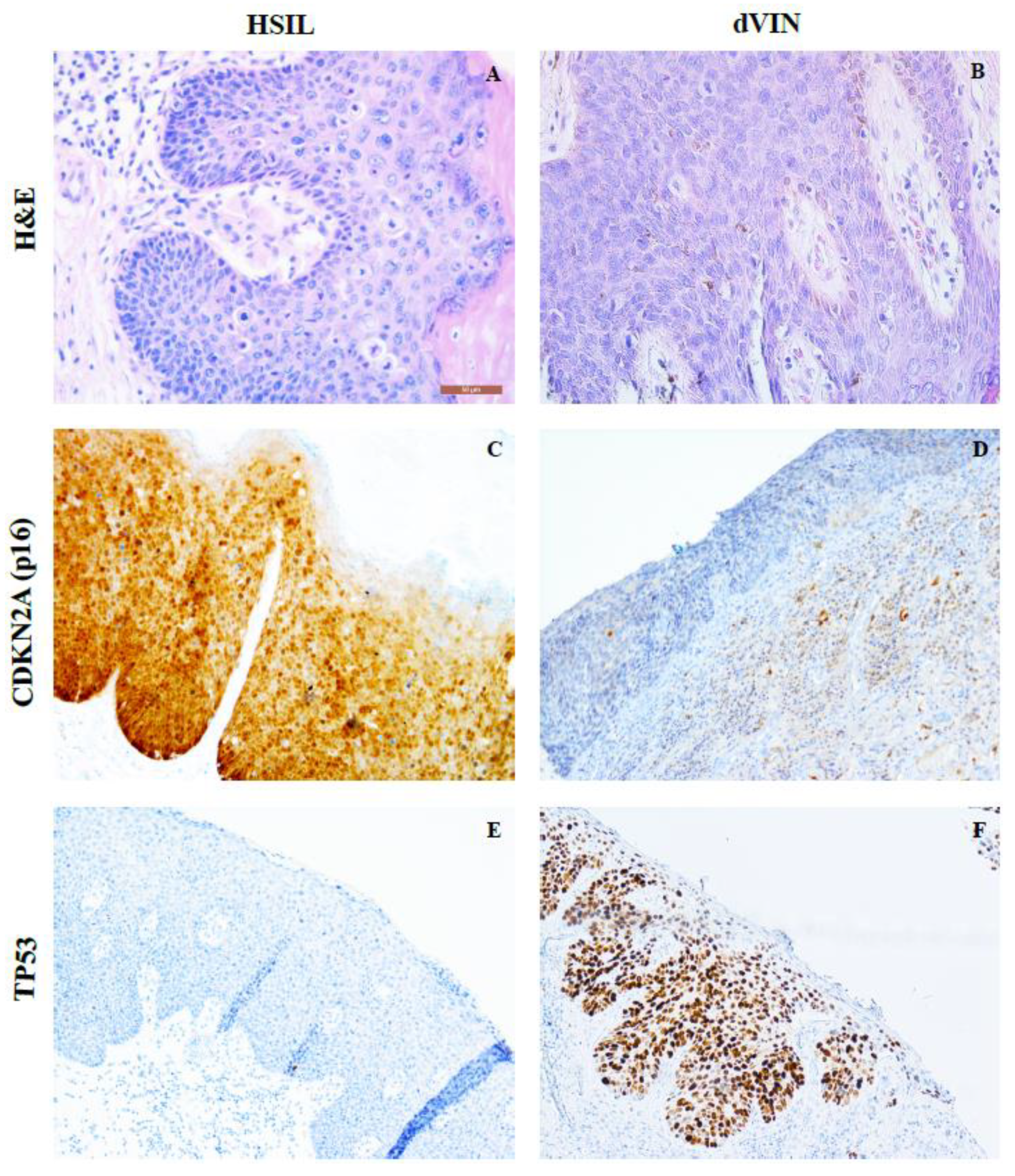
2.1. HPV Genotyping; p16 and p53 Negative Staining Results
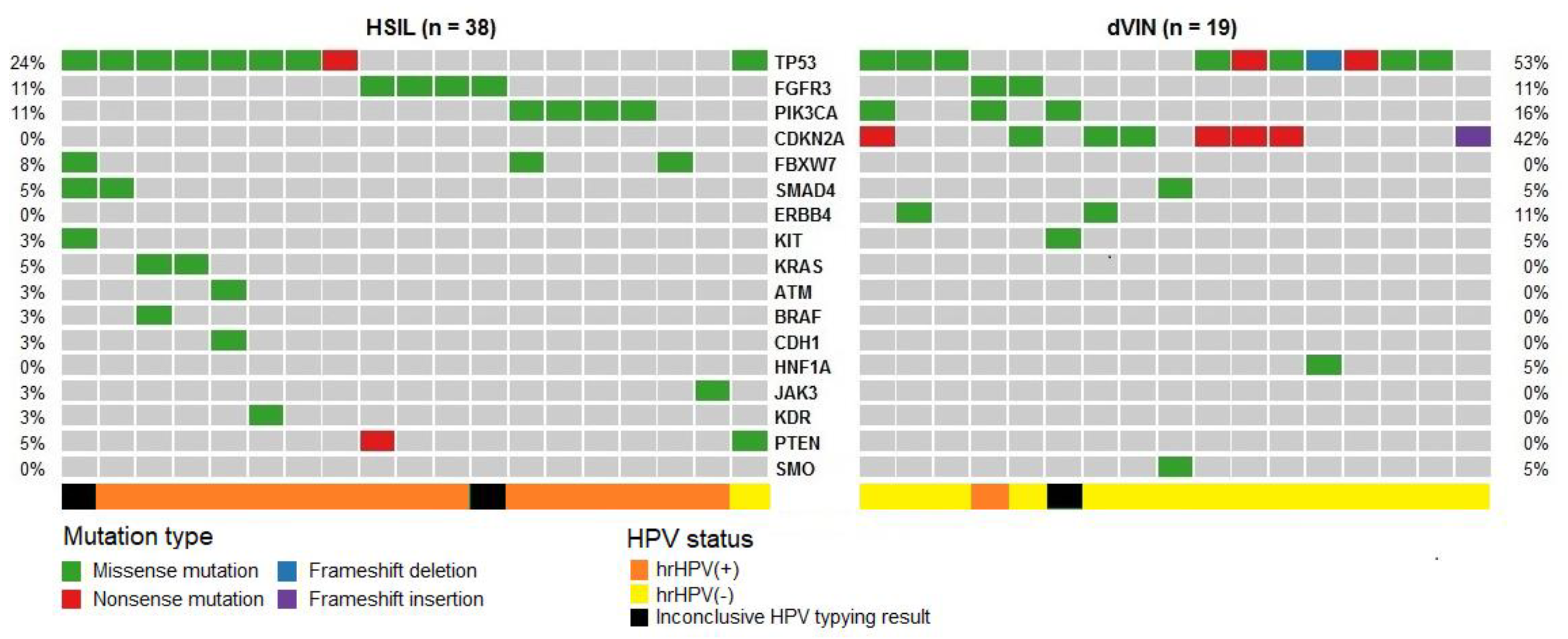
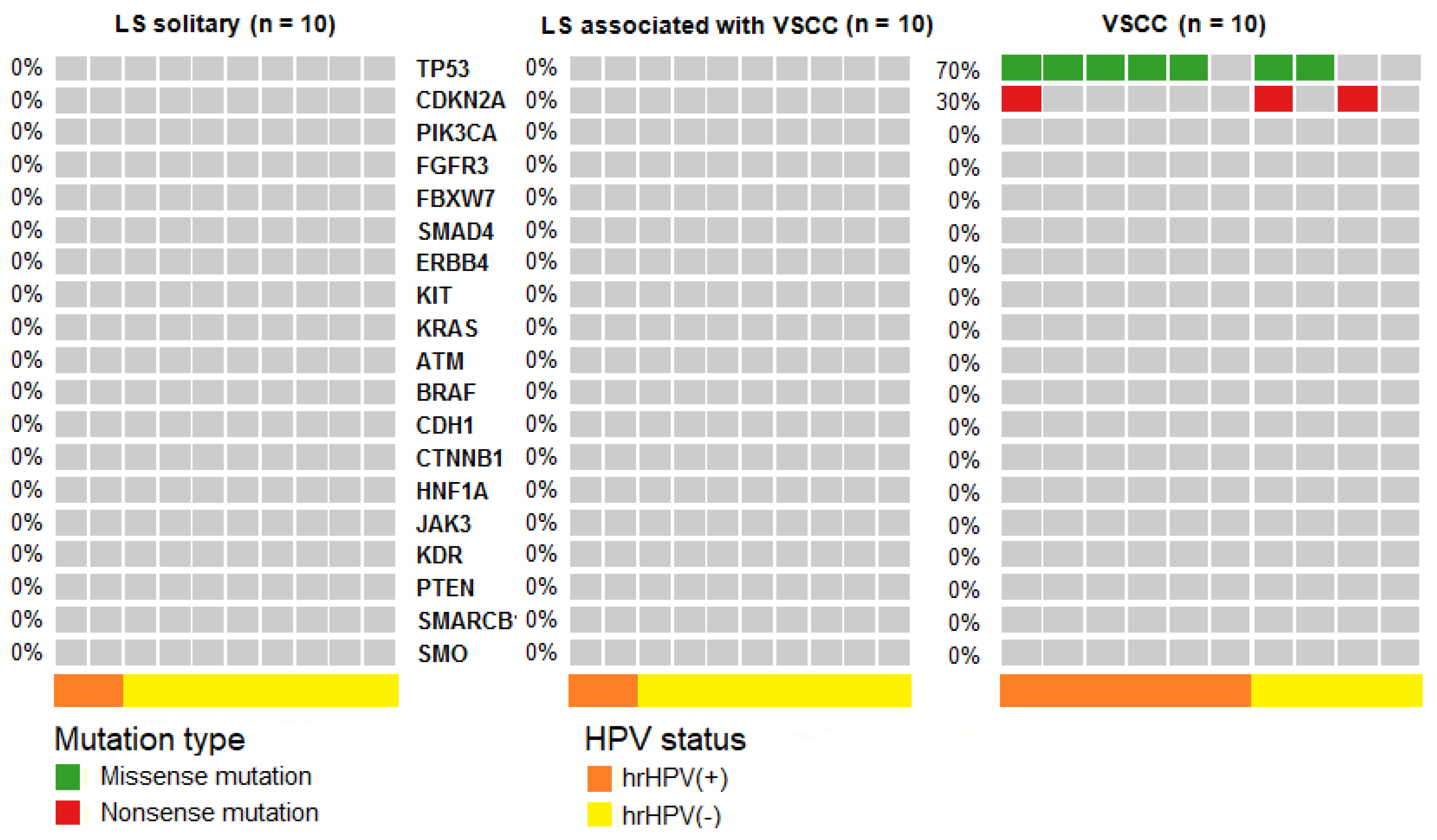
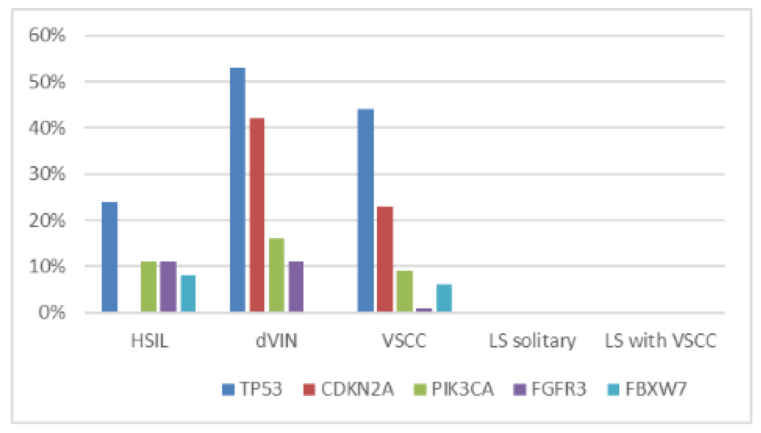
Similarly to cervical squamous lesions associated with hrHPV, immunostaining can assist in distinguishing HSIL from dVIN. Therefore, Immunohistochemistry (IHC) analysis was used to confirm the diagnosis of premalignant lesions of the vulva, as described previously [23] (Figure 1, Table S1). HSIL specimen were strongly CDKN2A (p16)- positive, showing CDKN2A expression in the middle and upper epithelial layers, whereas dVIN was either CDKN2A-negative or contained minimal CDKN2A expression in parabasal cells. Overall, 91% of (30/33) of HSIL and 12% (2/17) of dVIN samples were found to harbor hrHPV (in five cases of HSIL and two cases of dVIN, HPV genotyping results were inconclusive). Meanwhile, 20% (2/10) of solitary LS samples were hrHPV-positive. Equally, 20% (2/10) of LS associated with VSCC were hrHPV- positive, whereas in matching VSCC samples, hrHPV was detected in 60% (6/10) of cases.

2.2. NGS Results

DNA isolated from the analyzed samples (20 LS, 38 HSIL, 19 dVIN and 10 VSCC tumors) was subjected to next generation sequencing using the Ion AmpliSeq Cancer Hotspot Panel v2. Genetic changes were identified in 19 out of 50 genes examined, i.e., TP53, CDKN2A, FGFR3, PIK3CA, FBXW7, ERBB4, KIT, KRAS, PTEN, SMAD4, STK11, MET, ATM, BRAF, CDH1, HNF1A, JAK3, KDR, and SMO. Generally, according to the ClinVar (ncbi.nlm.nih.gov/clinvar/), dbSNP (https://0-www-ncbi-nlm-nih-gov.brum.beds.ac.uk/SNP/) and COSMIC (http://cancer.sanger.ac.uk/cosmic) databases, in the 241 changes identified in the studied samples, 70 pathogenic mutations, 9 variants of uncertain significance (VUS) (S1) and 162 polymorphisms (provided in Table S2) were classified.

Pathogenic Mutations Detected in HSIL, dVIN and LS

Pathogenic mutations, according to the ClinVar and COSMIC databases, were detected in 50% (19/38) of HSIL and 89% (17/19) of dVIN samples. HSIL samples carried 29 mutations in 11 genes: TP53, FGFR3, PIK3CA, FBXW7, KRAS, SMAD4, ERBB4, JAK3, PTEN, BRAF and KIT. dVIN samples harbored 20 mutations in six genes: TP53, CDKN2A, PIK3CA, SMAD4, FGFR3 and KIT. The obtained results reveal the prevailing mutations in TP53 in both HSIL and dVIN samples (at 24% and 53% frequencies, respectively), while CDKN2A mutations were absent in HSIL and present in 42% of dVIN cases (Table 1). The obtained frequencies of detected pathogenic mutations for the two subgroups of premalignant lesions are depicted in Figure 2. In LS, no pathogenic mutations were detected in the analyzed genes (including TP53 and CDKN2A)—neither in the 10 LS associated with VSCC nor in the 10 solitary LS samples (Table 1, Figure 3).
The comparison of distribution of pathogenic mutations in vulvar premalignant lesions with the data obtained for 10 cases of VSCC analyzed in the present study along with 81 VSCC cases analyzed previously using the same methods [23] is visualized in Figure 4. Frequencies of these mutations in HSIL and dVIN cases are very similar to those observed in VSCC.
A total of 163 polymorphic changes in HSIL, dVIN and LS samples were localized in seven genes, namely: TP53 (p.P72R), PIK3CA (p.I391M), KDR (p.Q472H), MET (p.N375S, p.T992I), KIT (p.M541L), ATM (p.S333F), and STK11 (p.F354L) (Table S2). The frequencies of these polymorphisms were similar in hrHPV(+) and hrHPV(-) samples. All the analyzed samples carried at least one polymorphism and the most frequent were found in TP53, KDR and KIT genes (Table 1).

3. Discussion

Our study aimed to expand knowledge on VSCC etiopathogenesis by analyzing somatic mutations in patients with VSCC and its precursors with cancer gene-targeted next-generation sequencing. The current understanding of VSCC pathogenesis is that it follows two alternative routes: i) alterations of vulvar epithelium caused by hrHPV infections may lead to the development of HSIL and then progression to VSCC or ii) alterations of vulvar epithelium caused by mutations, mainly of TP53 and CDKN2A genes, may provide basis for dVIN formation and subsequent development of VSCC. Recently, Nooij et al. [25] proposed an additional subgroup of vulvar cancers—HPV-negative and TP53 wild-type being characterized by frequent NOTCH1 mutations. Based on their findings, it may be hypothesized that mutations of NOTCH1 and HRAS are probable drivers of vulvar carcinogenesis without TP53 mutations. LS and/or squamous cell hyperplasia are proposed to precede dVIN formation [3]. This view is supported by the fact that dVIN is very often associated with LS and develops in the background of it. On the other hand, a very low risk of LS progression to VSCC means that LS should not be considered as a premalignancy. The continuum of these two pathologies, LS followed by dVIN, remains to be examined in more detail.
We analyzed solitary LS and LS associated with VSCC, HSIL and dVIN specimens based on the same experimental approach as used previously [23]. Premalignant vulvar lesions, both dVIN and HSIL, were found to harbor mutational profiles very similar to that of VSCC. Mutations of TP53 and CDKN2A are the most common genetic alterations identified in VSCC and are already present in its premalignant lesions. Therefore, mutations of TP53 and CDKN2A may be considered as early events during VSCC carcinogenesis. The higher mutation rate in dVIN as compared to HSIL as observed in our study and recently by Nooij et al. [25] may at least partially explain the fact that (despite of histological differentiation) dVIN exhibits high oncogenic potential [4,8], whereas the malignant potential of HSIL lesions is considered to be low. The significance of the polymorphisms detected in our sample set remains to be verified in a case control study.
It must be noted, however, as it was already pointed out by Singh et al. [21] in their study on dVIN that the real frequencies of TP53 mutations could be even higher in our sample set because we did not assess the all coding exons of TP53. Our analysis was limited to hot spots in exon 2 (excluding codons:p.21-25), exon 4 (excluding codons: p.40-67), exon 5, exon 6 (excluding codons: p.223–224); exon 7 (excluding codons: p.258-263), exon 8 and exon 11 of the TP53 gene. Additionally, due to limitations of the NGS method, we could not detect larger (> ~30bp) deletions. These are also the reasons for the 31% inconsistency between the p53-immunopositivity and TP53 mutation detection observed in our sample set. Additionally, DO7, the routinely used anti-p53, does not differentiate between mutant and wild-type p53 protein, and several disturbances in p53 pathway can result in abnormal p53 protein expression [26]. DO-7 recognizes the p53 N-terminal region (p.21–25) and the coding sequence for this epitope was excluded from our sequencing analysis. Moreover, as previously shown by Murnyák and Hortobágyi [26] in their analysis of the IARC TP53 Database, p53 IHC cannot be reliably used as a surrogate for mutation analysis (not only for nonsense mutations or deletions but also for missense TP53 mutations that were revealed in the absence of p53 protein expression).
Similarly, our p16 IHC staining results do not correlate with CDKN2A mutation status. This finding is in line with the data obtained in, for example, head and neck cancer studies [27,28], and this lack of correlation is due to diverse mechanisms of regulation of p16 function in human cancer [29]. Notably, all the examined HSIL specimens were p16-positive in IHC examination and none of them harbored CDKN2A mutations. On the contrary, all the examined dVIN cases were p16-negative and nearly half of them harbored CDKN2A mutations. In VSCC, the sensitivity and specificity of p16 IHC for detecting HPV-associated carcinomas were reported as close to 100% [19]. In AGO CaRE-1, a retrospective survey of VSCC patients, HPV DNA was detected in 78% of the p16-postitive tumors [30]. However, CDKN2A mutations were detected at low frequencies and similar in HPV(−) and HPV(+) VSCC (approximately 16% of cases) [31]. p16 protein induction may by mediated by inactivation of p53 and pRB by HPV oncoproteins and via epigenetic de-repression of p16 by KDM6B (JMJD3) histone demethylase in HPV infected cells. De-repression of p16 is required to maintain viability of hrHPV-infected cells [32]. In HPV-negative VSCC, CDKN2A promoter methylation is a frequent mechanism of p16 inactivation. Therefore, besides mutations of CDKN2A coding for p16, a plethora of other mechanisms generally leading to cell cycle deregulation affect p16 expression, and it seems that p16 may function as either a tumor suppressor or an oncogene in HPV-independent and HPV-associated carcinogenesis, respectively.
In 50% of HSIL and 11% of dVIN samples, we did not detect mutations of the analyzed genes. These rates can be compared to the respective percentages of 35% and 41% of previously analyzed hrHPV(+) and hrHPV(-) VSCC samples with no mutations detected in the same genes [23]. Our findings are in agreement with the conclusions of a recent genetic study on HPV(+) and HPV(-) vulvar carcinogenesis revealing similarities in copy number variation (CNV) patterns at the cancer stage [33] but divergence at its premalignant stages depending on HPV status of the lesions [34]. Based on CNV analysis and TP53 sequencing, Pouwer et al. [35] recently provided preliminary genetic evidence for a clonal relationship between LS, dVIN and VSCC.
The progression of LS to HPV-negative vulvar cancer was previously hypothesized by some authors [19,36,37]. The reported rate of TP53 mutations in LS varies widely—from 0% to 70% [37,38]—but the overall frequency of the combined results of the studies analyzed previously by Trietsch et al. [20] is 6%. Our data do not support notion that TP53 mutations are already present in the LS. As dVIN often resembles LS, and its histopathological diagnosis is challenging, the reported higher rates of TP53 mutations in LS might have been caused by the samples misdiagnosis caused by inadequate interpretation of morphological data as well as by the lack of unified guidelines for p53 expression assessment by immunohistochemistry [3,14,21,39]. However, the conclusions from our study are limited by the small LS sample number, and the same diagnostic challenges apply to our study. Moreover, the lack of TP53 mutations in LS does not preclude its role as a VSCC precursor, as causes other than genetic could promote LS progression, such as epigenetic (hypermethylation or hydroxymethylation) [40,41] or immune factors [42]. Hypermethylation of MGMT and RASSF2A (coding for DNA repair protein and cell cycle regulator, respectively) was detected in VSCC and LS associated with VSCC but not in isolated LS [40]. Additionally, global methylation as well as hydroxymethylation aberrations were observed in LS, confirming its epigenetic pathogenesis background [41]. Both LS and vulvar pre-malignant and malignant lesions are infiltrated with M2 macrophages and Tregs, which can inhibit tumor-specific T cell responses and promote carcinogenesis, adding more complexity to the understanding of the sequence of molecular events in vulvar precancers [42].
The recognition of the malignant potential of LS, dVIN, HSIL adjacent to the VSCC is highly relevant for the choice of primary surgical approach and the surgical resection margin. Despite the need of a distance of more than 8 mm initially proposed, recent studies have confirmed the safety of margins < 8 mm in node-negative VSCC patients [43,44,45]. Therefore, the question for reliable risk factors, other than lymph node status, is ongoing. Yap et al. [46] reported that women with VSCC arising in a field of LS are at an increased risk of developing local relapses and second field tumors (SFT) after resection of the primary tumor. Te Grootenhuis et al. [44] showed an actuarial local recurrence rates ten years after primary treatment of 42.5%, ranging from 28.1% for patients with HSIL, to 30.7% for patients with no precursor lesion, 44.2% for patients with LS, 44.8% for patients with dVIN, and 76.4% for patients with both LS and dVIN in the resection margin 10 years after treatment, respectively. Thus, the knowledge on the biology of premalignant lesions is important not only to know what risks they carry for the progression of primary lesions but also for VSCC recurrence. As SFT are believed to be genetically related to the primary tumor, our data do not support the notion of LS contribution to SFT. It could be speculated that the increased rate of local VSCC recurrences associated with LS is rather caused by second primary tumors (SPT), i.e., the new tumors genetically unrelated to the primary tumor. However, factors other than genetic could contribute to local relapses, such as epigenetic changes [47] or immune microenvironment [42].
The significance of PIK3CA, FGFR3 and FBXW7 mutations for vulvar carcinogenesis and vulvar cancer patient management remains to elucidated. PIK3CA mutations are most frequently (25.6%) found in HPV-related cancers, such as oropharyngeal, cervical, anal and vulvar squamous cell carcinomas [48]. Almost all of the identified mutations in PIK3CA in our sample set were detected in hrHPV-positive specimens. In two HSIL samples, we detected the p.H1047R mutation of PIK3CA, which was shown to be associated with the response to PI3K/AKT/mTOR signaling pathway inhibitors administered to patients with diverse advanced cancers [49]. The most frequent mutation of FGFR3 detected in premalignant vulvar lesions was p.S249C, described previously in VSCC hrHPV(+) (2-14%) [23,50] and other HPV-associated cancers [51,52]. In hrHPV+ head and neck cancer, the p.S249C mutation of FGFR3 was shown to be associated with poor prognosis [52]. In our study, FBXW7 mutations were observed in 8% of HSIL samples (three hrHPV+), but were absent in dVIN and LS samples. The presence of FBXW7 mutations in hrHPV(+) VSCC [23,33] suggests the involvement of FBXW7 alternations in HSIL progression to HPV-dependent VSCC. Indeed, the rates of FBXW7 gene mutation detection are noticeable in HPV related cancers [48].
Our preliminary data do not support a genetic background for the notion of LS as the VSCC premalignant lesion. Based on the current understanding of the relevance of the tumor microenvironment (TME), as well as the inflammatory processes in LS, future perspectives will focus on combined approaches taking into account to impact of ageing on the TME [53]. The ultimate goal would be to identify LS patients at risk for the development of VSCC and tailor the treatment accordingly. Our findings also suggest that patients with vulvar pre-cancers could potentially benefit from therapy targeted against cell cycle regulatory molecules, as previously proposed for VSCC [54], including the PI3K-Akt pathway members [31]. Numerous strategies for such targeted treatment modalities have been proposed, and some are being examined in ongoing clinical trials for other cancer types [55].

4. Materials and Methods

4.1. Patients

Clinical material was obtained from 20 randomly selected patients treated for LS ((10 associated with VSCC (median age 74.5 years, rage 37–81)) and 10 solitary LS)) and ten for early VSCC tumors (IB) at the Radboud university medical center, Nijmegen, the Netherlands, as well as from 38 HSIL (patients’ median age 60 years, range 27–84), and 19 dVIN (patients’ median age 68.7 years, range 41.4–89.0) patients treated at the Maria Sklodowska-Curie National Research Institute of Oncology in Warsaw, Poland and at the Holycross Cancer Center in Kielce, Poland. Histological diagnoses and assessment of IHC staining results were performed by the two independent gyne-pathologists working in the participating centers. Any ambiguities in histopathological findings were cross-reviewed.
HSIL and dVIN patients with no previous topical therapy were treated according to the local protocol with excision as a primary modality of choice. Patients operated on between April 2001 and September 2014 were enrolled. Research ethics board approval was received for the study with institutional authorization of the Radboud University Medical Center (No.: 2017-3996), the Maria Sklodowska-Curie National Research Institute of Oncology (No. 44/2002, 16/2015) and Holycross Cancer Center (No. 15/2014).

4.2. DNA Isolation and HPV Genotyping

Genomic DNA was isolated from paraffin-embedded tissue specimens using Maxwell® 16 FFPE DNA Purification Kit (Promega, Madison, WI, USA) according to the manufacturer’s protocol. The hrHPV status was determined as previously stated [23] using the AmpliSens HPV HCR-genotype-titre-FRT test (InterLabService Ltd., Moscow, Russian Federation), which detects 14 hrHPV genotypes, namely: HPV16, 18, 31, 33, 35, 39, 45, 51, 52, 56, 58, 59, 66, and 68, following the manufacturer’s instructions.

4.3. Next Generation Sequencing (NGS)

Library preparation was performed from 10 ng of DNA from each sample, which was added to the multiplex PCR reaction for library preparation using the Ion AmpliSeq Library Kit 2.0, Ion AmpliSeq Cancer Hotspot Panel v2 Kit (CHPv2), according to the manufacturer’s instructions (Thermo Fisher Scientific, Waltham, MA, USA). CHPv2 contains 207 pairs of primers, covering hotspots in the following 50 genes: ABL1, EZH2, JAK3, PTEN, ACT1, FBXW7, IDH2, PTPN11, ALK, FGFR1, KDR, RB1, APC, FGFR2, KIT, RET, ATM, FGFR3, KRAS, SMAD4, BRAF, FLT3, MET, SMARCB1, CDH1, GNA11, MLH1, SMO, CDKN2A, GNAS, MPL, SRC, CSF1R, GNAQ, NOTCH1, STK11, CTNNB1, HNF1A, NPM1, TP53, EGFR, HRAS, NRAS, VHL, ERBB2, IDH1, PDGFR, ERBB4, JAK2 and PIK3CA. Clonally amplified templates for NGS were prepared using the Ion Chef System and Ion 520 and Ion 530 Kit-Chef according to the manufacturer’s instructions (Thermo Fisher Scientific). The obtained barcoded libraries were loaded onto four Ion 530 chips and sequenced using Ion 520 and Ion 530 Kit-Chef and Ion S5 System according to the manufacturer’s instructions (Thermo Fisher Scientific, Waltham, MA, USA).

4.4. Next Generation Sequencing (NGS)

The analysis of NGS data was performed using three different programs to cross-validate the results. The raw data generated during sequencing were processed using the Torrent Server Suite 5.6 (Thermo Fisher Scientific, Waltham, MA, USA). The obtained sequences were aligned (mapped) to the reference sequence of the human genome (hg19) with the Torrent Server Suite 5.6. Variant calling was performed using the Variant Caller v5.6 embedded in the Torrent Server Suite 5.6. The default parameters used for CHPv2 data analysis were: minimum allele frequency - SNP = 0.02 / INDEL = 0.05, minimum quality—10, and minimum coverage—20×. The called variants were viewed by the Integrative Genomics Viewer (Broad Institute) (http://software.broadinstitute.org/software/igv/). In addition, the Torrent Server Suite 5.6 was used to generate sequencing files in the FASTQ format. The FASTQ files were analyzed using the Biomedical Genomics Workbench 4.0 (QIAGEN) and GALAXY [56] platform (www.usegalaxy.org). Default parameters used in the analysis by the Biomedical Genomics Workbench 4.0 software were as follows: minimum allele frequency—0.02, the minimum quality—10 and minimum coverage—10x. The Biomedical Genomics Workbench 4.0 was also used for annotation. Data analysis with the GALAXY was performed using FASTQ Groomer tool to generate the fastqsanger format and then the Bowtie2 tool (with default parameters) was used to map the reads to the reference sequence hg19. After mapping, SAMtool was used to generate mpileup files. Mpileup format files were then used in the variant detection step. For variant detection, the VarScan tool was applied with the following parameters: minimum allele frequency of 0.05, minimum quality of 25 and minimum coverage—40x. The annotation of detected variants by the Torrent Server Suite 5.6 and the GALAXY was done with the wANNOVAR tool (http://wannovar.wglab.org/). In cases without mutations within the RB, TP53 or PTEN (genes with a confirmed role in vulvar carcinogenesis) as assigned by the three programs used, NGS sequencing results were additionally reviewed using the Integrative Genomics Viewer.
The NGS results were visualized as CoOncoplots plotted using Maftools from the R Bioconductor package [57].

4.5. Immunohistochemistry

Immunohistochemical staining (IHC) with antibodies against p16 (E6H4, Ventana-Roche Diagnostic, IN, USA) and p53 (clone DO-7, Dako Denmark A/S, An Agilent Technologies Company) and subsequent IHC assessment of vulvar premalignant lesions (dVIN and HSIL) was performed as described previously [23].

5. Conclusions

We conclude that our data support the notion that TP53 mutations can be considered as early events during VSCC carcinogenesis. TP53 mutations are present in HSIL and dVIN lesions, with higher frequency in the latter premalignancy, thus possibly contributing to its higher oncogenic potential as compared to HSIL. CDKN2A mutations absent in HSIL and detected in dVIN with high frequency may further increase the oncogenic potential of dVIN. In terms of incidence, we identified PIK3CA, FGFR3 and FBXW7 as genes which mutated less frequently. Importantly, our results provide no genetic hints for the contribution of LS to vulvar carcinogenesis, as LS samples remained non-mutated in both solitary LS and LS associated with VSCC. However, further research using whole genome sequencing as well as examination epigenetic and other non-genetic factors is needed in order to verify biological mechanisms leading to LS progression, either directly or via dVIN formation.

Supplementary Materials

Supplementary materials can be found at https://0-www-mdpi-com.brum.beds.ac.uk/1422-0067/21/14/4880/s1. Table S1: Pathogenic mutations and variants of uncertain significance detected by next generation sequencing in hrHPV(+) (n = 42) and hrHPV(−) (n=40) and samples where hrHPV genotyping results were inconclusive (n=7) in premalignant lesions and VSCC tumors. Table S2: List of polymorphisms in hrHPV(+) (n = 42 and hrHPV(−) (n=40) and samples where hrHPV genotyping results were inconclusive (n=7) in premalignant lesions and VSCC tumors determined by next generation sequencing.

Author Contributions

Conceptualization, A.K. and M.K.; methodology, A.K.; software, S.Z.; formal analysis, S.Z.; investigation, S.Z., N.R., E.B.-Z. and J.K.; resources, A.-F.W.P., K.Z. and J.K.; data curation, S.Z. and A.-F.W.P.; writing—original draft preparation, S.Z., M.K.; writing—review and editing, J.M.A.P., J.A.d.H. and M.K.; visualization, S.Z. and N.R.; supervision, M.K.; project administration, M.K.; funding acquisition, M.K. All authors have read and agreed to the published version of the manuscript.

Funding

This research was funded by the Polish National Science Centre, grant number 2013/10/E/NZ5/00663.

Acknowledgments

The authors gratefully acknowledge the assistance of Transition Technologies S.A. in patient data analysis using the MedStream Designer (MSD) application on the Onko.Sys platform.

Conflicts of Interest

The authors declare no conflict of interest.

Abbreviations

dVINdifferentiated-type vulvar intraepithelial neoplasia
H&Ehematoxylin and eosin staining
HSILhigh-grade squamous intraepithelial lesion
HPVhuman papilloma virus
hrHPVhigh-risk HPV
LSlichen sclerosus
VSCCvulvar squamous cell carcinoma

References

  1. International Agency for Research on Cancer: Lyon, France, 2014.
  2. van de Nieuwenhof, H.P.; van der Avoort, I.A.; de Hullu, J.A. Review of squamous premalignant vulvar lesions. Crit Rev. Oncol. Hematol. 2008, 68, 131–156. [Google Scholar] [CrossRef] [PubMed]
  3. Hoang, L.N.; Park, K.J.; Soslow, R.A.; Murali, R. Squamous precursor lesions of the vulva: Current classification and diagnostic challenges. Pathology 2016, 48, 291–302. [Google Scholar] [CrossRef] [PubMed] [Green Version]
  4. Eva, L.J.; Ganesan, R.; Chan, K.K.; Honest, H.; Luesley, D.M. Differentiated-type vulval intraepithelial neoplasia has a high-risk association with vulval squamous cell carcinoma. Int. J. Gynecol. Cancer 2009, 19, 741–744. [Google Scholar] [CrossRef] [PubMed]
  5. van de Nieuwenhof, H.P.; Massuger, L.F.; van der Avoort, I.A.; Bekkers, R.L.; Casparie, M.; Abma, W.; van Kempen, L.C.; de Hullu, J.A. Vulvar squamous cell carcinoma development after diagnosis of VIN increases with age. Eur. J. Cancer 2009, 45, 851–856. [Google Scholar] [CrossRef]
  6. Bigby, S.M.; Eva, L.J.; Fong, K.L.; Jones, R.W. The Natural History of Vulvar Intraepithelial Neoplasia, Differentiated Type: Evidence for Progression and Diagnostic Challenges. Int. J. Gynecol. Pathol. 2016, 35, 574–584. [Google Scholar] [CrossRef]
  7. Judson, P.L.; Habermann, E.B.; Baxter, N.N.; Durham, S.B.; Virnig, B.A. Trends in the incidence of invasive and in situ vulvar carcinoma. Obstet. Gynecol. 2006, 107, 1018–1022. [Google Scholar] [CrossRef]
  8. Women and Health: Today’s Evidence Tomorrow’s Agenda; World Health Organization: Geneva, Switzerland, 2009.
  9. van den Einden, L.C.; de Hullu, J.A.; Massuger, L.F.; Grefte, J.M.; Bult, P.; Wiersma, A.; van Engen-van Grunsven, A.C.; Sturm, B.; Bosch, S.L.; Hollema, H.; et al. Interobserver variability and the effect of education in the histopathological diagnosis of differentiated vulvar intraepithelial neoplasia. Mod. Pathol. 2013, 26, 874–880. [Google Scholar] [CrossRef] [Green Version]
  10. Schuurman, M.S.; van den Einden, L.C.; Massuger, L.F.; Kiemeney, L.A.; van der Aa, M.A.; de Hullu, J.A. Trends in incidence and survival of Dutch women with vulvar squamous cell carcinoma. Eur. J. Cancer 2013, 49, 3872–3880. [Google Scholar] [CrossRef]
  11. Akhtar-Danesh, N.; Elit, L.; Lytwyn, A. Trends in incidence and survival of women with invasive vulvar cancer in the United States and Canada: A population-based study. Gynecol. Oncol. 2014, 134, 314–318. [Google Scholar] [CrossRef]
  12. Micheletti, L.; Preti, M.; Radici, G.; Boveri, S.; Di Pumpo, O.; Privitera, S.S.; Ghiringhello, B.; Benedetto, C. Vulvar Lichen Sclerosus and Neoplastic Transformation: A Retrospective Study of 976 Cases. J. Low Genit. Tract. Dis. 2016, 20, 180–183. [Google Scholar] [CrossRef] [Green Version]
  13. Halonen, P.; Jakobsson, M.; Heikinheimo, O.; Riska, A.; Gissler, M.; Pukkala, E. Lichen sclerosus and risk of cancer. Int. J. Cancer 2017, 140, 1998–2002. [Google Scholar] [CrossRef] [PubMed] [Green Version]
  14. van de Nieuwenhof, H.P.; Bulten, J.; Hollema, H.; Dommerholt, R.G.; Massuger, L.F.; van der Zee, A.G.; de Hullu, J.A.; van Kempen, L.C. Differentiated vulvar intraepithelial neoplasia is often found in lesions, previously diagnosed as lichen sclerosus, which have progressed to vulvar squamous cell carcinoma. Mod. Pathol. 2011, 24, 297–305. [Google Scholar] [CrossRef] [PubMed]
  15. Neill, S.M.; Tatnall, F.M.; Cox, N.H. Guidelines for the management of lichen sclerosus. Br. J. Dermatol. 2002, 147, 640–649. [Google Scholar] [CrossRef] [PubMed]
  16. Carli, P.; De Magnis, A.; Mannone, F.; Botti, E.; Taddei, G.; Cattaneo, A. Vulvar carcinoma associated with lichen sclerosus. Experience at the Florence, Italy, Vulvar Clinic. J. Reprod. Med. 2003, 48, 313–318. [Google Scholar] [PubMed]
  17. Dong, F.; Kojiro, S.; Borger, D.R.; Growdon, W.B.; Oliva, E. Squamous Cell Carcinoma of the Vulva: A Subclassification of 97 Cases by Clinicopathologic, Immunohistochemical, and Molecular Features (p16, p53, and EGFR). Am. J. Surg. Pathol. 2015, 39, 1045–1053. [Google Scholar] [CrossRef]
  18. Cheng, A.S.; Karnezis, A.N.; Jordan, S.; Singh, N.; McAlpine, J.N.; Gilks, C.B. p16 Immunostaining Allows for Accurate Subclassification of Vulvar Squamous Cell Carcinoma Into HPV-Associated and HPV-Independent Cases. Int. J. Gynecol. Pathol. 2016, 35, 385–393. [Google Scholar] [CrossRef]
  19. Del Pino, M.; Rodriguez-Carunchio, L.; Ordi, J. Pathways of vulvar intraepithelial neoplasia and squamous cell carcinoma. Histopathology 2013, 62, 161–175. [Google Scholar] [CrossRef]
  20. Trietsch, M.D.; Nooij, L.S.; Gaarenstroom, K.N.; van Poelgeest, M.I. Genetic and epigenetic changes in vulvar squamous cell carcinoma and its precursor lesions: A review of the current literature. Gynecol. Oncol. 2015, 136, 143–157. [Google Scholar] [CrossRef]
  21. Singh, N.; Leen, S.L.; Han, G.; Faruqi, A.; Kokka, F.; Rosenthal, A.; Jiang, X.R.; Kim, R.; McAlpine, J.N.; Gilks, C.B. Expanding the morphologic spectrum of differentiated VIN (dVIN) through detailed mapping of cases with p53 loss. Am. J. Surg. Pathol. 2015, 39, 52–60. [Google Scholar] [CrossRef]
  22. Pinto, A.P.; Miron, A.; Yassin, Y.; Monte, N.; Woo, T.Y.; Mehra, K.K.; Medeiros, F.; Crum, C.P. Differentiated vulvar intraepithelial neoplasia contains Tp53 mutations and is genetically linked to vulvar squamous cell carcinoma. Mod. Pathol. 2010, 23, 404–412. [Google Scholar] [CrossRef] [Green Version]
  23. Zieba, S.; Kowalik, A.; Zalewski, K.; Rusetska, N.; Goryca, K.; Piascik, A.; Misiek, M.; Bakula-Zalewska, E.; Kopczynski, J.; Kowalski, K.; et al. Somatic mutation profiling of vulvar cancer: Exploring therapeutic targets. Gynecol. Oncol. 2018, 150, 552–561. [Google Scholar] [CrossRef] [PubMed]
  24. Vogelstein, B.; Kinzler, K.W. The Path to Cancer—Three Strikes and You’re Out. N. Engl. J. Med. 2015, 373, 1895–1898. [Google Scholar] [CrossRef] [PubMed]
  25. Nooij, L.S.; Ter Haar, N.T.; Ruano, D.; Rakislova, N.; van Wezel, T.; Smit, V.; Trimbos, B.; Ordi, J.; van Poelgeest, M.I.E.; Bosse, T. Genomic Characterization of Vulvar (Pre)cancers Identifies Distinct Molecular Subtypes with Prognostic Significance. Clin. Cancer Res. 2017, 23, 6781–6789. [Google Scholar] [CrossRef] [PubMed] [Green Version]
  26. Murnyak, B.; Hortobagyi, T. Immunohistochemical correlates of TP53 somatic mutations in cancer. Oncotarget 2016, 7, 64910–64920. [Google Scholar] [CrossRef] [Green Version]
  27. Lim, A.M.; Do, H.; Young, R.J.; Wong, S.Q.; Angel, C.; Collins, M.; Takano, E.A.; Corry, J.; Wiesenfeld, D.; Kleid, S.; et al. Differential mechanisms of CDKN2A (p16) alteration in oral tongue squamous cell carcinomas and correlation with patient outcome. Int. J. Cancer 2014, 135, 887–895. [Google Scholar] [CrossRef]
  28. Larque, A.B.; Conde, L.; Hakim, S.; Alos, L.; Jares, P.; Vilaseca, I.; Cardesa, A.; Nadal, A. P16(INK(4)a) overexpression is associated with CDKN2A mutation and worse prognosis in HPV-negative laryngeal squamous cell carcinomas. Virchows Arch. 2015, 466, 375–382. [Google Scholar] [CrossRef]
  29. Li, J.; Poi, M.J.; Tsai, M.D. Regulatory mechanisms of tumor suppressor P16(INK4A) and their relevance to cancer. Biochemistry 2011, 50, 5566–5582. [Google Scholar] [CrossRef] [Green Version]
  30. Woelber, L.L.P.K.; Eulenburg, C.H.; de Gregorio, N.; Klapdor, R.; Kalder, M.; Braicu, E.I.; Fuerst, S.; Klar, M.; Strauss, H.-G.; Mehlhorn, G.; et al. p53 and p16 expression profiles reveal three prognostically relevant subgroups in vulvar cancer: A TMA based study by the AGO-CaRE-translational study group. J. Clin. Oncol. 2019, 37, 5592. [Google Scholar] [CrossRef]
  31. Zieba, S.; Chechlinska, M.; Kowalik, A.; Kowalewska, M. Genes pathways and vulvar carcinoma—New insights from next-generation sequencing studies. Gynecol. Oncol. 2020. ahead of print. [Google Scholar] [CrossRef]
  32. McLaughlin-Drubin, M.E.; Park, D.; Munger, K. Tumor suppressor p16INK4A is necessary for survival of cervical carcinoma cell lines. Proc. Natl. Acad Sci. USA 2013, 110, 16175–16180. [Google Scholar] [CrossRef] [Green Version]
  33. Han, M.R.; Shin, S.; Park, H.C.; Kim, M.S.; Lee, S.H.; Jung, S.H.; Song, S.Y.; Lee, S.H.; Chung, Y.J. Mutational signatures and chromosome alteration profiles of squamous cell carcinomas of the vulva. Exp. Mol. Med. 2018, 50, e442. [Google Scholar] [CrossRef] [PubMed]
  34. Swarts, D.R.A.; Voorham, Q.J.M.; van Splunter, A.P.; Wilting, S.M.; Sie, D.; Pronk, D.; van Beurden, M.; Heideman, D.A.M.; Snijders, P.J.F.; Meijer, C.; et al. Molecular heterogeneity in human papillomavirus-dependent and -independent vulvar carcinogenesis. Cancer Med. 2018, 7, 4542–4553. [Google Scholar] [CrossRef] [PubMed]
  35. Pouwer, A.W.; Loes van den, E.; van der Linden, M.; Hehir-Kwa, J.Y.; Yu, J.; Hendriks, K.M.; Kamping, E.J.; Eijkelenboom, A.; Massuger, L.; Bulten, J.; et al. Clonal Relationship Between Lichen Sclerosus, Differentiated Vulvar Intra-epithelial Neoplasia and Non HPV-related Vulvar Squamous Cell Carcinoma. Cancer Genom. Proteom. 2020, 17, 151–160. [Google Scholar] [CrossRef] [PubMed] [Green Version]
  36. Carlson, J.A.; Ambros, R.; Malfetano, J.; Ross, J.; Grabowski, R.; Lamb, P.; Figge, H.; Mihm, M.C., Jr. Vulvar lichen sclerosus and squamous cell carcinoma: A cohort, case control, and investigational study with historical perspective; implications for chronic inflammation and sclerosis in the development of neoplasia. Hum. Pathol. 1998, 29, 932–948. [Google Scholar] [CrossRef]
  37. Vanin, K.; Scurry, J.; Thorne, H.; Yuen, K.; Ramsay, R.G. Overexpression of wild-type p53 in lichen sclerosus adjacent to human papillomavirus-negative vulvar cancer. J. Invest. Dermatol. 2002, 119, 1027–1033. [Google Scholar] [CrossRef] [Green Version]
  38. Rolfe, K.J.; MacLean, A.B.; Crow, J.C.; Benjamin, E.; Reid, W.M.; Perrett, C.W. TP53 mutations in vulval lichen sclerosus adjacent to squamous cell carcinoma of the vulva. Br. J. Cancer 2003, 89, 2249–2253. [Google Scholar] [CrossRef]
  39. Dasgupta, S.; Ewing-Graham, P.C.; van Kemenade, F.J.; van Doorn, H.C.; Noordhoek Hegt, V.; Koljenovic, S. Differentiated vulvar intraepithelial neoplasia (dVIN): The most helpful histological features and the utility of cytokeratins 13 and 17. Virchows Arch. 2018, 473, 739–747. [Google Scholar] [CrossRef] [Green Version]
  40. Guerrero, D.; Guarch, R.; Ojer, A.; Casas, J.M.; Mendez-Meca, C.; Esteller, M.; Barba-Ramos, E.; Garcia-Bragado, F.; Puras, A. Differential hypermethylation of genes in vulvar cancer and lichen sclerosus coexisting or not with vulvar cancer. Int. J. Cancer 2011, 128, 2853–2864. [Google Scholar] [CrossRef]
  41. Gambichler, T.; Terras, S.; Kreuter, A.; Skrygan, M. Altered global methylation and hydroxymethylation status in vulvar lichen sclerosus: Further support for epigenetic mechanisms. Br. J. Dermatol. 2014, 170, 687–693. [Google Scholar] [CrossRef] [Green Version]
  42. Abdulrahman, Z.; Kortekaas, K.E.; De Vos Van Steenwijk, P.J.; Van Der Burg, S.H.; Van Poelgeest, M.I. The immune microenvironment in vulvar (pre)cancer: Review of literature and implications for immunotherapy. Expert Opin. Biol. Ther. 2018, 18, 1223–1233. [Google Scholar] [CrossRef]
  43. Woelber, L.; Griebel, L.F.; Eulenburg, C.; Sehouli, J.; Jueckstock, J.; Hilpert, F.; de Gregorio, N.; Hasenburg, A.; Ignatov, A.; Hillemanns, P.; et al. Role of tumour-free margin distance for loco-regional control in vulvar cancer-a subset analysis of the Arbeitsgemeinschaft Gynakologische Onkologie CaRE-1 multicenter study. Eur. J. Cancer 2016, 69, 180–188. [Google Scholar] [CrossRef] [PubMed]
  44. Te Grootenhuis, N.C.; Pouwer, A.W.; de Bock, G.H.; Hollema, H.; Bulten, J.; van der Zee, A.G.J.; de Hullu, J.A.; Oonk, M.H.M. Prognostic factors for local recurrence of squamous cell carcinoma of the vulva: A systematic review. Gynecol. Oncol. 2018, 148, 622–631. [Google Scholar] [CrossRef]
  45. Pleunis, N.; Leermakers, M.E.J.; van der Wurff, A.A.; Klinkhamer, P.; Ezendam, N.P.M.; Boll, D.; de Hullu, J.A.; Pijnenborg, J.M.A. Surgical margins in squamous cell carcinoma, different for the vulva? Eur. J. Surg. Oncol. 2018, 44, 1555–1561. [Google Scholar] [CrossRef]
  46. Yap, J.K.; Fox, R.; Leonard, S.; Ganesan, R.; Kehoe, S.T.; Dawson, C.W.; Woodman, C.B.; Luesley, D.M. Adjacent Lichen Sclerosis predicts local recurrence and second field tumour in women with vulvar squamous cell carcinoma. Gynecol. Oncol. 2016, 142, 420–426. [Google Scholar] [CrossRef]
  47. Leonard, S.; Pereira, M.; Fox, R.; Gordon, N.; Yap, J.; Kehoe, S.; Luesley, D.; Woodman, C.; Ganesan, R. Over-expression of DNMT3A predicts the risk of recurrent vulvar squamous cell carcinomas. Gynecol. Oncol. 2016, 143, 414–420. [Google Scholar] [CrossRef] [Green Version]
  48. Koncar, R.F.; Feldman, R.; Bahassi, E.M.; Hashemi Sadraei, N. Comparative molecular profiling of HPV-induced squamous cell carcinomas. Cancer Med. 2017, 6, 1673–1685. [Google Scholar] [CrossRef]
  49. Janku, F.; Wheler, J.J.; Naing, A.; Falchook, G.S.; Hong, D.S.; Stepanek, V.M.; Fu, S.; Piha-Paul, S.A.; Lee, J.J.; Luthra, R.; et al. PIK3CA mutation H1047R is associated with response to PI3K/AKT/mTOR signaling pathway inhibitors in early-phase clinical trials. Cancer Res. 2013, 73, 276–284. [Google Scholar] [CrossRef] [Green Version]
  50. Weberpals, J.I.; Lo, B.; Duciaume, M.M.; Spaans, J.N.; Clancy, A.A.; Dimitroulakos, J.; Goss, G.D.; Sekhon, H.S. Vulvar Squamous Cell Carcinoma (VSCC) as Two Diseases: HPV Status Identifies Distinct Mutational Profiles Including Oncogenic Fibroblast Growth Factor Receptor 3. Clin. Cancer Res. 2017, 23, 4501–4510. [Google Scholar] [CrossRef] [Green Version]
  51. Rosty, C.; Aubriot, M.H.; Cappellen, D.; Bourdin, J.; Cartier, I.; Thiery, J.P.; Sastre-Garau, X.; Radvanyi, F. Clinical and biological characteristics of cervical neoplasias with FGFR3 mutation. Mol. Cancer 2005, 4, 15. [Google Scholar] [CrossRef] [Green Version]
  52. Bersani, C.; Sivars, L.; Haeggblom, L.; DiLorenzo, S.; Mints, M.; Ahrlund-Richter, A.; Tertipis, N.; Munck-Wikland, E.; Nasman, A.; Ramqvist, T.; et al. Targeted sequencing of tonsillar and base of tongue cancer and human papillomavirus positive unknown primary of the head and neck reveals prognostic effects of mutated FGFR3. Oncotarget 2017, 8, 35339–35350. [Google Scholar] [CrossRef]
  53. Elkhattouti, A.; Hassan, M.; Gomez, C.R. Stromal Fibroblast in Age-Related Cancer: Role in Tumorigenesis and Potential as Novel Therapeutic Target. Front. Oncol. 2015, 5, 158. [Google Scholar] [CrossRef] [Green Version]
  54. Clancy, A.A.; Spaans, J.N.; Weberpals, J.I. The forgotten woman’s cancer: Vulvar squamous cell carcinoma (VSCC) and a targeted approach to therapy. Ann. Oncol. 2016, 27, 1696–1705. [Google Scholar] [CrossRef]
  55. Gatzka, M.V. Targeted Tumor Therapy Remixed—An Update on the Use of Small-Molecule Drugs in Combination Therapies. Cancers 2018, 10, 155. [Google Scholar] [CrossRef] [Green Version]
  56. Afgan, E.; Baker, D.; Batut, B.; van den Beek, M.; Bouvier, D.; Cech, M.; Chilton, J.; Clements, D.; Coraor, N.; Gruning, B.A.; et al. The Galaxy platform for accessible, reproducible and collaborative biomedical analyses: 2018 update. Nucleic. Acids Res. 2018, 46, W537–W544. [Google Scholar] [CrossRef] [Green Version]
  57. Mayakonda, A.; Lin, D.C.; Assenov, Y.; Plass, C.; Koeffler, H.P. Maftools: Efficient and comprehensive analysis of somatic variants in cancer. Genome Res. 2018, 28, 1747–1756. [Google Scholar] [CrossRef] [Green Version]
Figure 1. Examples of immunohistochemical CDKN2A (middle panel) and TP53 (lower panel) staining performed on tissue sections of high-grade squamous intraepithelial lesion (HSIL) and differentiated-type vulvar intraepithelial neoplasia (dVIN). H&E—hematoxylin and eosin staining (upper panel); 40x magnification—(A,B); 20x magnification—(CF).
Ijms 21 04880 g001
Figure 2. Distribution of pathogenic mutations and variants of uncertain significance in vulvar premalignant lesions. Samples were classified as HSIL (n = 38) and dVIN (n = 19) based on histopathological and IHC assessment. Each column corresponds to an individual tumor case, while each row corresponds to the mutated gene. Samples with no mutations detected are depicted in grey, missense mutations are highlighted in green, whereas red hits indicate nonsense mutations, indel mutation are violet and blue. hrHPV-positive and hrHPV-negative samples are marked in the bottom line with orange and yellow color, respectively.
Ijms 21 04880 g002
Figure 3. Distribution of pathogenic mutations in solitary lichen sclerosus (LS) (n = 10) and LS (n=10) associated with VSCC (n = 10). Each column corresponds to an individual LS or VSCC case, while each row corresponds to the mutated gene. Samples with no mutations detected are depicted in grey, missense mutations are highlighted in green, whereas red hits indicate nonsense mutations. hrHPV-positive and hrHPV-negative samples are marked in the bottom line with orange and yellow color, respectively.
Ijms 21 04880 g003
Figure 4. Mutation prevalence of the most often mutated genes in dVIN, HSIL, VSCC and LS samples.
Ijms 21 04880 g004
Table 1. Most frequent mutation and polymorphism rates in HSIL, dVIN, lichen sclerosus (LS) and vulvar squamous cell carcinoma (VSCC) samples.
Sample TypeMutations (% of Cases)Polymorphisms (% of Cases)
TP53CDKN2ATP53 P72RKDR Q472HKIT M541L
HSIL (n = 38)240100428
dVIN (n = 19)53421006316
solitary LS (n = 10)001004020
LS associated with VSCC (n = 10)001005030
VSCC (n = 10)70301005030

