Article

A Role for Mycobacterium tuberculosis Sigma Factor C in Copper Nutritional Immunity

by
Benjamin T. Grosse-Siestrup
1,
Tuhina Gupta
1,
Shelly Helms
1,
Samantha L. Tucker
1,
Martin I. Voskuil
2,
Frederick D. Quinn
1 and
Russell K. Karls
1,*
1
Department of Infectious Diseases, College of Veterinary Medicine, University of Georgia, Athens, GA 30602, USA
2
Department of Immunology and Microbiology, University of Colorado Anschutz Medical Campus, Aurora, CO 80045, USA
*
Author to whom correspondence should be addressed.
Int. J. Mol. Sci. 2021, 22(4), 2118; https://0-doi-org.brum.beds.ac.uk/10.3390/ijms22042118
Submission received: 25 December 2020 / Revised: 9 February 2021 / Accepted: 17 February 2021 / Published: 20 February 2021

Abstract

:
Sigma factor C (SigC) contributes to Mycobacterium tuberculosis virulence in various animal models, but the stress response coordinated by this transcription factor was undefined. The results presented here indicate that SigC prevents copper starvation. Whole genome expression studies demonstrate short-term (4-h) induction of sigC, controlled from a tetracycline-inducible promoter, upregulates ctpB and genes in the nonribosomal peptide synthase (nrp) operon. These genes are expressed at higher levels after 48-h sigC induction, but also elevated are genes encoding copper-responsive regulator RicR and RicR-regulated copper toxicity response operon genes rv0846–rv0850, suggesting prolonged sigC induction results in excessive copper uptake. No growth and global transcriptional differences are observed between a sigC null mutant relative to its parent strain in 7H9 medium. In a copper-deficient medium, however, growth of the sigC deletion strain lags the parent, and 40 genes (including those in the nrp operon) are differentially expressed. Copper supplementation reverses the growth defect and silences most transcriptional differences. Together, these data support SigC as a transcriptional regulator of copper acquisition when the metal is scarce. Attenuation of sigC mutants in severe combined immunodeficient mice is consistent with an inability to overcome innate host defenses that sequester copper ions to deprive invading microbes of this essential micronutrient.

1. Introduction

Mycobacterium tuberculosis is the primary cause of death in humans by a single bacterial pathogen. In 2019, an estimated 10 million people developed tuberculosis (TB) disease and 1.4 million individuals died from TB; 208,000 of the deaths occurred among persons co-infected with HIV [1]. Globally, latent tuberculosis infections have been estimated at 1.7 billion [2].
The success of M. tuberculosis as a pathogen is in part due to sensing stress cues in the host and responding through implementation of effective countermeasures. In M. tuberculosis, sigma factor C (SigC) is one of 13 transcription sigma factors [3,4]. Mutants lacking intact sigC are attenuated in murine and guinea pig infection models [5,6,7]; however, these null mutants failed to yield phenotypes in vitro such as slower intracellular replication in phagocytyic cells [5,7]. Transcription from two start sites mapped upstream of sigC is detected in a sigC null mutant suggesting that this gene is not transcriptionally autoregulated [8]. Elevated sigC expression was not detected in response to a variety of stress stimuli [9]. Although sigC is fully conserved in M. tuberculosis strains CDC1551 and H37Rv, and in M. bovis BCG, none of the genes defined as members of the SigC regulon by transcriptome analyses of a CDC1551 sigC mutant [7] were upregulated after 18-h induction of sigC controlled from a tetracycline-inducible promoter in strain H37Rv [10] nor were any found with upstream promoters bound by SigC-RNA polymerase in M. bovis BCG chromosome immunoprecipitation/DNA microarray studies [11]. In the present study, phenotypes and gene expression associated with sigC were examined in derivatives of M. tuberculosis strain Erdman.
Transition metals are essential for all living organisms. Iron and copper commonly function as cofactors of enzymes that catalyze electron transfer reactions [12]. The electrostatic properties of these trace elements can stabilize reaction intermediates, but toxicity can result if such metals are present in excess [13]. Acquisition of trace elements from a host is a requirement for pathogenic bacteria. The term nutritional immunity was first used to define host processes of iron sequestration to prevent the growth of invading microbes [14], but subsequently expanded to encompass host sequestration of any essential trace metal from potential pathogens (reviewed in [15]). From an antimicrobial perspective, levels of free copper ions are maintained at extremely low levels by complexing or chaperoning the metal in most eukaryotic systems, an exception being phagosomes where copper ions are concentrated to target engulfed microbes [16,17]. The M. tuberculosis cytochrome aa3–bc1 supercomplex is a member of the heme-copper respiratory oxidase family. All saturation transposon mutagenesis studies indicate ctaD (rv3043), encoding cytochrome aa3 subunit 1 that contains the copper-B catalytic site, is an essential gene in M. tuberculosis [18,19,20]. As an obligate aerobe [21], M. tuberculosis must acquire copper to replicate. The data presented herein support SigC as a transcription factor required by the obligate human pathogen M. tuberculosis to acquire copper from environments where levels of the free metal are very low.

2. Results and Discussion

2.1. Absence of Significant Growth or Global Gene Expression Differences after Deletion of sigC from M. tuberculosis Strain Erdman Cultured in 7H9 Medium

To aid in identifying sigC-associated phenotypes, an internal sigC deletion mutant (ΔsigC) was generated in M. tuberculosis strain Erdman (Figure 1). Since sigC is transcribed at high levels under many conditions [9], to minimize altering potential antisense effects on the downstream divergently transcribed cobK gene, an in-frame, internal portion of sigC was removed. The deletion removes bases encoding amino acids 123–168, which comprise most of sigma factor region 4 (amino acids 123–175) [22]. SigC has conserved sigma-70 family regions 2 and 4 for SigC-RNA polymerase binding to the -10 and -35 promoter regions, respectively, but lacks region 3 that aids promoter binding through contact with the extended -10 region [22]. Our previous studies showed reduced virulence in guinea pigs of an M. tuberculosis strain H37Rv derivative with the same sigC lesion, but the parent strain was more attenuated than expected [5]. We subsequently learned that this H37Rv strain was passaged on solid medium many years longer than standard freezer-stocked H37Rv isolates. Thus, to minimize the likelihood that a SigC-regulated gene may have been mutated during long-term passage, we used strain Erdman to generate the new mutant. The Erdman ΔsigC strain exhibited growth kinetics in Middlebrook 7H9 broth indistinguishable from strain Erdman and the complemented sigC mutant (Supplementary materials Figure S1). This was anticipated as sigC mutants of CDC1551 and H37Rv had no growth defects in 7H9 medium [5]. Transcriptomic studies using DNA microarrays with probes to the open reading frames in H37Rv examined global gene expression differences between ΔsigC and parent Erdman cultured to OD600 = 1 in 7H9 medium. Data obtained from four biological replicates hybridized on a total of ten slides revealed that no genes had significant differential expression of twofold or more after the data were analyzed using Significance Analysis of Microarrays (SAM) software (Table S1). The absence of significant gene expression differences was unexpected based on microarray results reported for a CDC1551ΔsigC mutant cultured in 7H9 medium, which identified over a dozen different genes repressed >2-fold in the mutant at OD600 = 0.5, OD600 = 2.0, and in stationary phase, but only sigC was downregulated in all growth conditions [7]. That study reported that two biological replicates were tested per strain per condition, which may underlie the inconsistent gene expression differences. The experiments to follow support why SigC function may not be required in 7H9 medium.

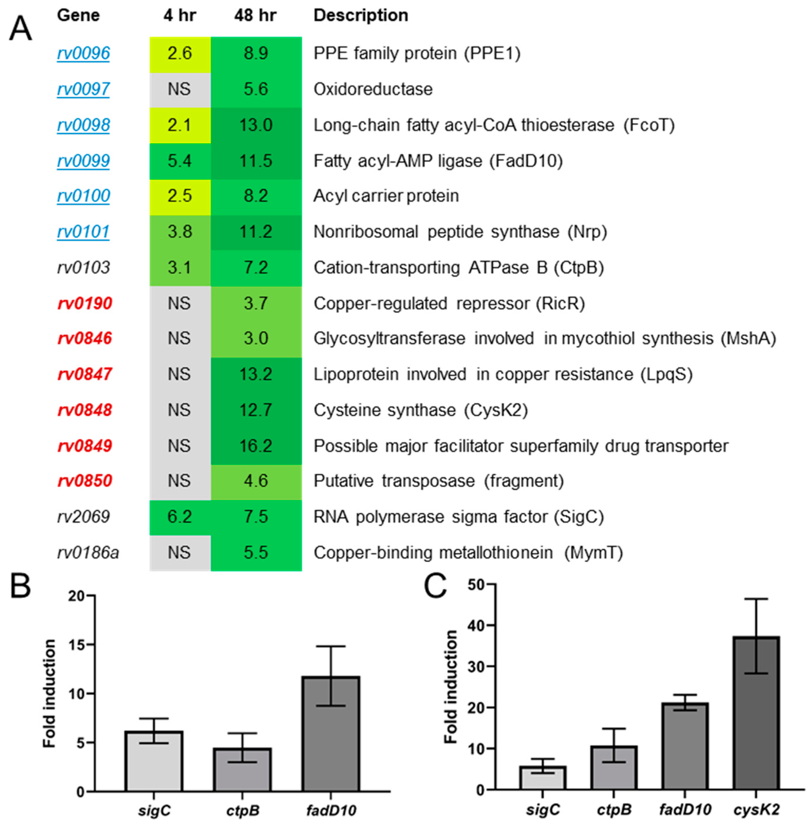
2.2. Artificial sigC Induction Points to a Role for SigC in Metal Transport

As significant gene expression differences were not detected between ΔsigC and strain Erdman in 7H9 cultures, transcriptome studies were performed with strain Erdman transformed with plasmid pSR173 encoding SigC with an N-terminal myc tag transcriptionally controlled from a tetracycline-inducible promoter. DNA microarray studies compared transcription of uninduced and anhydrotetracycline (aTc)-induced cells. After 4-h aTc induction, seven genes were significantly upregulated at least twofold (Figure 2A, column 2, Table S2). The artificially induced sigC gene was elevated 6.2-fold. The other genes with increased transcription were ctpB (rv0103), elevated 3.1-fold, and five of the six genes in the nearby ppe1 (rv0096)–nrp (rv0101) operon, increased 2.1- to 5.4-fold. The microarray results were confirmed by qRT-PCR of sigC, ctpB, and fadD10 (rv0099) only (Figure 2B). The ctpB gene encodes a cytoplasmic membrane P1B-type ATPase associated with transport of copper from mycobacterial cells [23]. Orthologs of genes in the ppe1–nrp operon are present in slow-growing pathogenic mycobacteria and in actinomyces [24]. The rv0097–nrp orthologous region from Mycobacterium marinum expressed in Escherichia coli enables synthesis of isonitrile lipopeptides when 2-decanoic or 2-dodecanoic acid is present in the cultures [24]. An isonitrile product from Streptomyces thioluteus binds copper and functions as a chalkophore for copper acquisition [25], while an M. marinum mutant lacking the nrp region was reported to accumulate less intracellular zinc [24].
To determine if the transcriptional profile changes after prolonged induction of sigC, expression after 48-h growth was examined. Consistent with the 4-h results, sigC, ctpB, and ppe1–nrp operon genes were upregulated, but to greater extents, ranging from 5.6- to 11.5-fold, (Figure 2A, Column 3, Table S3). Seven additional genes were significantly upregulated in response to 48-h sigC induction in Erdman (Figure 2A, Table S3). The regulated-in-copper repressor gene ricR was elevated (3.7-fold) as were five genes in the RicR-controlled rv0846–rv0850 genomic region (ranging from 3.0- to 16.2-fold, Figure 2A). This region encodes a multi-copper oxidase (rv0846), a lipoprotein LpqS (rv0847), cysteine synthase CysK2 (rv0848), and a membrane permease (rv0849) that reduce the toxic effects of elevated intracellular copper levels [26]. Gene rv0186a encoding the copper-binding metallothionein MymT was increased 5.5-fold (Figure 2A). MymT protein binds up to six Cu(I) ions and mymT partially protects M. tuberculosis from copper toxicity [27]. Quantitative RT-PCR of sigC, ctpB, rv0099, and rv0846 confirmed elevated transcription after 48-h sigC induction (Figure 2C). The 48-h sigC induction results are very similar to findings from a transcriptome study in which sigC was artificially induced for 18 h in an H37Rv background from a tetracycline-inducible promoter [10]. In that report, genes increased more than 2-fold included all six genes in the ppe1–nrp operon (4.2- to 5.9-fold), all five genes in the rv0846–rv0850 region (2.6- to 3.7-fold), and ctpB (2.2-fold). The only other gene elevated after 18-h sigC induction was rv1813 (2.0-fold) [10]; this gene was not increased in our studies. Finally, the mymT gene that we detected elevated after 48-h sigC induction was the only gene not upregulated after 18-h sigC induction [10]. This fits with the concept that bacteria accumulate more copper the longer sigC is artificially induced. Consistent with our 4- and 48-h sigC-induction data, Rustad and colleagues also did not detect any genes downregulated more than 2-fold after inducing sigC for 18 h.
Transcription of ctpB and the ppe1–nrp operon by SigC is consistent with results from in vivo chromosome immunoprecipitation/DNA microarray studies performed in M. bovis BCG that identified myc-SigC binding hotspots and almost identical predicted SigC-35 and -10 promoter elements immediately upstream of both regions [11]; note that the promoter region shown in Figure 3, panel C of that publication is mislabeled as rv0095c, instead of rv0096 (ppe1). That study also demonstrated in vitro transcription of ctpB by RNA polymerase reconstituted with SigC, but did not report myc-SigC binding upstream of homologs of ricR or RicR-regulated genes. This suggests that ctpB and the ppe1–nrp operon are directly transcribed by Sig-RNA polymerase, but the copper toxicity-response genes are likely indirectly upregulated after prolonged artificial induction of sigC and actions of the encoded products of the ppe1–nrp operon and/or ctpB. Taken together, these results suggest that SigC functions in metal acquisition.

2.3. Identification of an In Vitro Phenotype for ΔsigC

To test the hypothesis that SigC functions in metal uptake, growth of the sigC mutant, sigC-complemented mutant, and strain Erdman was examined in a defined medium lacking copper, zinc, and calcium. To minimize nutrient carryover, cells initially cultured in 7H9 broth were washed with and subcultured into Sauton medium containing 0.025% of dispersant tyloxapol (SMT). After 3–4 passages in SMT medium, a phenotype developed in which growth of ΔsigC lagged the parent strain and complemented mutant (Figure 3A). To determine if the phenotype was due to lack of a specific metal present in 7H9 medium but lacking in SMT, growth was examined in SMT supplemented with the concentrations of copper, zinc, or calcium salts indicated in the formula for Middlebrook 7H9 medium in the BD Difco and BBL Manual [28]. Growth of ΔsigC was almost fully restored to wild type levels by supplementation with all three metals or with 6 µM copper sulfate alone, but not with either zinc or calcium salts (Figure 3B–E). These data confirm that SigC functions in copper acquisition.

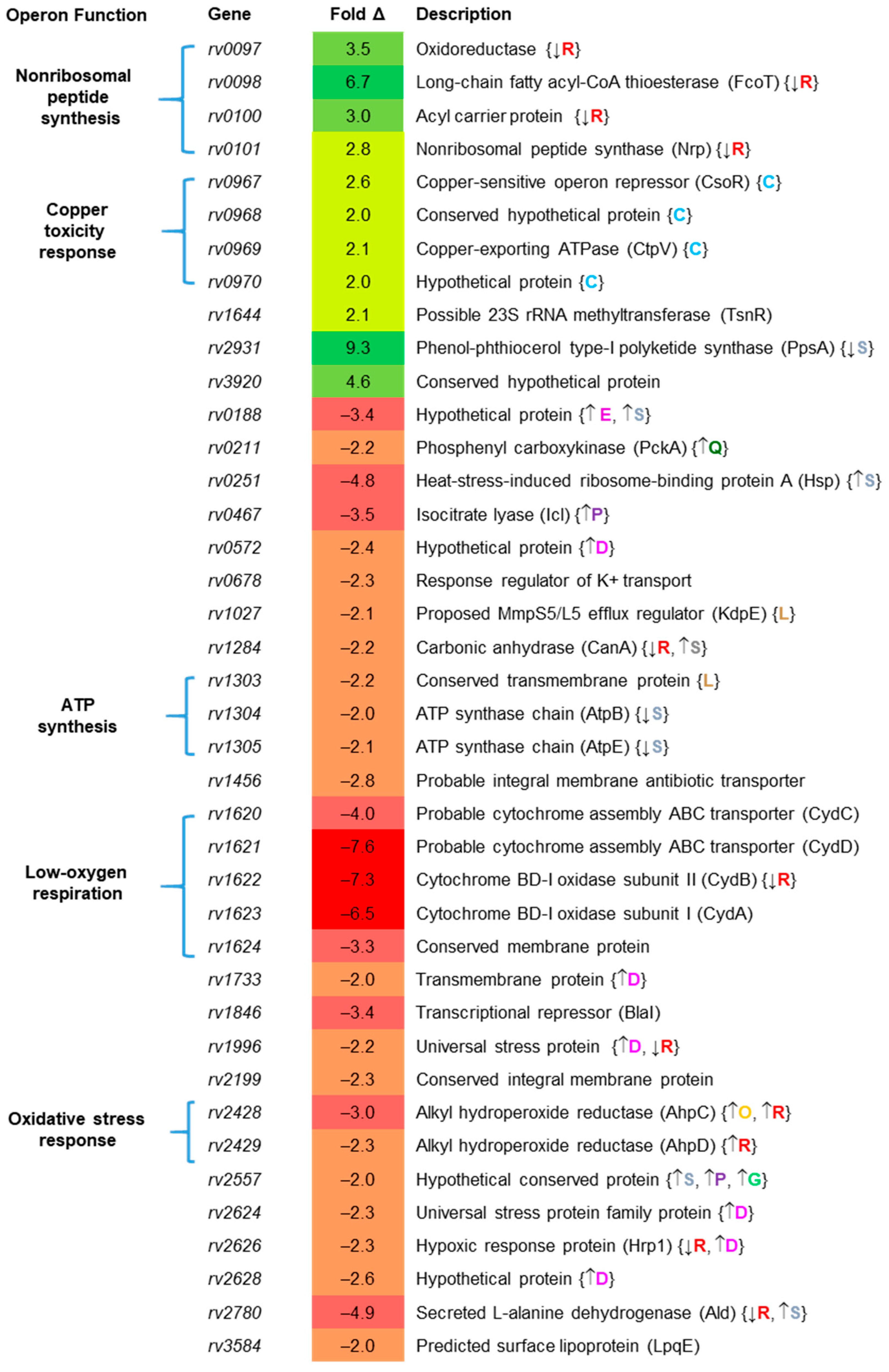
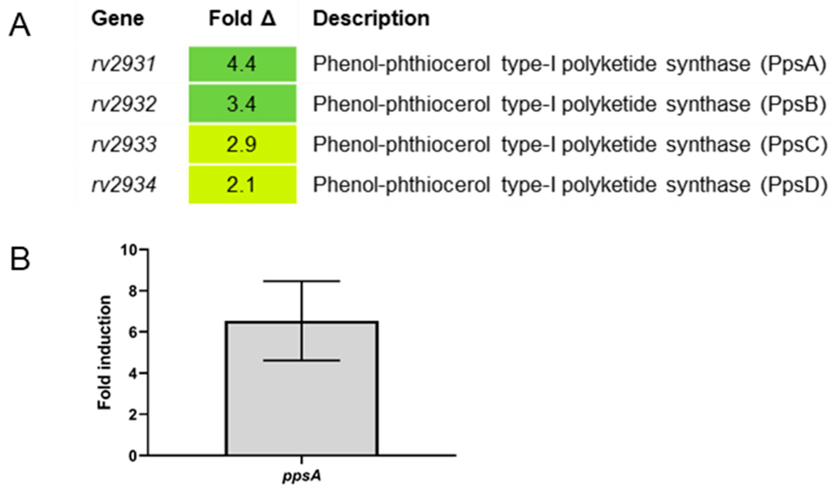
2.4. Differential Gene Expression between Erdman and ΔsigC in SMT Supports SigC Function in Copper Uptake

To investigate how the growth defect of the sigC mutant in SMT lacking copper impacts gene expression, global transcriptional analysis between ΔsigC and parent Erdman was examined in SMT cultures grown to log phase (OD600 = 1). Cells were cultured as described for the growth studies (Section 2.3). Significant differential expression of twofold or greater was observed for 40 genes (Figure 4, Figure S2, Table S4). Of the 11 genes expressed at higher levels in Erdman, four were in the ppe1–nrp operon upregulated after artificial sigC induction in 7H9 medium (Figure 2). This operon is also upregulated in an M. tuberculosis mutant with a deletion in the rv0490–rv0491 locus encoding the SenX3–RegX3 two-component regulator [29] which senses and responds to low phosphate levels [30]. In a ΔsigC mutant of M. tuberculosis strain CDC1551, expression of the rv0490 homolog mt0509 was reported to be downregulated in stationary phase 7H9 cultures [7]. Together, these observations indicate links between sigC, metal transport, and phosphate availability.
Other genes more highly expressed in Erdman relative to ΔsigC included the csoR–rv968–ctpV–rv970 operon (Figure 4, Figure S2, Table S4). This operon is controlled by the copper-sensitive operon repressor, CsoR, which is distinct from the RicR-controlled operon that was upregulated in 7H9 cultures after 48-h sigC induction (Figure 2A). The csoR operon may be upregulated as a homeostatic mechanism after internalization of sufficient copper ions to bind CsoR and de-repression of its operon leading to production of the copper-exporting P-type ATPase CtpV to prevent toxic levels of copper from accumulating in the cytoplasm [31]. The rv2931/ppsA gene was the most-highly upregulated (9.3-fold) in Erdman relative to ΔsigC (Figure 4). It encodes a phenol-phthiocerol polyketide synthase that functions in phthiocerol dimycocerosates (PDIM) biosynthesis [32]. The elevated expression of ppsA in Erdman cultured in SMT medium but not when sigC was artificially induced in 7H9 medium suggests an indirect effect of sigC on ppsA expression, which will be discussed later. Gene rv3920, encoding a conserved protein of unknown function, was also significantly upregulated (4.6-fold). The only other gene significantly upregulated in the parent strain was rv1644/tnsR (2.1-fold) encoding a predicted 23S rRNA methyltransferase. It is unclear why these genes are elevated in a strain wild type for sigC under copper-limiting conditions. Of note, ctpB expression was not significantly upregulated in Erdman versus ΔsigC cultured in SMT despite ctpB being upregulated after 4- or 48-h sigC induction in 7H9 medium (Figure 2).
Among the 29 genes more highly expressed in ΔsigC relative to Erdman in SMT were operons that function in production of cytochrome bd oxidase (rv1620–rv1624), ATP synthesis (rv1303–rv1305), and alkyl hydroperoxide reductase (rv2428–rv2429) (Figure 4). Other genes upregulated in the mutant include several involved in central carbon metabolism, such as icl (encoding isocitrate lyase), pckA (encoding phosphoenol carboxykinase), ald (encoding alanine/glycine dehydrogenase), and canA (encoding carbonic anhydrase) (Figure 4). Elevated transcription of cydB and icl in the mutant was confirmed by qRT-PCR (Figure S2).
Genes upregulated in the sigC mutant in SMT cultures indicate alterations in central carbon metabolism and bioenergetics to alleviate reductive stress. Reductive stress results from the abnormal increase in the reduced forms of redox carriers such as NADH, NADPH, and FADH2 [41]. For detailed descriptions of M. tuberculosis central carbon metabolism and respiration, a number of reviews are recommended [42,43,44,45,46]. Under growth conditions with adequate aeration, the M. tuberculosis TCA cycle generates CO2 as well as NADH and FADH2. These carrier molecules are reoxidized by transfer of electrons through the menaquinone pool and the Qcr–Cta cytochrome aa3–bc1 supercomplex to oxygen with concomitant pumping of protons across the cytoplasmic membrane to provide the proton motive force to drive ATP synthesis ([46], Figure S3). If the TCA cycle or electron transport chain is disrupted, then the reductive stress resulting from the buildup of reduced TCA cycle intermediates could lead to induction of metabolic processes in an effort to restore redox balance [41]. This appears to be the case for the sigC mutant (Figure S3). Increased icl expression allows the glyoxylate shunt to bypass the NADH- and CO2-generating steps in the TCA cycle that function in the production of succinate from α-ketoglutarate. Upregulation of ald enables production of amino acids glycine and alanine with concomitant oxidation of NADH to NAD. Increased transcription of pckA supplies phosphoenolpyruvate carboxykinase required for gluconeogenesis [47]. Expression of pckA is elevated by mildly acidic and hypoxic conditions [39]. The carbonic anhydrase encoded by rv1284 is sensitive to oxidizing conditions [48]; thus, rv1284 upregulation suggests a reduced redox state in which carbon fixation could serve as an electron sink to help restore redox balance. Metabolic flux analyses in carbon-limited chemostat studies have demonstrated that M. tuberculosis and M. bovis BCG can fix CO2 and utilize glyoxylate shunt and anapleurotic reactions for oxidation of pyruvate and production of succinyl CoA [49]. Elevated transcription of cydABCD is clearly a response to reductive stress. This operon is induced under hypoxic conditions and encodes a cytochrome bd oxidase (Cyd) which has higher oxygen affinity than the Qcr–Cta supercomplex, but is bioenergetically inefficient [46]. Importantly, unlike the Qcr–Cta supercomplexes, which are heme-copper oxygen reductases essential for efficient M. tuberculosis aerobic respiration, Cyd complexes only have heme centers for electron transfer to oxygen [50]. Upregulated Cyd production in response to copper starvation by ΔsigC in SMT would provide some copper-independent respiration, albeit with much less proton pumping across the membrane needed for ATP synthesis than respiration through the Qcr–Cta supercomplex (Figure S3). Upregulation of ATP synthesis genes rv1303–rv1305 in the mutant suggests energy generation is affected.
Genes upregulated in the sigC mutant may also indicate responses to copper deficiency through inducing expression of copper-independent enzymes to mitigate respiratory distress. Alkyl hydroperoxide reductase genes ahpCD (rv2428 and rv2429) upregulated by oxidating stress [37] may be upregulated in ΔsigC to provide an alternate defense against reactive oxygen species if insufficient copper was available to cofactor the mycobacterial Cu-Zn superoxide dismutase or other copper-dependent reductases. Genes rv0572, rv1733, rv1996, rv2624, hrp1, and rv2628 are also induced as part of the DosR hypoxic response regulon [33,51]. Gene rv0188 is a member of the enduring hypoxic response [34], while rv2557 and rv0467/icl are upregulated during anaerobic persistence [38], with rv2557 also elevated in the transition zone of human necrotic lung granulomas [35]. Hypoxia is expected from prolonged lack of aerobic respiration from copper-starved Qcr–Cta supercomplexes in the sigC mutant as cytochrome bd oxidases require three times more oxygen than heme-copper supercomplexes to pump the same number of protons to generate an equivalent amount of ATP (Figure S3).
Upregulation of transcriptional regulators and genes controlled by other regulators in ΔsigC may be efforts to bypass different copper-dependent processes. Multiple redox-sensing systems would be expected to be affected by reductive stress and ATP depletion. The encoded product of rv0572 is a predicted regulator the MmpS5/L5 efflux system [52]. The blaI gene encoding the beta-lactamase repressor is part of the iron-regulated repressor (IdeR) regulon [53], while rv1305 and atpE are predicted members of the BlaI regulon [54]. Genes ahpC, ahpD, ald, cydB, hrpI, rv1996, and rv2626 are regulated by the phosphate-responsive SenX3/RegX3 regulatory system [55]. Genes rv0188, rv1284, hsp, atpB, atpE, rv2557, and ald were reported to be upregulated after 96-h starvation [40], while kdpE and rv1303 are part of the RelA-regulated stringent response [36]. Taken together, the seemingly disparate sets of genes with elevated expression in ΔsigC in copper-deficient SMT medium is consistent with a model in which these bacteria are upregulating the production of enzymes and processes that do not require copper for function.

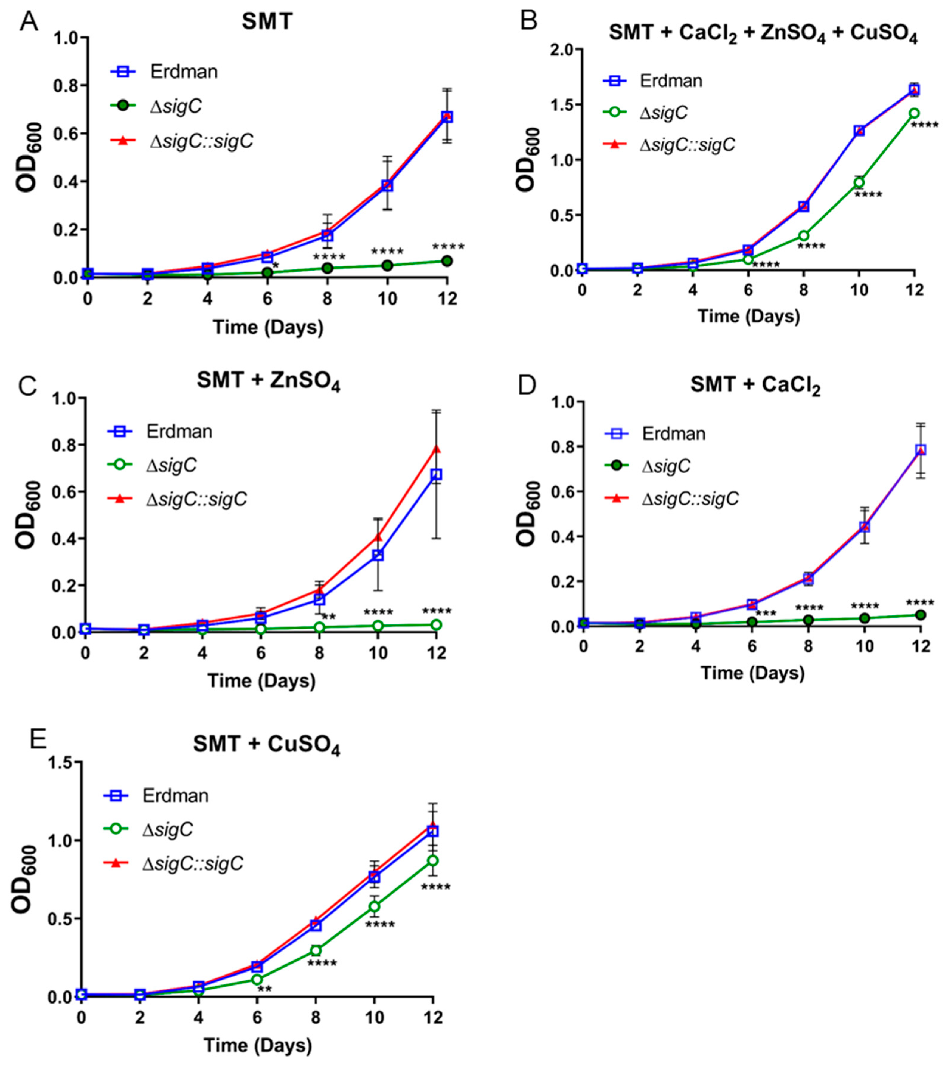
2.5. Copper Supplementation Eliminates Most Transcriptional Differences between ΔsigC and Erdman

Based on the improved growth of ΔsigC in SMT supplemented with 6 μM copper sulfate (Figure 3E), we hypothesized that similar supplementation would eliminate many of the observed gene expression differences between Erdman and ΔsigC grown in SMT. This is precisely what we observed. Only 1 of the 40 genes that were differentially regulated twofold or more between Erdman and ΔsigC from SMT cultures (Figure 4) remained significantly elevated after 6 µM copper sulfate supplementation (Figure 5A, Table S5).
The gene ppsA was again elevated in Erdman relative to ΔsigC, but also increased were genes ppsB, ppsC, and ppsD, located immediately downstream of ppsA and each encoding different phenol-phthiocerol polyketide synthases that function in PDIM lipid synthesis (Figure 5A). Elevated expression of this operon was confirmed by examination of ppsA by qRT-PCR (Figure 5B). It is unclear why PDIM synthesis operon genes were elevated in strain Erdman relative to the sigC mutant. No SigC promoters comparable to those preceding ctpB and ppe1 were detected upstream of ppsA. The sigC mutant does not have a smooth colony morphology such as those observed for PDIM mutants, which indicates that sigC is not required for PDIM synthesis under all conditions. PDIM synthesis is upregulated in response to reductive stress by serving as an electron sink [56]. It is possible that mutations in PDIM synthesis develop in the sigC mutant upon passage in low-copper medium but eliminating a mechanism that mitigates reductive stress is unlikely to confer a growth advantage as ΔsigC depletes copper stores needed for respiration. Genes in the pps operon function in production of a common lipid core used for PDIM and glycosylated phenolphthiocerol dimycocerosate surface lipids [57]; thus, it is possible that under copper-starved conditions, the copper-chelating product of the ppe1–nrp operon enzymes is bound to the same common lipid core. The net effect would be a surface-exposed copper receptor for collecting trace amounts of copper from the environment. Reduced expression of pps genes in the sigC mutant in suboptimal copper conditions may reflect reduced need for the common lipid core due to an inability to produce the copper-chelating molecule. The transcriptional connection between sigC and PDIM genes may be indirect through redox-sensing factors. Transcription factor WhiB3 directly senses the redox state in M. tuberculosis through its thiol-disulfide redox switch and modulates the production of virulence lipids including PDIM [56]. PDIM synthesis is also impacted by one or more genes in the sigma factor M (SigM) operon. Deletion/replacement of H37Rv sigM with a kanamycin-resistance gene in the same orientation resulted in increased PDIM levels and expression of PDIM synthesis genes ppsABCDE, ddrABC, and mas [58]. As sigM is located upstream of genes encoding redox proteins thioredoxin and thioredoxin reductase, it is unclear if increased PDIM gene expression in the sigM mutant was due to loss of sigM or to altered transcription of downstream redox protein genes. It is also of note that artificial induction of sigM in the sigM mutant background resulted in 10-fold increased transcription of ppe1–nrp operon genes rv0098–rv0100 [58]. Further studies are needed to understand the link between PDIM gene expression and sigC and potential interplay with other regulatory factors. Since 6 µM copper supplementation of SMT did not fully restore the growth rate of ΔsigC to that of parent strain Erdman (Figure 3E), a lower redox state in the sigC mutant due to a suboptimal level of copper-containing cytochrome aa3–bc1 supercomplexes may continue to underlie the growth deficiency. If so, supplementing with a sufficient level of copper should fully restore growth of the mutant strain to equal that of its parent.

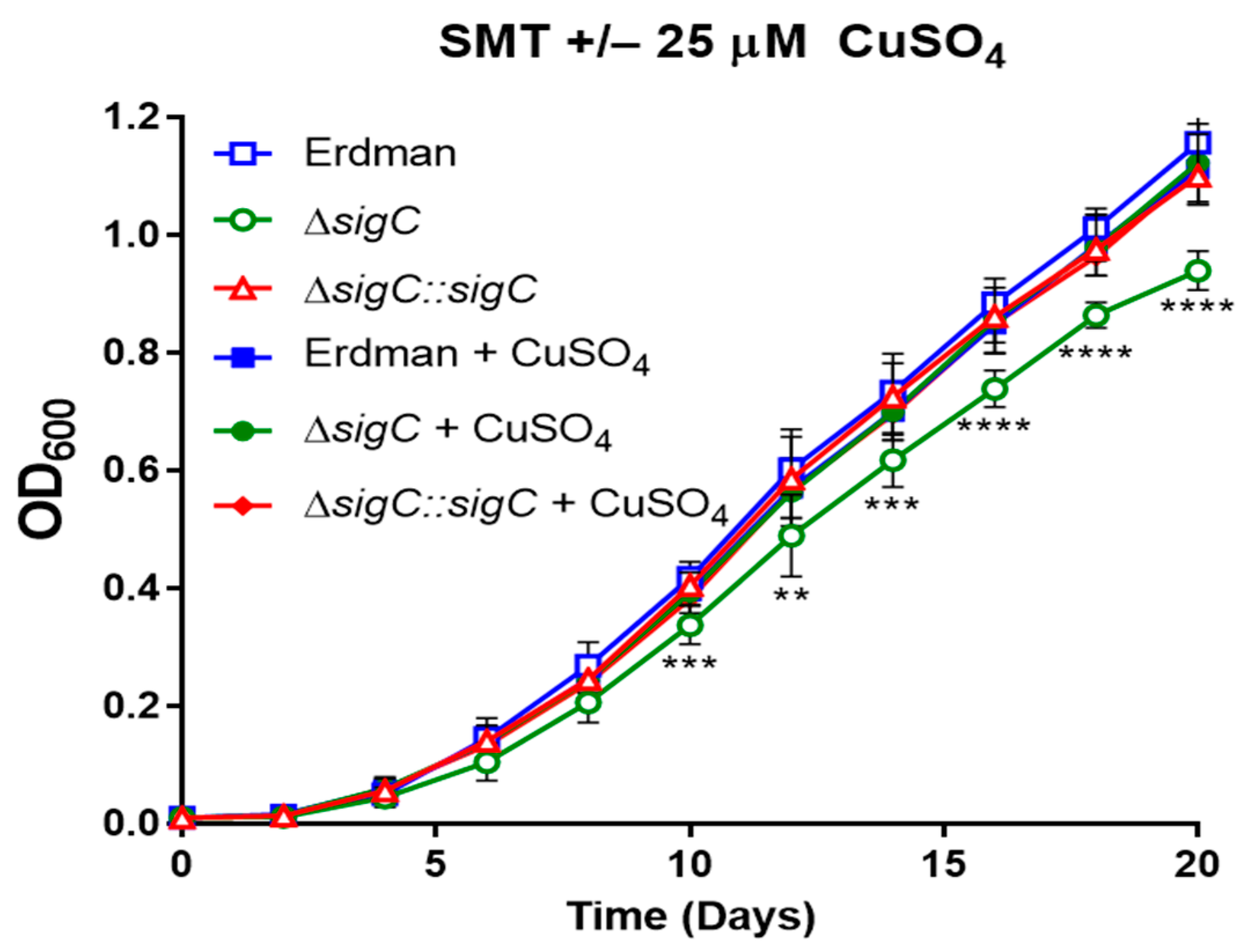
2.6. Complete Reversal of the ΔsigC Growth Defect by 25 µM Copper Sulfate Addition

We assessed whether a higher concentration of copper greater than 6 µM would fully reverse the growth defect of ΔsigC to equal the replication rate of parent Erdman. We previously observed that growth of ΔsigC equals that of Erdman in 7H9 medium (Figure S1). While 6 µM copper sulfate is specifically added in the 7H9 broth base, additional copper is likely present associated with the post autoclave addition of bovine serum albumin. The average concentration of copper in healthy human serum is estimated at 17 µM [59]. We compared growth of ΔsigC, the complemented mutant, and Erdman in SMT in the presence or absence of 25 µM copper sulfate and observed that this level of supplementation fully restored the ΔsigC growth rate to equal those of wild type and the complemented mutant (Figure 6).

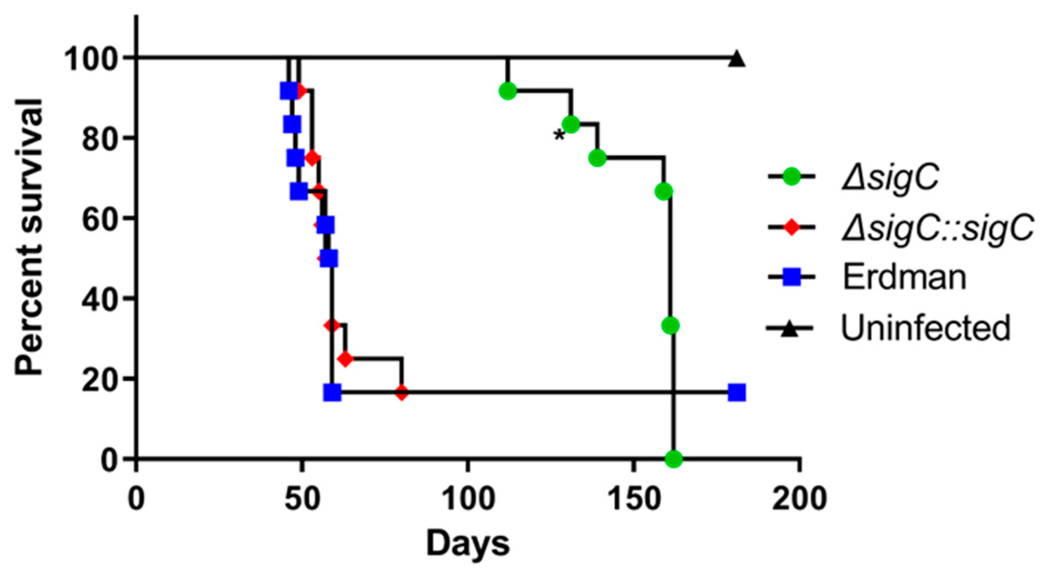
2.7. Attenuation of M. tuberculosis Erdman ΔsigC Virulence in SCID Mice

To determine if deletion of sigC from strain Erdman resulted in attenuation, groups of twelve SCID mice were infected in parallel by intratracheal instillation with 0.025 mL of 104 CFU/mL of ΔsigC, Erdman, or the sigC-complemented mutant. Animals infected with Erdman or the complement had median survival times of 58 and 58.5 days, respectively (Figure 7). The ΔsigC-infected mice survived more than twice as long with a median survival of 161 days. Attenuation of Erdman ΔsigC in SCID mice is consistent with that reported for a CDC1551 ΔsigC strain wherein the mean survival time was 86 days following aerosol infection with 100 CFU of the mutant and only 26 days for animals infected with CDC1551 or the complemented CDC1551ΔsigC mutant [6]. The accelerated decline of animals aerosol-infected with parent strain CDC1551 relative to those infected with Erdman bacilli delivered intratracheally might be due to genetic differences between the strains or to more-efficient delivery of bacilli to the alveoli when bacterial suspensions are aerosolized.
Since sigC is required for transcription of the ppe1–nrp operon under copper starvation conditions and sigC mutant strains are attenuated in animal models, mutants in the ppe1–nrp operon would also be expected to be highly attenuated. Studies with various mutant strains indicate this is the case. An M. tuberculosis nrp mutant was shown to be highly attenuated after infection in both immunocompetent (C57BL/6) and immunodeficient (SCID-, RAG2-, or IFNγ-deficient) murine models [60]. Mutant strains with disruption of the M. bovis ppe1–nrp operon by transposon insertion into the rv0097 homolog resulted in a smooth colony phenotype, loss of PDIM synthesis, and avirulence upon infection of guinea pigs [61]. Transposon disruption of rv0097 in M. tuberculosis reduced bacterial persistence in infected C57BL/6 mice, but surprisingly rendered the bacteria resistant to isoniazid in drug-treated mice, but not in axenic cultures [62]. Mutants of M. tuberculosis that fail to produce PDIMs display increased membrane permeability [63], reduced receptor-mediated macrophage infection [64], and reduced replication in murine lungs [65,66] and attenuated virulence [66]. It is unclear if all mutations that disrupt the ppe1–nrp operon directly result in loss of PDIM synthesis or if culture in copper-deficient conditions selects for suppressor mutations in PDIM synthesis genes to increase membrane permeability to more efficiently access copper. The Erdman ΔsigC stocks used for the animal studies were prepared in 7H9 medium prior to any studies involving growth in copper-deficient SMT. The CDC1551ΔsigC strain was also cultured in 7H9 medium [7]. As complementation of both mutants fully restored virulence, it is unlikely that spontaneous mutations blocking PDIM synthesis were responsible for the attenuation of sigC mutants in either M. tuberculosis background.
The attenuation of sigC mutants in animal models and its essentiality in scavenging copper when the metal is scarce in vitro strongly supports its significance to an obligate pathogen like M. tuberculosis to overcome host copper nutritional immunity. The essentiality of the copper-requiring cytochrome aa3–bc1 supercomplex underscores the importance of a copper-scavenging system to compete effectively for host copper. This is reflected in the conservation of the ppe1–nrp operon in slow-growing pathogenic mycobacteria, but not in fast-growing mycobacteria. The product(s) of the enzymes encoded in the M. tuberculosis ppe1–nrp operon remain to be elucidated. Although rv0097–rv0100 substitute for the corresponding genes from M. marinum in isonitrile lipopeptide synthesis when co-expressed in E. coli with the M. marinum nrp ortholog, no products were detected with M. tuberculosis rv0097–nrp [24]. From the larger size of M. tuberculosis nrp (7539 bp) relative to the truncated M. marinum ortholog (4250 bp), the chemical structures of the final products are predicted to differ. While our data suggest that the M. tuberculosis product will function in copper acquisition, a role in the uptake of zinc or other transition metal has yet to be fully explored.

2.8. Future Directions

A number of open questions remain in addition to defining the product of M. tuberculosis nrp operon enzymes. How does M. tuberculosis sense copper deficiency? The lack of differential expression of full-length sigC in Erdman versus sigC with an internal 138 bp deletion in ΔsigC suggests post transcriptional control of SigC. Activity of other sigma factors is often regulated by binding of an anti-sigma factor encoded in the same operon, but sigC is monocistronic and no anti-SigC factor has been identified to date. What is the relationship between control of ppe1–nrp operon genes by SigC and SigM? Why did overexpression of sigM in M. tuberculosis result in approximately ten-fold increased transcription of rv0098–rv0100, but not other genes in the operon [58], whereas sigM deletion had no effect on the same genes, but induced PDIM synthesis genes and production of the lipids [58]? Does SigM direct production of metal-binding isonitrile lipopeptides in response to metal starvation or other stress conditions or does it only function in the expression of lipid-transfer genes in the ppe1–nrp operon? What role does ctpB play in metal transport? Although ctpB is transcribed in vitro by SigC-RNA polymerase, the gene is not differentially expressed between a sigC mutant and its parent in copper-deficient medium. While this may suggest CtpB functions in copper export, it is also possible that the sigC-expressing bacteria had accumulated adequate amounts of copper prior to harvesting the cells for RNA analysis and elevated transcription was no longer necessary. These are active areas of investigation.

3. Materials and Methods

3.1. Bacterial Strains and Culture Conditions

Escherichia coli strains were cultured in LB medium with 50 µg/mL kanamycin or 200 µg/mL hygromycin, as appropriate, for plasmid maintenance. Mycobacterium tuberculosis strain Erdman and derivatives were cultured at 37 °C with slow shaking (70 rpm) in Middlebrook 7H9 medium sterilized by autoclaving and then supplemented with 0.05% Tween 80, 0.5% glycerol, and 10% ADS (albumin, dextrose, NaCl) (7H9tgADS) [67] or in Sauton medium (0.05% KH2PO4, 0.05% MgSO4, 0.2% citric acid, 0.005% ferric ammonium citrate, 6% glycerol, 0.4% asparagine, pH adjusted to 7.4) with 0.025% Tyloxapol (SMT) added as a dispersant and sterilized by filtration. Water used for preparation of media and additives was obtained from a Type 1 ultrapure system (ELGA ULTRA GE MK2). Mycobacteria liquid culture vessels used were either (i) glass 125-mL sidearm flasks prepared by autoclaving for 90 min submerged in 1% Vesphene II solution, hand cleaned with a bristle brush, rinsed with Type 1 water, autoclaved for 90 minutes submerged in Type 1 water, rinsed with Type 1 water, and sterilized by autoclaving or (ii) new 250-mL wide-mouth polypropylene bottles (VWR) rinsed with Type 1 water and sterilized by autoclaving. The ΔsigC growth defect phenotype developed after 3–4 passages in SMT in either vessel type. For RNA isolation, bacteria were cultured in 7H9tgADS or SMT. For specific experiments, SMT was supplemented with the indicated concentrations of copper sulfate, zinc sulfate, and/or calcium chloride. Middlebrook 7H10 agar supplemented with 0.05% Tween 80, 0.5% glycerol, and 10% ADS (7H10tgADS) was used for plating mycobacteria. For M. tuberculosis strains harboring plasmids, 50 µg/mL hygromycin or 25 µg/mL kanamycin was used, as appropriate.
A mutant with a 138-bp deletion within sigC was generated in M. tuberculosis strain Erdman by homologous recombination using methods previously described [5]. The ΔsigC mutant strain was confirmed by PCR of the sigC gene with primers 5′-GGT GGT TGC TCT TCC AAC ATG ACC GCG ACG GCA AGC-3′ and 5′-GGT GGT CTG CAG CTA GCC GGT GAG GTC GTC G-3′.
For complementation, plasmid pMV306sigC was constructed by PCR of sigC and ~600 bp of upstream DNA with primers 5′-GCT AAG CTT GCT CGT CCG TAG TCA C-3′ and 5′-CGC AAG CTT CGG TGG TCA TGA TAG C-3′ and inserted into the HindIII site of plasmid pMV306, which integrates into the M. tuberculosis chromosome at the mycobacteriophage L5 attB site [68]. After, pMV306sigC was electroporated into ∆sigC cultured in 7H9 medium and the plasmid was maintained with kanamycin selection.
For tetracycline-inducible sigC transcription, plasmid pSR173 which encodes SigC with an N-terminal myc tag and previously used in the myc-SigC chromosome immunoprecipitation/DNA microarray studies in M. bovis BCG [11] was kindly provided by Drs. Rodrigue and Gaudreau (University of Sherbrooke, Sherbrooke, QC, Canada) and maintained with hygromycin selection.

3.2. RNA Isolation

For RNA isolation from M. tuberculosis cultures, cells were harvested by centrifugation (3500× g, 5 min) and cell pellets were frozen on dry ice and stored at −80 °C. Cells were thawed in the presence of Trizol (Gibco/Thermo Fisher Scientific, Waltham, MA, USA) and disrupted with 0.1 mm zirconium beads in a BeadBeater (Cole-Parmer, Vernon Hills, IL, USA) for three cycles of 40 s each at 4800 rpm with one minute cooling on ice between cycles. Next, chloroform was added and the tubes were mixed by inversion, incubated for three minutes, and the phases separated by centrifugation (12,000× g, 15 min). The aqueous phase was transferred to a new tube and extracted with an equal volume of acid phenol/chloroform. Following centrifugation (12,000× g, 10 min), the aqueous phase was transferred to a new tube and the RNA was isopropanol precipitated. Pelleted RNA was washed twice with 75% ethanol and suspended in diethyl pyrocarbonate-treated water. For DNA removal, each sample was treated twice with Turbo RNAse-free DNAse (Ambion/Thermo Fisher Scientific, Waltham, MA, USA) and RNA purified by RNeasy (Qiagen, Hilden, Germany) using the manufacturers’ instructions. After elution from RNeasy columns, 25 units RNaseOUT (Invitrogen/Thermo Fisher Scientific, Waltham, MA, USA) was added to each RNA sample. Residual genomic DNA contamination was assessed by PCR (40 cycles) with sigA-specific primers: 5′-AAC AGA TCG GCA AGG TAG-3′ and 5′AAC TTG TAC CCC TTG GTG-3′.

3.3. Microarray Analyses

The DNA microarray slides used in these studies were supplied by the TB Vaccine Testing and Research Materials Contract administered by Colorado State University, Fort Collins, CO (for sigC induction studies) and the M.I. Voskuil laboratory, University of Colorado Denver (for Erdman versus sigC mutant studies). All slides contained 70mer oligonucleotide probes for 4269 M. tuberculosis open reading frames (ORFs) from strains H37Rv and CDC with 26 controls. Complementary DNA (cDNA) was prepared using methods from the Pathogen Functional Genomics Resource Center at the J. Craig Venter Institute. Briefly, 2 µg RNA samples were incubated with 6 µg random hexamers (Invitrogen/Thermo Fisher Scientific, Waltham, MA, USA), 0.5 mM dNTP/aminoallyl-dUTPs, 2 µL SuperScript III Reverse Transcriptase, and 10 mM DTT overnight at 42 °C. The aminoallyl-labeled cDNAs produced were purified by MinElute PCR purification (Qiagen, Hilden, Germany) using manufacturer instructions except that the wash and elution buffers in the kit were replaced with phosphate wash buffer (5 mM KH2PO4, pH 8.0, 80% EtOH) and phosphate elution buffer (4 mM KH2PO4, pH 8.5), respectively. The cDNAs were next coupled to Cy3 or Cy5 dyes (Amersham Biosciences/Thermo Fisher Scientific, Waltham, MA, USA) and unbound dye was removed by MinElute PCR purification. The Cy3- and Cy5-labeled cDNAs were combined in pairs, dried, and suspended in hybridization buffer (5× SSC, 25% formamide, 0.1% SDS) with 0.4 mg/mL yeast tRNA, heated (98 °C, 2 min) and cooled briefly on ice. Microarray slides were prehybridized for a minimum of one hour at 42 °C by incubation in prehybridization buffer (5× SSC, 0.1% SDS, 1% BSA), washed stepwise with water and isopropanol, dried, and then hybridized with the labeled cDNA mixture 12–18 h at 42 °C. Slides were washed with high (0.1× SSC), medium (0.1× SSC, 0.1% SDS), and low (2× SSC, 0.1% SDS) stringency buffers, and fluorescence measured on a ProScanArray microarray scanner (Perkin Elmer, Waltham, MA, USA). Images collected were analyzed with ScanArrayExpress software and exported to Microsoft Excel. Each experiment consisted of a minimum of four biological replicates with at least two hybridizations performed per replicate. The dyes were swapped for the two hybridizations of each biological replicate. For each gene probe, eight intensity values or more were obtained. For normalization of the data, the LOWESS algorithm was used [69]. For identification of open reading frames (ORFs) with significant differential expression, the Significance Analysis of Microarrays (SAM) test procedure was applied using Excel SAM version 2.1 R package (http://statweb.stanford.edu/~tibs/SAM/Rdist/index.html (accessed on 24 December 2020)) set to a stringent false-discovery rate of zero. To ensure stringency, genes differentially regulated less than 2-fold have been excluded.

3.4. Quantitative RT-PCR Assays

For cDNA generation, 500 ng RNA from each biological replicate was incubated with gene-specific primers and ImProm-II Reverse Transcriptase (Promega Corporation, Madison, WI, USA) following the manufacturer’s instructions. Quantitation of target transcripts was determined by PCR of 0.5 µL cDNA with Platinum SYBR Green Mastermix (Invitrogen/Thermo Fisher Scientific, Waltham, MA, USA) using a BioRad iCycler, and applying the ∆∆Ct method with normalization to sigA [70]. Three or more biological replicates were assayed for each gene.

3.5. Animal Infection Studies

Procedures involving animals were conducted in accordance with protocols approved by the University of Georgia Institutional Animal Care and Use Committee (A2007-10135-0, approval date 28 February 2007) and in accordance with American Association for Accreditation of Laboratory Animal Care (AAALAS) policy. Severe-combined immunodeficient mice, female, 8–10 weeks of age, were purchased from Jackson Laboratories. All animals received ad libitum food and water. To assess the relative virulence of M. tuberculosis strains ΔsigC, Erdman, and the complemented sigC mutant, each strain was cultured in 7H9tgADS to OD600 = 1, counted microscopically, diluted in PBS + 0.05% Tween 80. Twelve mice per strain were infected by intratracheal instillation with 0.025 mL of 104 CFU/mL stocks or sham-infected with 0.025 mL of phosphate-buffered saline (PBS). For survival studies, the mice were closely monitored for signs of disease or distress and humanely euthanized if moribund. The statistical significance of the Kaplan–Meier survival curve data was evaluated using the Mantel–Cox test within GraphPad Prism 9 software.

3.6. Statistical Analysis

One-way ANOVA was used to assess statistical significance between groups for qRT-PCR studies (Figure 2B,C and Figure 6, Figure S2) and bacterial growth curves (Figure 3 and Figure 5, Figure S1). For animal survival studies, the Mantel–Cox test was employed. For microarray studies (Figure 2A, Figure 4, and Figure 6), the Statistical Analysis of Microarrays test (Excel SAM version 2.1 R package, Stanford University) was utilized to identify genes with significant differential expression. Numbers of sample replicates are indicated for each experiment. With the exception of the microarray studies, all statistical analyses were performed with GraphPad Prism ver. 9.0.0 (121) (GraphPad Software, Inc., San Diego, CA, USA).

Supplementary Materials

Supplementary materials can be found at https://0-www-mdpi-com.brum.beds.ac.uk/1422-0067/22/4/2118/s1.

Author Contributions

Conceptualization, B.T.G.-S. and R.K.K.; data curation, B.T.G.-S., T.G. and R.K.K.; formal analysis, B.T.G.-S., T.G., S.L.T. and R.K.K.; funding acquisition, F.D.Q. and R.K.K.; investigation, B.T.G.-S., T.G., S.H. and S.L.T.; methodology, B.T.G.-S., T.G., S.H., S.L.T., M.I.V., F.D.Q. and R.K.K.; project administration, R.K.K.; resources, M.I.V.; supervision, F.D.Q. and R.K.K.; validation, B.T.G.-S., T.G., S.L.T. and R.K.K.; visualization, B.T.G.-S., T.G., S.L.T., M.I.V. and R.K.K.; writing—original draft, B.T.G.-S. and R.K.K.; writing—review and editing, B.T.G.-S., T.G., S.H., S.L.T., M.I.V., F.D.Q. and R.K.K. All authors have read and agreed to the published version of the manuscript.

Funding

This work was supported by a Collaboration Planning Grant from the Georgia Research Alliance, Atlanta, GA, USA.

Institutional Review Board Statement

The animal studies were conducted in accordance with protocols approved by the University of Georgia Animal Care and Use Committee (A2007-10135-0, approval date 28 February 2007) and in accordance with the AAALAS policy. The study design, procedures, animal monitoring, statistical analysis, and data reporting fulfill the criteria of the Animal Research: Reporting of In Vivo Experiments (ARRIVE) guidelines (https://www.nc3rs.org.uk/arrive-guidelines (accessed date 24 December 2020)). The number of animals used in this study was kept to an essential minimum in accordance with the 3Rs policy. All experiments with viable M. tuberculosis bacteria were conducted in biosafety level 3/animal biosafety level 3 facilities with protocols and safety practices approved by the University of Georgia Institutional Biosafety Committee (protocol 2007-0025, approved 23 August 2007).

Data Availability Statement

All data are contained within the article and supplementary materials.

Acknowledgments

We thank the Pathogen Functional Genomics Laboratory at the J. Craig Venter Institute for the opportunity for B.T.G.-S. to obtain microarray training. We are grateful to the Michael W. Adams laboratory at the University of Georgia for microarray scanner use and helpful technical expertise. We thank Rebecca L. Pavlicek for assistance with the animal studies. This work includes re-analysis of dissertation research data from the first author [71].

Conflicts of Interest

The authors declare no conflict of interest.

References

  1. WHO. Global Tuberculosis Report; World Health Organization: Geneva, Switzerland, 2020. [Google Scholar]
  2. Houben, R.M.; Dodd, P.J. The Global Burden of Latent Tuberculosis Infection: A Re-estimation Using Mathematical Modelling. PLoS Med. 2016, 13, e1002152. [Google Scholar] [CrossRef] [Green Version]
  3. Cole, S.T.; Brosch, R.; Parkhill, J.; Garnier, T.; Churcher, C.; Harris, D.; Gordon, S.V.; Eiglmeier, K.; Gas, S.; Barry, C.E., 3rd; et al. Deciphering the biology of Mycobacterium tuberculosis from the complete genome sequence. Nature 1998, 393, 537–544. [Google Scholar] [CrossRef] [PubMed]
  4. Manganelli, R. Sigma Factors: Key Molecules in Mycobacterium tuberculosis Physiology and Virulence. Microbiol. Spectr. 2014, 2, MGM2-0007-2013. [Google Scholar] [CrossRef] [Green Version]
  5. Karls, R.K.; Guarner, J.; McMurray, D.N.; Birkness, K.A.; Quinn, F.D. Examination of Mycobacterium tuberculosis sigma factor mutants using low-dose aerosol infection of guinea pigs suggests a role for SigC in pathogenesis. Microbiology 2006, 152, 1591–1600. [Google Scholar] [CrossRef] [PubMed] [Green Version]
  6. Abdul-Majid, K.B.; Ly, L.H.; Converse, P.J.; Geiman, D.E.; McMurray, D.N.; Bishai, W.R. Altered cellular infiltration and cytokine levels during early Mycobacterium tuberculosis sigC mutant infection are associated with late-stage disease attenuation and milder immunopathology in mice. BMC Microbiol. 2008, 8, 151. [Google Scholar] [CrossRef] [Green Version]
  7. Sun, R.; Converse, P.J.; Ko, C.; Tyagi, S.; Morrison, N.E.; Bishai, W.R. Mycobacterium tuberculosis ECF sigma factor sigC is required for lethality in mice and for the conditional expression of a defined gene set. Mol. Microbiol. 2004, 52, 25–38. [Google Scholar] [CrossRef] [PubMed]
  8. Chang, A.; Smollett, K.L.; Gopaul, K.K.; Chan, B.H.; Davis, E.O. Mycobacterium tuberculosis H37Rv sigC is expressed from two promoters but is not auto-regulatory. Tuberculosis 2012, 92, 48–55. [Google Scholar] [CrossRef] [PubMed]
  9. Manganelli, R.; Dubnau, E.; Tyagi, S.; Kramer, F.R.; Smith, I. Differential expression of 10 sigma factor genes in Mycobacterium tuberculosis. Mol. Microbiol. 1999, 31, 715–724. [Google Scholar] [CrossRef]
  10. Rustad, T.R.; Minch, K.J.; Ma, S.; Winkler, J.K.; Hobbs, S.; Hickey, M.; Brabant, W.; Turkarslan, S.; Price, N.D.; Baliga, N.S.; et al. Mapping and manipulating the Mycobacterium tuberculosis transcriptome using a transcription factor overexpression-derived regulatory network. Genome Biol. 2014, 15, 502. [Google Scholar] [CrossRef]
  11. Rodrigue, S.; Brodeur, J.; Jacques, P.E.; Gervais, A.L.; Brzezinski, R.; Gaudreau, L. Identification of mycobacterial sigma factor binding sites by chromatin immunoprecipitation assays. J. Bacteriol. 2007, 189, 1505–1513. [Google Scholar] [CrossRef] [Green Version]
  12. Andreini, C.; Bertini, I.; Cavallaro, G.; Holliday, G.L.; Thornton, J.M. Metal ions in biological catalysis: From enzyme databases to general principles. J. Biol. Inorg. Chem. 2008, 13, 1205–1218. [Google Scholar] [CrossRef]
  13. Hood, M.I.; Skaar, E.P. Nutritional immunity: Transition metals at the pathogen-host interface. Nat. Rev. Microbiol. 2012, 10, 525–537. [Google Scholar] [CrossRef]
  14. Weinberg, E.D. Nutritional immunity. Host’s attempt to withold iron from microbial invaders. JAMA 1975, 231, 39–41. [Google Scholar] [CrossRef] [PubMed]
  15. Hennigar, S.R.; McClung, J.P. Nutritional Immunity: Starving Pathogens of Trace Minerals. Am. J. Lifestyle Med. 2016, 10, 170–173. [Google Scholar] [CrossRef] [PubMed]
  16. Darwin, K.H. Mycobacterium tuberculosis and Copper: A Newly Appreciated Defense against an Old Foe? J. Biol. Chem. 2015, 290, 18962–18966. [Google Scholar] [CrossRef] [Green Version]
  17. Fu, Y.; Chang, F.M.; Giedroc, D.P. Copper transport and trafficking at the host-bacterial pathogen interface. Acc. Chem. Res. 2014, 47, 3605–3613. [Google Scholar] [CrossRef]
  18. Sassetti, C.M.; Boyd, D.H.; Rubin, E.J. Genes required for mycobacterial growth defined by high density mutagenesis. Mol. Microbiol. 2003, 48, 77–84. [Google Scholar] [CrossRef]
  19. Griffin, J.E.; Gawronski, J.D.; Dejesus, M.A.; Ioerger, T.R.; Akerley, B.J.; Sassetti, C.M. High-resolution phenotypic profiling defines genes essential for mycobacterial growth and cholesterol catabolism. PLoS Pathog. 2011, 7, e1002251. [Google Scholar] [CrossRef] [Green Version]
  20. DeJesus, M.A.; Gerrick, E.R.; Xu, W.; Park, S.W.; Long, J.E.; Boutte, C.C.; Rubin, E.J.; Schnappinger, D.; Ehrt, S.; Fortune, S.M.; et al. Comprehensive Essentiality Analysis of the Mycobacterium tuberculosis Genome via Saturating Transposon Mutagenesis. mBio 2017, 8, e02133-16. [Google Scholar] [CrossRef] [Green Version]
  21. Koch, A.; Mizrahi, V. Mycobacterium tuberculosis. Trends Microbiol. 2018, 26, 555–556. [Google Scholar] [CrossRef] [PubMed]
  22. Rodrigue, S.; Provvedi, R.; Jacques, P.E.; Gaudreau, L.; Manganelli, R. The sigma factors of Mycobacterium tuberculosis. FEMS Microbiol. Rev. 2006, 30, 926–941. [Google Scholar] [CrossRef] [Green Version]
  23. Leon-Torres, A.; Arango, E.; Castillo, E.; Soto, C.Y. CtpB is a plasma membrane copper (I) transporting P-type ATPase of Mycobacterium tuberculosis. Biol. Res. 2020, 53, 6. [Google Scholar] [CrossRef]
  24. Harris, N.C.; Sato, M.; Herman, N.A.; Twigg, F.; Cai, W.; Liu, J.; Zhu, X.; Downey, J.; Khalaf, R.; Martin, J.; et al. Biosynthesis of isonitrile lipopeptides by conserved nonribosomal peptide synthetase gene clusters in Actinobacteria. Proc. Natl. Acad. Sci. USA 2017, 114, 7025–7030. [Google Scholar] [CrossRef] [PubMed] [Green Version]
  25. Wang, L.; Zhu, M.; Zhang, Q.; Zhang, X.; Yang, P.; Liu, Z.; Deng, Y.; Zhu, Y.; Huang, X.; Han, L.; et al. Diisonitrile Natural Product SF2768 Functions As a Chalkophore That Mediates Copper Acquisition in Streptomyces thioluteus. ACS Chem. Biol. 2017, 12, 3067–3075. [Google Scholar] [CrossRef]
  26. Festa, R.A.; Jones, M.B.; Butler-Wu, S.; Sinsimer, D.; Gerads, R.; Bishai, W.R.; Peterson, S.N.; Darwin, K.H. A novel copper-responsive regulon in Mycobacterium tuberculosis. Mol. Microbiol. 2011, 79, 133–148. [Google Scholar] [CrossRef] [PubMed] [Green Version]
  27. Gold, B.; Deng, H.; Bryk, R.; Vargas, D.; Eliezer, D.; Roberts, J.; Jiang, X.; Nathan, C. Identification of a copper-binding metallothionein in pathogenic mycobacteria. Nat. Chem. Biol. 2008, 4, 609–616. [Google Scholar] [CrossRef] [PubMed] [Green Version]
  28. Zimbro, M.J. Difco & BBL Manual: Manual of Microbiological Culture Media, 2nd ed.; Becton, Dickinson and Company: Sparks, MD, USA, 2009. [Google Scholar]
  29. Parish, T.; Smith, D.A.; Roberts, G.; Betts, J.; Stoker, N.G. The senX3-regX3 two-component regulatory system of Mycobacterium tuberculosis is required for virulence. Microbiology 2003, 149, 1423–1435. [Google Scholar] [CrossRef] [PubMed] [Green Version]
  30. Rifat, D.; Belchis, D.A.; Karakousis, P.C. senX3-independent contribution of regX3 to Mycobacterium tuberculosis virulence. BMC Microbiol. 2014, 14, 265. [Google Scholar] [CrossRef] [Green Version]
  31. Marcus, S.A.; Sidiropoulos, S.W.; Steinberg, H.; Talaat, A.M. CsoR Is Essential for Maintaining Copper Homeostasis in Mycobacterium tuberculosis. PLoS ONE 2016, 11, e0151816. [Google Scholar] [CrossRef]
  32. He, W.; Soll, C.E.; Chavadi, S.S.; Zhang, G.; Warren, J.D.; Quadri, L.E. Cooperation between a coenzyme A-independent stand-alone initiation module and an iterative type I polyketide synthase during synthesis of mycobacterial phenolic glycolipids. J. Am. Chem. Soc. 2009, 131, 16744–16750. [Google Scholar] [CrossRef] [Green Version]
  33. Park, H.D.; Guinn, K.M.; Harrell, M.I.; Liao, R.; Voskuil, M.I.; Tompa, M.; Schoolnik, G.K.; Sherman, D.R. Rv3133c/dosR is a transcription factor that mediates the hypoxic response of Mycobacterium tuberculosis. Mol. Microbiol. 2003, 48, 833–843. [Google Scholar] [CrossRef] [Green Version]
  34. Rustad, T.R.; Harrell, M.I.; Liao, R.; Sherman, D.R. The enduring hypoxic response of Mycobacterium tuberculosis. PLoS ONE 2008, 3, e1502. [Google Scholar] [CrossRef] [PubMed]
  35. Fenhalls, G.; Stevens, L.; Moses, L.; Bezuidenhout, J.; Betts, J.C.; Helden Pv, P.; Lukey, P.T.; Duncan, K. In situ detection of Mycobacterium tuberculosis transcripts in human lung granulomas reveals differential gene expression in necrotic lesions. Infect. Immun. 2002, 70, 6330–6338. [Google Scholar] [CrossRef] [PubMed] [Green Version]
  36. Dahl, J.L.; Kraus, C.N.; Boshoff, H.I.; Doan, B.; Foley, K.; Avarbock, D.; Kaplan, G.; Mizrahi, V.; Rubin, H.; Barry, C.E., 3rd. The role of RelMtb-mediated adaptation to stationary phase in long-term persistence of Mycobacterium tuberculosis in mice. Proc. Natl. Acad. Sci. USA 2003, 100, 10026–10031. [Google Scholar] [CrossRef] [Green Version]
  37. Sherman, D.R.; Mdluli, K.; Hickey, M.J.; Barry, C.E., 3rd; Stover, C.K. AhpC, oxidative stress and drug resistance in Mycobacterium tuberculosis. Biofactors 1999, 10, 211–217. [Google Scholar] [CrossRef]
  38. Saxena, A.; Srivastava, V.; Srivastava, R.; Srivastava, B.S. Identification of genes of Mycobacterium tuberculosis upregulated during anaerobic persistence by fluorescence and kanamycin resistance selection. Tuberculosis 2008, 88, 518–525. [Google Scholar] [CrossRef] [PubMed]
  39. Kim, S.Y.; Lee, B.S.; Shin, S.J.; Kim, H.J.; Park, J.K. Differentially expressed genes in Mycobacterium tuberculosis H37Rv under mild acidic and hypoxic conditions. J. Med. Microbiol. 2008, 57, 1473–1480. [Google Scholar] [CrossRef]
  40. Betts, J.C.; Lukey, P.T.; Robb, L.C.; McAdam, R.A.; Duncan, K. Evaluation of a nutrient starvation model of Mycobacterium tuberculosis persistence by gene and protein expression profiling. Mol. Microbiol. 2002, 43, 717–731. [Google Scholar] [CrossRef]
  41. Farhana, A.; Guidry, L.; Srivastava, A.; Singh, A.; Hondalus, M.K.; Steyn, A.J.C. Reductive Stress in Microbes: Implications for Understanding Mycobacterium tuberculosis Diseases and Persistence. In Advances in Microbial Physiology; Poole, R.K., Ed.; Elsevier Academic Press: Oxford, UK, 2010; Volume 57, pp. 43–117. [Google Scholar]
  42. Rhee, K.Y.; de Carvalho, L.P.; Bryk, R.; Ehrt, S.; Marrero, J.; Park, S.W.; Schnappinger, D.; Venugopal, A.; Nathan, C. Central carbon metabolism in Mycobacterium tuberculosis: An unexpected frontier. Trends Microbiol. 2011, 19, 307–314. [Google Scholar] [CrossRef] [Green Version]
  43. Ehrt, S.; Rhee, K. Mycobacterium tuberculosis metabolism and host interaction: Mysteries and paradoxes. Curr. Top. Microbiol. Immunol. 2013, 374, 163–188. [Google Scholar] [CrossRef]
  44. Baughn, A.D.; Rhee, K.Y. Metabolomics of Central Carbon Metabolism in Mycobacterium tuberculosis. Microbiol. Spectr. 2014, 2, 323–339. [Google Scholar] [CrossRef] [PubMed] [Green Version]
  45. Ehrt, S.; Schnappinger, D.; Rhee, K.Y. Metabolic principles of persistence and pathogenicity in Mycobacterium tuberculosis. Nat. Rev. Microbiol. 2018, 16, 496–507. [Google Scholar] [CrossRef] [PubMed]
  46. Cook, G.M.; Hards, K.; Vilcheze, C.; Hartman, T.; Berney, M. Energetics of Respiration and Oxidative Phosphorylation in Mycobacteria. Microbiol. Spectr. 2014, 2, 389–409. [Google Scholar] [CrossRef] [PubMed] [Green Version]
  47. Marrero, J.; Rhee, K.Y.; Schnappinger, D.; Pethe, K.; Ehrt, S. Gluconeogenic carbon flow of tricarboxylic acid cycle intermediates is critical for Mycobacterium tuberculosis to establish and maintain infection. Proc. Natl. Acad. Sci. USA 2010, 107, 9819–9824. [Google Scholar] [CrossRef] [Green Version]
  48. Nienaber, L.; Cave-Freeman, E.; Cross, M.; Mason, L.; Bailey, U.M.; Amani, P.; Davis, R.A.; Taylor, P.; Hofmann, A. Chemical probing suggests redox-regulation of the carbonic anhydrase activity of mycobacterial Rv1284. FEBS J. 2015, 282, 2708–2721. [Google Scholar] [CrossRef] [PubMed] [Green Version]
  49. Beste, D.J.; Bonde, B.; Hawkins, N.; Ward, J.L.; Beale, M.H.; Noack, S.; Noh, K.; Kruger, N.J.; Ratcliffe, R.G.; McFadden, J. (1)(3)C metabolic flux analysis identifies an unusual route for pyruvate dissimilation in mycobacteria which requires isocitrate lyase and carbon dioxide fixation. PLoS Pathog. 2011, 7, e1002091. [Google Scholar] [CrossRef] [Green Version]
  50. Giuffre, A.; Borisov, V.B.; Arese, M.; Sarti, P.; Forte, E. Cytochrome bd oxidase and bacterial tolerance to oxidative and nitrosative stress. Biochim. Biophys. Acta 2014, 1837, 1178–1187. [Google Scholar] [CrossRef] [Green Version]
  51. Voskuil, M.I.; Visconti, K.C.; Schoolnik, G.K. Mycobacterium tuberculosis gene expression during adaptation to stationary phase and low-oxygen dormancy. Tuberculosis 2004, 84, 218–227. [Google Scholar] [CrossRef]
  52. Milano, A.; Pasca, M.R.; Provvedi, R.; Lucarelli, A.P.; Manina, G.; Ribeiro, A.L.; Manganelli, R.; Riccardi, G. Azole resistance in Mycobacterium tuberculosis is mediated by the MmpS5-MmpL5 efflux system. Tuberculosis 2009, 89, 84–90. [Google Scholar] [CrossRef]
  53. Gold, B.; Rodriguez, G.M.; Marras, S.A.; Pentecost, M.; Smith, I. The Mycobacterium tuberculosis IdeR is a dual functional regulator that controls transcription of genes involved in iron acquisition, iron storage and survival in macrophages. Mol. Microbiol. 2001, 42, 851–865. [Google Scholar] [CrossRef] [PubMed] [Green Version]
  54. Sala, C.; Haouz, A.; Saul, F.A.; Miras, I.; Rosenkrands, I.; Alzari, P.M.; Cole, S.T. Genome-wide regulon and crystal structure of BlaI (Rv1846c) from Mycobacterium tuberculosis. Mol. Microbiol. 2009, 71, 1102–1116. [Google Scholar] [CrossRef]
  55. Roberts, G.; Vadrevu, I.S.; Madiraju, M.V.; Parish, T. Control of CydB and GltA1 expression by the SenX3 RegX3 two component regulatory system of Mycobacterium tuberculosis. PLoS ONE 2011, 6, e21090. [Google Scholar] [CrossRef]
  56. Singh, A.; Crossman, D.K.; Mai, D.; Guidry, L.; Voskuil, M.I.; Renfrow, M.B.; Steyn, A.J. Mycobacterium tuberculosis WhiB3 maintains redox homeostasis by regulating virulence lipid anabolism to modulate macrophage response. PLoS Pathog. 2009, 5, e1000545. [Google Scholar] [CrossRef] [PubMed] [Green Version]
  57. Simeone, R.; Constant, P.; Malaga, W.; Guilhot, C.; Daffe, M.; Chalut, C. Molecular dissection of the biosynthetic relationship between phthiocerol and phthiodiolone dimycocerosates and their critical role in the virulence and permeability of Mycobacterium tuberculosis. FEBS J. 2007, 274, 1957–1969. [Google Scholar] [CrossRef]
  58. Raman, S.; Puyang, X.; Cheng, T.Y.; Young, D.C.; Moody, D.B.; Husson, R.N. Mycobacterium tuberculosis SigM positively regulates Esx secreted protein and nonribosomal peptide synthetase genes and down regulates virulence-associated surface lipid synthesis. J. Bacteriol. 2006, 188, 8460–8468. [Google Scholar] [CrossRef] [Green Version]
  59. Li, D.D.; Zhang, W.; Wang, Z.Y.; Zhao, P. Serum Copper, Zinc, and Iron Levels in Patients with Alzheimer’s Disease: A Meta-Analysis of Case-Control Studies. Front. Aging. Neurosci. 2017, 9, 300. [Google Scholar] [CrossRef] [PubMed] [Green Version]
  60. Bhatt, K.; Machado, H.; Osorio, N.S.; Sousa, J.; Cardoso, F.; Magalhaes, C.; Chen, B.; Chen, M.; Kim, J.; Singh, A.; et al. A Nonribosomal Peptide Synthase Gene Driving Virulence in Mycobacterium tuberculosis. mSphere 2018, 3, e00352-18. [Google Scholar] [CrossRef] [PubMed] [Green Version]
  61. Hotter, G.S.; Wards, B.J.; Mouat, P.; Besra, G.S.; Gomes, J.; Singh, M.; Bassett, S.; Kawakami, P.; Wheeler, P.R.; de Lisle, G.W.; et al. Transposon mutagenesis of Mb0100 at the ppe1-nrp locus in Mycobacterium bovis disrupts phthiocerol dimycocerosate (PDIM) and glycosylphenol-PDIM biosynthesis, producing an avirulent strain with vaccine properties at least equal to those of M. bovis BCG. J. Bacteriol. 2005, 187, 2267–2277. [Google Scholar] [CrossRef] [PubMed] [Green Version]
  62. Dhar, N.; McKinney, J.D. Mycobacterium tuberculosis persistence mutants identified by screening in isoniazid-treated mice. Proc. Natl. Acad. Sci. USA 2010, 107, 12275–12280. [Google Scholar] [CrossRef] [Green Version]
  63. Camacho, L.R.; Constant, P.; Raynaud, C.; Laneelle, M.A.; Triccas, J.A.; Gicquel, B.; Daffe, M.; Guilhot, C. Analysis of the phthiocerol dimycocerosate locus of Mycobacterium tuberculosis. Evidence that this lipid is involved in the cell wall permeability barrier. J. Biol. Chem. 2001, 276, 19845–19854. [Google Scholar] [CrossRef] [Green Version]
  64. Astarie-Dequeker, C.; Le Guyader, L.; Malaga, W.; Seaphanh, F.K.; Chalut, C.; Lopez, A.; Guilhot, C. Phthiocerol dimycocerosates of M. tuberculosis participate in macrophage invasion by inducing changes in the organization of plasma membrane lipids. PLoS Pathog. 2009, 5, e1000289. [Google Scholar] [CrossRef] [PubMed] [Green Version]
  65. Cox, J.S.; Chen, B.; McNeil, M.; Jacobs, W.R., Jr. Complex lipid determines tissue-specific replication of Mycobacterium tuberculosis in mice. Nature 1999, 402, 79–83. [Google Scholar] [CrossRef] [PubMed]
  66. Kirksey, M.A.; Tischler, A.D.; Simeone, R.; Hisert, K.B.; Uplekar, S.; Guilhot, C.; McKinney, J.D. Spontaneous phthiocerol dimycocerosate-deficient variants of Mycobacterium tuberculosis are susceptible to gamma interferon-mediated immunity. Infect. Immun. 2011, 79, 2829–2838. [Google Scholar] [CrossRef] [PubMed] [Green Version]
  67. Braunstein, M.; Bardarov, S.S.; Jacobs, W.R., Jr. Genetic methods for deciphering virulence determinants of Mycobacterium tuberculosis. Methods Enzymol. 2002, 358, 67–99. [Google Scholar] [CrossRef] [PubMed]
  68. Kong, D.; Kunimoto, D.Y. Secretion of human interleukin 2 by recombinant Mycobacterium bovis BCG. Infect. Immun. 1995, 63, 799–803. [Google Scholar] [CrossRef] [Green Version]
  69. Yang, Y.H.; Dudoit, S.; Luu, P.; Lin, D.M.; Peng, V.; Ngai, J.; Speed, T.P. Normalization for cDNA microarray data: A robust composite method addressing single and multiple slide systematic variation. Nucleic Acids Res. 2002, 30, e15. [Google Scholar] [CrossRef] [PubMed] [Green Version]
  70. Livak, K.J.; Schmittgen, T.D. Analysis of relative gene expression data using real-time quantitative PCR and the 2(-Delta Delta C(T)) Method. Methods 2001, 25, 402–408. [Google Scholar] [CrossRef]
  71. Grosse-Siestrup, B.T. Examination of the SigC Regulon and Cobalamin Biosynthesis in Mycobacterium tuberculosis. Ph.D. Thesis, University of Georgia, Athens, Georgia, 2012. [Google Scholar]
Figure 1. Confirmation of Mycobacterium tuberculosis Erdman ΔsigC. PCR analysis was used to confirm the 138-bp deletion within sigC of mutant (ΔsigC) relative to parent strain Erdman (WT). PCR bands of 587 and 449 bp are expected for WT and ΔsigC, respectively.
Ijms 22 02118 g001
Figure 2. (AC). Genes expressed >2-fold after 4- or 48-h sigC induction in Erdman in 7H9 medium. (A) Global gene expression studies using DNA microarrays in strain Erdman carrying a plasmid encoding sigC under control of a tetracycline-inducible promoter were performed in cultures grown to OD600 = 0.4 in 7H9tgADS prior to 4- or 48-h incubation with and without 50 ng/mL anhydrotetracycline induction. Genes with 2-fold or greater differential expression determined to be significant using Statistical Analysis for Microarrays software are shown. Genes in the ppe1–nrp operon are indicated (underlined, blue font). No genes were significantly downregulated more than 2-fold following artificial sigC induction. Genes regulated by RicR are shown (emboldened, red font). NS—not significant. Confirmation of microarray results by qRT-PCR of RNA from 4-h (B) and 48-h (C) sigC induction experiments. Results for rv2069 (sigC), rv0103 (ctpB), rv0099 (fadD10), and rv0848 (cysK2) were normalized to housekeeping gene sigA. Error bars indicate the standard deviation from the mean from a minimum of 3 biological replicates.
Ijms 22 02118 g002
Figure 3. (AE). Examination of metals on ΔsigC growth. Strains Erdman, ΔsigC, and the complemented sigC mutant (ΔsigC::sigC) were cultured in Sauton Medium with 0.025% Tyloxapol (SMT) alone (A) and after addition of one or more copper (6 µM), zinc (6 µM), or calcium (4.5 µM) salts (BE). Culture densities shown are the average of two experiments with six independent replicates. One-way ANOVA was used to evaluate statistical significance (* p < 0.05, ** p < 0.01, *** p < 0.001, **** p < 0.0001).
Ijms 22 02118 g003
Figure 4. Genes differentially expressed >2-fold in Erdman and ΔsigC cultured in SMT. Genes predicted to be in the same operon are grouped by proposed operon function. Some of the identified conditions/regulators and direction (arrows) of specific gene are indicated (within {braces}). Code: C = copper/CsoR (light blue font) [31], D = DosR/hypoxia (pink font) [33], E = enduring hypoxic response (dark blue font) [34], G = in granulomas (green font) [35], L = RelMtb stringent response (brown font) [36], O = oxidative stress (orange font) [37], P = anaerobic persistence (purple font) [38], Q = acid and hypoxia (dark green font) [39], R = SenX3/RegX3 (red font) [29], S = in vitro starvation (gray font) [40]. Fold-changes for the genes shown are the average of 4 independent experiments. If fold-change was less than one, the (negative) reciprocal is listed. Genes with >2-fold significant differential expression after SAM analysis are shown.
Ijms 22 02118 g004
Figure 5. (A,B). Genes differentially expressed >2-fold in Erdman vs. ΔsigC cultured in copper-supplemented SMT medium. (A) Microarray studies were performed to examine global expression differences between strains Erdman and ΔsigC cultured to OD600 = 1 in SMT supplemented with 6 µM CuSO4. Microarrays used contained probes to the OFSs from M. tuberculosis strain H37Rv. Only genes with significant differential expression after SAM analysis of more than 2-fold are shown. (B) Expression of rv2931 (ppsA) from strains cultured similarly was examined by qRT-PCR normalizing to sigA. Mean and standard error of 3 biological replicates is shown.
Ijms 22 02118 g005
Figure 6. Restoration of wildtype growth of ΔsigC in SMT with 25 µM copper. Growth was monitored for strains Erdman, ΔsigC, and ΔsigC::sigC cultured in SMT medium without or with 25 µM copper sulfate supplementation. Results shown are the averages of 2 experiments with 3 replicates per strain. One-way ANOVA was used to assess statistical significance between groups (** p < 0.01, *** p < 0.001, **** p < 0.0001).
Ijms 22 02118 g006
Figure 7. Survival of SCID mice infected with M. tuberculosis strains. Groups of 12 SCID mice were infected by intratracheal instillation with 0.025 mL of 104 CFU/mL of strain Erdman, ΔsigC, or ΔsigC::sigC, or instilled with an equal volume of PBS. Animal survival differences between the sigC mutant and either the parent or complement group was analyzed by the Mantel–Cox test (* p < 0.05).
Ijms 22 02118 g007
Publisher’s Note: MDPI stays neutral with regard to jurisdictional claims in published maps and institutional affiliations.

