
Exogenous Integrin αIIbβ3 Inhibitors Revisited: Past, Present and Future Applications


Abstract
:1. Introduction
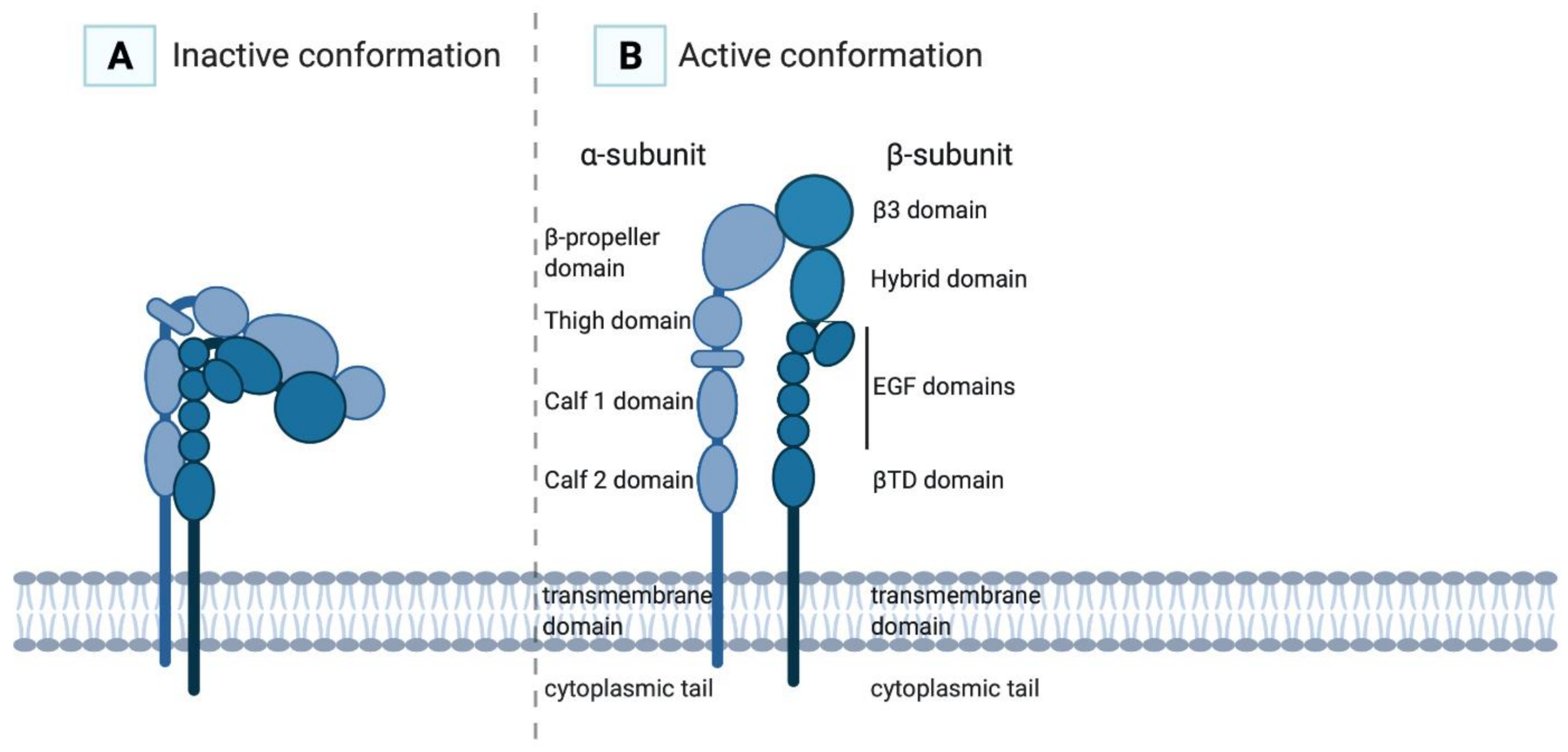
2. Integrin αIIbβ3GPIIb/IIIa
3. Ligands
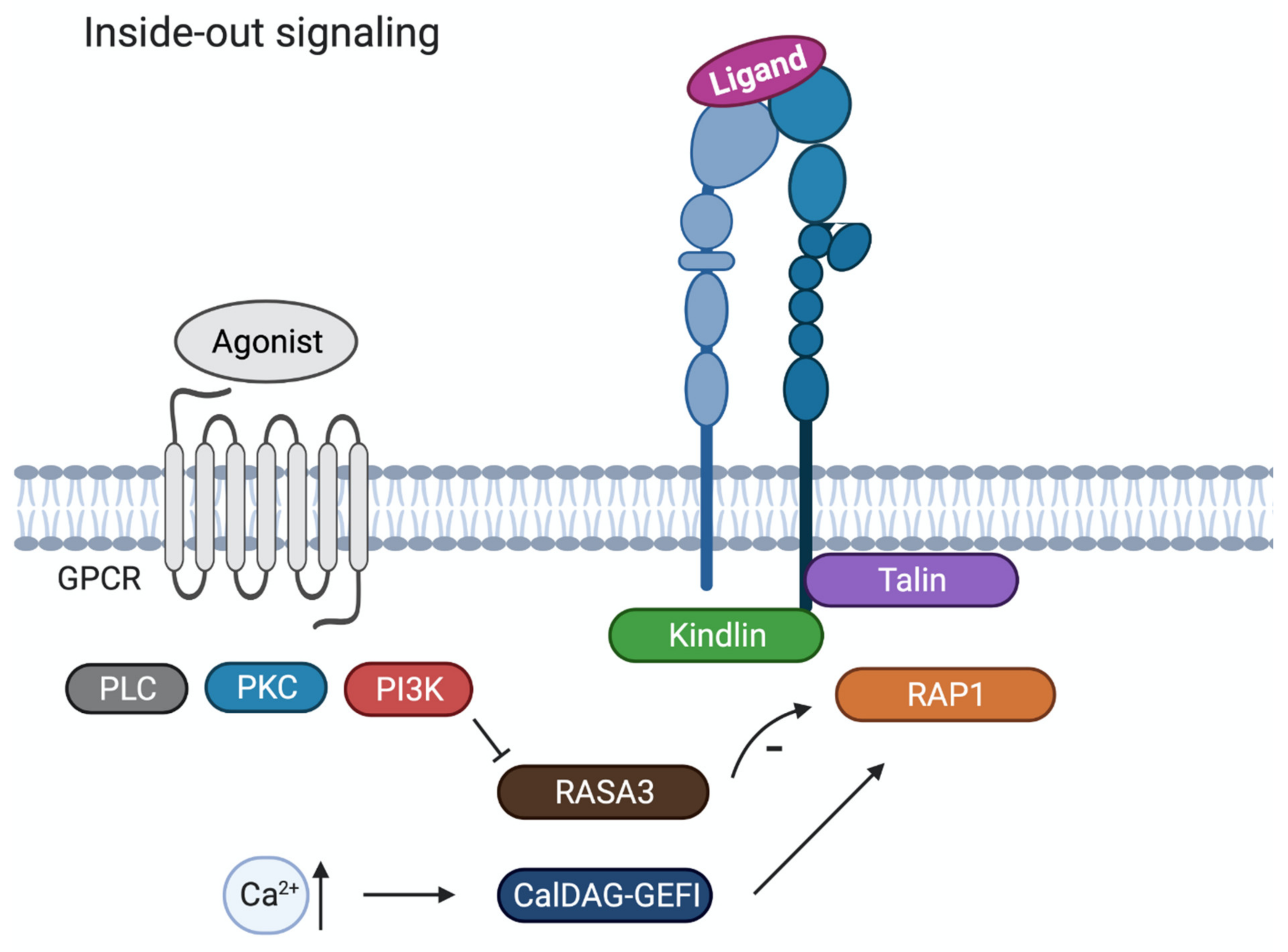
4. Inside-out Signalling
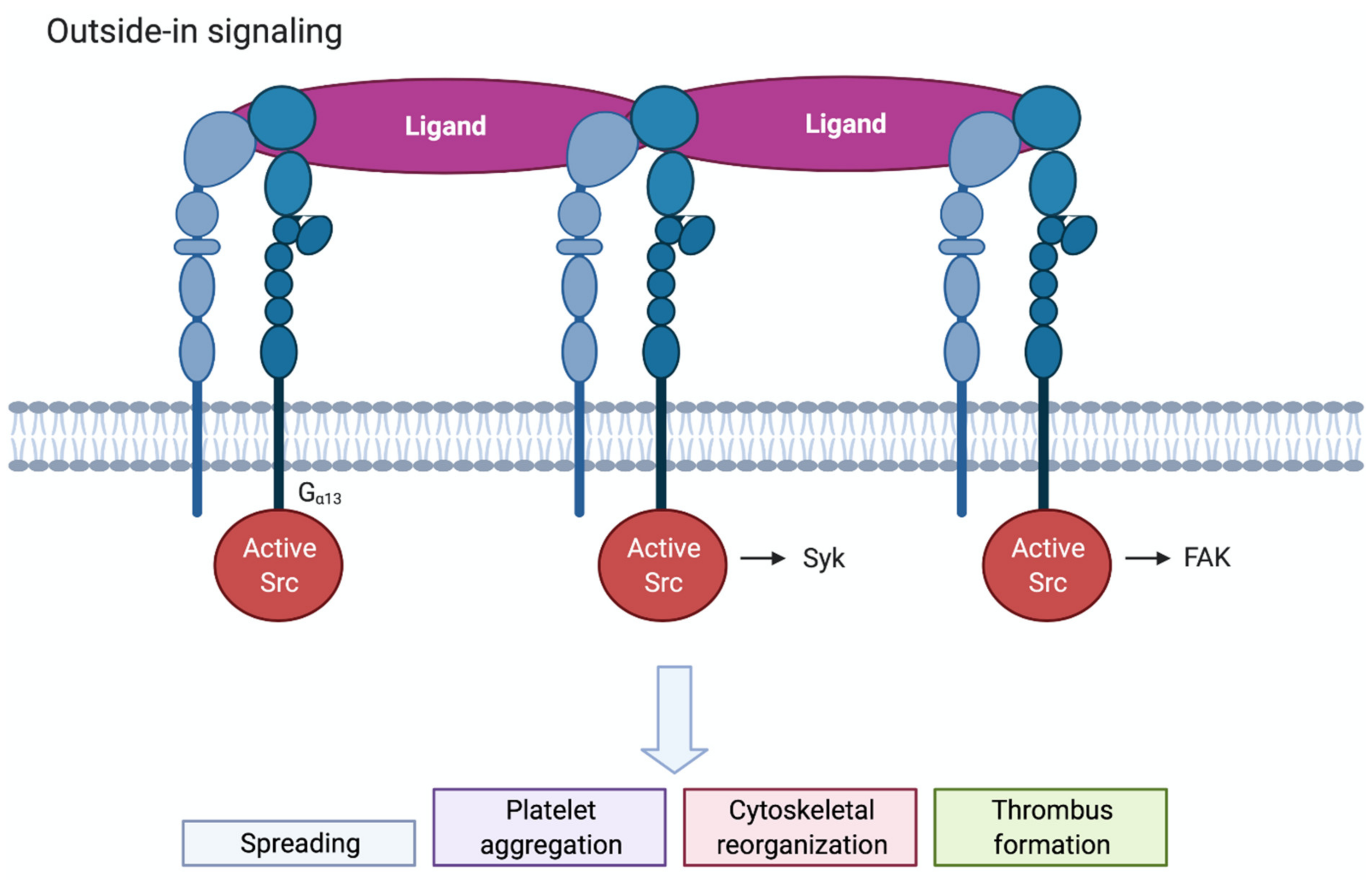
5. Outside-in Signalling
6. Antithrombotic Agents from Nature
7. Disintegrins from Snakes
7.1. Echistatin
7.2. Rhodostomin/Kistrin
8. Disintegrins from Ticks
8.1. Disagregin
8.2. Variabilin
9. Disintegrins from Leeches
9.1. Decorsin
9.2. Ornatin E
10. Disintegrins from Worms
Hookworm Platelet Inhibitor (HPI)
11. Disintegrins from Horseflies
11.1. Tabinhibitin
11.2. Tablysin-15
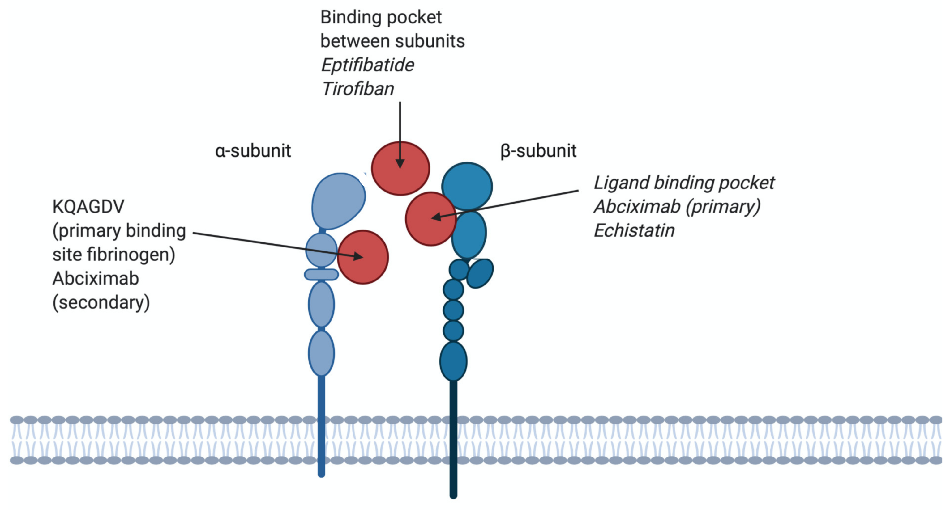
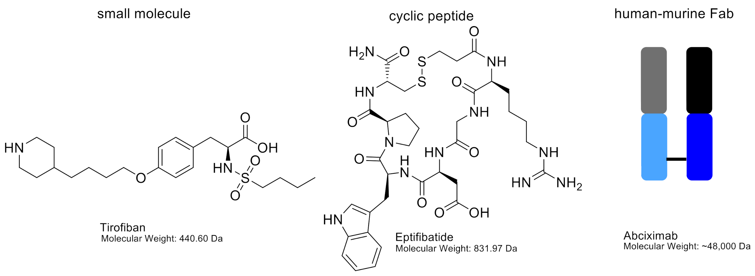
12. Antagonist Binding to the αIIbβ3 Receptor; Used in (Pre)Clinical Setting
12.1. β3. Chain Binding
Abciximab
12.2. Binding Pocket between αIIb and β3 Subunits
12.2.1. Eptifibatide
12.2.2. Tirofiban
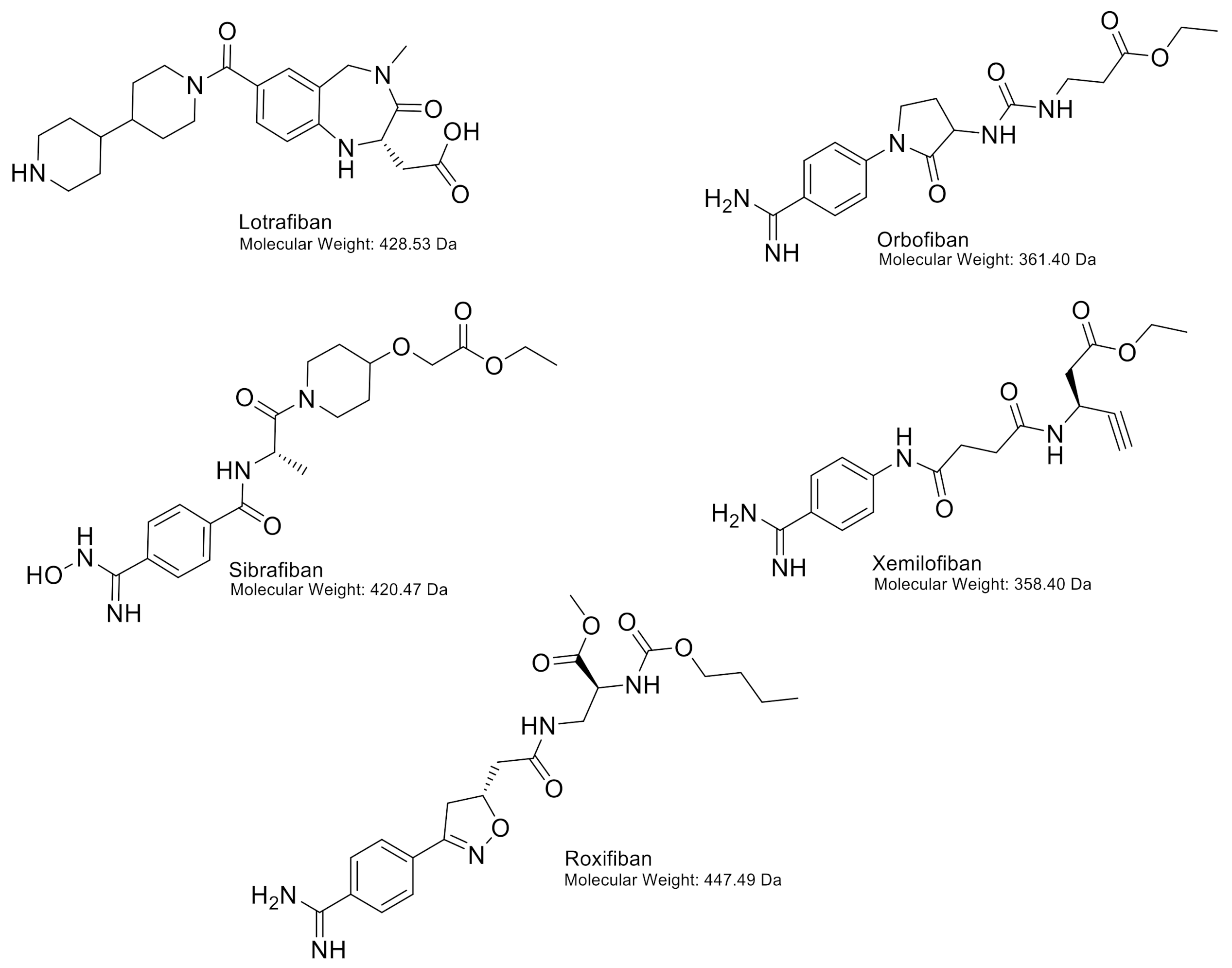
13. Oral Antagonists
13.1. Orbofiban
13.2. Sibrafiban
13.3. Xemilofiban
13.4. Lotrafiban
13.5. Roxifiban
14. Conclusions
Author Contributions
Funding
Institutional Review Board Statement
Informed Consent Statement
Data Availability Statement
Acknowledgments
Conflicts of Interest
References
- Huang, J.; Li, X.; Shi, X.; Zhu, M.; Wang, J.; Huang, S.; Huang, X.; Wang, H.; Li, L.; Deng, H.; et al. Platelet integrin alphaIIbbeta3: Signal transduction, regulation, and its therapeutic targeting. J. Hematol. Oncol. 2019, 12, 26. [Google Scholar] [CrossRef] [PubMed] [Green Version]
- Anderson, L.R.; Owens, T.W.; Naylor, M.J. Structural and mechanical functions of integrins. Biophys. Rev. 2014, 6, 203–213. [Google Scholar] [CrossRef] [Green Version]
- Zhang, K.; Chen, J. The regulation of integrin function by divalent cations. Cell Adhes. Migr. 2012, 6, 20–29. [Google Scholar] [CrossRef] [Green Version]
- Bazzoni, G.; Shih, D.T.; Buck, C.A.; Hemler, M.E. Monoclonal antibody 9EG7 defines a novel beta 1 integrin epitope induced by soluble ligand and manganese, but inhibited by calcium. J. Biol. Chem. 1995, 270, 25570–25577. [Google Scholar] [CrossRef] [Green Version]
- Heino, J. The collagen family members as cell adhesion proteins. Bioessays 2007, 29, 1001–1010. [Google Scholar] [CrossRef] [PubMed]
- Rose, D.M.; Alon, R.; Ginsberg, M.H. Integrin modulation and signaling in leukocyte adhesion and migration. Immunol. Rev. 2007, 218, 126–134. [Google Scholar] [CrossRef]
- Takada, Y.; Ye, X.; Simon, S. The integrins. Genome Biol. 2007, 8, 215. [Google Scholar] [CrossRef] [PubMed] [Green Version]
- Bennett, J.S. Structure and function of the platelet integrin alphaIIbbeta3. J. Clin. Investig. 2005, 115, 3363–3369. [Google Scholar] [CrossRef] [PubMed] [Green Version]
- Coller, B.S.; Shattil, S.J. The GPIIb/IIIa (integrin alphaIIbbeta3) odyssey: A technology-driven saga of a receptor with twists, turns, and even a bend. Blood 2008, 112, 3011–3025. [Google Scholar] [CrossRef] [Green Version]
- Rupp, P.A.; Czirok, A.; Little, C.D. alphavbeta3 integrin-dependent endothelial cell dynamics in vivo. Development 2004, 131, 2887–2897. [Google Scholar] [CrossRef] [Green Version]
- Bury, L.; Zetterberg, E.; Leinoe, E.B.; Falcinelli, E.; Marturano, A.; Manni, G.; Nurden, A.T.; Gresele, P. A novel variant Glanzmann thrombasthenia due to co-inheritance of a loss- and a gain-of-function mutation of ITGB3: Evidence of a dominant effect of gain-of-function mutations. Haematologica 2018, 103, e259–e263. [Google Scholar] [CrossRef] [PubMed] [Green Version]
- Nurden, A.T. Glanzmann thrombasthenia. Orphanet J. Rare Dis. 2006, 1, 10. [Google Scholar] [CrossRef]
- Mehrbod, M.; Trisno, S.; Mofrad, M.R. On the activation of integrin alphaIIbbeta3: Outside-in and inside-out pathways. Biophys. J. 2013, 105, 1304–1315. [Google Scholar] [CrossRef] [PubMed] [Green Version]
- Bury, L.; Malara, A.; Gresele, P.; Balduini, A. Outside-in signalling generated by a constitutively activated integrin alphaIIbbeta3 impairs proplatelet formation in human megakaryocytes. PLoS ONE 2012, 7, e34449. [Google Scholar] [CrossRef] [PubMed] [Green Version]
- Ma, Y.Q.; Qin, J.; Plow, E.F. Platelet integrin alpha(IIb)beta: Activation mechanisms. J. Thromb. Haemost. 2007, 5, 1345–1352. [Google Scholar] [CrossRef] [PubMed]
- Jordan, P.A.; Gibbins, J.M. Extracellular disulfide exchange and the regulation of cellular function. Antioxid. Redox Signal. 2006, 8, 312–324. [Google Scholar] [CrossRef] [PubMed]
- Levin, L.; Zelzion, E.; Nachliel, E.; Gutman, M.; Tsfadia, Y.; Einav, Y. A single disulfide bond disruption in the beta3 integrin subunit promotes thiol/disulfide exchange, a molecular dynamics study. PLoS ONE 2013, 8, e59175. [Google Scholar] [CrossRef]
- Dorris, S.L.; Peebles, R.S., Jr. PGI2 as a regulator of inflammatory diseases. Mediators Inflamm. 2012, 2012, 926968. [Google Scholar] [CrossRef] [Green Version]
- Radomski, M.W.; Palmer, R.M.; Moncada, S. An L-arginine/nitric oxide pathway present in human platelets regulates aggregation. Proc. Natl. Acad. Sci. USA 1990, 87, 5193–5197. [Google Scholar] [CrossRef] [PubMed] [Green Version]
- Versteeg, H.H.; Heemskerk, J.W.; Levi, M.; Reitsma, P.H. New fundamentals in hemostasis. Physiol. Rev. 2013, 93, 327–358. [Google Scholar] [CrossRef] [Green Version]
- Kanaji, S.; Fahs, S.A.; Shi, Q.; Haberichter, S.L.; Montgomery, R.R. Contribution of platelet vs. endothelial VWF to platelet adhesion and hemostasis. J. Thromb Haemost. 2012, 10, 1646–1652. [Google Scholar] [CrossRef] [PubMed]
- Sharda, A.; Flaumenhaft, R. The life cycle of platelet granules. F1000Res 2018, 7, 236. [Google Scholar] [CrossRef] [PubMed]
- Filkova, A.A.; Martyanov, A.A.; Garzon Dasgupta, A.K.; Panteleev, M.A.; Sveshnikova, A.N. Quantitative dynamics of reversible platelet aggregation: Mathematical modelling and experiments. Sci. Rep. 2019, 9, 6217. [Google Scholar] [CrossRef] [PubMed]
- Hu, D.D.; White, C.A.; Panzer-Knodle, S.; Page, J.D.; Nicholson, N.; Smith, J.W. A new model of dual interacting ligand binding sites on integrin alphaIIbbeta3. J. Biol. Chem. 1999, 274, 4633–4639. [Google Scholar] [CrossRef] [Green Version]
- Durrant, T.N.; van den Bosch, M.T.; Hers, I. Integrin alphaIIbbeta3 outside-in signaling. Blood 2017, 130, 1607–1619. [Google Scholar] [CrossRef] [Green Version]
- Ye, F.; Kim, C.; Ginsberg, M.H. Reconstruction of integrin activation. Blood 2012, 119, 26–33. [Google Scholar] [CrossRef] [PubMed] [Green Version]
- Bye, A.P.; Unsworth, A.J.; Gibbins, J.M. Platelet signaling: A complex interplay between inhibitory and activatory networks. J. Thromb. Haemost. 2016, 14, 918–930. [Google Scholar] [CrossRef] [Green Version]
- Stefanini, L.; Bergmeier, W. RAP1-GTPase signaling and platelet function. J. Mol. Med. 2016, 94, 13–19. [Google Scholar] [CrossRef]
- Harburger, D.S.; Bouaouina, M.; Calderwood, D.A. Kindlin-1 and -2 directly bind the C-terminal region of beta integrin cytoplasmic tails and exert integrin-specific activation effects. J. Biol. Chem. 2009, 284, 11485–11497. [Google Scholar] [CrossRef] [Green Version]
- Gong, H.; Shen, B.; Flevaris, P.; Chow, C.; Lam, S.C.; Voyno-Yasenetskaya, T.A.; Kozasa, T.; Du, X. G protein subunit Galpha13 binds to integrin alphaIIbbeta3 and mediates integrin “outside-in” signaling. Science 2010, 327, 340–343. [Google Scholar] [CrossRef] [Green Version]
- Buensuceso, C.S.; Arias-Salgado, E.G.; Shattil, S.J. Protein-protein interactions in platelet alphaIIbbeta3 signaling. Semin. Thromb. Hemost. 2004, 30, 427–439. [Google Scholar] [CrossRef] [PubMed] [Green Version]
- Martin, V.; Guillermet-Guibert, J.; Chicanne, G.; Cabou, C.; Jandrot-Perrus, M.; Plantavid, M.; Vanhaesebroeck, B.; Payrastre, B.; Gratacap, M.-P. Deletion of the p110beta isoform of phosphoinositide 3-kinase in platelets reveals its central role in Akt activation and thrombus formation in vitro and in vivo. Blood 2010, 115, 2008–2013. [Google Scholar] [CrossRef] [PubMed] [Green Version]
- Canobbio, I.; Stefanini, L.; Cipolla, L.; Ciraolo, E.; Gruppi, C.; Balduini, C.; Hirsch, E.; Torti, M. Genetic evidence for a predominant role of PI3Kbeta catalytic activity in ITAM- and integrin-mediated signaling in platelets. Blood 2009, 114, 2193–2196. [Google Scholar] [CrossRef] [PubMed] [Green Version]
- Das, M.; Ithychanda, S.; Qin, J.; Plow, E.F. Mechanisms of talin-dependent integrin signaling and crosstalk. Biochim. Biophys. Acta. 2014, 1838, 579–588. [Google Scholar] [CrossRef] [PubMed] [Green Version]
- Ruggeri, Z.M. Platelets in atherothrombosis. Nat. Med. 2002, 8, 1227–1234. [Google Scholar] [CrossRef]
- Koupenova, M.; Kehrel, B.E.; Corkrey, H.A.; Freedman, J.E. Thrombosis and platelets: An update. Eur. Heart J. 2017, 38, 785–791. [Google Scholar] [CrossRef] [PubMed]
- Previtali, E.; Bucciarelli, P.; Passamonti, S.M.; Martinelli, I. Risk factors for venous and arterial thrombosis. Blood Transfus. 2011, 9, 120–138. [Google Scholar] [PubMed]
- Gurbel, P.A.; Fox, K.A.A.; Tantry, U.S.; Ten Cate, H.; Weitz, J.I. Combination Antiplatelet and Oral Anticoagulant Therapy in Patients With Coronary and Peripheral Artery Disease. Circulation 2019, 139, 2170–2185. [Google Scholar] [CrossRef]
- Nowak, G. Pharmacology of recombinant hirudin. Semin. Thromb. Hemost. 2002, 28, 415–424. [Google Scholar] [CrossRef]
- Yeh, C.H.; Peng, H.C.; Huang, T.F. Accutin, a new disintegrin, inhibits angiogenesis in vitro and in vivo by acting as integrin alphavbeta3 antagonist and inducing apoptosis. Blood 1998, 92, 3268–3276. [Google Scholar] [CrossRef]
- Yeh, C.H.; Peng, H.C.; Yih, J.B.; Huang, T.F. A new short chain RGD-containing disintegrin, accutin, inhibits the common pathway of human platelet aggregation. Biochim. Biophys. Acta 1998, 1425, 493–504. [Google Scholar] [CrossRef]
- Okuda, D.; Koike, H.; Morita, T. A new gene structure of the disintegrin family: A subunit of dimeric disintegrin has a short coding region. Biochemistry 2002, 41, 14248–14254. [Google Scholar] [CrossRef] [PubMed]
- Calvete, J.J.; Schäfer, W.; Soszka, T.; Lu, W.Q.; Cook, J.J.; Jameson, B.A.; Niewiarowski, S. Identification of the disulfide bond pattern in albolabrin, an RGD-containing peptide from the venom of Trimeresurus albolabris: Significance for the expression of platelet aggregation inhibitory activity. Biochemistry 1991, 30, 5225–5229. [Google Scholar] [CrossRef]
- Singhamatr, P.; Rojnuckarin, P. Molecular cloning of albolatin, a novel snake venom metalloprotease from green pit viper (Trimeresurus albolabris), and expression of its disintegrin domain. Toxicon 2007, 50, 1192–1200. [Google Scholar] [CrossRef] [PubMed]
- Chao, B.H.; Jakubowski, J.A.; Savage, B.; Chow, E.P.; Marzec, U.M.; Harker, L.A.; Maraganore, J.M. Agkistrodon piscivorus piscivorus platelet aggregation inhibitor: A potent inhibitor of platelet activation. Proc. Natl. Acad. Sci. USA 1989, 86, 8050–8054. [Google Scholar] [CrossRef] [PubMed] [Green Version]
- Huang, T.F.; Wang, W.J.; Teng, C.M.; Ouyang, C. Mechanism of action of the antiplatelet peptide, arietin, from Bitis arietans venom. Biochim. Biophys. Acta 1991, 1074, 144–150. [Google Scholar] [CrossRef]
- Shimokawa, K.; Jia, L.G.; Shannon, J.D.; Fox, J.W. Isolation, sequence analysis, and biological activity of atrolysin E/D, the non-RGD disintegrin domain from Crotalus atrox venom. Arch. Biochem. Biophys. 1998, 354, 239–246. [Google Scholar] [CrossRef]
- Angulo, Y.; Castro, A.; Lomonte, B.; Rucavado, A.; Fernandez, J.; Calvete, J.J.; Gutiérrez, J.M. Isolation and characterization of four medium-size disintegrins from the venoms of Central American viperid snakes of the genera Atropoides, Bothrops, Cerrophidion and Crotalus. Biochimie 2014, 107, 376–384. [Google Scholar] [CrossRef]
- Soto, J.G.; White, S.A.; Reyes, S.R.; Regalado, R.; Sanchez, E.E.; Perez, J.C. Molecular evolution of PIII-SVMP and RGD disintegrin genes from the genus Crotalus. Gene 2007, 389, 66–72. [Google Scholar] [CrossRef] [PubMed]
- Scarborough, R.; Rose, J.; Hsu, M.; Phillips, D.; Fried, V.; Campbell, A.; Nannizzi, L.; Charo, I. Barbourin. A GPIIb-IIIa-specific integrin antagonist from the venom of Sistrurus m. barbouri. J. Biol. Chem. 1991, 266, 9359–9362. [Google Scholar] [CrossRef]
- Scarborough, R.; Rose, J.; Naughton, M.; Phillips, D.; Nannizzi, L.; Arfsten, A.; Campbell, A.; Charo, I. Characterization of the integrin specificities of disintegrins isolated from American pit viper venoms. J. Biol. Chem. 1993, 268, 1058–1065. [Google Scholar] [CrossRef]
- Rucinski, B.; Niewiarowski, S.; Holt, J.C.; Soszka, T.; Knudsen, K.A. Batroxostatin, an Arg-Gly-Asp-containing peptide from Bothrops atrox, is a potent inhibitor of platelet aggregation and cell interaction with fibronectin. Biochim. Biophys. Acta 1990, 1054, 257–262. [Google Scholar] [CrossRef]
- Shebuski, R.J.; Ramjit, D.R.; Bencen, G.H.; Polokoff, M.A. Characterization and platelet inhibitory activity of bitistatin, a potent arginine-glycine-aspartic acid-containing peptide from the venom of the viper Bitis arietans. J. Biol. Chem. 1989, 264, 21550–21556. [Google Scholar] [CrossRef]
- Pinto, A.; Angulo, Y.; Jiménez, R.; Lomonte, B. Isolation of bothrasperin, a disintegrin with potent platelet aggregation inhibitory activity, from the venom of the snake Bothrops asper. Rev. Biol. Trop. 2003, 51, 253–259. [Google Scholar] [PubMed]
- Sánchez, E.E.; Rodríguez-Acosta, A.; Palomar, R.; Lucena, S.E.; Bashir, S.; Soto, J.G.; Pérez, J.C. Colombistatin: A disintegrin isolated from the venom of the South American snake (Bothrops colombiensis) that effectively inhibits platelet aggregation and SK-Mel-28 cell adhesion. Arch. Toxicol. 2008, 83, 271–279. [Google Scholar] [CrossRef] [PubMed]
- Trikha, M.; Rote, W.E.; Manley, P.J.; Lucchesi, B.R.; Markland, F.S. Purification and characterization of platelet aggregation inhibitors from snake venoms. Thromb. Res. 1994, 73, 39–52. [Google Scholar] [CrossRef] [Green Version]
- Zhou, Q.; Hu, P.; Ritter, M.R.; Swenson, S.D.; Argounova, S.; Epstein, A.L.; Markland, F.S. Molecular Cloning and Functional Expression of Contortrostatin, a Homodimeric Disintegrin from Southern Copperhead Snake Venom. Arch. Biochem. Biophys. 2000, 375, 278–288. [Google Scholar] [CrossRef] [PubMed]
- Liu, C.Z.; Peng, H.C.; Huang, T.F. Crotavirin, a potent platelet aggregation inhibitor purified from the venom of the snake Crotalus viridis. Toxicon 1995, 33, 1289–1298. [Google Scholar] [CrossRef]
- Da Silva, M.; Lucena, S.; Aguilar, I.; Rodríguez-Acosta, A.; Salazar, A.M.; Sánchez, E.E.; Girón, M.E.; Carvajal, Z.; Arocha-Piñango, C.L.; Guerrero, B. Anti-platelet effect of cumanastatin 1, a disintegrin isolated from venom of South American Crotalus rattlesnake. Thromb. Res. 2009, 123, 731–739. [Google Scholar] [CrossRef]
- Williams, J.A.; Lu, X.; Rahman, S.; Keating, C.; Kakkar, V. Dendroaspin: A potent integrin receptor inhibitor from the venoms of Dendroaspis viridis and D. jamesonii. Biochem. Soc. Trans. 1993, 21, 73. [Google Scholar] [CrossRef]
- Cheng, C.H.; Chen, Y.C.; Shiu, J.H.; Chang, Y.T.; Chang, Y.S.; Huang, C.H.; Chen, C.H.; Chuang, W.J. Dynamics and functional differences between dendroaspin and rhodostomin: Insights into protein scaffolds in integrin recognition. Protein Sci. 2012, 21, 1872–1884. [Google Scholar] [CrossRef] [Green Version]
- Gan, Z.R.; Gould, R.J.; Jacobs, J.W.; Friedman, P.A.; Polokoff, M.A. Echistatin. A potent platelet aggregation inhibitor from the venom of the viper, Echis carinatus. J. Biol. Chem. 1988, 263, 19827–19832. [Google Scholar] [CrossRef]
- Kapp, T.G.; Rechenmacher, F.; Neubauer, S.; Maltsev, O.V.; Cavalcanti-Adam, E.A.; Zarka, R.; Reuning, U.; Notni, J.; Wester, H.-J.; Mas-Moruno, C.; et al. A Comprehensive Evaluation of the Activity and Selectivity Profile of Ligands for RGD-binding Integrins. Sci. Rep. 2017, 7, 39805. [Google Scholar] [CrossRef] [PubMed] [Green Version]
- Williams, J.A.; Ashby, B.; Daniel, J.L. Ligands to the platelet fibrinogen receptor glycoprotein IIb-IIIa do not affect agonist-induced second messengers Ca2+ or cyclic AMP. Biochem. J. 1990, 270, 149–155. [Google Scholar] [CrossRef] [Green Version]
- Senn, H.; Klaus, W. The nuclear magnetic resonance solution structure of flavoridin, an antagonist of the platelet GP IIb-IIIa receptor. J. Mol. Biol. 1993, 232, 907–925. [Google Scholar] [CrossRef] [PubMed]
- Kawasaki, T.; Sakai, Y.; Taniuchi, Y.; Sato, K.; Maruyama, K.; Shimizu, M.; Kaku, S.; Yano, S.; Inagaki, O.; Tomioka, K.; et al. Biochemical characterization of a new disintegrin, flavostatin, isolated from Trimeresurus flavoviridis venom. Biochimie 1996, 78, 245–252. [Google Scholar] [CrossRef]
- Huang, T.F.; Peng, H.C.; Peng, I.S.; Teng, C.M.; Ouyang, C. An antiplatelet peptide, gabonin, from Bitis gabonica snake venom. Arch. Biochem. Biophys. 1992, 298, 13–20. [Google Scholar] [CrossRef]
- Huang, T.F.; Liu, C.Z.; Ouyang, C.H.; Teng, C.M. Halysin, an antiplatelet Arg-Gly-Asp-containing snake venom peptide, as fibrinogen receptor antagonist. Biochem. Pharmacol. 1991, 42, 1209–1219. [Google Scholar]
- Bazaa, A.; Juarez, P.; Marrakchi, N.; Lasfer, Z.B.; El Ayeb, M.; Harrison, R.A.; Calvete, J.J.; Sanz, L. Loss of Introns Along the Evolutionary Diversification Pathway of Snake Venom Disintegrins Evidenced by Sequence Analysis of Genomic DNA from Macrovipera lebetina transmediterranea and Echis ocellatus. J. Mol. Evol. 2006, 64, 261–271. [Google Scholar] [CrossRef]
- Della-Casa, M.S.; Junqueira-De-Azevedo, I.; Butera, D.; Clissa, P.B.; Lopes, D.S.; Serrano, S.M.; Pimenta, D.C.; Magalhães, G.S.; Ho, P.L.; Moura-Da-Silva, A.M. Insularin, a disintegrin from Bothrops insularis venom: Inhibition of platelet aggregation and endothelial cell adhesion by the native and recombinant GST-insularin proteins. Toxicon 2011, 57, 125–133. [Google Scholar] [CrossRef]
- Wermelinger, L.S.; Geraldo, R.B.; Frattani, F.S.; Rodrigues, C.R.; Juliano, M.A.; Castro, H.C.; Zingali, R.B. Integrin inhibitors from snake venom: Exploring the relationship between the structure and activity of RGD-peptides. Arch. Biochem. Biophys. 2009, 482, 25–32. [Google Scholar] [CrossRef] [PubMed]
- Zhou, X.-D.; Ding, C.-H.; Tai, H.; Jin, Y.; Chen, R.-Q.; Lu, Q.-M.; Wang, W.-Y.; Xiong, Y.-L. A novel disintegrin, jerdonatin, inhibits platelet aggregation and sperm–egg binding. Comp. Biochem. Physiol. Part B Biochem. Mol. Biol. 2004, 139, 117–122. [Google Scholar] [CrossRef]
- Okuda, D.; Nozaki, C.; Sekiya, F.; Morita, T. Comparative biochemistry of disintegrins isolated from snake venom: Consideration of the taxonomy and geographical distribution of snakes in the genus Echis. J. Biochem. 2001, 129, 615–620. [Google Scholar] [CrossRef]
- Sánchez, E.E.; Galán, J.A.; Russell, W.K.; Soto, J.G.; Russell, D.H.; Pérez, J.C. Isolation and characterization of two disintegrins inhibiting ADP-induced human platelet aggregation from the venom of Crotalus scutulatus scutulatus (Mohave Rattlesnake). Toxicol. Appl. Pharmacol. 2006, 212, 59–68. [Google Scholar] [CrossRef]
- Borja, M.; Galan, J.A.; Cantu, E.; Zugasti-Cruz, A.; Rodríguez-Acosta, A.; Lazcano, D.; Lucena, S.; Suntravat, M.; Sánchez, Y.E.E. Morulustatin, A. Disintegrin that Inhibits ADP-Induced Platelet Aggregation, Isolated from the Mexican Tamaulipan Rock Rattlesnake (Crotalus lepidus morulus). Rev. Cient. 2016, 26, 86–94. [Google Scholar]
- Chernyshenko, V.; Petruk, N.; Korolova, D.; Kasatkina, L.; Gornytska, O.; Platonova, T.; Chernyshenko, T.; Rebriev, A.; Dzhus, O.; Garmanchuk, L.; et al. Antiplatelet and anti-proliferative action of disintegrin from Echis multisquamatis snake venom. Croat. Med. J. 2017, 58, 118–127. [Google Scholar] [CrossRef] [Green Version]
- Okuda, D.; Morita, T. Purification and characterization of a new RGD/KGD-containing dimeric disintegrin, piscivostatin, from the venom of Agkistrodon piscivorus piscivorus: The unique effect of piscivostatin on platelet aggregation. J. Biochem. 2001, 130, 407–415. [Google Scholar] [CrossRef] [PubMed]
- Carey, C.M.; Bueno, R.; Gutierrez, D.A.; Petro, C.; Lucena, S.E.; Sánchez, E.E.; Soto, J.G. Recombinant rubistatin (r-Rub), an MVD disintegrin, inhibits cell migration and proliferation, and is a strong apoptotic inducer of the human melanoma cell line SK-Mel-28. Toxicon 2012, 59, 241–248. [Google Scholar] [CrossRef] [Green Version]
- Kang, I.C.; Chung, K.H.; Lee, S.J.; Yun, Y.; Moon, H.M.; Kim, D.S. Purification and molecular cloning of a platelet aggregation inhibitor from the snake (Agkistrodon halys brevicaudus) venom. Thromb. Res. 1998, 91, 65–73. [Google Scholar] [CrossRef]
- Park, D.; Kang, I.; Kim, H.; Chung, K.; Kim, D.S.; Yun, Y. Cloning and characterization of novel disintegrins from Agkistrodon halys venom. Mol. Cells 1998, 8, 578–584. [Google Scholar] [PubMed]
- Hong, S.Y.; Koh, Y.S.; Chung, K.H.; Kim, D.S. Snake venom disintegrin, saxatilin, inhibits platelet aggregation, human umbilical vein endothelial cell proliferation, and smooth muscle cell migration. Thromb. Res. 2002, 105, 79–86. [Google Scholar] [CrossRef]
- Bilgrami, S.; Tomar, S.; Yadav, S.; Kaur, P.; Kumar, J.; Jabeen, T.; Sharma, S.; Singh, T.P. Crystal Structure of Schistatin, a Disintegrin Homodimer from Saw-scaled Viper (Echis carinatus) at 2.5 Å Resolution. J. Mol. Biol. 2004, 341, 829–837. [Google Scholar] [CrossRef] [PubMed]
- Huang, T.F.; Sheu, J.R.; Teng, C.M.; Chen, S.W.; Liu, C.S. Triflavin, an antiplatelet Arg-Gly-Asp-containing peptide, is a specific antagonist of platelet membrane glycoprotein IIb-IIIa complex. J. Biochem. 1991, 109, 328–334. [Google Scholar] [PubMed]
- Huang, T.F.; Holt, J.C.; Lukasiewicz, H.; Niewiarowski, S. Trigramin. A low molecular weight peptide inhibiting fibrinogen interaction with platelet receptors expressed on glycoprotein IIb-IIIa complex. J. Biol. Chem. 1987, 262, 16157–16163. [Google Scholar] [CrossRef]
- Knudsen, K.A.; Tuszynski, G.P.; Huang, T.F.; Niewiarowski, S. Trigramin, an RGD-containing peptide from snake venom, inhibits cell-substratum adhesion of human melanoma cells. Exp. Cell Res. 1988, 179, 42–49. [Google Scholar] [CrossRef]
- Hung, Y.C.; Hsu, C.C.; Chung, C.H.; Huang, T.F. The disintegrin, trimucrin, suppresses LPS-induced activation of phagocytes primarily through blockade of NF-κB and MAPK activation. Naunyn Schmiedebergs Arch. Pharmacol. 2016, 389, 723–737. [Google Scholar] [CrossRef]
- Oshikawa, K.; Terada, S. Ussuristatin 2, a novel KGD-bearing disintegrin from Agkistrodon ussuriensis venom. J. Biochem. 1999, 125, 31–35. [Google Scholar] [CrossRef]
- Minea, R.O.; Helchowski, C.M.; Zidovetzki, S.J.; Costa, F.K.; Swenson, S.D.; Markland, F.S., Jr. Vicrostatin—An anti-invasive multi-integrin targeting chimeric disintegrin with tumor anti-angiogenic and pro-apoptotic activities. PLoS ONE 2010, 5, e10929. [Google Scholar] [CrossRef] [PubMed]
- Karczewski, J.; Endris, R.; Connolly, T.M. Disagregin is a fibrinogen receptor antagonist lacking the Arg-Gly-Asp sequence from the tick, Ornithodoros moubata. J. Biol. Chem. 1994, 269, 6702–6708. [Google Scholar] [CrossRef]
- Francischetti, I.M.B.; Pham, V.M.; Mans, B.J.; Andersen, J.F.; Mather, T.N.; Lane, R.S.; Ribeiro, J.M.C. The transcriptome of the salivary glands of the female western black-legged tick Ixodes pacificus (Acari: Ixodidae). Insect Biochem. Mol. Biol. 2005, 35, 1142–1161. [Google Scholar] [CrossRef] [Green Version]
- Mans, B.J.; Andersen, J.F.; Schwan, T.G.; Ribeiro, J.M. Characterization of anti-hemostatic factors in the argasid, Argas monolakensis: Implications for the evolution of blood-feeding in the soft tick family. Insect Biochem. Mol. Biol. 2008, 38, 22–41. [Google Scholar] [CrossRef] [PubMed] [Green Version]
- Mans, B.J.; Louw, A.I.; Neitz, A.W. Savignygrin, a platelet aggregation inhibitor from the soft tick Ornithodoros savignyi, presents the RGD integrin recognition motif on the Kunitz-BPTI fold. J. Biol. Chem. 2002, 277, 21371–21378. [Google Scholar] [CrossRef] [PubMed] [Green Version]
- Wang, X.; Coons, L.B.; Taylor, D.B.; Stevens, S.E.; Gartner, T.K., Jr. Variabilin, a novel RGD-containing antagonist of glycoprotein IIb-IIIa and platelet aggregation inhibitor from the hard tick Dermacentor variabilis. J. Biol. Chem. 1996, 271, 17785–17790. [Google Scholar] [CrossRef] [PubMed] [Green Version]
- Seymour, J.L.; Henzel, W.J.; Nevins, B.; Stults, J.T.; Lazarus, R.A. Decorsin. A potent glycoprotein IIb-IIIa antagonist and platelet aggregation inhibitor from the leech Macrobdella decora. J. Biol. Chem. 1990, 265, 10143–10147. [Google Scholar] [CrossRef]
- Mazur, P.; Henzel, W.J.; Seymour, J.L.; Lazarus, R.A. Ornatins: Potent glycoprotein IIb-IIIa antagonists and platelet aggregation inhibitors from the leech Placobdella ornata. Eur. J. Biochem. 1991, 202, 1073–1082. [Google Scholar] [CrossRef] [PubMed]
- Del Valle, A.; Jones, B.F.; Harrison, L.M.; Chadderdon, R.C.; Cappello, M. Isolation and molecular cloning of a secreted hookworm platelet inhibitor from adult Ancylostoma caninum. Mol. Biochem. Parasitol. 2003, 129, 167–177. [Google Scholar] [CrossRef]
- Chadderdon, R.C.; Cappello, M. The hookworm platelet inhibitor: Functional blockade of integrins GPIIb/IIIa (alphaIIbbeta3) and GPIa/IIa (alpha2beta1) inhibits platelet aggregation and adhesion in vitro. J. Infect. Dis. 1999, 179, 1235–1241. [Google Scholar] [CrossRef] [Green Version]
- Ma, D.; Wang, Y.; Yang, H.; Wu, J.; An, S.; Gao, L.; Xu, X.; Lai, R. Anti-thrombosis Repertoire of Blood-feeding Horsefly Salivary Glands. Mol. Cell. Proteom. 2009, 8, 2071–2079. [Google Scholar] [CrossRef] [Green Version]
- Liu, H.; Yang, X.; Andersen, J.F.; Wang, Y.; Tokumasu, F.; Ribeiro, J.M.C.; Ma, D.; Xu, X.; An, S.; Francischetti, I.M.B.; et al. A novel family of RGD-containing disintegrins (Tablysin-15) from the salivary gland of the horsefly Tabanus yao targets αIIbβ3 or αVβ3 and inhibits platelet aggregation and angiogenesis. Thromb. Haemost. 2011, 105, 1032–1045. [Google Scholar] [CrossRef] [PubMed] [Green Version]
- Marcinkiewicz, C.; Rosenthal, L.A.; Mosser, D.M.; Kunicki, T.J.; Niewiarowski, S. Immunological characterization of eristostatin and echistatin binding sites on alpha IIb beta 3 and alpha V beta 3 integrins. Biochem. J. 1996, 317, 817–825. [Google Scholar] [CrossRef] [Green Version]
- Van den Kerkhof, D.L.; Nagy, M.; Wichapong, K.; Brouns, S.L.N.; Heemskerk, J.W.M.; Hackeng, T.M.; Dijkgraaf, I. Inhibition of platelet adhesion, thrombus formation, and fibrin formation by a potent alphaIIbbeta3 integrin inhibitor from ticks. Res. Pract. Thromb. Haemost. 2021, 5, 231–242. [Google Scholar] [CrossRef] [PubMed]
- Mazur, P.; Dennis, M.S.; Seymour, J.L.; Lazarus, R.A. Expression, purification, and characterization of recombinant ornatin E, a potent glycoprotein IIb-IIIa antagonist. Protein Expr. Purif. 1993, 4, 282–289. [Google Scholar] [CrossRef] [PubMed]
- Schror, K.; Weber, A.A. Comparative pharmacology of GP IIb/IIIa antagonists. J. Thromb. Thrombolysis 2003, 15, 71–80. [Google Scholar] [CrossRef] [PubMed]
- Mascelli, M.A.; Lance, E.T.; Damaraju, L.; Wagner, C.L.; Weisman, H.F.; Jordan, R.E. Pharmacodynamic profile of short-term abciximab treatment demonstrates prolonged platelet inhibition with gradual recovery from GP IIb/IIIa receptor blockade. Circulation 1998, 97, 1680–1688. [Google Scholar] [CrossRef] [PubMed]
- Schwarz, M.; Meade, G.; Stoll, P.; Ylanne, J.; Bassler, N.; Chen, Y.C.; Hagemeyer, C.E.; Ahrens, I.; Moran, N.; Kenny, D.; et al. Conformation-specific blockade of the integrin GPIIb/IIIa: A novel antiplatelet strategy that selectively targets activated platelets. Circ. Res. 2006, 99, 25–33. [Google Scholar] [CrossRef] [PubMed] [Green Version]
- Bedjaoui, A.; Allal, K.; Lounes, M.S.; Belhadi, C.E.; Mekarnia, A.; Sediki, S.; Kara, M.; Azaza, A.; Monsuez, J.-J.; Benkhedda, S. Intracoronary or intravenous abciximab after aspiration thrombectomy in patients with STEMI undergoing primary percutaneous coronary intervention. Cardiovasc. J. Afr. 2019, 30, 45–51. [Google Scholar] [CrossRef] [PubMed] [Green Version]
- Stoffer, K.; Bistas, K.G.; Reddy, V.; Shah, S. Abciximab; StatPearls: Treasure Island, FL, USA, 2020. [Google Scholar]
- (FDA) US. FDA Drug Shortages. Available online: https://www.accessdata.fda.gov/scripts/drugshortages/dsp_ActiveIngredientDetails.cfm?AI=Abciximab%20(ReoPro)%20Injection&st=c&tab=tabs-1 (accessed on 8 February 2021).
- Phillips, D.R.; Scarborough, R.M. Clinical pharmacology of eptifibatide. Am. J. Cardiol. 1997, 80, 11B–20B. [Google Scholar] [CrossRef]
- Hashemzadeh, M.; Furukawa, M.; Goldsberry, S.; Movahed, M.R. Chemical structures and mode of action of intravenous glycoprotein IIb/IIIa receptor blockers: A review. Exp. Clin. Cardiol. 2008, 13, 192–197. [Google Scholar]
- Valgimigli, M.; Campo, G.; Tebaldi, M.; Carletti, R.; Arcozzi, C.; Ferrari, R.; Percoco, G. Abciximab: A reappraisal of its use in coronary care. Biologics 2008, 2, 29–39. [Google Scholar] [CrossRef] [Green Version]
- Di, L. Strategic approaches to optimizing peptide ADME properties. AAPS J. 2015, 17, 134–143. [Google Scholar] [CrossRef] [PubMed]
- Gang, D.; Kim, D.W.; Park, H.S. Cyclic Peptides: Promising Scaffolds for Biopharmaceuticals. Genes 2018, 9, 557. [Google Scholar] [CrossRef] [PubMed] [Green Version]
- Lele, M.; Sajid, M.; Wajih, N.; Stouffer, G.A. Eptifibatide and 7E3, but not tirofiban, inhibit alpha(v)beta integrin-mediated binding of smooth muscle cells to thrombospondin and prothrombin. Circulation 2001, 104, 582–587. [Google Scholar] [CrossRef] [PubMed] [Green Version]
- Tcheng, J.E. Clinical challenges of platelet glycoprotein IIb/IIIa receptor inhibitor therapy: Bleeding, reversal, thrombocytopenia, and retreatment. Am. Heart J. 2000, 139, S38–S45. [Google Scholar] [CrossRef] [PubMed]
- Bansal, A.B.; Sattar, Y.; Jamil, R.T. Eptifibatide; StatPearls: Treasure Island, FL, USA, 2020. [Google Scholar]
- The PURSUIT Trial Investigators. Inhibition of platelet glycoprotein IIb/IIIa with eptifibatide in patients with acute coronary syndromes. N. Engl. J. Med. 1998, 339, 436–443. [Google Scholar] [CrossRef]
- Valgimigli, M.; Percoco, G.; Barbieri, D.; Ferrari, F.; Guardigli, G.; Parrinello, G.; Soukhomovskaia, O.; Ferrari, R. The additive value of tirofiban administered with the high-dose bolus in the prevention of ischemic complications during high-risk coronary angioplasty: The ADVANCE Trial. J. Am. Coll. Cardiol. 2004, 44, 14–19. [Google Scholar] [CrossRef] [Green Version]
- Tcheng, J.E. Enhancing safety and outcomes with the newer antithrombotic and antiplatelet agents. Am. Heart J. 1995, 130, 673–679. [Google Scholar] [CrossRef]
- Yeh, E.T.; Khan, B.V. The potential role of antiplatelet agents in modulating inflammatory markers in atherothrombosis. J. Thromb. Haemost. 2006, 4, 2308–2316. [Google Scholar] [CrossRef]
- Nicholson, N.S.; Abood, N.A.; Panzer-Knodle, S.G.; Frederick, L.G.; Page, J.D.; Salyers, A.K.; Suleymanov, O.D.; Szalony, J.A.; Taite, B.B.; Anders, R.J. Orbofiban: An orally active GPIIb/IIIa platelet receptor antagonist. Med. Res. Rev. 2001, 21, 211–226. [Google Scholar] [CrossRef] [PubMed]
- Cannon, C.P.; McCabe, C.H.; Wilcox, R.G.; Langer, A.; Caspi, A.; Berink, P.; Lopez-Sendon, J.; Toman, J.; Charlesworth, A.; Anders, R.J.; et al. Oral Glycoprotein IIb/IIIa Inhibition With Orbofiban in Patients With Unstable Coronary Syndromes (OPUS-TIMI 16) Trial. Circulation 2000, 102, 149–156. [Google Scholar] [CrossRef] [Green Version]
- Holmes, M.B.; Sobel, B.E.; Cannon, C.P.; Schneider, D.J. Increased platelet reactivity in patients given orbofiban after an acute coronary syndrome: An OPUS-TIMI 16 substudy. Orbofiban in Patients with Unstable coronary syndromes. Thrombolysis In Myocardial Infarction. Am. J. Cardiol. 2000, 85, 491–493. [Google Scholar] [CrossRef]
- Serrano, C.V., Jr.; Nicolau, J.C.; Venturinelli, M.; Baracioli, L.M.; Anders, R.J.; Cannon, C.P.; Ramires, J.A. Role of oral blockade of platelet glycoprotein IIb/IIIa on neutrophil activation in patients with acute coronary syndromes. Cardiovasc. Drugs Ther. 2003, 17, 129–132. [Google Scholar] [CrossRef] [PubMed]
- Dooley, M.; Goa, K.L. Sibrafiban. Drugs 1999, 57, 225–230. [Google Scholar] [CrossRef] [PubMed]
- Newby, L.K. Long-term oral platelet glycoprotein IIb/IIIa receptor antagonism with sibrafiban after acute coronary syndromes: Study design of the sibrafiban versus aspirin to yield maximum protection from ischemic heart events post-acute coronary syndromes (SYMPHONY) trial. Symphony Steering Committee. Am. Heart J. 1999, 138, 210–218. [Google Scholar]
- The SYMPHONY Investigators. Comparison of sibrafiban with aspirin for prevention of cardiovascular events after acute coronary syndromes: A randomised trial. Lancet 2000, 355, 337–345. [Google Scholar] [CrossRef]
- Anders, R.; Kleiman, J.; Nicholson, N.; Wazowicz, B.; Burns, D. Xemilofiban/orbofiban: Insight into drug development. Cardiovasc. Drug Rev. 2001, 19, 116–132. [Google Scholar] [CrossRef] [PubMed]
- O’Neill, W.W.; Serruys, P.; Knudtson, M.; Van Es, G.-A.; Timmis, G.C.; Van Der Zwaan, C.; Kleiman, J.; Gong, J.; Roecker, E.B.; Dreiling, R.; et al. Long-Term Treatment with a Platelet Glycoprotein-Receptor Antagonist after Percutaneous Coronary Revascularization. N. Engl. J. Med. 2000, 342, 1316–1324. [Google Scholar] [CrossRef] [PubMed]
- Liu, F.; Craft, R.M.; Morris, S.A.; Carroll, R.C. Lotrafiban: An oral platelet glycoprotein IIb/IIIa blocker. Exp. Opin. Investig. Drugs 2000, 9, 2673–2687. [Google Scholar] [CrossRef]
- Topol, E.J.; Easton, D.; Harrington, R.A.; Amarenco, P.; Califf, R.M.; Graffagnino, C.; Davis, S.; Diener, H.-C.; Ferguson, J.; Fitzgerald, D.; et al. Randomized, Double-Blind, Placebo-Controlled, International Trial of the Oral IIb/IIIa Antagonist Lotrafiban in Coronary and Cerebrovascular Disease. Circulation 2003, 108, 399–406. [Google Scholar] [CrossRef] [Green Version]
- Mousa, S.A.; Kapil, R.; Mu, D.X. Intravenous and oral antithrombotic efficacy of the novel platelet GPIIb/IIIa antagonist roxifiban (DMP754) and its free acid form, XV459. Arterioscler. Thromb. Vasc. Biol. 1999, 19, 2535–2541. [Google Scholar] [CrossRef] [PubMed] [Green Version]
- Mousa, S.A.; Bozarth, J.M.; Lorelli, W.; Forsythe, M.S.; Thoolen, M.J.; Slee, A.M.; Reilly, T.M.; A Friedman, P. Antiplatelet efficacy of XV459, a novel nonpeptide platelet GPIIb/IIIa antagonist: Comparative platelet binding profiles with c7E3. J. Pharmacol. Exp. Ther. 1998, 286, 1277–1284. [Google Scholar]
- Serebruany, V.L.; Malinin, A.I.; O’Connor, C.M.; Gurbel, P.A. Roxifiban Oral Compound Kinetics Evaluation Trial IPS. Effects of roxifiban on platelet aggregation and major receptor expression in patients with coronary artery disease for the Roxifiban Oral Compound Kinetics Evaluation Trial-I (ROCKET-I Platelet Substudy). Am. Heart J. 2003, 146, 91–98. [Google Scholar]
- Armstrong, P.C.; Peter, K. GPIIb/IIIa inhibitors: From bench to bedside and back to bench again. Thromb. Haemost. 2012, 107, 808–814. [Google Scholar] [CrossRef]
- Merlini, P.A.; Rossi, M.; Menozzi, A.; Buratti, S.; Brennan, D.M.; Moliterno, D.J.; Topol, E.J.; Ardissino, D. Thrombocytopenia Caused by Abciximab or Tirofiban and Its Association With Clinical Outcome in Patients Undergoing Coronary Stenting. Circulation 2004, 109, 2203–2206. [Google Scholar] [CrossRef] [Green Version]
- Schmid-Hempel, P. Immune defence, parasite evasion strategies and their relevance for ‘macroscopic phenomena’ such as virulence. Philos. Trans. R. Soc. Lond. B Biol. Sci. 2009, 364, 85–98. [Google Scholar] [CrossRef] [PubMed] [Green Version]
- Kuo, Y.J.; Chung, C.H.; Pan, T.Y.; Chuang, W.J.; Huang, T.F. A Novel alphaIIbbeta3 Antagonist from Snake Venom Prevents Thrombosis without Causing Bleeding. Toxins 2019, 12, 11. [Google Scholar] [CrossRef] [PubMed] [Green Version]
- Kam, P.C.; Egan, M.K. Platelet glycoprotein IIb/IIIa antagonists: Pharmacology and clinical developments. Anesthesiology. 2002, 96, 1237–1249. [Google Scholar] [PubMed]
- Theroux, P. Oral inhibitors of platelet membrane receptor glycoprotein IIb/IIIa in clinical cardiology: Issues and opportunities. Am. Heart J. 1998, 135, 107–112. [Google Scholar] [CrossRef]
- Olie, R.H.; van der Meijden, P.E.J.; Ten Cate, H. The coagulation system in atherothrombosis: Implications for new therapeutic strategies. Res. Pract. Thromb. Haemost. 2018, 2, 188–198. [Google Scholar] [CrossRef]
- Gaziano, J.M.; Brotons, C.; Coppolecchia, R.; Cricelli, C.; Darius, H.; Gorelick, P.B.; Howard, G.H.; Pearson, T.A.; Rothwell, P.M.; Ruilope, L.M.; et al. Use of aspirin to reduce risk of initial vascular events in patients at moderate risk of cardiovascular disease (ARRIVE): A randomised, double-blind, placebo-controlled trial. Lancet 2018, 392, 1036–1046. [Google Scholar] [CrossRef]
- McFadyen, J.D.; Schaff, M.; Peter, K. Current and future antiplatelet therapies: Emphasis on preserving haemostasis. Nat. Rev. Cardiol. 2018, 15, 181–191. [Google Scholar] [CrossRef]
- Ziegler, M.; Wang, X.; Peter, K. Platelets in cardiac ischaemia/reperfusion injury: A promising therapeutic target. Cardiovasc. Res. 2019, 115, 1178–1188. [Google Scholar] [CrossRef] [PubMed]


| Name | Species | Target | Integrin | Sequence | Ref. |
|---|---|---|---|---|---|
| Accutin | Snake | platelets | αIIbβ3/αvβ3 | RGD | [40,41] |
| Acostatin 1 | Snake | platelets | αIIbβ3 | RGD | [42] |
| Acostatin 2 | Snake | platelets | αIIbβ3 | RGD | [42] |
| Albolabrin | Snake | platelets | αIIbβ3 | RGD | [43] |
| Albolatin | Snake | platelets | αIIbβ3/α2β1 | KGD | [44] |
| Applagin | Snake | platelets | αIIbβ3 | RGD | [45] |
| Arietin | Snake | platelets | αIIbβ3 | RGD | [46] |
| Atrolysin E | Snake | platelets | αIIbβ3?/others? | MVD | [47] |
| Atropoimin | Snake | platelets | αvβ3 | RGD | [48] |
| Atroxatin | Snake | platelets | αIIbβ3 | RGD | [49] |
| Barbourin | Snake | platelets | αIIbβ3 | KGD | [50] |
| Basicilin | Snake | platelets | αIIbβ3/αvβ3/α5β1 | RGD | [51] |
| Batroxostatin | Snake | platelets | αIIbβ3 | RGD | [52] |
| Bitistatin | Snake | platelets | αIIbβ3/αvβ3 | RGD | [53] |
| Bothrasperin | Snake | platelets | αIIbβ3?/αvβ3 | RGD | [54] |
| Cerastin | Snake | platelets | αIIbβ3/αvβ3 | RGD | [51] |
| Cereberin | Snake | platelets | αIIbβ3/αvβ3 | RGD | [51] |
| Colombistatin | Snake | platelets/human urinary and skin melanoma cancer cells | αIIbβ3/α5β1 | RGD | [55] |
| Contortrostatin | Snake | platelets/cancer cells | αIIbβ3/αvβ3/ α5β1/αvβ5 | RGD | [56,57] |
| Cotiarin | Snake | platelets | αIIbβ3/α5β1 | RGD | [51] |
| Crotavirin | Snake | platelets | αIIbβ3 | RGD? | [58] |
| Crotratroxin | Snake | platelets | αIIbβ3/α5β1 | RGD | [51] |
| Cumanastatin 1 | Snake | platelets | αIIbβ3 | RGD? | [59] |
| Dendroaspin | Snake | platelets/endothelial cells | αIIbβ3/αvβ3/α5β1 | RGD | [60,61] |
| Echistatin | Snake | platelets/osteoclasts | αIIbβ3/αvβ3/α5β1 | RGD | [62,63] |
| Elegantin | Snake | platelets | αIIbβ3 | RGD | [64] |
| Flavoridin | Snake | platelets | αIIbβ3 | RGD | [65] |
| Flavostatin | Snake | platelets | αIIbβ3 | RGD | [66] |
| Gabonin | Snake | platelets | αIIbβ3 | RGD | [67] |
| Halysin | Snake | platelets | αIIbβ3 | RGD | [68] |
| Halystatin | Snake | platelets | αIIbβ3? | RGD | [69] |
| Insularin | Snake | platelets/endothelial cells | αIIbβ3/αvβ3 | RGD | [70] |
| Jararacin | Snake | platelets | αIIbβ3/αvβ3 | RGD | [51,71] |
| Jarastatin | Snake | platelets | αIIbβ3 | RGD | [71] |
| Jerdonatin | Snake | platelets | αIIbβ3 | RGD | [72] |
| Lachesin | Snake | platelets | αIIbβ3/αvβ3/α5β1 | RGD | [51] |
| Leucogastin A | Snake | platelets | αIIbβ3? | RGD | [73] |
| Leucogastin B | Snake | platelets | αIIbβ3? | RGD | [73] |
| Lutosin | Snake | platelets | αIIbβ3/αvβ3/α5β1 | RGD | [51] |
| Mojastin 1 | Snake | platelets/human urinary bladder cell adhesion to fibronectin | αIIbβ3/α5β1 | RGD | [74] |
| Mojastin 2 | Snake | platelets/human urinary bladder cell adhesion to fibronectin | αIIbβ3/α5β1 | RGD | [74] |
| Molossin | Snake | platelets | αIIbβ3/αvβ3 | RGD | [51] |
| Morulustatin | Snake | platelets | αIIbβ3? | ? | [75] |
| Multisquamatin | Snake | platelets | αIIbβ3 | RGD | [56,73] |
| Ocelatin | Snake | platelets | αIIbβ3? | RGD | [73] |
| PAIEM | Snake | platelets | αIIbβ3 | RGD | [76] |
| Piscivostatin | Snake | platelets | αIIbβ3 | RGD/KGD | [77] |
| Pyramidin A | Snake | platelets | αIIbβ3? | RGD | [73] |
| Pyramidin B | Snake | platelets | αIIbβ3? | RGD | [73] |
| Rhodostomin/Kistrin | Snake | platelets/endothelial cells | αIIbβ3/αvβ3/α5β1 | RGD | [61] |
| Rubistatin | Snake | platelets/SK-Mel-28 cells | αIIbβ3? | MVD | [78] |
| Salmosin 1 | Snake | platelets/endothelial cells | αIIbβ3/αvβ3 | RGD | [79] |
| Salmosin 2 | Snake | platelets | αIIbβ3 | KGD | [80] |
| Saxatilin | Snake | platelets | αIIbβ3/αvβ3 | RGD | [81] |
| Schistatin | Snake | platelets | αIIbβ3/αvβ3 | RGD | [82] |
| Tergeminin | Snake | platelets | αIIbβ3 | RGD | [50] |
| Triflavin | Snake | platelets | αIIbβ3 | RGD | [83] |
| Trigramin | Snake | platelets/human melanoma cells | αIIbβ3 | RGD | [84,85] |
| Trimucrin | Snake | platelets/endothelial cells | αIIbβ3/αvβ3 | RGD | [86] |
| Ussuristatin-1 | Snake | platelets | αIIbβ3 | RGD | [87] |
| Ussuristatin-2 | Snake | platelets | αIIbβ3/α5β1 | KGD | [87] |
| Vicrostatin | Snake | platelets | αIIbβ3/α5β1 | RGD | [88] |
| Viridin | Snake | platelets | αIIbβ3/αvβ3 | RGD | [51] |
| Disagregin | Tick | platelets | αIIbβ3 | RED | [89] |
| Ixodegrin | Tick | platelets? | αIIbβ3? | RGD | [90] |
| Monogrin | Tick | platelets | αIIbβ3 | RGD | [91] |
| Savignygrin | Tick | platelets | αIIbβ3 | RGD/RED | [92] |
| Variabilin | Tick | platelets | αIIbβ3/αvβ3 | RGD | [93] |
| Decorsin | Leech | platelets | αIIbβ3 | RGD | [94] |
| Ornatin E | Leech | platelets | αIIbβ3 | RGD | [95] |
| Hookworm platelet inhibitor (HPI) | Worm | platelets | αIIbβ3/α2β1 | KGD | [96,97] |
| Tabinhibitin | Horsefly | platelets | αIIbβ3 | RGD | [98] |
| Tablysin-15 | Horsefly | platelets/endothelial cells | αIIbβ3/αvβ3/α5β1 | RGD | [99] |
Publisher’s Note: MDPI stays neutral with regard to jurisdictional claims in published maps and institutional affiliations. |
© 2021 by the authors. Licensee MDPI, Basel, Switzerland. This article is an open access article distributed under the terms and conditions of the Creative Commons Attribution (CC BY) license (http://creativecommons.org/licenses/by/4.0/).
Share and Cite
van den Kerkhof, D.L.; van der Meijden, P.E.J.; Hackeng, T.M.; Dijkgraaf, I. Exogenous Integrin αIIbβ3 Inhibitors Revisited: Past, Present and Future Applications. Int. J. Mol. Sci. 2021, 22, 3366. https://0-doi-org.brum.beds.ac.uk/10.3390/ijms22073366
van den Kerkhof DL, van der Meijden PEJ, Hackeng TM, Dijkgraaf I. Exogenous Integrin αIIbβ3 Inhibitors Revisited: Past, Present and Future Applications. International Journal of Molecular Sciences. 2021; 22(7):3366. https://0-doi-org.brum.beds.ac.uk/10.3390/ijms22073366
Chicago/Turabian Stylevan den Kerkhof, Danique L., Paola E.J. van der Meijden, Tilman M. Hackeng, and Ingrid Dijkgraaf. 2021. "Exogenous Integrin αIIbβ3 Inhibitors Revisited: Past, Present and Future Applications" International Journal of Molecular Sciences 22, no. 7: 3366. https://0-doi-org.brum.beds.ac.uk/10.3390/ijms22073366