Review

Rhizobial Exopolysaccharides: Genetic Regulation of Their Synthesis and Relevance in Symbiosis with Legumes

by
Sebastián Acosta-Jurado
1,
Francisco Fuentes-Romero
1,
Jose-Enrique Ruiz-Sainz
1,
Monika Janczarek
2,* and
José-María Vinardell
1,*
1
Department of Microbiology, University of Sevilla, Avda. Reina Mercedes 6, 41012 Seville, Spain
2
Department of Industrial and Environmental Microbiology, Institute of Biological Sciences, Faculty of Biology and Biotechnology, Maria Curie-Skłodowska University, Akademicka 19, 20-033 Lublin, Poland
*
Authors to whom correspondence should be addressed.
Int. J. Mol. Sci. 2021, 22(12), 6233; https://0-doi-org.brum.beds.ac.uk/10.3390/ijms22126233
Submission received: 5 May 2021 / Revised: 3 June 2021 / Accepted: 6 June 2021 / Published: 9 June 2021
(This article belongs to the Special Issue Molecular Plant-Microbe Interactions)

Abstract

:
Rhizobia are soil proteobacteria able to engage in a nitrogen-fixing symbiotic interaction with legumes that involves the rhizobial infection of roots and the bacterial invasion of new organs formed by the plant in response to the presence of appropriate bacterial partners. This interaction relies on a complex molecular dialogue between both symbionts. Bacterial N-acetyl-glucosamine oligomers called Nod factors are indispensable in most cases for early steps of the symbiotic interaction. In addition, different rhizobial surface polysaccharides, such as exopolysaccharides (EPS), may also be symbiotically relevant. EPS are acidic polysaccharides located out of the cell with little or no cell association that carry out important roles both in free-life and in symbiosis. EPS production is very complexly modulated and, frequently, co-regulated with Nod factors, but the type of co-regulation varies depending on the rhizobial strain. Many studies point out a signalling role for EPS-derived oligosaccharides in root infection and nodule invasion but, in certain symbiotic couples, EPS can be dispensable for a successful interaction. In summary, the complex regulation of the production of rhizobial EPS varies in different rhizobia, and the relevance of this polysaccharide in symbiosis with legumes depends on the specific interacting couple.

1. Introduction

Rhizobia is the common name for a group of soil proteobacteria able to enter into symbiosis with legume plants [1]. As part of this interaction, rhizobia infect legume roots, the plant forms new organs (root nodules), and these organs are invaded by rhizobia: all together, these events are referred as the nodulation process. Inside nodules, rhizobia are hosted by symbiotic plant cells, in which they differentiate into bacteroids able to fix N2 into ammonia that, mostly, is assimilated by the plant [2,3]. Thanks to this symbiosis, legumes can grow in soils poor in nitrogen, which is important from both an ecological and an agricultural point of view. The inoculation of legumes with appropriate rhizobia can reduce or even eliminate the necessity of the application of nitrogen fertilizers (expensive and highly polluting), which makes the use of rhizobial inoculants very appropriate for sustainable agricultural practices [4,5].
The rhizobia–legume symbiotic interaction is characterized by being specific: each rhizobial strain can nodulate with a definite set of legumes (known as the nodulation range), which can be narrow or broad, and each legume species can be nodulated by a set of rhizobial strains [6]. This specificity relies on a complex molecular dialogue that is established between both symbionts [6,7,8,9]. Although there are exceptions [1,10], in most of the rhizobia–legume symbiotic interactions this dialogue can be summarized as follows [9,10]: plant root exudates contain different nutrients that can feed rhizobia, including flavonoids, which, when appropriate and through the activation of the bacterial LysR regulator NodD, trigger the expression of bacterial nodulation genes and thus, the production and secretion of a set of different Nod factors. These rhizobial molecules are N-acetyl-glucosamine oligosaccharides harbouring different chemical decorations that, when appropriate, are perceived by LysM receptors present in root hair membranes, eliciting different responses necessary for bacterial infection and the start of the nodule organogenesis program. The most evolved rhizobial infection pathway is through infection threads (IT): briefly, following cytoskeletal reorganization, root hairs curve and trap rhizobial cells that previously adhered to their tips. These bacteria become enclosed in a kind of pocket wherein the action of lytic enzymes produced by both symbionts leads to local plant cell-wall degradation. At that point, root hair membrane invaginates and originates a channel, the IT, that will be used by rhizobia to penetrate into the root. These channels grow towards the root cortex, where, in parallel to the infection process, nodule organogenesis had started. Several cortical cells dedifferentiated into meristematic cells and gave rise to the formation of the nodule primordium. Later, certain cells inside the nodule differentiate into high-size, symbiotic cells through a process of endoreplication (replication of the genome in the absence of cell division). IT, which have continued to grow and have also branched out, eventually enter into the forming nodule and liberate rhizobia into the nodule endoreduplicated cells by endocytosis. Inside these cells, rhizobia differentiate into bacteroids, able to fix nitrogen.
In addition to Nod factors, other rhizobial molecules play important roles in symbiotic interaction with legumes [7,8]. On the one hand, some rhizobia, such as S. fredii or B. japonicum, deliver effector proteins into plant cells through a type 3 secretion system [11]. These effector proteins mainly alter host signalling and suppress plant defences. On the other hand, different rhizobial surface polysaccharides participate in the nodulation process, most probably acting as signals required for the progression of the interaction and/or preventing plant defence mechanisms [7,8,12,13]. Rhizobia are Gram-negative bacteria, so they have an inner membrane (cytoplasmic membrane) and an outer membrane, separated by the periplasmic space that also contains the peptidoglycan layer. The main rhizobial surface polysaccharides studied so far are cyclic glucans (CG), lipopolysaccharides (LPS), capsular polysaccharides (CPS), K-antigen capsular polysaccharides (KPS) and exopolysaccharides (EPS).
CG are cyclic homopolymers of glucose residues that can harbour different substitutions such as 1-phosphoglycerol or succinate. CG are mainly present in the periplasmic space but can also be secreted to the extracellular milieu. LPS are very complex glycolipid molecules located in the outer leaflet of the outer membrane of Gram-negative bacteria. KPS are acidic polysaccharides analogous to the group II of K-antigens described in Escherichia coli that, among rhizobia, are present in the Sinorhizobium (=Ensifer) genus and located, as LPS, in the outer leaflet of the outer membrane. In Rhizobium leguminosarum, the term “capsular polysaccharide” (CPS) refers to different polysaccharides, either neutral or acidic, that form a matrix surrounding the bacteria [14]. Finally, EPS are acidic polysaccharides secreted to the cellular environment and located on the cell surface. The symbiotic relevance of each of these polysaccharides notably varies depending on the symbiotic couple analysed, with the only exception of CG, which have been shown to be essential in all the rhizobia–legume interactions studied so far [8,15].
There are other rhizobial surface polysaccharides, and some of them may play a role in symbiosis, reviewed by [7]. In Rhizobium leguminosarum, glucomannan (a water-soluble surface heteropolymer, consisting of 95% of glucose and mannose and small amounts of galactose and rhamnose), also known as neutral polysaccharide, is essential for the attachment of this bacterium to a lectin present in Vicia sativa and Pisum sativum root hairs. Also in R. leguminosarum, a neutral gel-forming polysaccharide containing galactose, mannose, and glucose in a molar ratio 4:1:1 is formed in in vitro cultures at the late stationary phase, but, apparently, it does not play a role in symbiosis. Finally, cellulose fibrils produced by rhizobia may participate in bacterial attachment to host root hairs and biofilm formation.

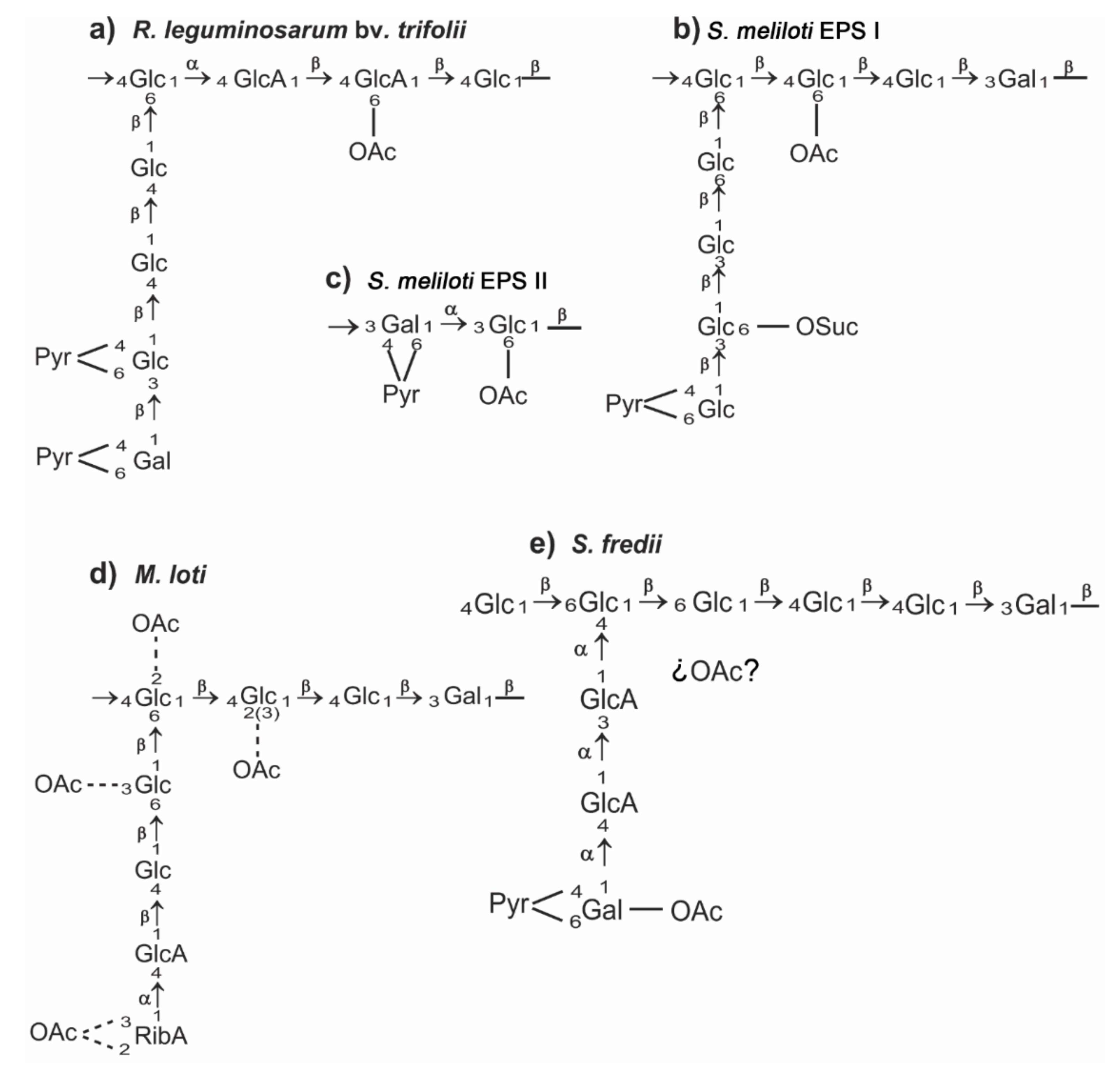
2. A General View of Rhizobial Exopolysaccharides

As mentioned above, EPS are acidic polysaccharides located out of the cell with little or no cell association [8]. These polysaccharides perform different functions, such as protection against environmental stresses, attachment to biotic and abiotic surfaces, and nutrient gathering [7,16]. During interactions with plants, diverse functions have been proposed for EPS from both pathogenic and symbiotic bacteria reviewed by [13], such as the sequestration of calcium ions, ROS (reactive oxygen species) scavenging, prevention of cellulose-mediated cell agglutination, tolerance to acidic pH, participation in biofilm formation, and host surface attachment. In addition, there is evidence of the role of EPS as a key signal for rhizobial infection in several rhizobia–legume couples as Rhizobium leguminosarum bv. trifolii/Trifolium spp., Sinorhizobium meliloti/Medicago sativa and Mesorhizobium loti/Lotus japonicus [7,16,17,18,19].
Rhizobial EPS are complex and high-molecular-mass heteropolymers whose structure varies at the species level. The repeating units are constituted of a variable number of hexose and uronic acid residues showing either alpha or beta glycosidic linkages. They can also be either linear or side-branched. In addition to sugars, several non-carbohydrate substituents can appear, mostly succinate, pyruvate, or acetate, which contribute to the acidic character of this polysaccharide [8,16,20]. Figure 1 shows the structure of the rhizobial EPS that will be treated in detail in this review. As it will be mentioned later, rhizobial EPS can be produced as either high- or low-molecular-mass forms (HMM and LMM).
Traditionally, the symbiotic relevance of rhizobial EPS has been related to the type of nodule ontogeny [16,17]. Thus, this polysaccharide appeared to be essential for indeterminate nodule-forming symbiosis, such as S. meliloti/M. sativa or R. leguminosarum bv. trifolii/Trifolium spp., but dispensable for determinate nodule-forming symbiosis, such as Sinorhizobium fredii/Glycine max and M. loti/L. japonicus. According to Stacey et al. [22] this difference may be due to the fact that the IT of indeterminate nodules are narrower than those of determinate nodules, and that the EPS-mediated matrix could be more important in the former ones.
In this review, we summarize the current knowledge about rhizobial EPS, focusing on the complex regulation of its production and on the symbiotic relevance of this polysaccharide. For the sake of clarity, we will restrict this review to four rhizobial species where EPS has been studied in more detail: R. leguminosarum, S. meliloti, S. fredii, and M. loti. Supplementary Material Table S1 lists all of the genes (including regulatory ones) involved in EPS production that are mentioned in this work. These studies will show that the symbiotic importance of rhizobial EPS depends more on the specific couple analysed than on the type of nodule, determinate or indeterminate, formed by the host legume.

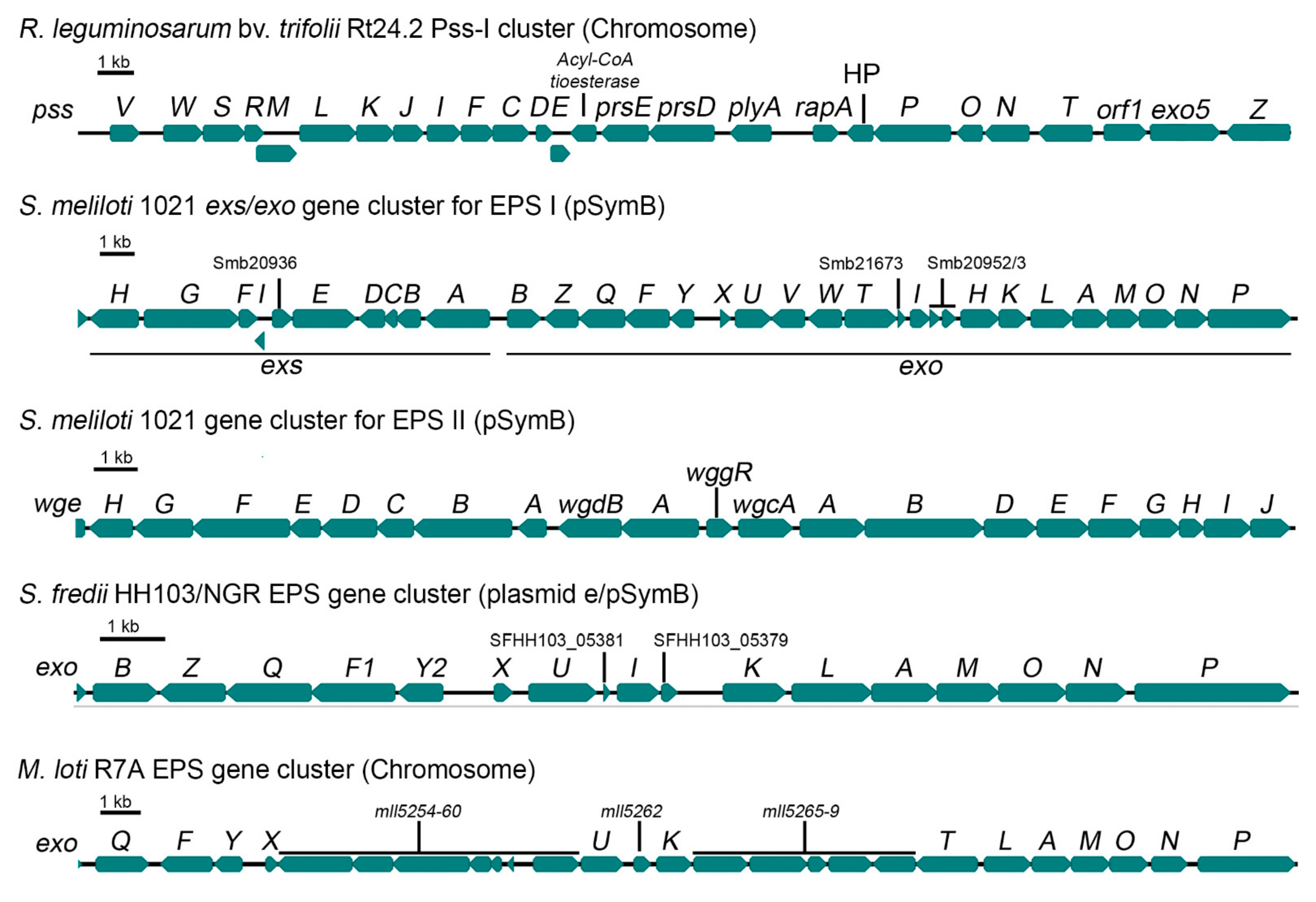
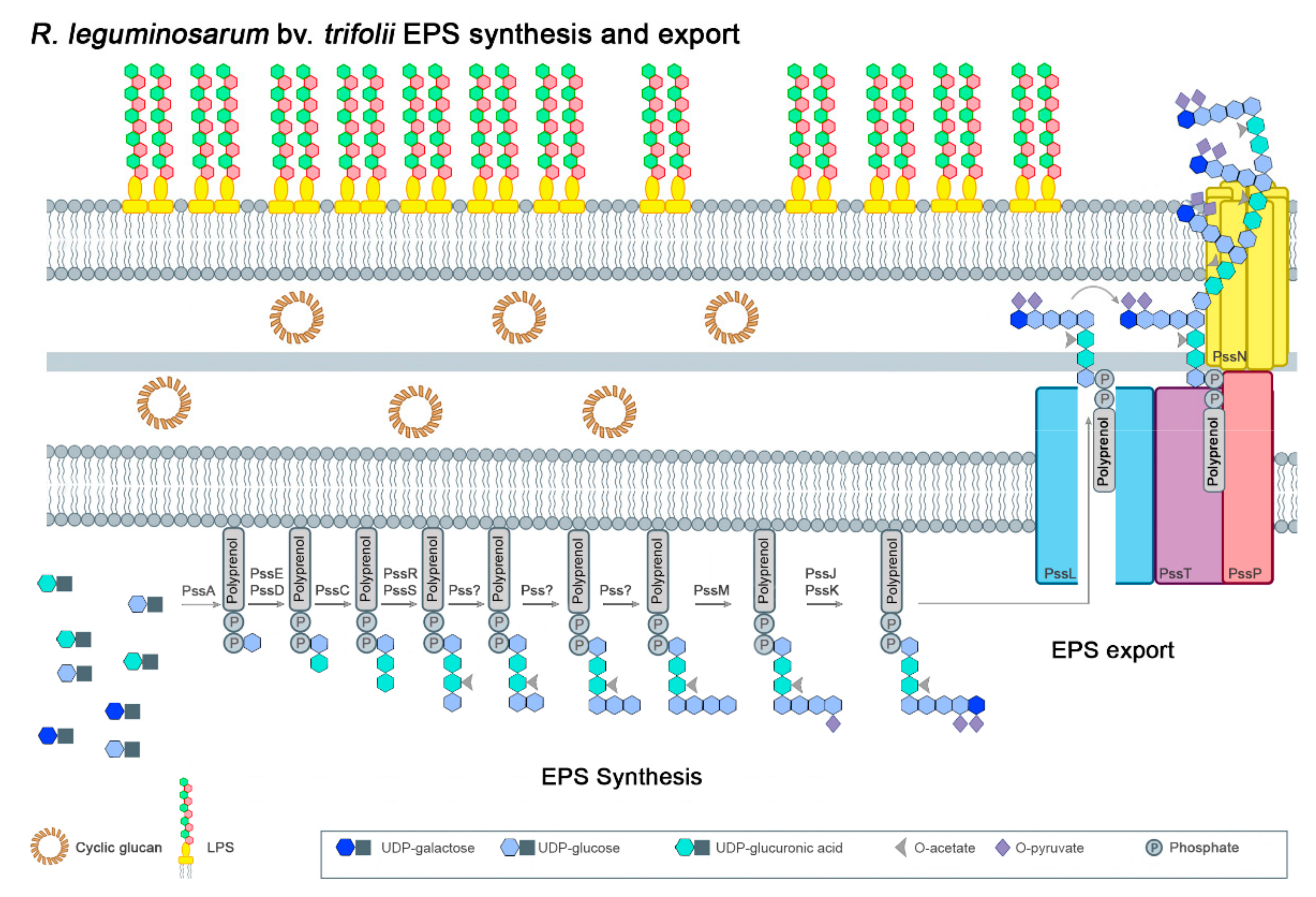
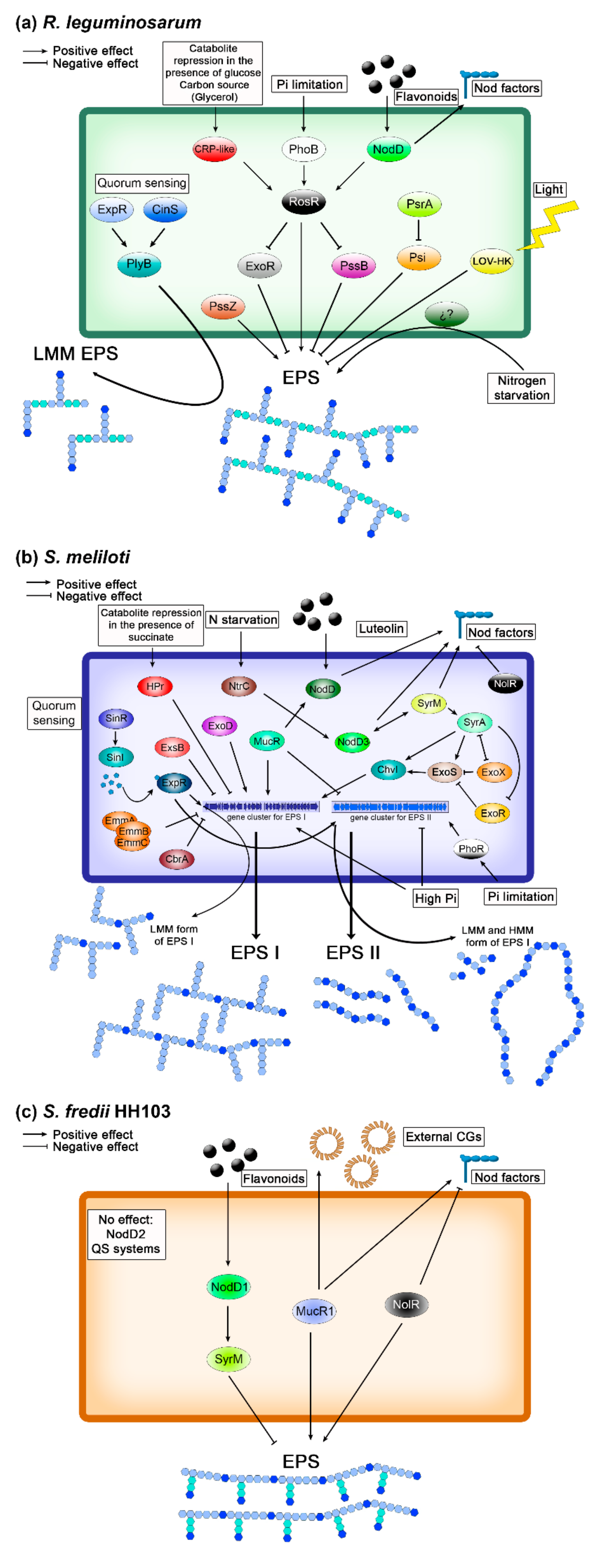
3. Rhizobium leguminosarum 

Rhizobium leguminosarum is a rhizobial species in which three biovars, trifolii, viciae and phaseoli, can be distinguished based on the type of legumes infected. Each of these biovars exhibit a narrow host-range. R. leguminosarum bv. trifolii strains establish symbiosis with Trifolium, bv. viciae with Pisum, Vicia, Lens, and Lathyrus, and bv. phaseoli with Phaseolus spp. plants. The symbiotic importance of EPS in R. leguminosarum has been related to the type of nodules formed by these host plants [6,7,23,24]. Trifolium, Pisum, Vicia, Lathyrus, Lens, belonging to the galegoid clade of subfamily Papilionoideae, form indeterminate nodules, whereas determinate nodules are formed by phaseolid legumes such as Phaseolus. It is generally accepted that, in general, the production of EPS is required for the effective symbiosis of R. leguminosarum trifolii and viciae strains with their host plants, which form indeterminate nodules, but not for bv. phaseoli strains, which establish symbiosis with bean plants forming determinate nodules.
Despite these differences in the symbiotic importance of EPS, R. leguminosarum strains produce EPS showing similar, but not identical, structures [16] In most R. leguminosarum bv. trifolii strains (such as Rt42.2, RBL5599, LPR5, etc.), this polymer consists of octasaccharide repeating units which contain D-glucose, D-glucuronic acid and D-galactose in a molar ratio 5:2:1, joined by β-1,3 and β-1,4 glycosidic bonds, and are modified by non-sugar (acetyl and pyruvyl) groups (Figure 1), but in other strains (such as 4S), the galactose residue is not present [16,25,26,27,28]. In R. leguminosarum bv. viciae, the EPS repeating unit is very similar to the R. leguminosarum bv. trifolii octasaccharide but including an additional D-glucuronic acid residue [16,28].
Similarly to other microorganisms, EPS synthesis in rhizobia is a complex process requiring the coordinated activity of many enzymatic and transporter proteins [29]. Genes involved in the synthesis and secretion of rhizobial EPS are usually grouped in large clusters located on chromosomes or megaplasmids [30,31,32,33]. In the case of R. leguminosarum, a great majority of genes engaged in EPS production are located on a large chromosomal region, called Pss-I [34,35,36,37], that encompasses nearly 30 genes. This region is highly conserved among all the so far sequenced R. leguminosarum genomes, although differences in the genetic organization, due to rearrangements, can be found at the strain level [31,34,35,38,39,40]. Figure 2 shows the Pss-I region from R. leguminosarum bv. trifolii Rt24.2. This clustering of genes involved in EPS synthesis most probably reflects their coordinated regulation of expression, including the influence of various environmental factors (light, root exudates, nitrogen and phosphate limitation, catabolite repression, stress conditions) [41,42,43,44,45,46]. The Pss-I region contains the exo5 gene, responsible for the synthesis of glucuronic acid (GlcA), genes encoding glycosyl transferases responsible for EPS subunit synthesis (pssCDE, pssJIFGH, pssS), genes involved in the addition of non-sugar modifications to EPS subunits (pssRMK), and genes involved in EPS assembly and secretion (pssTNOP, pssL) (Figure 2). The exo5 gene codes for a UDP-glucose dehydrogenase which converts UDP-glucose to UDP-GlcA, which, in turns, is the source for the synthesis of galacturonic acid (GalA). A mutant in this gene displays pleiotropic effects (i.e., changes in bacterial cell envelope and ineffective symbiosis), because its inability to synthesize uronic acids results in the absence of EPS and capsular polysaccharide (CPS) production and in the synthesis of an altered LPS that lacks GalA residues [14,47].
Some chromosomal genes crucial for EPS production, such as exoB and pssA, are located outside the Pss-I region. The exoB gene codes for a UDP-glucose 4-epimerase that synthesizes UDP-galactose, being a donor of this sugar in the synthesis of EPS and other galactose-containing polysaccharides [51,52]. An exoB mutant produces EPS lacking galactose residues and is unable to properly invade host roots (abnormal nodules inefficient in nitrogen fixation were formed on clover roots). Figure 3 shows a putative model for the different steps of EPS subunit assembly and export in R. leguminosarum bv. trifolii strains in which this subunit is an octasaccharide containing D-glucose, D-glucuronic acid and D-galactose in a molar ratio 5:2:1. The pssA gene is involved in the first step of the EPS subunit assembly. This gene encodes a glucosyl-isoprenylphosphate transferase that transfers glucose-1-phosphate from UDP-glucose to the lipid carrier [53]. Mutations in pssA totally abolish EPS synthesis, leading to the induction of empty (without bacteria), non-nitrogen-fixing root nodules on host plants (clover, pea and vetch) [44,54,55,56,57,58]. The transcription of pssA in both ex planta and in planta conditions is at a low level, indicating that the expression of this gene, which is crucial for EPS synthesis, is under very stringent regulation [44,59,60,61].
All pss genes involved in subsequent steps of the EPS subunit synthesis belong to the Pss-I region. The addition of two glucuronosyl residues to the growing subunit is performed by a glucuronosyl-β-1,4-glucosyltransferase and a glucuronosyl-β-1,4-glucuronosyltransferase encoded by pssDE and pssC, respectively [36,53,55,62]. Interestingly, a pssD mutant shows very similar phenotypic traits as the pssA mutant (lack of EPS synthesis and non-nitrogen-fixing nodules induced on clover and vetch roots). The fourth step of the unit assembly is most probably conducted by PssS, which is a unique enzyme among R. leguminosarum glycosyltransferases engaged in the EPS synthesis because it generates an exclusive α-1,4-glucosyl bound [35]. However, there is little information about glycosyl transferases involved in the subsequent steps of EPS synthesis. Based on the CAZy database (Carbohydrate Active Enzymes), some pss genes located in the Pss-I region are predicted to be engaged in this process (pssF, pssG, pssH, pssI) [63,64]. As recently evidenced, pssJ codes for a galactosyl transferase that is involved in the addition of the terminal sugar residue to the octasaccharide subunits [65]. A ΔpssJ deletion mutant produces EPS lacking terminal galactose in the side chain of the subunit (Figure 1). However, the lack of addition of this galactose residue does not block EPS polymerization.
Three pss genes from the Pss-I region are involved in non-sugar modifications of the EPS structure (Figure 3); pssR codes for a putative acetyl transferase, whereas pssM and pssK code for ketal pyruvate transferases [66]. Mutation of pssM resulted in the absence of pyruvyl groups in the EPS subunits (at the sub-terminal glucose) (Figure 1) and the induction of non-nitrogen-fixing nodules on the host plant: in these nodules, the normal invasion and release of bacteria into plant cells were observed, but their differentiation into bacteroids was essentially impaired, revealing the essentiality of the addition of pyruvyl residues for the symbiotic function of this polysaccharide.
The polymerization and secretion of EPS in R. leguminosarum are carried out by proteins encoded by the pssL and pssTNOP genes from the Pss-I cluster (Figure 3). Detailed information about these processes can be found in recent reviews [29,64]. Briefly, the polymerization of EPS subunits is performed by a Wzx/Wzy system that is composed of a Wzx-type translocase (flippase), a Wzy-type polysaccharide polymerase and a co-polymerase. In R. leguminosarum, PssL is most probably a Wzx translocase involved in the transfer of the EPS subunits across the inner membrane. This type of polysaccharide transporter proteins (Wzx translocases) is characterized by a specific topology containing numerous (10–14) predicted transmembrane (TM) segments. PssT, a Wzy-type protein containing 12 TMs, is the polysaccharide polymerase responsible for the polymerization of the EPS subunits [67]. PssP is another component of this system (PCP, polysaccharide co-polymerase) that is involved in the determination of the EPS chains length [68]. This protein has been classified as a bacterial tyrosine kinase [69,70,71]. PssP displays a significant identity to S. meliloti ExoP and other membrane-periplasmic auxiliary (OMA) proteins that are involved in the synthesis of HMM surface polysaccharides. Transport of EPS outside bacterial cells is conducted through a channel in the outer membrane formed by Wza-type proteins that are lipoproteins forming octameric α-helical channels spanning both inner and outer membranes [72]. In R. leguminosarum, PssN plays the role of this outer membrane translocase that is able to form homooligomeric structures and interact with PssP [73].
Other genes related to EPS synthesis are also located in the Pss-I region, such as prsDE (coding for components of a type 1 secretion system) and plyA (coding for a glycanase that cleaves EPS, affecting its processing) [35,62,74]. This secretion system shows an atypical broad substrate specificity, exporting at least 13 protein substrates (e.g., glycanases (PlyA, PlyB, and PlyC), rhizobial adhesion proteins (RapA2, RapB, and RapC), and the nodulation protein NodO) [75,76,77,78]. A prsD mutant synthesizes EPS of a higher polymerization degree than that of the wild-type strain and elicits a higher number of nodules incapable of fixing nitrogen [75].
Recently, the role in EPS synthesis of another gene belonging to the Pss-I region, pssZ, has been shown (Figure 2). This gene codes for a protein belonging to the bacterial serine/threonine protein phosphatases family, and its inactivation totally abolished EPS production in R. leguminosarum bv. trifolii [79]. Comparative transcriptomic analysis of the pssZ mutant and the wild-type strain identified a large number of genes differentially expressed in these two backgrounds, including genes related to several cellular processes (signalling, transcription regulation, synthesis of cell-surface polysaccharides, cell division and motility, bacterial metabolism) [61]. Interestingly, expression of almost all pss genes located in the Pss-I region was totally inhibited in the pssZ mutant. These data confirmed the important role of the serine/threonine protein phosphatase PssZ in EPS synthesis, most probably via its participation in a regulatory network [71]. In bacteria, in addition to two-component signalling systems, alternative regulatory pathways controlled by Hanks-type serine/threonine kinases and serine/threonine phosphatases also play an essential role in regulation of many processes, such as growth and cell division, cell-wall biogenesis, sporulation, biofilm formation, stress response, and metabolic and developmental processes, as well as either symbiotic or pathogenic interactions with higher host organisms [70,80]. In fact, the pssZ mutation resulted in pleiotropic effects in rhizobial cells: a lack of EPS production, decreased growth kinetics and motility, altered cell-surface properties, and failure to infect the host plant.
The global regulator RosR is involved in the positive regulation of EPS synthesis in R. leguminosarum. This protein is encoded by the chromosomal rosR gene that is not linked to the Pss-I region [81,82] and that is present in the genomes of all strains belonging to the three R. leguminosarum biovars and the closely related species R. etli and R. gallicum [83,84]. R. leguminosarum rosR also exhibits a significant similarity with A. tumefaciens ros [85], mucR of S. meliloti [86] and S. fredii [87,88], as well as with mucR of pathogenic Brucella abortus [89]. All these genes code for transcriptional regulators belonging to the α-proteobacteria Ros/MucR family of zinc-finger proteins which regulate the expression of genes required for the successful interactions of these bacteria with eukaryotic hosts. The R. leguminosarum bv. trifolii rosR mutant produces substantially less EPS (including a decreased LMM to HMM ratio) than the wild-type strain and establishes impaired symbiosis with clover plants [81,90]. In addition, alterations in the polysaccharide constituent of LPS, changes in membrane and secreted proteins, and decreased motility were observed in the rosR mutant [90,91]. On the other hand, additional rosR copies (similarly to multiple pssA copies) resulted in a nearly twofold increase of EPS synthesis and enhanced nodulation and symbiotic effectiveness [92]. Transcriptome profiling of the rosR mutant revealed the additional role of RosR in motility, synthesis of cell-surface components, carbon and nitrogen transport and metabolism, and other cellular processes [37]. Among a large group of genes (1106) differentially transcribed in the rosR mutant, a majority (63%) were up-regulated (suggesting that RosR functions mainly as a negative regulator), including prsD, rapA1 (autoaggregation protein), ndvA (CG transport protein to the periplasm), and genes of various transcriptional regulators (i.e., nitrogen regulatory protein P-II, phosphate regulatory system PhoBR, LacI, LuxR-type regulator RaiR). In contrast, several genes involved in EPS synthesis (pssA, pssC, pssI, and pssS) and cell motility (including the regulator rem and several genes coding for flagellar proteins) were down-regulated in the rosR mutant. This finding suggests the participation of RosR in a complex regulatory network. Regulation of rosR expression is very complex and involves four regulatory proteins: RosR, CRP, NodD, and PhoB, which modulate rosR transcription in response to various environmental factors (phosphate limitation, flavonoids, carbon source) [81,82,93] (Figure 4). A high rosR expression is ensured by the action of its strong promoter and two additional (upstream promoter and TGN-extended −10) elements [82,94]. Moreover, other sequence motifs (RosR-box bound by RosR, a LysR motif recognized by NodD, cAMP-CRP, PHO boxes recognized by PhoB, and numerous inverted repeats important for rosR mRNA stability) are engaged in the regulation of rosR expression [81,82,93,94]. Regarding the effect of the C source, a slight repression of the rosR gene is observed in the presence of glucose but not in that of glycerol [81].
Some environmental factors (root exudates, phosphate limitation, and nitrogen starvation) positively affect the transcription of pssA, pssB, pssO and pssP [60,95]. Flavonoids and NodD enhance EPS synthesis in R. leguminosarum through the activation of rosR [93], suggesting linked positive regulation of the synthesis of both symbiotic signals (EPS and Nod factors) [96], as also happens in S. meliloti.
There are different regulatory proteins (ExoR, Psi, PsrA, ExpR, and PssB) that have a negative effect on EPS production in R. leguminosarum. ExoR is an orthologue of S. meliloti ExoR and is involved in pssA repression [44,58]. An exoR mutant produces 3-fold more EPS than the wild-type strain and induces the formation of both effective and ineffective nodules on the host plant [97]. The pssB gene is located upstream of pssA and negatively affects EPS production, although its precise role in this process remains to be established. pssB encodes an inositol monophosphate phosphatase, and mutants in this gene produce more EPS than the wild-type R. leguminosarum bv. trifolii and viciae strains and induce non-nitrogen-fixing nodules on their host plants [82,98,99,100]. The psi (a polysaccharide inhibition) and psrA (a polysaccharide restoration) genes have been found on the symbiotic plasmid (pSym) of R. leguminosarum bv. phaseoli, near the nod-nif region required for nodulation and nitrogen fixation [101]. Interestingly, these genes are absent in R. leguminosarum bv. trifolii and viciae genomes [83], indicating that some regulators of EPS production are biovar-specific. An interesting effect was observed for psi, psrA, and pssA genes. Multiple psi copies inhibited EPS synthesis and abolished Phaseolus nodulation, whereas additional copies of psrA or pssA overcame this inhibitory effect, indicating that balanced numbers of these genes are required for proper EPS synthesis [59,101,102,103].
EPS synthesis in R. leguminosarum is also influenced by quorum sensing (QS) (Figure 4). So far, four N-acyl homoserine lactone (AHL)-based QS systems have been identified (cin, rai, rhi and tra) in this rhizobial species; they are involved in several processes (e.g., plasmid transfer, nodulation, and EPS production) [104]. The cin system, encompassing the cinR, cinI and cinS genes, is at the top of this hierarchical regulatory network and controls the induction of the remaining QS (rai, rhi, and tra) systems. CinR is a transcription regulatory protein inducing cinI expression in response to a signal molecule AHL, whereas CinI is responsible for the synthesis of specific AHL (N-(3-hydroxy-7-cis-tetradecenoyl)-L-homoserine lactone). CinS is a small protein with an anti-repressor function that induces the expression of the two regulatory genes of the rhi and rai systems (rhiR and raiR) and also that of plyB, a gene coding for a glycosyl hydrolase that degrades HMM EPS [105,106]. Another protein, the LuxR-type regulator ExpR, which functions independently of AHL, is also involved in QS and EPS production (it induces expression of raiR and plyB). This protein is an orthologue of S. meliloti ExpR, although it regulates the expression of a lower gene number than S. meliloti (among them only one gene to with EPS synthesis, plyB) [105,106].
In R. leguminosarum, light negatively influences exopolysaccharide synthesis as well as attachment and nodulation. This environmental factor is recognized by a histidine kinase photoreceptor containing a light, oxygen, and voltage (LOV)-domain [41]. In the absence of light, as well as in a strain carrying an inactivated LOV-histidine kinase gene, the production of EPS is significantly higher than in the wild-type strain under light conditions. However, this regulatory pathway is not common, since this LOV-histidine kinase photoreceptor gene is present only in some genomes of R. leguminosarum bv. viciae and trifolii strains (e.g., Rlv3841, RtWSM2304 and RtWSM1325).
EPS plays an important role in the adaptation of R. leguminosarum cells to various environmental stresses in their free-living stage, providing protection against desiccation, heavy metals, salinity, and nutrient limitation [43,46,107]. This polymer is also essential for biofilm formation on both abiotic surfaces and plant roots, being a major component of the biofilm matrix. The presence of Zn2+ ions positively affects EPS production and biofilm formation by R. leguminosarum cells, although the exact mechanism is still unknown. EPS-deficient mutants are significantly more sensitive to these stress factors in comparison to the wild-type strains. Regarding symbiosis, EPS is crucial for effective interactions of R. leguminosarum bv. trifolii and viciae strains with their hosts. It is required for the initiation and propagation of long IT and protection against H2O2 generated by the host during bacterial invasion [46,108]. All mutants of R. leguminosarum bv. trifolii and viciae that do not produce EPS have an identical phenotype: on compatible host plant roots these mutants only induce the formation of small nodule-like structures devoid of rhizobia that are ineffective in nitrogen fixation, confirming that EPS is indispensable for the proper interactions of these rhizobia with their macrosymbionts [35,44,54,55,56,57,58,62,79]. On the contrary, the overproduction of EPS by R. leguminosarum bv. trifolii strains enhances symbiotic competitiveness and nodulation in clover [92].

4. Sinorhizobium meliloti 

Sinorhizobium meliloti is a rhizobial species characterized by its narrow-host range for nodulation and is restricted to a few legume genera such as Trigonella, Medicago and Melilotus, reviewed by [109]. Despite this fact, the high amount of tools and resources that have been generated along the last 20 years in the study of the interaction between S. meliloti and Medicago truncatula has made this interaction a model system for the study of rhizobia–legume symbiosis, particularly of the symbiotic interactions forming indeterminate nodules [10].
The genomic sequence of S. meliloti 1021 became available in 2001 [110] and shows that this genome is tripartite and composed of a 3.65-Mb chromosome and the 1.35-Mb pSymA and 1.68-Mb pSymB megaplasmids. Nodulation genes are harboured by the pSymA, whereas genes involved in the production of symbiotically relevant polysaccharides are distributed among the chromosome and the pSymB. The expression of nod genes is complexly regulated and includes three different copies of NodD (NodD1, NodD2, NodD3), and another LysR regulator, SyrM [111]. NodD1 and NodD2 are constitutively expressed and activate the expression of nod genes in the presence of flavonoids (such as luteolin or methoxychalcone) and plant betaines (such as trigonelline and stachydrine), respectively, although NodD2 can also interact with methoxychalcone. NodD3 induces nod genes in the absence of external compounds. However, NodD3 expression is activated by SyrM, and vice versa, thus forming a self-amplifying circuit. In addition to NodD3, SyrM also activates the expression of syrA, whose encoded product is a small protein located in the cytoplasmic membrane and that, lacking an apparent DNA binding domain, mediates the transcriptional up-regulation of genes involved in the biosynthesis of one of the two forms of EPS present in S. meliloti: the succinoglycan (or EPS I) [112]. Thus, S. meliloti NodD3 connects the production of two different symbiotic signals: Nod factors and EPS I. The global regulator NolR is not present in 1021, but in other S. meliloti strains, such as Rm41, it represses NodD1 and NodD2 and, thus, Nod factors production [113].
S. meliloti strains produce two different kinds of EPS (Figure 1) [114,115]: EPS I or succinoglycan, whose repeating unit is a octasaccharide composed of seven residues of D-glucose and one residue of D-galactose joined by β-1,3, β-1,4 and β-1,6 glycosidic linkages, and EPS II or galactoglucan, composed of disaccharide repeating units containing D-glucose and D-galactose in a molar ratio 1:1 linked by α-1,3 and β-1,3 bonds. In both cases there are non-sugar substitutions: acetyl, pyruvyl and succinyl groups in EPS I, whereas in EPS II most of the glucosyl residues are 6-O-acetylated and all the galactosyl residues are substituted with 4,6-O-pyruvyl groups. Both kind of EPS can be produced in HMM and LMM fractions [116], but only the LMM forms of these polysaccharides are symbiotically active, as will be discussed later. EPS I is constitutively produced by S. meliloti 1021 in a complex medium, whereas the production of EPS II is stimulated by phosphate-limiting conditions [117].
The production of both types of EPS relies on two different gene clusters that are harboured by the pSymB (Figure 2), reviewed by [20,114,115,118]. The biosynthesis of EPS I is directed by a 27 kb region composed of 19 exo and 9 exs genes and affected by different regulators, whose encoding genes are located on different replicons, including the exo/exs cluster. The gene cluster responsible for the production of EPS II is 30 kb long and is constituted by 30 wgx (formerly exp) genes. Both exo/exs and wgx genes code for the different proteins required for the production of precursors, the synthesis and decoration of the repeating units, polymerization, and transport to the external medium. The chromosomal exoC gene codes for a phosphoglucomutase that is also required for the conversion of glucose-6-phosphate into glucose-1-phosphate, a precursor of EPS I.
The regulation of the production of both S. meliloti EPS is extremely complex, taking place at the transcriptional, post-transcriptional, and post-translational levels, and is subjected to external signals, such as plant flavonoids, abiotic stresses, and the level of nutrients such as nitrogen and phosphate (Figure 4) [7,115,119,120]. The primary regulatory circuit controlling exo gene expression is called RSI (for ExoR, ExoS, ChvI) and is composed of three elements: ExoR, the histidine kinase ExoS, and the response regulator ChvI, which are located in the periplasm, the membrane, and the cytoplasm, respectively [121,122,123]. ExoS activates ChvI through phosphorylation, which, in turns, activate exo gene expression but also the chromosomal exoR gene, which codes for a repressor that physically interacts with ExoS, inhibiting the ExoS/ChvI two-component signalling and, thus, EPS production. This tripartite system is also involved in the regulation of flagellum biosynthesis genes [121], as well as in other important cellular processes, such as carbon source utilization [124]. ChvI, when phosphorylated, represses motility. The chromosomal gene cbrA also represses EPS production but enhances motility [125]. Similarly, the tripartite EmmA/EmmB/EmmC system, also encoded by chromosomal genes, increases motility and decreases EPS production [126]. Thus, in S. meliloti there are different pathways that regulate bacterial motility and EPS production in opposite ways. Another negative regulator of EPS I production is encoded by exoX, which belongs to the exo/exs cluster [127]. The ExoX protein is a small, inner membrane-attached protein that presumably interacts with and inhibits ExoY, the protein responsible for the addition of the first sugar residue (UDP-Gal) of the octasaccharide repeating unit to the lipid carrier located in the inner membrane. The exsB gene, also located in the exo/exs cluster, codes for another negative regulator of EPS I production, which, most probably, acts at the post-transcriptional level [115,128]. S. meliloti EPS I production is also negatively regulated by a Succinate-Mediated Catabolite Repression that operates in the presence of succinate (the preferred carbon and energy source for this bacterium) and involves the participation of a phosphotransferase system (PTS) and the HPr protein [129].
EPS I production is positively influenced by two other proteins that also act as positive regulators of the synthesis of another symbiotic signal, Nod factors. On the one hand, SyrM, as mentioned before, is a positive activator of NodD3 and, consequently, enhances Nod factor synthesis and also activates the synthesis of SyrA that, in turns, stimulates EPS I production through the RSI circuit [111]. On the other hand, the global regulator MucR regulates differentially the production of EPS I and EPS II through the transcriptional activation of exo genes and the repression of the wgx cluster. MucR also stimulates Nod factor production and represses bacterial motility and genes involved in nitrogen fixation, thus acting as an enhancer of the first steps of the nodulation process [96,130]. Another chromosomal gene, exoD, has also a positive effect on EPS I production, although the exact mechanism remains to be elucidated [127].
Phosphate (Pi) levels have a clear influence in the production of EPS II by S. meliloti [115]. Thus, EPS II production is repressed in the presence of high Pi concentrations (above 10 mM, found in nodules), whereas low Pi concentrations, such as those typically found in natural environments (0.1 to 10 µM), induces the production of this polysaccharide. This regulation is achieved through the two component system formed by the sensor kinase PhoR and the response regulator PhoB, which, under Pi limitation, induces the expression of the wgx transcriptional activator WggR (that also shows autoinduction activity), whereas that, as mentioned above, MucR represses the expression of wgx genes (including that of wggR) under Pi sufficiency [130].
In S. meliloti, the Sin QS system is also involved in the regulation of EPS production [115,131]. This QS system is composed of the LuxR-type transcriptional regulator SinR, and SinI, an autoinducer synthase responsible for the production of different long-chain AHL, and, in conjunction with another LuxR-type regulator called ExpR, regulates many different processes, such as the production of EPS I and EPS II, motility, chemotaxis, nitrogen fixation, and metal transport [96,132]. The expression of sinI is SinR-dependent and enhanced by AHL-ExpR. Regarding EPS production, ExpR, when activated by the AHL produced by SinI, induces the biosynthesis of EPS II but also the production of LMM forms of EPS I through the induction of exsH, which codes for an endo-1,3-1,4-β-glycanase that depolymerizes HMM succinoglycan to produce symbiotically active LMM succinoglycan [133,134]. It is important to remark that one of the most-studied S. meliloti strains, 1021, lacks this QS-mediated regulation of EPS production because this strain harbours a disrupted copy of expR [110]. Because of this, most of the studies carried out analysing the role of S. meliloti ExpR in EPS production have been carried out in strain Rm8530 [96].
Interestingly, in S. meliloti there is an inverse correlation between EPS production and motility functions reviewed by [115]: three different but linked regulatory systems that enhance EPS production (MucR, and the ExoR/ExoS/ChvI and ExpR/Sin regulatory systems) also suppress motility, showing that both processes are linked by global regulatory pathways. In S. fredii HH103, MucR1 also positively regulates EPS production and negatively genes involved in motility [87].
As in R. leguminosarum bv. trifolii, EPS and LPS production in S. meliloti are connected through the exoB gene, which codes for a UDP-glucose 4-epimerase that is responsible for the production of galactose, a sugar that is present in both polysaccharides [135].
The work carried out in late 80 s and early 90s by the German group headed by Dr. Pühler demonstrated the essentiality of S. meliloti EPS for the successful infection of Medicago roots and invasion of nodules [136,137,138]. Following Tn5 mutagenesis, they identified different S. meliloti mutants that were unable to produce EPS and showed an Inf-phenotype with alfalfa. In addition, they identified many of the exo genes involved in EPS I production. In the model symbiosis S. meliloti/M. sativa, LMM forms of EPS are indispensable for a successful interaction, in such a way that the absence of EPS production leads to a lack of formation of IT, resulting in infection arrest [17]. In a very elegant work, it was showed that EPS I is more efficient than EPS II mediating both IT initiation and extension, and also that both kinds of EPS can be functionally replaced, although with lower efficiency, by another surface polysaccharide, KPS [139]. In fact, succinoglycan is the only form of S. meliloti EPS that can mediate the formation of IT on M. truncatula. KPS are typically formed by a disaccharide repeating unit, one of them being a 3-deoxy-D-manno-oct-2-ulosonic acid (Kdo) residue or a Kdo-derivative (Kdx). However, not all the S. meliloti strains produce a symbiotically active form of KPS, as in the case of S. meliloti 1021, which only contains a poly-Kdo that does not play any role in symbiosis [140]. Thus, this strain absolutely requires EPS, either succinoglycan or galactoglucan, for a successful interaction with alfalfa. In contrast, S. meliloti Rm41 exo mutants (such as AK631 and the exoB derivative of Rm41) are still able to induce the formation of nitrogen-fixing nodules on Medicago because this strain produces a biologically active KPS [8,141].
In addition to playing a key role in symbiotic signalling with the host plant, which most probably suppresses plant defence responses [142], a number of functional studies indicate that both S. meliloti EPS play a protective role against not only different abiotic stresses, such as detergents, salt, acidic pH, and heat [143] but also certain responses from the host plant, such as ROS that are present within IT [144] and, maybe, against antimicrobial peptides that are produced inside nodules of certain host legumes belonging to the IRLC (inverting-repeat lacking clade) such as Medicago [145]. S. meliloti EPS, especially EPS II, are also important for biofilm formation [146].

5. Sinorhizobium fredii 

The Sinorhizobium fredii species is constituted by fast-growing rhizobial strains with a broad host-range that usually, with exceptions such as NGR234, includes soybean (Glycine max) [147]. Among S. fredii soybean-nodulating strains, two groups can be distinguished for their ability (or not) to nodulate American commercial soybeans [148]. Thus, the most studied S. fredii strains vary in their symbiotic behaviour with soybeans. Strain NGR234 fails to nodulate both American and Asiatic soybeans [149], although recently it has been shown that this strain can effectively nodulate certain varieties of Glycine soja, the ancestor of soybeans [150]. Strains USDA257 and CCBAU 45436 [149,151] efficiently nodulated Asiatic varieties of soybean, such as Peking or Jing Dou 19, but failed to fix nitrogen on American soybeans such as Williams82. Strain HH103, instead, is able to induce the formation of nitrogen-fixing nodules on both Asiatic and American varieties of soybean [147]. The broad host range of HH103 and its ability to efficiently nodulate commercial varieties of soybean make this strain very attractive both for this study and its potential application as a legume inoculant. The HH103 complete genome sequence [152,153], as well as transcriptomic analyses of the effect of genistein in global gene expression in the wild-type strain and in mutants lacking the symbiotic regulators NodD1, TtsI, MucR1, or SyrM [87,154,155], have been published. These analyses have been complemented with studies on other symbiotic regulators, such as NolR or NodD2, as well as the analysis of the effect of inducer flavonoids on EPS production [156,157,158,159], and they will be commented on later.
Rodríguez-Navarro et al. [48] reported that the HH103 EPS structure is composed of D-glucose, D-galactose, D-GlcA, and pyruvic acid, in a 5:2:2:1 ratio and that it is partially acetylated (Figure 1). This structure is identical to that reported previously for strain NGR234 [49]. However, it is necessary to remark that an incorrect figure of the NGR234 EPS structure (in which the first GlcA residue is incorrectly linked to the main chain through a β-glycosidic linkage instead of the correct α one) has been published in later articles and reviews but was corrected in a recent review [8]. To our knowledge, S. fredii strains, in contrast to S. meliloti, produce one single type of EPS.
Genes responsible for EPS production in HH103 constitute a cluster located on the largest plasmid (pSfHH103e, 2096125-bp, Accession: NC_016815.1) and show the same organization (Figure 2) as that present in NGR234 [160]. This cluster includes genes involved in regulation (exoX), EPS polymerization and transport (exoF, exoP, exoQ, exoK), and structural genes coding for glucosyl or galactosyl transferases (exoA, exoL, exoM, exoO, exoU, exoY), an UDP-glucose 4-epimerase (exoB), an acetyl transferase (exoZ), and the protein responsible for the synthesis of UDP-glucose from glucose-1-phosphate (exoN). The S. fredii EPS and the S. meliloti EPS I are structurally very similar, but still there are some differences between them, such as the fact that the repeating unit is an octasaccharide in S. meliloti and a nonasaccharide in S. fredii, as well as the absence of succinyl decorations in S. fredii and GlcA residues in S. meliloti. Thus, although the organization of the genetic clusters responsible for the production of these two exopolysaccharides are pretty similar, there are 4 genes that are present in S. meliloti 1021 but absent in S. fredii strains: exoV (coding for a pyruvyl transferase), exoW (glycosyl transferase), exoT (Wzx-type transport protein), and exoH (succinyl transferase). The presence of GlcA in the S. fredii EPS is provided by the encoded product of rkpK, which belongs to the so-called rkp-2 region, and codes for a UDP-glucose 6-dehydrogenase [161]. Interestingly, the absence of GlcA in a HH103 rkpK mutant, as previously described for an R. leguminosarum exo5 derivative, results in the blocking of EPS molecule assembly and, thus, in an EPS-deficient phenotype [14,161]. The other gene that is present in the rkp-2 region, lpsL, codes for a UDP-glucuronate 4-epimerase which catalyzes the conversion of GlcA into GalA. Both uronic acids are present in the S. fredii HH103 LPS [162], which justifies the fact that HH103 mutants in either rkpK or lpsL exhibited altered forms of LPS. As mentioned before, the R. leguminosarum bv. trifolii orthologue of rkpK, exo5, is also involved in CPS, EPS, and LPS production since GlcA is present in CPS and EPS, whereas GalA is present in LPS [14,47]. Interestingly, S. meliloti Sm21 also harbour the rkp-2 region, since uronic acids are present in its LPS as well as, in contrast to HH103, in its K-antigen type capsular polysaccharide (KPS) [163]. However, the S. meliloti rkp-2 region does not influence the production of EPS, since neither EPS I nor EPS II harbour uronic acids. Thus, the rkpK (or exo5) and lpsL genes constitute a really good example for illustrating how the same genes can be required for the production of different surface polysaccharides in different rhizobia, but also how interconnected can be the synthesis of these biomolecules (Figure 5). To the best of our knowledge, the R. leguminosarum gene (s) involved in UDP-GlcA conversion into UDP-GalA has not been described yet.
The regulation of EPS production in S. fredii, although it remains less studied than in the closely related species S. meliloti, has been shown to be directly connected to nod regulon (Figure 4). In S. fredii HH103, the presence of flavonoids able to induce nod genes led to a full repression of EPS production, as determined by NMR (nuclear magnetic resonance) experiments, this process being dependent on the symbiotic regulator NodD1, which positively regulates Nod factor production and the symbiotic type 3 secretion system, T3SS [159]. To our knowledge, this is the only case of EPS repression mediated by inducing flavonoids described in rhizobia so far. In fact, this kind of repression does not take place in other S. fredii strains where it has been analysed, such as NGR234 or USDA257 [159]. Previous works showed that, in S. fredii USDA193, genistein provoked changes in EPS production [164], but these changes were qualitative rather than quantitative, and it was not established whether they depend on NodD or not. Interestingly, the global repressor NolR, which negatively regulates the production of Nod factors and the symbiotic T3SS, is a positive regulator of EPS synthesis in HH103, since the lack of NolR results in a clear reduction of EPS production, which is almost complete even in the absence of flavonoids [157]. Thus, EPS production in S. fredii is connected to the nod regulon and modulated in a completely opposite way to that of Nod factors and the symbiotic T3SS. qPCR studies showed that two crucial genes for EPS production, exoY2 (coding for the protein that should mediate the addition of the first sugar to the lipid carrier) and exoK (a glucanase that could be involved in the cleavage of HMM to LMM forms of EPS) are strongly repressed when genistein and NodD1 are present and also when nolR is inactivated [159]. Recent studies have shown that S. fredii SyrM, whose orthologue in S. meliloti is involved in the activation of EPS production, participates in this genistein-mediated repression, although the mechanism remains obscure [155]. In clear contrast, flavonoids enhance EPS production in S. meliloti 1021 in a process mediated by SyrM through the SyrA protein [111]. In R. leguminosarum bv. trifolii 24.2, the presence of flavonoids has also been shown to enhance EPS production, in this case through the RosR transcriptional regulator [93]. In S. fredii HH103 and CCBAU45536, MucR1, an orthologue of RosR, is also a positive regulator of EPS production [87,88], but its effect is not related to flavonoids and cannot counterpart the presence of this phenolic compounds in strain HH103. Transcriptomic studies performed in HH103 revealed that, among genes involved in EPS synthesis, MucR only affects the expression of an operon constituted by exoY2, exoF1 (whose products are responsible for the addition of the first sugar residue, UDP-Gal, to the lipid carrier located in the plasmatic membrane) and exoQ (involved in EPS polymerization). Curiously, in the absence of flavonoids, the HH103 MucR1 protein is also an activator of different genes, most of unknown function, belonging to the nod regulon. This is a new indication of how different HH103 regulators (NolR, MucR1, SyrM) connect the production of EPS and the expression of at least part of the genes constituting the nod regulon. The production of S. fredii HH103 EPS is also somehow interconnected with that of CG [15,87]. Thus, the inactivation of the HH103 gene coding for the CG synthase (cgs) led to an increased transcription of exoA and the production of EPS, whereas the mutation of exoA not only resulted in the abolition of EPS synthesis but also in an increased production of external CG. The MucR1 regulator is clearly involved in this connection, since its inactivation negatively affects exo gene expression but also that of ndvA, which codes for the transporter involved in the transfer of CG from the cytoplasm to the periplasm [87].
In contrast to that found in R. leguminosarum and S. meliloti, S. fredii HH103 EPS production is not influenced by QS mechanisms since this strain lacks a functional copy of the expR gene [165]. However, the two QS systems identified in strain NGR234 have a positive effect on EPS production and repress genes involved in motility and chemotaxis [166].
The S. fredii NGR234 EPS has revealed to be indispensable for the formation of nitrogen-fixing nodules with several host-plants, such as Albizia lebbeck and Leucaena leucocephala, but the lack of this polysaccharide did not affect symbiosis with other hosts, such as cowpea (Vigna unguiculata). Interestingly, the inactivation of the NGR234 exoK gene, coding for a glycanase, resulted in a lack of production of EPS-derived oligosaccharides and in the same symbiotic phenotype as EPS- mutants, indicating that, as described previously for S. meliloti, these NGR234 exo-oligosaccharides are crucial for symbiosis with certain host plants [167]. The symbiotic role of HH103 EPS has been studied in different host plants, either forming determinate or indeterminate nodules, and in no case has this polysaccharide has proven to be crucial. In determinate-nodule-forming legumes, the absence of HH103 EPS has different effects in symbiosis depending on the host legume: positive with soybean (slight increase in plant-top dry weight and significant increase in competitiveness ability), neutral with Lotus burttii (no differences in plant development) or negative with cowpea (slight decrease in plant-top dry weight and significant decrease in competitiveness ability) [48,168,169,170]. The fact that EPS appears to be slightly detrimental for the symbiosis of HH103 with its natural host, soybean, opens the question of whether EPS repression through nod-gene-inducing flavonoids is a mechanism for optimizing symbiosis with this legume. Regarding indeterminate-nodule-forming legumes, the HH103 exoA mutant induces the formation of nitrogen-fixing nodules with a very similar performance to those of the parental strain, on pigeon-pea (Cajanus cajan) and Glycyrrhiza uralensis [168,171]. The last legume belongs, as Medicago, to the IRLC clade [172]. In S. meliloti, EPS can be functionally replaced by KPS for effective symbiosis with alfalfa [139]. However, a double exoA rkpH mutant (producing no EPS nor KPS), although negatively affected when compared to HH103, is still able to induce the formation of nitrogen-fixing nodules on both pigeon pea and G. uralensis [168,171]. This fact is a good example of how “rules” (strict requirement of either EPS or KPS for effective symbiosis with an indeterminate nodule-forming legume) extracted for a model symbiotic system (S. meliloti/alfalfa) cannot always be applied to other symbiotic relations.
In free-living HH103 cells, EPS has proven to be essential for biofilm formation on abiotic surfaces [48,159], but it is not implied in attachment to soybean roots. In fact, and in contrast to Bradyrhizobium diazoefficiens USDA110, HH103 EPS (as in all S. fredii strains described so far) is unable to bind to soybean lectin. Regarding other physiological traits, the absence of EPS impairs the low surface motility exhibited by S. fredii HH103 and negatively affects tolerance to osmotic stress (either 50–100 mM NaCl or 5–10% sucrose m/v) but has no effect on sensitivity to the oxidizing agents H2O2 or paraquat [48].

6. Mesorhizobium loti 

The Mesorhizobium loti R7A and MAFF303099 strains, recently reclassified as M. japonicum [173], were isolated from Lotus corniculatus and Lotus japonicus nodules, respectively [174,175]. Both strains have been extensively studied since they are able to induce nitrogen-fixing nodules on the model legume plant L. japonicus Gifu [176,177]. The M. loti MAFF303099 and R7A genomes have been sequenced [178,179]; in both strains, the symbiotic genes are grouped in the symbiosis island, a mobilizable 500 kb DNA region able to make a non-symbiotic mesorhizobia into a Lotus symbiont. The symbiosis islands of these strains share a conserved backbone sequence of 248 kb with about 98% DNA sequence identity [180]. Interestingly, a comparison of the genomes of these two strains has shown that they have different secretion systems to introduce protein effectors into the plant cells. While a T4SS is present in R7A, the MAFF303099 strain possesses a T3SS, albeit both secretion systems seem to be involved in the determination of the host range and would be functionally exchangeable [180,181].
As has been described by Kelly et al. [50], M. loti R7A produces both HMM and LMM forms of EPS. The repeating subunit consists of a highly O-acetylated octosaccharide composed of D-glucose, D-galactose, D-GlcA and D-riburonic acid in a 5:1:1:1 ratio (Figure 1). The presence of the riburonic acid as a terminal residue has only previously been reported in S. meliloti IFO13336, but in the case of this strain, it is α-d-Rib and not β-d-Rib as occurs in R7A [21].
The genes responsible for EPS production in M. loti R7A and MAFF303099 are clustered in the chromosome (Figure 2), similarly as in R. leguminosarum strains. The cluster exoQFYXUKTLAMONP (from mlr5249 to mlr5276) genes is present in both strains, whereas other genes that may be involved in EPS biosynthesis, such as possible orthologues of exoI (mll0560, mll8119 and mlr0479), exoZ (mlr6758 and mlr8032) and exoB (mll7878) are dispersed in the genome [178]. Although the acetylation of the M. loti R7A EPS was found in three out of six possible sites, the gene responsible for this substitution remains to be elucidated since, as mentioned, there are two different genes showing high identity to the S. meliloti 1021 exoZ gene, mlr6758 and mlr8032 [21].
To our knowledge, specific studies designed to analyse the regulation of the M. loti EPS have not been published. In contrast, different studies have been focused on the symbiotic role of this polysaccharide in different interactions, such as those of M. loti NZP2037 with Leucaena leucocephala and Lotus pedunculatus [182], M. loti MAFF303099 with L. japonicus B-129 [183], and M. loti R7A with L. corniculatus and L. japonicus Gifu [50]. In the case of M. loti NZP2037, the symbiotic phenotype of EPS-deficient mutants depended on the host plant: these mutants induced effective nodules with L. pedunculatus (a determinate-nodule-forming legume) but empty nodules unable to fix nitrogen with L. leucocephala (which forms indeterminate nodules). In the case of M. loti MAFF303099, mutants unable to produce EPS showed the same symbiotic phenotype as the wild-type strain in symbiosis with L. japonicus B-129. The most recent studies regarding the symbiotic relevance of M. loti EPS have been carried out on the R7A strain. In that strain, the symbiotic response with L. japonicus Gifu and L. corniculatus plants differs depending on the EPS biosynthesis step affected by the gene mutation. While those mutants affected in the early steps of the EPS production (such as exoA and exoB) were able to form effective nodules (although in the case of the exoA mutant, the number of nitrogen-fixing nodules induced on L. japonicus Gifu was clearly reduced), the mutants affected in the middle to late steps of EPS synthesis were clearly impaired on both plants with more or less severity depending on the mutant tested (exoO, exoU, exoK, mlr5265 and mlr5266) [50]. Based on these results, the M. loti EPS would act as a positive signal to modulate the plant response and carry out a normal rhizobial invasion (IT formation and bacterial release). Bacterial mutants that are affected at the early steps of the EPS production lacked this polysaccharide but were able to avoid the defence responses and induce effective nodules although with a delay. The more severe symbiotic impairment phenotypes were shown by bacterial mutants producing a truncated EPS that may be recognised as a negative signal that would activate plant defence responses and, therefore, disrupt IT progression [50].
The molecular basis of the results commented above was supported by the recent finding of the L. japonicus Gifu transmembrane LysM receptor kinase, EPR3. This receptor is able to recognise and bind monomeric EPS molecules, thus monitoring the bacterial EPS status during the colonization and infection processes [18]. The recognition of the M. loti wild-type EPS by the EPR3 receptor acts as a positive signal allowing epidermal infection. However, the truncated EPS form (a penta-glycan) produced by a M. loti exoU mutant is recognised as a non-compatible EPS, and this negative signal results in the arrest of IT formation and the absence of the formation of nitrogen-fixing nodules. In the case of an exoB mutant that does not produce EPS, the absence of a negative signal allows infection and the development of nitrogen-fixing nodules but to a lower extent than in the case of the wild-type strain because of the lack of the positive signal (recognition of the appropriate EPS by EPR3).
Thus, the presence of this EPS receptor establishes a double checkpoint in the infection process of M. loti with L. japonicus Gifu plants. The M. loti R7A Nod factors are recognised by the NFR1 and NFR5 receptors and trigger the symbiotic signal transduction leading to the early steps of infection and nodule devolvement [184,185]. At the first steps of this signal cascade, the epr3 gene is expressed, giving rise to EPS monitoring on the root infection zone and controlling the epidermal infection [18]. Recent results show that EPS3 is also expressed at the root cortex and nodule primordia, suggesting that EPS recognition is reiterated during symbiotic interaction, and it is also involved in intercellular cortical infection [19]. Recently, it has been shown that EPR3-like receptors, which present a structure that is quite different from that of LysM receptors involved in chitooligosaccharide (NF) perception, are ubiquitous in plants, suggesting that they might be surveillance receptors monitoring carbohydrates from different microbes associated with plant roots [186]. However, in the indeterminate-nodule-forming-legume M. truncatula, the orthologue of eps3 (Mtlyk10), although playing a relevant role in symbiosis, appears to be uninvolved in the perception of the S. meliloti EPS I [187].

7. Conclusions and Perspectives

In this review we have tried to present the current knowledge about one of the most studied rhizobial surface polysaccharides, the EPS. Currently, we can point out the following relevant facts:
(1)
Rhizobial EPS production has been found to be extremely complex in all the rhizobial strains in which this issue has been addressed. Usually, it involves the participation of different regulatory systems, either positive or negative ones, including the participation of global regulatory proteins and, in some cases, that of QS systems.
(2)
In many rhizobia-legume symbioses, EPS is absolutely essential or very important for a successful interaction. This is especially true in symbioses forming indeterminate nodules, although the interactions between S. fredii HH103 and G. uralensis and C. cajan are a clear exception. Also, the fact that EPS plays a very important role in the success of rhizobial infection in the M. loti/L. japonicus couple, which forms determinate nodules, makes it difficult to formulate a general rule. Interestingly, EPS appears to be crucial for the symbiotic performance of several rhizobia exhibiting a narrow host range (such as S. meliloti, R. leguminosarum, and M. loti).
(3)
In some rhizobia, EPS production is connected with the nod regulon, but the type of connection (positive or negative) varies depending on the rhizobial strain. Thus, in S. meliloti and R. leguminosarum bv. trifolii, appropriate flavonoids induce Nod factor production and stimulates EPS biosynthesis, which is enhanced in parallel to that of Nod factors, whereas in S. fredii HH103, appropriate flavonoids induce Nod factor production but suppress EPS biosynthesis.
(4)
Rhizobial EPS, in addition to being crucial for biofilm formation and for passive protection against abiotic stresses, may play a crucial role as a molecular signal required for suppressing plant defence responses and, thus, allowing a successful infection process. There is clear evidence that, at least in S. meliloti, R. leguminosarum bv. trifolii and S. fredii NGR234, this signalling function is carried out by oligosaccharides derived from EPS. The recent discovery of the Lotus japonicus EPR3 receptor as a detector of the presence of the “correct” oligosaccharides for granting a successful infection process supports this idea. However, in this context, it is surprising that S. fredii HH103, a broad host-range rhizobial strain, represses the production of its EPS in the presence of any nod genes inducing flavonoids and, thus, in the early steps of its symbiotic interactions with any potential host legume. Might this repression be a strategy for nodulating potential host legumes that would stop infection when they perceive the HH103 EPS to inadequate? Clearly, more research is required to elucidate this issue.
(5)
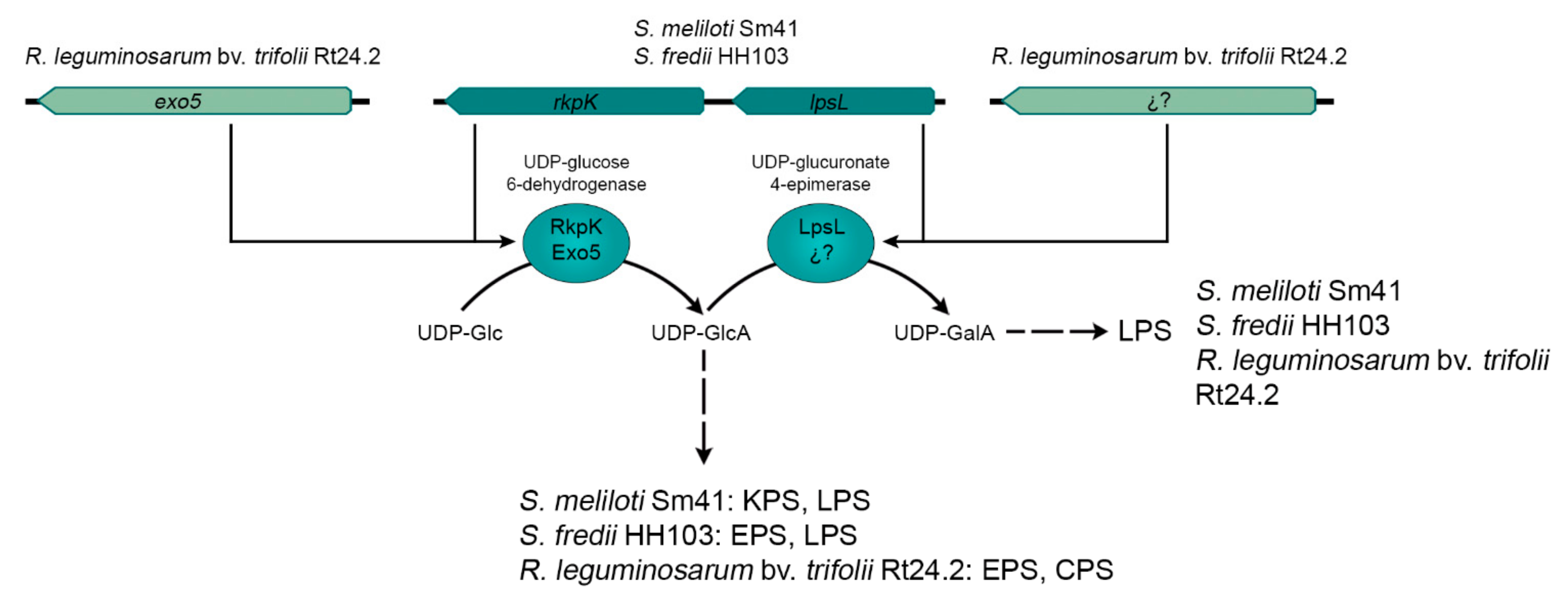
Rhizobial EPS production may be interconnected with the biosynthesis of other symbiotic relevant surface polysaccharides. For example, in R. leguminosarum bv. trifolii and S. fredii, the synthesis of EPS and LPS are interconnected through the rkp-2 region, whereas in S. meliloti, this genetic region participates in the production of KPS and LPS. At least in S. fredii, the production of EPS and extracellular CG is simultaneously controlled by the MucR1 regulator.
(6)
Rhizobial EPS symbiotic function can overlap with that of other surface polysaccharides, such KPS in the case of the interaction between S. meliloti and Medicago. However, this is not a general rule, as S. fredii mutants lacking EPS and KPS can still induce the formation of nitrogen-fixing nodules on G. uralensis and C. cajan.
In summary, EPS is a symbiotically relevant surface polysaccharide in most of the rhizobia–legume symbiotic interactions studied so far, and its production is complexly regulated together with that of other bacterial relevant characteristics, such as motility. However, the classical belief stating that EPS are specifically essential in interactions with indeterminate-nodule-forming legumes is not fully true, as demonstrated by the fact that they can also be highly important in determinate-nodule-forming interactions (such as M. loti/L. japonicus) and dispensable in the symbiosis between S. fredii and G. uralensis, which forms indeterminate nodules. Thus, in our opinion, the symbiotic importance of EPS cannot be predicted but must be analysed in every single rhizobial–legume interaction.

Supplementary Materials

Author Contributions

All authors (S.A.-J., F.F.-R., J.-E.R.-S., M.J., J.-M.V.) contributed to the manuscript writing and figures design. All authors have read and agreed to the published version of the manuscript.

Funding

This research was funded by the Polish National Science Centre, grant number 2018/31/B/NZ9/00663 and by the project PID2019-107634RB-I00 from the Spanish “Ministerio de Ciencia, Innovación y Universidades”.

Institutional Review Board Statement

Not applicable.

Informed Consent Statement

Not applicable.

Conflicts of Interest

The authors declare no conflict of interest.

Abbreviations

AHLN-acyl-homoserine lactone(s)
CGCyclic glucans
CPSCapsular polysaccharide
EPSExopolysaccharide
GlcAGlucuronic acid
GalAGalacturonic acid
HMMHigh molecular mass
IRLCInverting repeat-lacking clade
ITInfection thread(s)
KPSK-antigen capsular polysaccharide
LMMLow molecular mass
LOV-domainLight, oxygen, and voltage domain
LPSLipopolysaccharide
QSQuorum sensing
TMTransmembrane

References

  1. Poole, P.; Ramachandran, V.; Terpolilli, J. Rhizobia: From saprophytes to endosymbionts. Nat. Rev. Microbiol. 2018, 16, 291–303. [Google Scholar] [CrossRef] [PubMed]
  2. Tsyganova, A.V.; Kitaeva, A.B.; Tsyganov, V.E. Cell differentiation in nitrogen-fixing nodules hosting symbiosomes. Funct. Plant Biol. 2017, 45, 47–57. [Google Scholar] [CrossRef] [PubMed]
  3. Coba de la Peña, T.; Fedorova, E.; Pueyo, J.J.; Lucas, M.M. The symbiosome: Legume and rhizobia co-evolution toward a nitrogen-fixing organelle? Front. Plant Sci. 2018, 8, 2229. [Google Scholar] [CrossRef] [PubMed] [Green Version]
  4. Oldroyd, G.E.D.; Dixon, R. Biotechnological solutions to the nitrogen problem. Curr. Opin. Biotechnol. 2014, 26, 19–24. [Google Scholar] [CrossRef] [Green Version]
  5. Santos, M.S.; Nogueira, M.A.; Hungria, M. Microbial inoculants: Reviewing the past, discussing the present and previewing an outstanding future for the use of beneficial bacteria in agriculture. AMB Express 2019, 9, 205. [Google Scholar] [CrossRef]
  6. Walker, L.; Lagunas, B.; Gifford, M.L. Determinants of host range specificity in legume-rhizobia symbiosis. Front. Microbiol. 2020, 11, 3028. [Google Scholar] [CrossRef]
  7. Janczarek, M.; Rachwał, K.; Marzec, A.; Grzadziel, J.; Palusińska-Szysz, M. Signal molecules and cell-surface components involved in early stages of the legume-rhizobium interactions. Appl. Soil Ecol. 2015, 85, 94–113. [Google Scholar] [CrossRef]
  8. López-Baena, F.J.; Ruiz-Sainz, J.E.; Rodríguez-Carvajal, M.A.; Vinardell, J.M. Bacterial molecular signals in the Sinorhizobium fredii-soybean symbiosis. Int. J. Mol. Sci. 2016, 17, 755. [Google Scholar] [CrossRef] [Green Version]
  9. Oldroyd, G.E. Speak, friend, and enter: Signalling systems that promote beneficial symbiotic associations in plants. Nat. Rev. Microbiol. 2013, 11, 252–263. [Google Scholar] [CrossRef]
  10. Roy, S.; Liu, W.; Nandety, R.S.; Crook, A.; Mysore, K.S.; Pislariu, C.I.; Frugoli, J.; Dickstein, R.; Udvardi, M.K. Celebrating 20 Years of Genetic Discoveries in Legume Nodulation and Symbiotic Nitrogen Fixation. Plant Cell 2020, 32, 15–41. [Google Scholar] [CrossRef] [Green Version]
  11. Staehelin, C.; Krishnan, H.B. Nodulation outer proteins: Double-edged swords of symbiotic rhizobia. Biochem. J. 2015, 470, 263–274. [Google Scholar] [CrossRef] [Green Version]
  12. Downie, J.A. The roles of extracellular proteins, polysaccharides and signals in the interactions of rhizobia with legume roots. FEMS Microbiol. Rev. 2010, 34, 150–170. [Google Scholar] [CrossRef]
  13. Wanke, A.; Malisic, M.; Wawra, S.; Zuccaro, A. Unraveling the sugar code: The role of microbial extracellular glycans in plant-microbe interactions. J. Exp. Bot. 2021, 72, 15–35. [Google Scholar] [CrossRef]
  14. Laus, M.C.; Logman, T.J.; van Brussel, A.A.N.; Carlson, R.W.; Azadi, P.; Gao, M.; Kijne, J.W. Involvement of exo5 in production of surface polysaccharides in Rhizobium leguminosarum and its role in nodulation of Vicia sativa subsp. nigra. J. Bacteriol. 2004, 186, 6617–6625. [Google Scholar] [CrossRef] [Green Version]
  15. Crespo-Rivas, J.C.; Margaret, I.; Hidalgo, A.; Buendía-Clavería, A.M.; Ollero, F.J.; López-Baena, F.J.; del Socorro Murdoch, P.; Rodríguez-Carvajal, M.A.; Soria-Díaz, M.E.; Reguera, M.; et al. Sinorhizobium fredii HH103 cgs mutants are unable to nodulate determinate- and indeterminate nodule-forming legumes and overproduce an altered EPS. Mol. Plant Microbe Interact. 2009, 22, 575–588. [Google Scholar] [CrossRef] [Green Version]
  16. Fraysse, N.; Couderc, F.; Poinsot, V. Surface polysaccharide involvement in establishing the rhizobium-legume symbiosis. Eur. J. Biochem. 2003, 270, 1365–1380. [Google Scholar] [CrossRef]
  17. Jones, K.M.; Kobayashi, H.; Davies, B.W.; Taga, M.E.; Walker, G.C. How rhizobial symbionts invade plants: The Sinorhizobium-Medicago model. Nat. Rev. Microbiol. 2007, 5, 619–633. [Google Scholar] [CrossRef] [Green Version]
  18. Kawaharada, Y.; Kelly, S.; Nielsen, M.W.; Hjuler, C.T.; Gysel, K.; Muszynski, A.; Carlson, R.W.; Thygesen, M.B.; Sandal, N.; Asmussen, M.H.; et al. Receptor-mediated exopolysaccharide perception controls bacterial infection. Nature 2015, 523, 308–312. [Google Scholar] [CrossRef] [Green Version]
  19. Kawaharada, Y.; Nielsen, M.; Kelly, S.; James, E.; Andersen, K.; Rasmussen, S.; Füchtbauer, W.; Madsen, L.; Heckmann, A.; Radutoiu, S.; et al. Differential regulation of the Epr3 receptor coordinates membrane-restricted rhizobial colonization of root nodule primordia. Nat. Commun. 2017, 8, 14534–14545. [Google Scholar] [CrossRef]
  20. Becker, A.; Pühler, A. Production of exopolysaccharides. In The Rhizobiaceae. Molecular Biology of Model Plant-Associated Bacteria; Spaink, H.P., Kondorosi, A., Hooykaas, J.J., Eds.; Springer: Dordrecht, The Netherlands, 1998; pp. 97–118. [Google Scholar] [CrossRef]
  21. Muszyński, A.; Heiss, C.; Hjuler, C.T.; Sullivan, J.T.; Kelly, S.J.; Thygesen, M.B.; Stougaard, J.; Azadi, P.; Carlson, R.W.; Ronson, C.W. Structures of exopolysaccharides involved in receptor-mediated perception of Mesorhizobium loti by Lotus japonicus. J. Biol. Chem. 2016, 291, 20946–20961. [Google Scholar] [CrossRef] [Green Version]
  22. Stacey, G.; So, J.S.; Roth, L.E.; Lakshmi, S.K.B.; Carlson, R.W. A lipopolysaccharide mutant of Bradyrhizobium japonicum that uncouples plant from bacterial differentiation. Mol. Plant Microbe Interact. 1991, 4, 332–340. [Google Scholar] [CrossRef] [PubMed]
  23. Maroti, G.; Kondorosi, E. Nitrogen-fixing Rhizobium-legume symbiosis: Are polyploidy and host peptide-governed symbiont differentiation general principles of endosymbiosis? Front. Microbiol. 2014, 5, 326. [Google Scholar] [CrossRef] [PubMed] [Green Version]
  24. Ferguson, B.J.; Indrasumunar, A.; Hayashi, S.; Lin, M.H.; Lin, Y.H.; Reid, D.E.; Gresshoff, P.M. Molecular analysis of legume nodule development and autoregulation. J. Integr. Plant Biol. 2010, 52, 61–76. [Google Scholar] [CrossRef] [PubMed]
  25. Robertson, B.K.; Aman, P.; Darvill, A.G.; McNeil, M.; Albersheim, P. Host-symbiont interactions. The structure of acidic extracellular polysaccharides secreted by Rhizobium leguminosarum and Rhizobium trifolii. Plant Physiol. 1981, 67, 389–400. [Google Scholar] [CrossRef] [Green Version]
  26. Breedveld, M.W.; Canter Cremers, H.C.J.; Batley, M.; Posthumus, M.A.; Zevenhuizen, L.P.T.M.; Wijfelman, C.A.; Zehnder, A.J.B. Polysaccharide synthesis in relation to nodulation behaviour of Rhizobium leguminosarum. J. Bacteriol. 1993, 175, 750–757. [Google Scholar] [CrossRef] [Green Version]
  27. McNeil, M.; Darvill, J.; Darvill, A.; Albersheim, P.; van Veen, R.; Hooykaas, P.; Schilperoort, R.; Dell, A. The discernible structural features of the acidic exopolysaccharides secreted by different Rhizobium species are the same. Carbohydr. Res. 1986, 146, 307–326. [Google Scholar] [CrossRef]
  28. Spaink, H.P. Root nodulation and infection factors produced by rhizobial bacteria. Annu. Rev. Microbiol. 2000, 54, 257–288. [Google Scholar] [CrossRef] [Green Version]
  29. Schmid, J. Recent insights in microbial exopolysaccharide biosynthesis and engineering strategies. Curr. Opin. Biotechnol. 2018, 53, 130–136. [Google Scholar] [CrossRef]
  30. Finan, T.M.; Weidner, S.; Womg, K.; Buhrmester, J.; Chain, P.; Vorhölter, F.J.; Hernandez-Lucas, I.; Becker, A.; Cowie, A.; Gouzy, J.; et al. The complete sequence of the 1,683-kb pSymB megaplasmid from the N2-fixing endosymbiont Sinorhizobium meliloti. Proc. Natl. Acad. Sci. USA 2001, 98, 9889–9894. [Google Scholar] [CrossRef] [Green Version]
  31. Young, J.P.W.; Crossman, L.C.; Johnston, A.W.B.; Thomson, N.R.; Ghazoui, Z.F.; Hull, K.H.; Wexler, M.; Curson, A.R.; Todd, J.D.; Poole, P.S.; et al. The genome of Rhizobium leguminosarum has recognizable core and accessory components. Genome Biol. 2006, 7, R34. [Google Scholar] [CrossRef] [Green Version]
  32. González, V.; Santamaría, R.I.; Bustos, P.; Hernández-González, I.; Medrano-Soto, A.; Moreno-Hagelsieb, G.; Janga, S.C.; Ramírez, M.A.; Jiménez-Jacinto, V.; Collado-Vides, J.; et al. The partitioned Rhizobium etli genome: Genetic and metabolic redundancy in seven interacting replicons. Proc. Natl. Acad. Sci. USA 2006, 103, 3834–3839. [Google Scholar] [CrossRef] [Green Version]
  33. Kaneko, T.; Nakamura, Y.; Sato, S.; Minamisawa, K.; Uchiumi, T.; Sasamoto, S.; Watanabe, A.; Idesawa, K.; Iriguchi, M.; Kawashima, K.; et al. Complete genomic sequence of nitrogen-fixing symbiotic bacterium Bradyrhizobium japonicum USDA110. DNA Res. 2002, 9, 225–256. [Google Scholar] [CrossRef] [Green Version]
  34. Król, J.E.; Mazur, A.; Marczak, M.; Skorupska, A. Syntenic arrangements of the surface polysaccharide biosynthesis genes in Rhizobium leguminosarum. Genomics 2007, 89, 237–247. [Google Scholar] [CrossRef] [Green Version]
  35. Janczarek, M.; Rachwał, K.; Kopcińska, J. Genetic characterization of the Pss region and the role of PssS in exopolysaccharide production and symbiosis of Rhizobium leguminosarum bv. trifolii with clover. Plant Soil 2015, 396, 257–275. [Google Scholar] [CrossRef] [Green Version]
  36. Janczarek, M.; Rachwal, K.; Turska-Szewczuk, A. A mutation in pssE affects exopolysaccharide synthesis by Rhizobium leguminosarum bv. trifolii, its surface properties, and symbiosis with clover. Plant Soil 2017, 417, 331–347. [Google Scholar] [CrossRef]
  37. Rachwał, K.; Matczyńska, E.; Janczarek, M. Transcriptome profiling of a Rhizobium leguminosarum bv. trifolii rosR mutant reveals the role of the transcriptional regulator RosR in motility, synthesis of cell-surface components, and other cellular processes. BMC Genom. 2015, 16, 1111. [Google Scholar] [CrossRef] [Green Version]
  38. Reeve, W.; O’Hara, G.; Chain, P.; Ardley, J.; Bräu, L.; Nandesena, K.; Tiwari, R.; Copeland, A.; Nolan, M.; Han, C.; et al. Complete genome sequence of Rhizobium leguminosarum bv. trifolii strain WSM1325, an effective microsymbiont of annual Mediterranean clovers. Stand. Genom. Sci. 2010, 2, 347–356. [Google Scholar] [CrossRef]
  39. Reeve, W.; O’Hara, G.; Chain, P.; Ardley, J.; Bräu, L.; Nandesena, K.; Tiwari, R.; Malfatti, S.; Kiss, H.; Lapidus, A.; et al. Complete genome sequence of Rhizobium leguminosarum bv. trifolii strain WSM2304, an effective microsymbiont of the South American clover Trifolium polymorphum. Stand. Genom. Sci. 2010, 2, 66–76. [Google Scholar] [CrossRef]
  40. Sadykov, M.R.; Ivashina, T.V.; Kanapin, A.A.; Shlyapnikov, M.G.; Ksenzenko, V.N. Structural and functional organization of the exopolysaccharide biosynthesis genes in Rhizobium leguminosarum bv. viciae VF39. Mol. Biol. 1998, 32, 665–671. [Google Scholar]
  41. Bonomi, H.R.; Posadas, D.M.; Paris, G.; Carrica, M.D.; Frederickson, M.; Pietrasanta, L.I.; Bogomolni, R.A.; Zorreguieta, A.; Goldbaum, F.A. Light regulates attachment, exopolysaccharide production, and nodulation in Rhizobium leguminosarum through a LOV-histidine kinase photoreceptor. Proc. Natl. Acad. Sci. USA 2012, 109, 12135–12140. [Google Scholar] [CrossRef] [Green Version]
  42. Cieśla, J.; Kopycińska, M.; Łukowska, M.; Bieganowski, A.; Janczarek, M. Surface properties of wild-type Rhizobium leguminosarum bv. trifolii strain 24.2 and its derivatives with different extracellular polysaccharide content. PLoS ONE 2016, 11, e0165080. [Google Scholar] [CrossRef]
  43. Kopycińska, M.; Lipa, P.; Cieśla, J.; Kozieł, M.; Janczarek, M. Extracellular polysaccharide protects Rhizobium leguminosarum cells against zinc stress in vitro and during symbiosis with clover. Environ. Microbiol. Rep. 2018, 10, 355–368. [Google Scholar] [CrossRef] [PubMed]
  44. Janczarek, M.; Urbanik-Sypniewska, T. Expression of the Rhizobium leguminosarum bv. trifolii pssA gene involved in exopolysaccharide synthesis is regulated by RosR, phosphate and the carbon source. J. Bacteriol. 2013, 195, 3412–3423. [Google Scholar] [CrossRef] [PubMed] [Green Version]
  45. Janczarek, M.; Rachwał, K.; Cieśla, J.; Ginalska, G.; Bieganowski, J. Production of exopolysaccharide by Rhizobium leguminosarum bv. trifolii and its role in bacterial attachment and surface properties. Plant Soil 2015, 388, 211–227. [Google Scholar] [CrossRef] [Green Version]
  46. Jaszek, M.; Janczarek, M.; Kuczyński, K.; Piersiak, T.; Grzywnowicz, K. The response of the Rhizobium leguminosarum bv. trifolii wild-type and exopolysaccharide-deficient mutants to oxidative stress. Plant Soil 2014, 376, 75–94. [Google Scholar] [CrossRef] [Green Version]
  47. Muszyński, A.; Laus, M.; Kijne, J.W.; Carlson, R.W. The structures of the lipopolysaccharides from Rhizobium leguminosarum RBL5523 and its UDP-glucose dehydrogenase mutant (exo5). Glycobiology 2011, 21, 55–68. [Google Scholar] [CrossRef] [Green Version]
  48. Rodriguez-Navarro, D.N.; Rodriguez-Carvajal, M.A.; Acosta-Jurado, S.; Soto, M.J.; Margaret, I.; Crespo-Rivas, J.C.; Sanjuan, J.; Temprano, F.; Gil-Serrano, A.; Ruiz-Sainz, J.E.; et al. Structure and biological roles of Sinorhizobium fredii HH103 exopolysaccharide. PLoS ONE 2014, 9, e115391. [Google Scholar] [CrossRef] [Green Version]
  49. Djordjevic, S.P.; Batley, M.; Redmond, J.W.; Rolfe, B.G. The structure of the exopolysaccharide from Rhizobium sp. strain ANU280 (NGR234). Carbohydr. Res. 1986, 148, 87–99. [Google Scholar] [CrossRef]
  50. Kelly, S.J.; Muszyński, A.; Kawaharada, Y.; Hubber, A.M.; Sullivan, J.T.; Sandal, N.; Carlson, R.W.; Stougaard, J.; Ronson, C.W. Conditional requirement for exopolysaccharide in the MesorhizobiumLotus symbiosis. Mol. Plant-Microbe Interact. 2013, 26, 319–329. [Google Scholar] [CrossRef] [Green Version]
  51. Canter Cremers, H.C.J.; Batley, M.; Redmond, J.W.; Eysdems, L.; Breedveld, M.W.; Zevenhuizen, L.P.T.M.; Pees, E.; Wijffelman, C.A.; Lugtenberg, B.J.J. Rhizobium leguminosarum exoB mutants are deficient in the synthesis of UDP-glucose 4′-epimerase. J. Biol. Chem. 1990, 265, 21122–21127. [Google Scholar] [CrossRef]
  52. Sánchez-Andújar, B.; Coronado, C.; Philip-Hollingsworth, S.; Dazzo, F.B.; Palomares, A.J. Structure and role in symbiosis of the exoB gene of Rhizobium leguminosarum bv. trifolii. Mol. Gen. Genet. 1997, 255, 131–140. [Google Scholar] [CrossRef]
  53. Pollock, T.J.; Workum, W.A.; Thorne, L.; Mikolajczak, M.J.; Yamazaki, M.; Kijne, J.W.; Armentrout, R.W. Assignment of biochemical functions to glycosyl transferase genes which are essential for biosynthesis of exopolysaccharides in Sphingomonas strain S88 and Rhizobium leguminosarum. J. Bacteriol. 1998, 180, 586–593. [Google Scholar] [CrossRef] [Green Version]
  54. Rolfe, B.G.; Carlson, R.W.; Ridge, R.W.; Dazzo, F.B.; Mateos, P.F.; Pankhurst, C.E. Defective infection and nodulation of clovers by exopolysaccharide mutants of Rhizobium leguminosarum bv. trifolii. Aust. J. Plant Physiol. 1996, 23, 285–303. [Google Scholar] [CrossRef]
  55. van Workum, W.A.; Canter Cremers, H.C.J.; Wijfjes, A.H.M.; van der Kolk, C.; Wijffelman, C.A.; Kijne, J.W. Cloning and characterization of four genes of Rhizobium leguminosarum bv. trifolii involved in exopolysaccharide production and nodulation. Mol. Plant Microbe Interact. 1997, 10, 290–301. [Google Scholar] [CrossRef]
  56. Ivashina, T.V.; Khmelnitsky, M.I.; Shlyapnikov, M.G.; Kanapin, A.A.; Ksenzenko, V.N. The pss4 gene from Rhizobium leguminosarum biovar viciae VF39: Cloning, sequence and the possible role in polysaccharide production and nodule formation. Gene 1994, 50, 111–116. [Google Scholar] [CrossRef]
  57. Janczarek, M.; Król, J.; Skorupska, A. The pssB gene product of Rhizobium leguminosarum bv. trifolii is homologous to a family of inositol monophosphatases. FEMS Microbiol. Lett. 1999, 173, 319–325. [Google Scholar] [CrossRef] [Green Version]
  58. Janczarek, M.; Rachwał, K. Mutation in the pssA gene involved in exopolysaccharide synthesis leads to several physiological and symbiotic defects in Rhizobium leguminosarum bv. trifolii. Int. J. Mol. Sci. 2013, 14, 3711–3735. [Google Scholar] [CrossRef] [Green Version]
  59. Latchford, J.W.; Borthakur, D.; Johnston, A.W.B. The products of Rhizobium genes, psi and pss, which affect exopolysaccharide production, are associated with the bacterial cell surface. Mol. Microbiol. 1991, 5, 2107–2114. [Google Scholar] [CrossRef]
  60. Janczarek, M.; Skorupska, A. Regulation of pssA and pssB gene expression in R. leguminosarum bv. trifolii in response to environmental factors. Antonie Van Leeuwenhoek 2004, 85, 217–227. [Google Scholar] [CrossRef]
  61. Lipa, P.; Vinardell, J.M.; Janczarek, M. Transcriptomic studies reveal that the Rhizobium leguminosarum Serine/Threonine Protein Phosphatase PssZ has a role in the synthesis of cell-surface components, nutrient utilization, and other cellular processes. Int. J. Mol. Sci. 2019, 20, 2905. [Google Scholar] [CrossRef] [Green Version]
  62. Król, J.; Wielbo, J.; Mazur, A.; Kopcińska, J.; Łotocka, B.; Golinowski, W.; Skorupska, A. Molecular characterization of pssCDE Genes of Rhizobium leguminosarum bv. trifolii strain TA1: pssD mutant is affected in exopolysaccharide synthesis and endocytosis of bacteria. Mol. Plant Microbe Interact. 1998, 11, 1142–1148. [Google Scholar] [CrossRef] [PubMed]
  63. Ivashina, T.V.; Ksenzenko, V.N. Exopolysaccharide biosynthesis in Rhizobium leguminosarum: From genes to functions. In The Complex World of Polysaccharides; Karunaratne, D.N., Ed.; InTech: Rijeka, Croatia, 2012; pp. 99–126. [Google Scholar] [CrossRef] [Green Version]
  64. Marczak, M.; Mazur, A.; Koper, P.; Żebracki, K.; Skorupska, A. Synthesis of rhizobial exopolysaccharides and their importance for symbiosis with legume plants. Genes 2017, 8, 360. [Google Scholar] [CrossRef] [PubMed] [Green Version]
  65. Marczak, M.; Wójcik, M.; Żebracki, K.; Turska-Szewczuk, A.; Talarek, K.; Nowak, D.; Wawiórka, L.; Sieńczyk, M.; Łupicka-Słowik, A.; Bobrek, K.; et al. PssJ is a terminal galactosyltransferase involved in the assembly of the exopolysaccharide subunit in Rhizobium leguminosarum bv. trifolii. Int. J. Mol. Sci. 2020, 21, 7764. [Google Scholar] [CrossRef] [PubMed]
  66. Ivashina, T.V.; Fedorova, E.E.; Ashina, N.P.; Kalinchuk, N.A.; Druzhinina, T.N.; Shashkov, A.S.; Shibaev, V.N.; Ksenzenko, V.N. Mutation in the pssM gene encoding ketal pyruvate transferase leads to disruption of Rhizobium leguminosarum bv. viciae-Pisum sativum symbiosis. J. Appl. Microbiol. 2010, 109, 731–742. [Google Scholar] [CrossRef]
  67. Mazur, A.; Król, J.E.; Marczak, M.; Skorupska, A. Membrane topology of PssT, the transmembrane protein component of type I exopolysaccharide transport system in the Rhizobium leguminosarum bv. trifolii strain TA1. J. Bacteriol. 2003, 185, 2503–2511. [Google Scholar] [CrossRef] [Green Version]
  68. Mazur, A.; Król, J.E.; Wielbo, J.; Urbanik-Sypniewska, T.; Skorupska, A. Rhizobium leguminosarum bv. trifolii PssP protein is required for exopolysaccharide biosynthesis and polymerization. Mol. Plant Microbe Interact. 2002, 15, 388–397. [Google Scholar] [CrossRef] [Green Version]
  69. Grangeasse, C.; Terreux, R.; Nessler, S. Bacterial tyrosine-kinases: Structure-function analysis and therapeutic potential. Biochim. Biophys. Acta 2010, 1804, 628–634. [Google Scholar] [CrossRef]
  70. Janczarek, M.; Vinardell, J.M.; Lipa, P.; Karaś, M. Hanks-type serine/threonine protein kinases and phosphatases in bacteria: Roles in signaling and adaptation to various environments. Int. J. Mol. Sci. 2018, 19, 2872. [Google Scholar] [CrossRef] [Green Version]
  71. Lipa, M.; Janczarek, M. Phosphorylation systems in symbiotic nitrogen-fixing bacteria and their role in bacterial adaptation to various environmental stresses. PeerJ. 2020, 8, e8466. [Google Scholar] [CrossRef] [Green Version]
  72. Collins, R.F.; Derrick, J.P. Wza: A new structural paradigm for outer membrane secretory proteins? Trends Microbiol. 2007, 15, 96–100. [Google Scholar] [CrossRef]
  73. Marczak, M.; Mazur, A.; Król, J.E.; Gruszecki, W.I.; Skorupska, A. Lipoprotein PssN of Rhizobium leguminosarum bv. trifolii: Subcellular localization and possible involvement in exopolysaccharide export. J. Bacteriol. 2006, 188, 6943–6952. [Google Scholar] [CrossRef] [Green Version]
  74. Zorreguieta, A.; Finnie, C.; Downie, J.A. Extracellular glycanases of Rhizobium leguminosarum are activated on the cell surface by an exopolysaccharide-related component. J. Bacteriol. 2000, 182, 1304–1312. [Google Scholar] [CrossRef] [Green Version]
  75. Finnie, C.; Hartley, N.M.; Findlay, K.C.; Downie, J.A. The Rhizobium leguminosarum prsDE genes are required for secretion of several proteins, some of which influence nodulation, symbiotic nitrogen fixation and exopolysaccharide modification. Mol. Microbiol. 1997, 25, 135–146. [Google Scholar] [CrossRef]
  76. Finnie, C.; Zorreguieta, A.; Hartley, N.M.; Downie, J.A. Characterization of Rhizobium leguminosarum exopolysaccharide glycanases that are secreted via a type I exporter and have a novel heptapeptide repeat motif. J. Bacteriol. 1998, 180, 1691–1969. [Google Scholar] [CrossRef] [Green Version]
  77. Ausmees, N.; Jacobsson, K.; Lindberg, M. A unipolarly located, cell-surface-associated agglutinin, RapA, belongs to a family of Rhizobium-adhering proteins (Rap) in Rhizobium leguminosarum bv. trifolii. Microbiology 2001, 147, 549–559. [Google Scholar] [CrossRef] [Green Version]
  78. Krehenbrink, M.; Downie, J.A. Identification of protein secretion systems and novel secreted proteins in Rhizobium leguminosarum bv. viciae. BMC Genom. 2008, 9, 55. [Google Scholar] [CrossRef] [Green Version]
  79. Lipa, P.; Vinardell, J.M.; Kopcińska, J.; Zdybicka-Barabas, A.; Janczarek, M. Mutation in the pssZ gene negatively impacts exopolysaccharide synthesis, surface properties, and symbiosis of Rhizobium leguminosarum bv. trifolii with clover. Genes 2018, 9, 369. [Google Scholar] [CrossRef] [Green Version]
  80. Mijakovic, I.; Grangeasse, C.; Turgay, K. Exploring the diversity of protein modifications: Special bacterial phosphorylation systems. FEMS Microbiol. Rev. 2016, 40, 398–417. [Google Scholar] [CrossRef] [Green Version]
  81. Janczarek, M.; Skorupska, A. The Rhizobium leguminosarum bv. trifolii RosR: Transcriptional regulator involved in exopolysaccharide production. Mol. Plant Microbe Interact. 2007, 20, 867–881. [Google Scholar] [CrossRef] [Green Version]
  82. Janczarek, M.; Skorupska, A. Rhizobium leguminosarum bv. trifolii rosR gene expression is regulated by catabolic repression. FEMS Microbiol. Lett. 2009, 291, 112–119. [Google Scholar] [CrossRef]
  83. Janczarek, M.; Kalita, M.; Skorupska, A. New taxonomic markers for identification of Rhizobium leguminosarum and discrimination between closely related species. Arch. Microbiol. 2009, 191, 207–219. [Google Scholar] [CrossRef] [PubMed]
  84. Bittinger, M.A.; Handelsman, J. Identification of genes in the RosR regulon of Rhizobium etli. J. Bacteriol. 2000, 182, 1706–1713. [Google Scholar] [CrossRef] [PubMed] [Green Version]
  85. Chou, A.Y.; Archdeacon, J.; Kado, C.I. Agrobacterium transcriptional regulator Ros is a prokaryotic zinc finger protein that regulates the plant oncogene ipt. Proc. Natl. Acad. Sci. USA 1998, 95, 5293–5298. [Google Scholar] [CrossRef] [PubMed] [Green Version]
  86. Keller, M.; Roxlau, A.; Wenig, W.M.; Schmidt, M.; Quandt, J.; Niehaus, K.; Jording, D.; Arnold, W.; Pühler, A. Molecular analysis of the Rhizobium meliloti mucR gene regulating the biosynthesis of the exopolysaccharides succinoglycan and galactoglucan. Mol. Plant Microbe Interact. 1995, 8, 267–277. [Google Scholar] [CrossRef]
  87. Acosta-Jurado, S.; Alias-Villegas, C.; Navarro-Gomez, P.; Zehner, S.; Murdoch, P.D.; Rodriguez-Carvajal, M.A.; Soto, M.J.; Ollero, F.J.; Ruiz-Sainz, J.E.; Gottfert, M.; et al. The Sinorhizobium fredii HH103 MucR1 global regulator is connected with the nod regulon and is required for efficient symbiosis with Lotus burttii and Glycine max cv. Williams. Mol. Plant Microbe Interact. 2016, 29, 700–712. [Google Scholar] [CrossRef] [Green Version]
  88. Jiao, J.; Wu, L.J.; Zhang, B.; Hu, Y.; Li, Y.; Zhang, X.X.; Guo, H.J.; Liu, L.X.; Chen, W.X.; Zhang, Z.; et al. MucR is required for transcriptional activation of conserved ion transporters to support nitrogen fixation of Sinorhizobium fredii in soybean nodules. Mol. Plant Microbe Interact. 2016, 29, 352–361. [Google Scholar] [CrossRef] [Green Version]
  89. Pirone, L.; Pitzer, J.E.; D’Abrosca, G.; Fattorusso, R.; Malgieri, G.; Pedone, E.M.; Pedone, P.V.; Roop, R.M., II; Baglivo, I. Identifying the region responsible for Brucella abortus MucR higher-order oligomer formation and examining its role in gene regulation. Sci. Rep. 2018, 8, 17238. [Google Scholar] [CrossRef] [Green Version]
  90. Janczarek, M.; Kutkowska, J.; Piersiak, T.; Skorupska, A. Rhizobium leguminosarum bv. trifolii rosR is required for interaction with clover, biofilm formation and adaptation to the environment. BMC Microbiol. 2010, 10, 284. [Google Scholar] [CrossRef] [Green Version]
  91. Rachwał, K.; Boguszewska, A.; Kopcińska, J.; Karaś, M.; Tchórzewski, M.; Janczarek, M. The regulatory protein RosR affects Rhizobium leguminosarum bv. trifolii protein profiles, cell surface properties, and symbiosis with clover. Front. Microbiol. 2016, 7, 1302. [Google Scholar] [CrossRef] [Green Version]
  92. Janczarek, M.; Jaroszuk-Ściseł, J.; Skorupska, A. Multiple copies of rosR and pssA genes enhance exopolysaccharide production, symbiotic competitiveness and clover nodulation in Rhizobium leguminosarum bv. trifolii. Antonie Van Leeuwenhoek 2009, 96, 471–486. [Google Scholar] [CrossRef]
  93. Janczarek, M.; Skorupska, A. Modulation of rosR expression and exopolysaccharide production in Rhizobium leguminosarum bv. trifolii by phosphate and clover root exudates. Int. J. Mol. Sci. 2011, 12, 4132–4155. [Google Scholar] [CrossRef] [Green Version]
  94. Rachwał, K.; Lipa, P.; Wojda, I.; Vinardell, J.-M.; Janczarek, M. Regulatory elements located in the upstream region of the Rhizobium leguminosarum rosR global regulator are essential for its transcription and mRNA stability. Genes 2017, 8, 388. [Google Scholar] [CrossRef] [Green Version]
  95. Wielbo, J.; Mazur, A.; Krol, J.E.; Marczak, M.; Skorupska, A. Environmental modulation of the pssTNOP gene expression in Rhizobium leguminosarum bv. trifolii. Can. J. Microbiol. 2004, 50, 201–211. [Google Scholar] [CrossRef]
  96. Mueller, K.; González, J.E. Complex regulation of symbiotic functions is coordinated by MucR and quorum sensing in Sinorhizobium meliloti. J. Bact. 2011, 193, 485–496. [Google Scholar] [CrossRef] [Green Version]
  97. Reeve, W.G.; Dilworth, M.J.; Tiwari, R.P.; Glenn, A.R. Regulation of exopolysaccharide production in Rhizobium leguminosarum biovar viciae WSM710 involves exoR. Microbiology 1997, 143, 1951–1958. [Google Scholar] [CrossRef] [Green Version]
  98. Ivashina, T.; Sadykov, M.P.; Thatujev, B.M.; Kanapin, A.A.; Shlapnikov, M.G.; Ksenzenko, W. The Rhizobium leguminosarum bv viciae VF39 pssB gene product participates in symbiotic fixation of molecular nitrogen. Dokl. Akad. Nauk. 1996, 350, 712–715. [Google Scholar]
  99. Kutkowska, J.; Janczarek, M.; Kopcińska, J.; Urbanik-Sypniewska, T.; Skorupska, A. Effects of pssB mutation on surface polysaccharides and symbiotic phenotype of Rhizobium leguminosarum bv. trifolii. Acta Biol. Cracov. Bot. 2007, 49, 81–89. [Google Scholar]
  100. Janczarek, M.; Skorupska, A. The Rhizobium leguminosarum bv. trifolii pssB gene product is an inositol monophosphatase that influences exopolysaccharide synthesis. Arch. Microbiol. 2001, 175, 143–151. [Google Scholar] [CrossRef]
  101. Borthakur, D.; Johnston, A.W.B. Sequence of psi, a gene of the symbiotic plasmid of Rhizobium phaseoli which inhibits exopolysaccharide synthesis and nodulation and demonstration that its transcription is inhibited by psr, another gene on the symbiotic plasmid. Mol. Gen. Genet. 1987, 207, 149–154. [Google Scholar] [CrossRef]
  102. Mimmack, M.L.; Borthakur, D.; Jones, M.A.; Downie, J.A.; Johnston, A.W.B. The psi operon of Rhizobium leguminosarum biovar phaseoli: Identification of two genes whose products are located at the bacterial cell surface. Microbiology 1994, 140, 1223–1229. [Google Scholar] [CrossRef] [Green Version]
  103. Mimmack, M.L.; Hong, G.F.; Johnston, A.W.B. Sequence and regulation of psrA, a gene on the Sym plasmid of Rhizobium leguminosarum biovar phaseoli which inhibits transcription of the psi genes. Microbiology 1994, 140, 455–461. [Google Scholar] [CrossRef] [PubMed] [Green Version]
  104. Wisniewski-Dyé, F.; Downie, J.A. Quorum sensing in Rhizobium. Antonie Van Leeuwenhoek 2002, 81, 397–407. [Google Scholar] [CrossRef] [PubMed]
  105. Edwards, A.; Frederix, M.; Wisniewski-Dyé, F.; Jones, J.; Zorreguieta, A.; Downie, J.A. The cin and rai quorum-sensing regulatory systems in Rhizobium leguminosarum are coordinated by ExpR and CinS, a small regulatory protein coexpressed with CinI. J. Bacteriol. 2009, 191, 3059–3067. [Google Scholar] [CrossRef] [PubMed] [Green Version]
  106. Frederix, M.; Edwards, A.; McAnulla, C.; Downie, J.A. Co-ordination of quorum-sensing regulation in Rhizobium leguminosarum by induction of an anti-repressor. Mol. Microbiol. 2011, 81, 994–1007. [Google Scholar] [CrossRef]
  107. Bogino, P.C.; Oliva, M.M.; Sorroche, F.G.; Giordano, W. The role of bacterial biofilms and surface components in plant-bacterial associations. Int. J. Mol. Sci. 2013, 14, 5838–5859. [Google Scholar] [CrossRef] [Green Version]
  108. D’Haeze, W.; Glushka, J.; De Rycke, R.; Holsters, M.; Carlson, R.W. Structural characterization of extracellular polysaccharides of Azorhizobium caulinodans and importance for nodule initiation on Sesbania rostrata. Mol. Microbiol. 2004, 52, 485–500. [Google Scholar] [CrossRef]
  109. Jiménez-Guerrero, I.; Acosta-Jurado, S.; del Cerro, P.; Navarro-Gómez, P.; López-Baena, F.J.; Ollero, F.J.; Vinardell, J.M.; Pérez-Montaño, F. Transcriptomic studies of the effect of nod gene-inducing molecules in rhizobia: Different weapons, one purpose. Genes 2018, 9, 1. [Google Scholar] [CrossRef] [Green Version]
  110. Galibert, F.; Finan, T.M.; Long, S.R.; Puhler, A.; Abola, P.; Ampe, F.; Barloy-Hubler, F.; Barnett, M.J.; Becker, A.; Boistard, P.; et al. The composite genome of the legume symbiont Sinorhizobium meliloti. Science 2001, 293, 668–672. [Google Scholar] [CrossRef] [Green Version]
  111. Barnett, M.J.; Long, S.R. The Sinorhizobium meliloti SyrM regulon: Effects on global gene expression are mediated by syrA and nodD3. J. Bacteriol. 2015, 197, 1792–1806. [Google Scholar] [CrossRef] [Green Version]
  112. Barnett, M.J.; Swanson, J.A.; Long, S.R. Multiple genetic controls on Rhizobium meliloti syrA, a regulator of exopolysaccharide abundance. Genetics 1998, 148, 19–32. [Google Scholar] [CrossRef]
  113. Cren, M.; Kondorosi, A.; Kondorosi, E. NolR controls expression of the Rhizobium meliloti nodulation genes involved in the core Nod factor synthesis. Mol. Microbiol. 1995, 15, 733–747. [Google Scholar] [CrossRef]
  114. Becker, A.; Rüberg, S.; Baumgarth, B.; Bertram-Drogatz, P.A.; Quester, I.; Pühler, A. Regulation of succinoglycan and galactoglucan biosynthesis in Sinorhizobium meliloti. J. Mol. Microbiol. Biotechnol. 2002, 4, 187–190. [Google Scholar]
  115. Janczarek, M. Environmental signals and regulatory pathways that influence exopolysaccharide production in rhizobia. Int. J. Mol. Sci. 2011, 12, 7898–7933. [Google Scholar] [CrossRef] [Green Version]
  116. Glenn, S.A.; Gurich, N.; Feeney, M.A.; Gonzalez, J.E. The ExpR/Sin quorum-sensing system controls succinoglycan production in Sinorhizobium meliloti. J. Bacteriol. 2007, 189, 7077–7088. [Google Scholar] [CrossRef] [Green Version]
  117. Zhan, H.J.; Lee, C.C.; Leigh, J.A. Induction of the second exopolysaccharide (EPSb) in Rhizobium meliloti SU47 by low phosphate concentrations. J. Bacteriol. 1991, 173, 7391–7394. [Google Scholar] [CrossRef] [Green Version]
  118. Leigh, J.A.; Walker, G.C. Exopolysaccharides of Rhizobium: Synthesis, regulation and symbiotic function. Trends Genet. 1994, 10, 63–67. [Google Scholar] [CrossRef]
  119. Mendrygal, K.E.; González, J.E. Environmental regulation of exopolysaccharide production in Sinorhizobium meliloti. J. Bacteriol. 2000, 182, 599–606. [Google Scholar] [CrossRef] [Green Version]
  120. Krol, E.; Becker, A. Global transcriptional analysis of the phosphate starvation response in Sinorhizobium meliloti strains 1021 and 2011. Mol. Genet. Genom. 2004, 272, 1–17. [Google Scholar] [CrossRef]
  121. Yao, S.Y.; Luo, L.; Har, K.J.; Becker, A.; Ruberg, S.; Yu, G.Q.; Zhu, J.B.; Cheng, H.P. Sinorhizobium meliloti ExoR and ExoS proteins regulate both succinoglycan and flagellum production. J. Bacteriol. 2004, 186, 6042–6049. [Google Scholar] [CrossRef] [Green Version]
  122. Wells, D.H.; Chen, E.J.; Fisher, R.F.; Long, S.R. ExoR is genetically coupled to the ExoS-ChvI two-component system and located in the periplasm of Sinorhizobium meliloti. Mol. Microbiol. 2007, 64, 647–664. [Google Scholar] [CrossRef]
  123. Chen, E.J.; Fisher, R.F.; Perovich, V.M.; Sabio, E.A.; Long, S.R. Identification of direct transcriptional target genes of ExoS/ChvI two-component signaling in Sinorhizobium meliloti. J. Bacteriol. 2009, 191, 6833–6842. [Google Scholar] [CrossRef] [PubMed] [Green Version]
  124. Wang, C.; Kemp, J.; Da Fonseca, I.O.; Equi, R.C.; Sheng, X.; Charles, T.C.; Sobral, B.W.S. Sinorhizobium meliloti 1021 loss-of-function deletion mutation in chvI and its phenotypic characteristics. Mol. Plant Microbe Interact. 2010, 23, 153–160. [Google Scholar] [CrossRef] [PubMed] [Green Version]
  125. Gibson, K.E.; Barnett, M.J.; Toman, C.J.; Long, S.R.; Walker, G.C. The symbiosis regulator CbrA modulates a complex regulatory network affecting the flagellar apparatus and cell envelope proteins. J. Bacteriol. 2007, 189, 3591–3602. [Google Scholar] [CrossRef] [PubMed] [Green Version]
  126. Morris, J.; González, J.E. The novel genes emmABC are associated with exopolysaccharide production, motility, stress adaptation, and symbiosis in Sinorhizobium meliloti. J. Bacteriol. 2009, 191, 5890–5900. [Google Scholar] [CrossRef] [Green Version]
  127. Reed, J.W.; Glazebrook, J.; Walker, G.C. The exoR gene of Rhizobium meliloti affects RNA levels of other exo genes but lacks homology to known transcriptional regulators. J. Bacteriol. 1991, 173, 3789–3794. [Google Scholar] [CrossRef] [Green Version]
  128. Becker, A.; Küster, H.; Niehaus, K.; Pühler, A. Extension of the Rhizobium meliloti succinoglycan biosynthesis gene cluster: Identification of the exsA gene encoding an ABC transporter protein, and the exsB gene, which probably codes for a regulator of succinoglycan biosynthesis. Mol. Gen. Genet. 1995, 249, 487–497. [Google Scholar] [CrossRef]
  129. Pinedo, C.A.; Gage, D.J. HPrK regulates succinate-mediated catabolite repression in the gram-negative symbiont Sinorhizobium meliloti. J. Bacteriol. 2009, 191, 298–309. [Google Scholar] [CrossRef] [Green Version]
  130. Bahlawane, C.; Baumgarth, B.; Serrania, J.; Rüberg, S.; Becker, A. Fine-Tuning of galactoglucan biosynthesis in Sinorhizobium meliloti by differential WggR (ExpG)-, PhoB-, and MucR-dependent regulation of two promoters. J. Bacteriol. 2008, 190, 3456–3466. [Google Scholar] [CrossRef] [Green Version]
  131. Calatrava-Morales, N.; McIntosh, M.; Soto, M.J. Regulation mediated by N-acyl homoserine lactone quorum sensing signals in the rhizobium-legume symbiosis. Genes 2018, 9, 263. [Google Scholar] [CrossRef] [Green Version]
  132. Hoang, H.H.; Becker, A.; Gonzalez, J.E. The LuxR homolog ExpR, in combination with the Sin quorum sensing system, plays a central role in Sinorhizobium meliloti gene expression. J. Bacteriol. 2004, 186, 5460–5472. [Google Scholar] [CrossRef] [Green Version]
  133. Pellock, B.J.; Teplitski, M.; Boinay, R.P.; Bauer, W.D.; Walker, G.C. A LuxR homolog controls production of symbiotically active extracellular polysaccharide II by Sinorhizobium meliloti. J. Bacteriol. 2002, 184, 5067–5076. [Google Scholar] [CrossRef] [Green Version]
  134. McIntosh, M.; Krol, E.; Becker, A. Competitive and cooperative effects in quorum-sensing-regulated galactoglucan biosynthesis in Sinorhizobium meliloti. J. Bacteriol. 2008, 190, 5308–5317. [Google Scholar] [CrossRef] [Green Version]
  135. Buendia, A.M.; Enenkel, B.; Köplin, R.; Niehaus, K.; Arnold, W.; Pühler, A. The Rhizobium meliloti exoZ/exoB fragment of megaplasmid 2: ExoB functions as a UDP-glucose 4-epimerase and ExoZ shows homology to NodX of Rhizobium leguminosarum biovar viciae strain TOM. Mol. Microbiol. 1991, 5, 1519–1530. [Google Scholar] [CrossRef]
  136. Müller, P.; Hynes, M.; Kapp, D.; Niehaus, K.; Pühler, A. Two classes of Rhizobium meliloti infection mutants differ in exopolysaccharide production and in coinoculation properties with nodulation mutants. Mol. Gen. Genet. 1988, 211, 17–26. [Google Scholar] [CrossRef]
  137. Kapp, D.; Niehaus, K.; Quandt, J.; Muller, P.; Pühler, A. Cooperative action of Rhizobium meliloti nodulation and infection mutants during the process of forming mixed infected alfalfa nodules. Plant Cell 1990, 2, 139–151. [Google Scholar] [CrossRef] [Green Version]
  138. Becker, A.; Kleickmann, A.; Küster, H.; Keller, M.; Arnold, W.; Pühler, A. Analysis of the Rhizobium meliloti genes exoU, exoV, exoW, exoT, and exoI involved in exopolysaccharide biosynthesis and nodule invasion: exoU and exoW probably encode glucosyltransferases. Mol. Plant Microbe Interact. 1993, 6, 735–744. [Google Scholar] [CrossRef]
  139. Pellock, B.J.; Cheng, H.P.; Walker, G.C. Alfalfa root nodule invasion efficiency is dependent on Sinorhizobium meliloti polysaccharides. J. Bacteriol. 2000, 182, 4310–4318. [Google Scholar] [CrossRef] [Green Version]
  140. Fraysse, N.; Lindner, B.; Kaczynski, Z.; Sharypova, L.; Holst, O.; Niehaus, K.; Poinsot, V. Sinorhizobium meliloti strain 1021 produces a low-molecular-mass capsular polysaccharide that is a homopolymer of 3-deoxy-D-manno-oct-2-ulosonic acid harboring a phospholipid anchor. Glycobiology 2005, 15, 101–108. [Google Scholar] [CrossRef]
  141. Putnoky, P.; Petrovics, G.; Kereszt, A.; Grosskopf, E.; Ha, D.T.; Bánfalvi, Z.; Kondorosi, A. Rhizobium meliloti lipopolysaccharide and exopolysaccharide can have the same function in the plant-bacterium interaction. J. Bacteriol. 1990, 172, 5450–5458. [Google Scholar] [CrossRef] [Green Version]
  142. Jones, K.M.; Sharopova, N.; Lohar, D.P.; Zhang, J.Q.; VandenBosch, K.A.; Walker, G.C. Differential response of the plant Medicago truncatula to its symbiont Sinorhizobium meliloti or an exopolysaccharide- deficient mutant. Proc. Natl. Acad. Sci. USA 2008, 105, 704–709. [Google Scholar] [CrossRef] [Green Version]
  143. Barnett, M.J.; Long, S.R. Novel genes and regulators that influence production of cell surface exopolysaccharides in Sinorhizobium meliloti. J. Bacteriol. 2018, 200, e00501-17. [Google Scholar] [CrossRef] [PubMed] [Green Version]
  144. Santos, R.; Herouart, D.; Sigaud, S.; Touati, D.; Puppo, A. Oxidative burst in alfalfa–Sinorhizobium meliloti symbiotic interaction. Mol. Plant Microbe Interact. 2001, 14, 86–89. [Google Scholar] [CrossRef] [PubMed] [Green Version]
  145. Maroti, G.; Downie, J.A.; Kondorosi, E. Plant cysteine-rich peptides that inhibit pathogen growth and control rhizobial differentiation in legume nodules. Curr. Opin. Plant Biol. 2015, 26, 57–63. [Google Scholar] [CrossRef] [PubMed]
  146. Rinaudi, L.V.; Giordano, W. An integrated view of biofilm formation in rhizobia. FEMS Microbiol. Lett. 2010, 304, 1–11. [Google Scholar] [CrossRef] [PubMed] [Green Version]
  147. Margaret, I.; Becker, A.; Blom, J.; Bonilla, I.; Goesmann, A.; Göttfert, M.; Lloret, J.; Mittard-Runte, V.; Rückert, C.; Ruiz-Sainz, J.E.; et al. Symbiotic properties and first analyses of the genomic sequence of the fast growing model strain Sinorhizobium fredii HH103 nodulating soybean. J. Biotechnol. 2011, 155, 11–19. [Google Scholar] [CrossRef] [PubMed]
  148. Dowdle, S.F.; Bohlool, B.B. Predominance of fast-growing Rhizobium japonicum in a soybean field in the People’s Republic of China. Appl. Environ. Microbiol. 1985, 50, 1171–1176. [Google Scholar] [CrossRef] [Green Version]
  149. Pueppke, S.G.; Broughton, W.J. Rhizobium sp. strain NGR234 and R. fredii USDA257 share exceptionally broad: Nested host ranges. Mol. Plant Microbe Interact. 1999, 12, 293–318. [Google Scholar] [CrossRef] [Green Version]
  150. Temprano-Vera, F.; Rodríguez-Navarro, D.N.; Acosta-Jurado, S.; Perret, X.; Fossou, R.K.; Navarro-Gómez, P.; Zhen, T.; Yu, D.; An, Q.; Buendía-Clavería, A.M.; et al. Sinorhizobium fredii Strains HH103 and NGR234 form nitrogen fixing nodules with diverse wild soybeans (Glycine soja) from Central China but are ineffective on Northern China accessions. Front. Microbiol. 2018, 9, 2843. [Google Scholar] [CrossRef]
  151. Tian, C.F.; Zhou, Y.J.; Zhang, Y.M.; Li, Q.Q.; Zhang, Y.Z.; Li, D.F.; Wang, S.; Wang, J.; Gilbert, L.B.; Li, Y.R.; et al. Comparative genomics of rhizobia nodulating soybean suggests extensive recruitment of lineage-specific genes in adaptations. Proc. Natl. Acad. Sci. USA 2012, 109, 8629–8634. [Google Scholar] [CrossRef] [Green Version]
  152. Weidner, S.; Becker, A.; Bonilla, I.; Jaenicke, S.; Lloret, J.; Margaret, I.; Pühler, A.; Ruiz-Sainz, J.E.; Schneiker-Bekel, S.; Szczepanowski, R.; et al. Genome sequence of the soybean symbiont Sinorhizobium fredii HH103. J. Bacteriol. 2012, 194, 1617–1618. [Google Scholar] [CrossRef] [Green Version]
  153. Vinardell, J.M.; Acosta-Jurado, S.; Zehner, S.; Gottfert, M.; Becker, A.; Baena, I.; Blom, J.; Crespo-Rivas, J.C.; Goesmann, A.; Jaenicke, S.; et al. The Sinorhizobium fredii HH103 genome: A comparative analysis with S. fredii strains differing in their symbiotic behavior with soybean. Mol. Plant Microbe Interact. 2015, 28, 811–824. [Google Scholar] [CrossRef] [Green Version]
  154. Perez-Montano, F.; Jimenez-Guerrero, I.; Acosta-Jurado, S.; Navarro-Gomez, P.; Ollero, F.J.; Ruiz-Sainz, J.E.; Lopez-Baena, F.J.; Vinardell, J.M. A transcriptomic analysis of the effect of genistein on Sinorhizobium fredii HH103 reveals novel rhizobial genes putatively involved in symbiosis. Sci. Rep. 2016, 7, 31592. [Google Scholar] [CrossRef] [Green Version]
  155. Acosta-Jurado, S.; Alias-Villegas, C.; Navarro-Gomez, P.; Almozara, A.; Rodriguez-Carvajal, M.A.; Medina, C.; Vinardell, J.M. Sinorhizobium fredii HH103 syrM inactivation affects the expression of a large number of genes, impairs nodulation with soybean and extends the host-range to Lotus japonicus. Environ. Microbiol. 2020, 22, 1104–1124. [Google Scholar] [CrossRef]
  156. Machado, D.; Pueppke, S.G.; Vinardell, J.M.; Ruiz-Sainz, J.E.; Krishnan, H.B. Expression of nodD1 and nodD2 in Sinorhizobium fredii, a nitrogen-fixing symbiont of soybean and other legumes. Mol. Plant Microbe Interact. 1998, 11, 375–382. [Google Scholar] [CrossRef] [Green Version]
  157. Vinardell, J.M.; Ollero, F.J.; Hidalgo, A.; López-Baena, F.J.; Medina, C.; Ivanov-Vangelov, K.; Parada, M.; Madinabeitia, N.; Espuny, M.R.; Bellogín, R.A.; et al. NolR regulates diverse symbiotic signals of Sinorhizobium fredii HH103. Mol. Plant Microbe Interact. 2004, 17, 676–685. [Google Scholar] [CrossRef] [Green Version]
  158. Acosta-Jurado, S.; Rodriguez-Navarro, D.N.; Kawaharada, Y.; Rodriguez-Carvajal, M.A.; Gil-Serrano, A.; Soria-Diaz, M.E.; Pérez-Montaño, F.; Fernández-Perea, J.; Niu, Y.; Alias-Villegas, C.; et al. Sinorhizobium fredii HH103 nolR and nodD2 mutants gain capacity for infection thread invasion of Lotus japonicus Gifu and Lotus burttii. Environ. Microbiol. 2019, 21, 1718–1739. [Google Scholar] [CrossRef]
  159. Acosta-Jurado, S.; Navarro-Gomez, P.; Murdoch, P.S.; Crespo-Rivas, J.C.; Jie, S.; Cuesta-Berrio, L.; Ruiz-Sainz, J.E.; Rodriguez-Carvajal, M.A.; Vinardell, J.M. Exopolysaccharide production by Sinorhizobium fredii HH103 is repressed by genistein in a NodD1-dependent manner. PLoS ONE 2016, 11, e0160499. [Google Scholar] [CrossRef]
  160. Streit, W.R.; Schmitz, R.A.; Perret, X.; Staehelin, C.; Deakin, W.J.; Raasch, C.; Liesegang, H.; Broughton, W.J. An evolutionary hot spot: The pNGR234b replicon of Rhizobium sp. strain NGR234. J. Bacteriol. 2004, 186, 535–542. [Google Scholar] [CrossRef] [Green Version]
  161. Acosta-Jurado, S.; Navarro-Gómez, P.; Crespo-Rivas, J.C.; Medina, C.; del Socorro Murdoch, P.; Cuesta-Berrio, L.; Rodríguez-Carvajal, M.A.; Ruiz-Sainz, J.E.; Vinardell, J.M. The Sinorhizobium (Ensifer) fredii HH103 rkp-2 region is involved in the biosynthesis of lipopolysaccharide and exopolysaccharide but not in K-antigen polysaccharide production. Plant Soil 2017, 417, 415–431. [Google Scholar] [CrossRef]
  162. Di Lorenzo, F.; Speciale, I.; Silipo, A.; Alías-Villegas, C.; Acosta-Jurado, S.; Rodríguez-Carvajal, M.Á.; Dardanelli, M.S.; Palmigiano, A.; Garozzo, D.; Ruiz-Sainz, J.E.; et al. Structure of the unusual Sinorhizobium fredii HH103 lipopolysaccharide and its role in symbiosis. J. Biol. Chem. 2020, 295, 10969–10987. [Google Scholar] [CrossRef]
  163. Kereszt, A.; Kiss, E.; Reuhs, B.L.; Carlson, R.W.; Kondorosi, A.; Putnoky, P. Novel rkp gene clusters of Sinorhizobium meliloti involved in capsular polysaccharide production and invasion of the symbiotic nodule: The rkpK gene encodes a UDP-glucose dehydrogenase. J. Bacteriol. 1998, 180, 5426–5431. [Google Scholar] [CrossRef] [PubMed] [Green Version]
  164. Dunn, M.F.; Pueppke, A.G.; Krishnan, H.B. The nod gene inducer genistein alters the composition and molecular mass distribution of extracellular polysaccharides produced by Rhizobium fredii USDA 193. FEMS Microbiol. Lett. 1992, 97, 107–112. [Google Scholar] [CrossRef] [Green Version]
  165. Acosta-Jurado, S.; Alías-Villegas, C.; Almozara, A.; Espuny, M.R.; Vinardell, J.M.; Pérez-Montaño, F. Deciphering the symbiotic significance of quorum sensing systems of Sinorhizobium fredii HH103. Microorganisms 2020, 8, 68. [Google Scholar] [CrossRef] [PubMed] [Green Version]
  166. Krysciak, D.; Grote, J.; Rodriguez Orbegoso, M.; Utpatel, C.; Förstner, K.U.; Li, L.; Schmeisser, C.; Krishnan, H.B.; Streit, W.R. RNA sequencing analysis of the broad-host-range strain Sinorhizobium fredii NGR234 identifies a large set of genes linked to quorum sensing-dependent regulation in the background of a traI and ngrI deletion mutant. Appl. Environ. Microbiol. 2014, 80, 5655–5671. [Google Scholar] [CrossRef] [Green Version]
  167. Staehelin, C.; Forsberg, L.S.; D’Haeze, W.; Gao, M.Y.; Carlson, R.W.; Xie, Z.P.; Pellock, B.J.; Jones, K.M.; Walker, G.C.; Streit, W.R.; et al. Exo-oligosaccharides of Rhizobium sp. strain NGR234 are required for symbiosis with various legumes. J. Bacteriol. 2006, 188, 6168–6178. [Google Scholar] [CrossRef] [Green Version]
  168. Parada, M.; Vinardell, J.M.; Ollero, F.J.; Hidalgo, A.; Guitierrez, R.; Buendia-Claveria, A.M.; Lei, W.; Margaret, I.; Lopez-Baena, F.J.; Gil-Serrano, A.M.; et al. Sinorhizobium fredii HH103 mutants affected in capsular polysaccharide (KPS) are impaired for nodulation with soybean and Cajanus cajan. Mol. Plant Microbe Interact. 2006, 19, 43–52. [Google Scholar] [CrossRef] [Green Version]
  169. Hidalgo, A.; Margaret, I.; Crespo-Rivas, J.C.; Parada, M.; Murdoch, P.S.; Lopez, A.; Buendia-Claveria, A.M.; Moreno, J.; Albareda, M.; Gil- Serrano, A.M.; et al. The rkpU gene of Sinorhizobium fredii HH103 is required for bacterial K- antigen polysaccharide production and for efficient nodulation with soybean but not with cowpea. Microbiology 2010, 156, 3398–3411. [Google Scholar] [CrossRef]
  170. Acosta-Jurado, S.; Rodríguez-Navarro, D.N.; Kawaharada, Y.; Perea, J.F.; Gil-Serrano, A.; Jin, H.; An, Q.; Rodríguez-Carvajal, M.A.; Andersen, S.U.; Sandal, N.; et al. Sinorhizobium fredii HH103 invades Lotus burttii by crack entry in a Nod factor- and surface polysaccharide-dependent manner. Mol. Plant Microbe Interact. 2016, 29, 925–937. [Google Scholar] [CrossRef] [Green Version]
  171. Margaret-Oliver, I.; Lei, W.; Parada, M.; Rodríguez-Carvajal, M.A.; Crespo-Rivas, J.C.; Hidalgo, Á.; Gil-Serrano, A.; Moreno, J.; Rodríguez-Navarro, D.N.; Buendía-Clavería, A.; et al. Sinorhizobium fredii HH103 does not strictly require KPS and/or EPS to nodulate Glycyrrhiza uralensis, an indeterminate nodule-forming legume. Arch. Microbiol. 2012, 194, 87–102. [Google Scholar] [CrossRef]
  172. Crespo-Rivas, J.C.; Guefrachi, I.; Mok, K.C.; Villaécija-Aguilar, J.A.; Acosta-Jurado, S.; Pierre, O.; Ruiz-Sainz, J.E.; Taga, M.E.; Mergaert, P.; Vinardell, J.M. Sinorhizobium fredii HH103 bacteroids are not terminally differentiated and show altered O-antigen in nodules of the Inverted Repeat-Lacking Clade legume Glycyrrhiza uralensis. Environ. Microbiol. 2016, 18, 2392–2404. [Google Scholar] [CrossRef]
  173. Martínez-Hidalgo, P.; Ramírez-Bahena, M.H.; Flores-Félix, J.D.; Igual, J.M.; Sanjuán, J.; León-Barrios, M.; Peix, A.; Velázquez, E. Reclassification of strains MAFF 303099T and R7A into Mesorhizobium japonicum sp. nov. Int. J. Syst. Evol. Microbiol. 2016, 66, 4936–4941. [Google Scholar] [CrossRef]
  174. Sullivan, J.T.; Patrick, H.N.; Lowther, W.L.; Scott, D.B.; Ronson, C.W. Nodulating strain of Rhizobium loti arise through chromosomal symbiotic gene transfer in the environment. Proc. Natl. Acad. Sci. USA 1995, 92, 8985–8989. [Google Scholar] [CrossRef] [Green Version]
  175. Saeki, K.; Kouchi, H. The Lotus symbiont, Mesorhizobium loti: Molecular genetic techniques and application. J. Plant Res. 2000, 113, 457–465. [Google Scholar] [CrossRef]
  176. López-Lara, I.M.; van den Berg, J.D.J.; Thomas-Oates, J.E.; Glushka, J.; Lugtenberg, B.J.J.; Spaink, H.P. Structural identification of the lipo-chitin oligosaccharide nodulation signals of Rhizobium loti. Mol. Microbiol. 1995, 15, 627–638. [Google Scholar] [CrossRef]
  177. Imaizumi-Anraku, H.; Kawaguchi, M.; Koiwa, H.; Akao, S.; Syōno, K. Two ineffective-nodulating mutants of Lotus japonicus-different phenotypes caused by the blockage of endocytotic bacterial release and nodule maturation. Plant Cell Physiol. 1997, 38, 871–881. [Google Scholar] [CrossRef] [Green Version]
  178. Kaneko, T.; Nakamura, Y.; Sato, S.; Asamizu, E.; Kato, T.; Sasamoto, S.; Watanabe, A.; Idesawa, K.; Ishikawa, A.; Kawashima, K.; et al. Complete genome structure of the nitrogen-fixing symbiotic bacterium Mesorhizobium loti. DNA Res. 2000, 7, 331–338. [Google Scholar] [CrossRef] [Green Version]
  179. Kelly, S.; Sullivan, J.; Ronson, C.; Tian, R.; Bräu, L.; Munk, C.; Goodwin, L.; Han, C.; Woyke, T.; Reddy, T.; et al. Genome sequence of the Lotus spp. microsymbiont Mesorhizobium loti strain R7A. Stand. Genom. Sci. 2014, 9, 1–7. [Google Scholar] [CrossRef] [Green Version]
  180. Sullivan, J.T.; Trzebiatowski, J.R.; Cruickshank, R.W.; Gouzy, J.; Brown, S.D.; Elliot, R.M.; Fleetwood, D.J.; McCallum, N.G.; Rossbach, U.; Stuart, G.S.; et al. Comparative sequence analysis of the symbiosis island of Mesorhizobium loti strain R7A. J. Bacteriol. 2002, 184, 3086–3095. [Google Scholar] [CrossRef] [Green Version]
  181. Hubber, A.; Vergunst, A.C.; Sullivan, J.T.; Hooykaas, P.J.; Ronson, C.W. Symbiotic phenotypes and translocated effector proteins of the Mesorhizobium loti strain R7A VirB/D4 type IV secretion system. Mol. Microbiol. 2004, 54, 561–574. [Google Scholar] [CrossRef]
  182. Hotter, G.S.; Scott, D.B. Exopolysaccharide mutants of Rhizobium loti are fully effective on a determinate nodulating host but are ineffective on an indeterminate nodulating host. J. Bacteriol. 1991, 173, 851–859. [Google Scholar] [CrossRef] [Green Version]
  183. Mishima, E.; Hosokawa, A.; Imaizumi-Anraku, H.; Saito, K.; Kawaguchi, M.; Saeki, K. Requirement for Mesorhizobium loti ornithine transcarbamoylase for successful symbiosis with Lotus japonicus as revealed by an unexpected long-range genome deletion. Plant Cell Physiol. 2008, 49, 301–313. [Google Scholar] [CrossRef] [PubMed] [Green Version]
  184. Radutoiu, S.; Madsen, L.H.; Madsen, E.B.; Felle, H.H.; Umehara, Y.; Gronlund, M.; Sato, S.; Nakamura, Y.; Tabata, S.; Sandal, N.; et al. Plant recognition of symbiotic bacteria requires two LysM receptor-like kinases. Nature 2003, 425, 585–592. [Google Scholar] [CrossRef] [PubMed]
  185. Broghammer, A.; Krusell, L.; Blaise, M.; Sauer, J.; Sullivan, J.T.; Maolanon, N.; Vinther, M.; Lorentzen, A.; Madsen, E.B.; Jensen, K.J.; et al. Legume receptors perceive the rhizobial lipochitin oligosaccharide signal molecules by direct binding. Proc. Natl. Acad. Sci. USA 2012, 109, 13859–13864. [Google Scholar] [CrossRef] [PubMed] [Green Version]
  186. Wong, J.E.M.M.; Gysel, K.; Birkefeldt, T.G.; Vinther, M.; Muszyński, A.; Azadi, P.; Laursen, N.S.; Sullivan, J.T.; Ronson, C.W.; Stougaard, J.; et al. Structural signatures in EPR3 define a unique class of plant carbohydrate receptors. Nature 2020, 11, 3797. [Google Scholar] [CrossRef]
  187. Maillet, F.; Fournier, J.; Mendis, H.C.; Tadege, M.; Wen, J.; Ratet, P.; Mysore, K.S.; Gough, C.; Jones, K.M. Sinorhizobium meliloti succinylated high-molecular-weight succinoglycan and the Medicago truncatula LysM receptor-like kinase MtLYK10 participate independently in symbiotic infection. Plant J. 2020, 102, 311–326. [Google Scholar] [CrossRef] [Green Version]
Figure 1. EPS structures of different rhizobia [7,8,16,21]. (a) R. leguminosarum bv. trifolii; (b) EPS I (succinoglycan) of S. meliloti; (c) EPS II (galactoglucan) of S. meliloti; (d) M. loti; (e) S. fredii. EPS structure is normally conserved at the species level but, in R. leguminosarum, it varies at the biovar level and in biovar trifolii, it may vary at the strain level. The structure shown in this figure is that found in most R. leguminosarum bv. trifolii strains, such as Rt42.2, RBL5599, and LPR5. Ac, acetate; Gal, D-galactose; GalA, D-galacturonic acid; Glc, D-glucose; GlcA, D-glucuronic acid; Pyr, pyruvate; RibA, D-riburonic acid; Suc, succinate.
Ijms 22 06233 g001
Figure 2. Genetic organization of EPS synthesis clusters found in R. leguminosarum bv. trifolii Rt24.2, S. meliloti 1021, S. fredii HH103 and NGR234, and M. loti R7A [30,35,48,49,50].
Ijms 22 06233 g002
Figure 3. A model for EPS synthesis and export in R. leguminosarum bv. trifolii [29,35,36,53,55,62,63,64,65,66,67,68,69,70,71,72,73].
Ijms 22 06233 g003
Figure 4. Regulation of EPS synthesis in different rhizobia. (a) R. leguminosarum; (b) S. meliloti; (c) S. fredii HH103. Details and references used are provided in the text. For each species, not all the regulatory proteins showed in the figure may be present in all strains. In R. leguminosarum, Lov-HK is present in some strains of bvs. viciae and trifolii, whereas psi and psrA are only present in bv. phaseoli. In S. meliloti, expR is disrupted in strain 1021, avoiding the regulation of EPS production by QS. In the case of S. fredii, the positive effect of NolR and the negative effect of NodD1 and flavonoids on EPS production have only been proven for strain HH103.
Ijms 22 06233 g004
Figure 5. Genes involved in uronic acids production participate in the production of different surface polysaccharides depending on the rhizobial species [14,47,161,162,163].
Ijms 22 06233 g005
Publisher’s Note: MDPI stays neutral with regard to jurisdictional claims in published maps and institutional affiliations.

