Article

Arabidopsis thaliana Myb59 Gene Is Involved in the Response to Heterodera schachtii Infestation, and Its Overexpression Disturbs Regular Development of Nematode-Induced Syncytia

by
Anita Wiśniewska
1,*,
Kamila Wojszko
1,
Elżbieta Różańska
2,
Klaudia Lenarczyk
1,
Karol Kuczerski
1 and
Mirosław Sobczak
2
1
Department of Plant Physiology, Institute of Biology, Warsaw University of Life Sciences (SGGW), Nowoursynowska 159, 02-776 Warsaw, Poland
2
Department of Botany, Institute of Biology, Warsaw University of Life Sciences (SGGW), Nowoursynowska 159, 02-776 Warsaw, Poland
*
Author to whom correspondence should be addressed.
Int. J. Mol. Sci. 2021, 22(12), 6450; https://0-doi-org.brum.beds.ac.uk/10.3390/ijms22126450
Submission received: 15 April 2021 / Revised: 9 June 2021 / Accepted: 13 June 2021 / Published: 16 June 2021
(This article belongs to the Section Molecular Plant Sciences)

Abstract

:
Transcription factors are proteins that directly bind to regulatory sequences of genes to modulate and adjust plants’ responses to different stimuli including biotic and abiotic stresses. Sedentary plant parasitic nematodes, such as beet cyst nematode, Heterodera schachtii, have developed molecular tools to reprogram plant cell metabolism via the sophisticated manipulation of genes expression, to allow root invasion and the induction of a sequence of structural and physiological changes in plant tissues, leading to the formation of permanent feeding sites composed of modified plant cells (commonly called a syncytium). Here, we report on the AtMYB59 gene encoding putative MYB transcription factor that is downregulated in syncytia, as confirmed by RT-PCR and a promoter pMyb59::GUS activity assays. The constitutive overexpression of AtMYB59 led to the reduction in A. thaliana susceptibility, as indicated by decreased numbers of developed females, and to the disturbed development of nematode-induced syncytia. In contrast, mutant lines with a silenced expression of AtMYB59 were more susceptible to this parasite. The involvement of ABA in the modulation of AtMYB59 gene transcription appears feasible by several ABA-responsive cis regulatory elements, which were identified in silico in the gene promoter sequence, and experimental assays showed the induction of AtMYB59 transcription after ABA treatment. Based on these results, we suggest that AtMYB59 plays an important role in the successful parasitism of H. schachtii on A. thaliana roots.

1. Introduction

Sedentary plant parasitic nematodes employ the reprogramming of plant cell metabolism through a sophisticated manipulation of gene expression to achieve favorable conditions for the induction and development of their permanent feeding sites, which facilitate their own development and reproduction. They can cause extensive yield losses in almost all economically important crops [1]. The cyst-forming nematodes belong to Globodera and Heterodera genera, and they include some of the most economically harmful plant parasites such as potato cyst nematodes (Globodera pallida and G. rostochiensis), beet cyst nematode (Heterodera schachtii), soybean cyst nematode (H. glycines), and cereal cyst nematodes (H. avenae and H. filipjevi) [1]. Classical agrotechnical methods of cyst nematodes control (crop rotation, resistant cultivars, fallow, trap crops, solarization, intercropping, chemical nematicides, and/or biological control agents) are not effective enough, even when used in combination. The cropping of resistant cultivars is a method that is relatively easy, highly effective and inexpensive. However, its effectiveness and wide implementation are severely limited due to the scarcity of nematode-resistance genes that are species- and even pathotype-specific. Additionally, resistance granted by some nematode-resistance genes can easily be overcome by nematode populations during the permanent or repeated cultivation of the same crop, thus leading to the emergence of virulent pathotypes [1]. The infective second-stage juveniles (J2s) emerging from the eggs hidden in a protective cyst invade the root and migrate across the epidermis and cortex toward the vascular cylinder, where they select a single initial syncytial cell that incorporates neighboring parenchymatic and meristematic cells by the formation of local cell wall dissolution, thus giving rise to the syncytium. During the migration and selection of the initial syncytial cell, as well as during the feeding from the syncytium, the nematodes release secretions that are produced in their subventral and dorsal glands and promote the formation and functioning of the syncytium [2,3,4]. The syncytium is the only source of nutrients for nematodes during their whole lifetime. Two to three weeks after infection, after three molts, the juvenile develops into either an adult immobile female or a mobile vermiform male. After fertilization, females start to produce eggs and then die, turning into a protective cyst filled with eggs.
Transcription factors are proteins directly binding to cis regulatory elements mostly located in gene promoters in order to modify and regulate gene expression. Through the specific regulation of gene expression, they can control plant responses to biotic and abiotic stresses or modulate developmental processes. Most transcription factors have divergent functions in plants and animals. Approximately 45% of A. thaliana transcription factors belong to protein families specific to plants [5]. The A. thaliana genome contains 27,655 genes encoding proteins, among which 1700 (6%) genes encode transcription factors, including 339 genes encoding transcription factors with the MYB domain [5,6,7]. The MYB domain contains one to three repeats of 50–53 amino acids residues and is responsible for binding to DNA in a sequence-specific manner to regulate the expression of target genes [5,6,8]. Each of the MYB repeats, within the MYB domain, forms a helix–turn–helix secondary structure. The MYB domain is usually located at the N-terminus of MYB proteins. The C-terminal region of MYB proteins is highly variable and functions as either an activation or a repression domain. Among plant MYB family proteins, those with two MYB repeats predominate and are called R2R3-MYB proteins [5,6,8].
Phytohormones are involved in many aspects of plant development and responses to a wide range of biotic and abiotic stresses. The defense response of different plant species upon plant pathogen infection is often modulated by stress phytohormones: jasmonic acid (JA), ethylene (ET), salicylic acid (SA) and abscisic acid (ABA) [9,10]. The role of SA and JA in the plant regulation of basal defense response (pattern-triggered immunity (PTI)) or R-gene-mediated defense response (effector triggered immunity (ETI)) has been well characterized in the case of bacteria and fungi. The defense against biotrophic leaf pathogens generally involves SA-dependent signaling, whereas inducible defense against leaf-chewing insects and necrotrophic microbes was mediated by JA-dependent signaling [9]. However, exceptions and more complex events also exist. Furthermore, SA and JA signals frequently interact either antagonistically or synergistically [11].
The knowledge of the basic molecular mechanisms of interactions between plant and nematode may provide tools to develop new types of plant resistance or tolerance to nematode infection. Functional analyses of genes with different expression profiles during pathogenesis and the discovery of a relationship between a gene product and the nematode ability to develop in host roots can be useful for the selection of genes with fundamental importance for nematode parasitism. For detailed analysis, we selected one of the MYB genes on the basis of a list of differentially expressed genes published by Szakasits et al. [12] in a report concerning analyses of the transcriptome of syncytia induced by the beet cyst nematode H. schachtii in A. thaliana roots. Additionally, AtMyb59 was shown to be downregulated during A. thaliana root infestation by root-knot nematode Meloidogyne javanica, which induces the development of giant cells [13]. The selected gene, AtMYB59, is downregulated during syncytium development, which suggests that its product has an adverse impact on syncytium or nematode development. The aim of this work was to verify whether H. schachtii requires altered AtMYB59 gene expression to achieve proper developmental conditions for A. thaliana roots.

2. Results

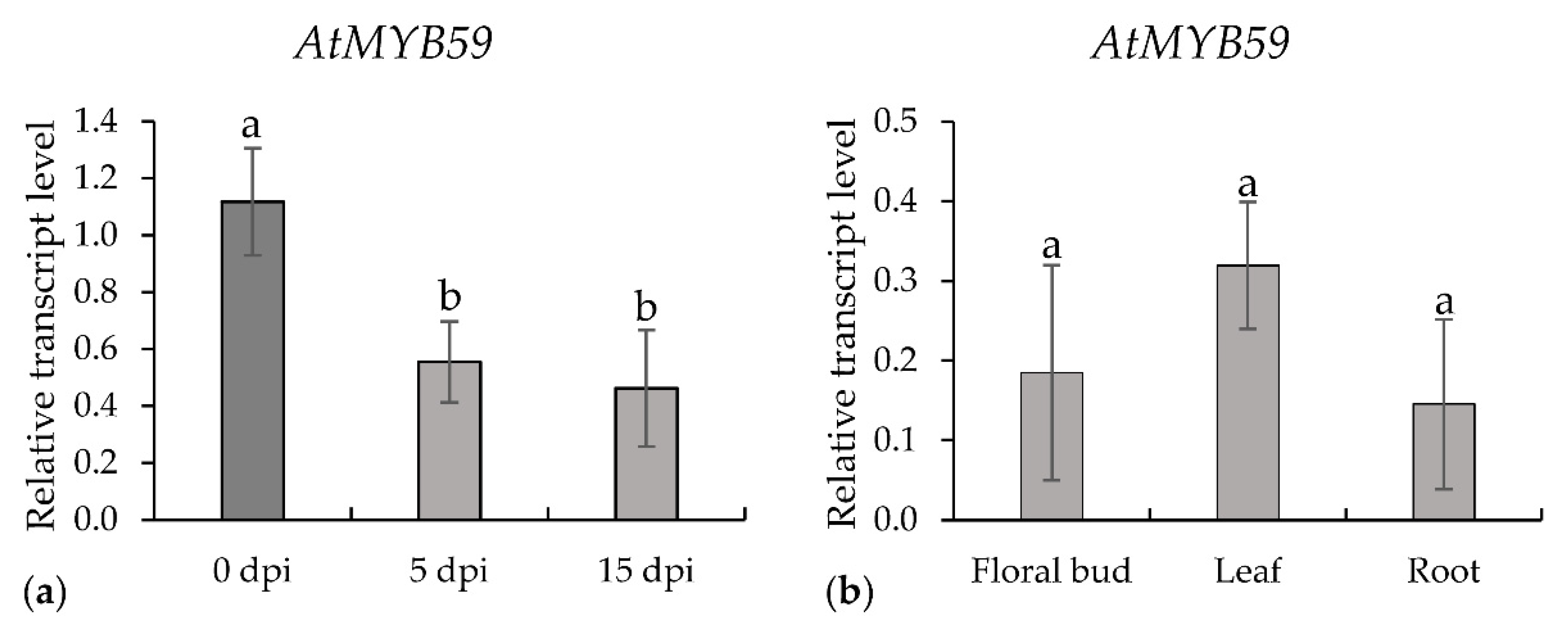
2.1. Expression of AtMYB59 Gene

Based on the transcriptome analysis of syncytia induced in A. thaliana roots by the beet cyst nematode H. schachtii [12], we found that AtMYB59 was significantly downregulated and presumed that its expression might have a negative impact on the development of syncytia and/or nematodes. Following Szakasits et al. [12], we analyzed the expression level of AtMYB59 in roots at 5 and 15 days post-infection (dpi), which confirmed the statistically significant downregulation of its expression up to more than 50% of its expression level in wild-type plants at both time points (Figure 1a). Concomitantly, we analyzed AtMYB59 transcript accumulation in the floral buds, leaves, and roots of one-month-old plants. The levels of AtMYB59 expression in the flower buds and roots were similar. A slightly higher expression level was found in leaves, but this difference was statistically insignificant compared with flower buds and roots (Figure 1b).
GUS activity was investigated in two transgenic lines, where GUS expression was controlled by two versions of AtMYB59 promoter differing in length: F1 is 704 bp and F2 is 507 bp (Figure 2). No differences were observed between these promoter fragments regarding the site and strength of GUS activity in pMyb59::GUS homozygotic lines. Both analyzed promoter sequences (F1 and F2) were not activated in the aerial organs (stem, buds, flowers, leaves, or siliques) of flowering four-week-old A. thaliana plants. Strong GUS activity was detected in the roots of three-day-old transgenic seedlings of both transgenic lines, and weak GUS activity appeared in the cotyledons (Figure 2a). However, in seven-day-old seedlings, with their first true leaves developed, GUS activity was identified in the vascular tissue of cotyledons and roots (Figure 2b). The promoter::GUS analyses showed that the activity of both promoter versions was mostly restricted to the roots. The GUS activity was detected in the vascular cylinder in the apical parts of the roots, but not in the root meristems or the primordia of the lateral roots in both transgenic lines (Figure 2c,e). The downregulation of GUS activity occurred in regions containing nematode infection sites at 5 dpi (Figure 2d,f) and persisted until 15 dpi in both pMyb59::GUS lines (data not shown). Both results, gene expression analysis and GUS activity analysis, confirmed the downregulation of AtMYB59 expression after nematode infection of A. thaliana roots.

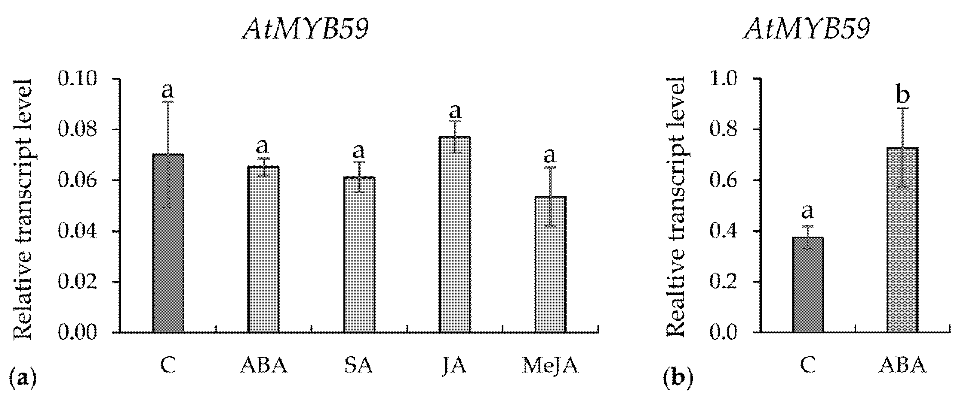
2.2. Expression of AtMYB59 Gene in Roots or Leaves Treated with Phytohormones

Expression level of AtMYB59 was examined in the uninfected roots of the 14-day-old wild-type plants after 24 h of root exposure to jasmonic acid (JA), (-)-methyl jasmonate (MeJA), salicylic acid (SA), or abscisic acid (ABA). There was no statistically significant difference between the AtMYB59 gene expression level in the control (water-treated) and hormone-treated roots (Figure 3a). In another experiment, the expression of AtMYB59 was investigated in the leaves of 14-day-old plants 24 h after the foliar application of the aforementioned hormones. AtMYB59 transcripts accumulation level increased almost two-fold after treatment with ABA (Figure 3b) and this reaction was statistically significant in contrast with the responses to other phytohormones (data not shown).

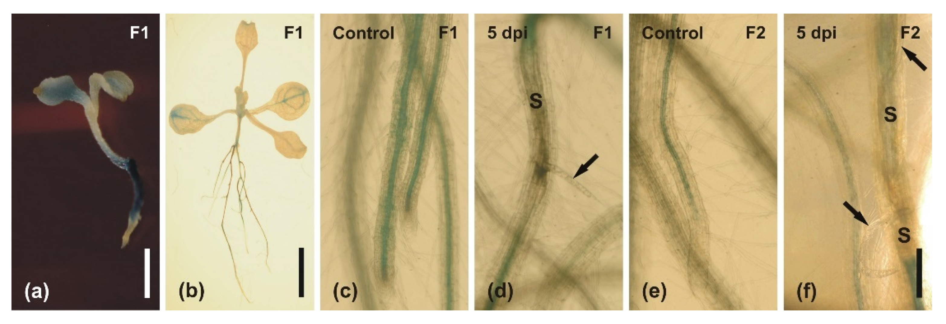
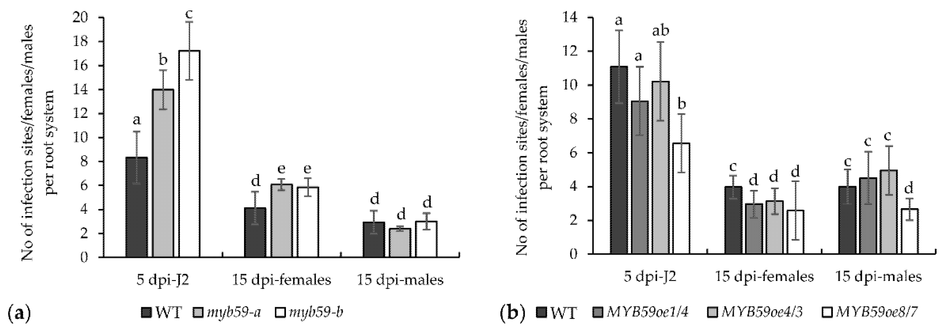
2.3. Infection of Roots with H. schachtii

The potential influence of AtMYB59 on the H. schachtii infection of A. thaliana was explored using two T-DNA mutants (myb59-a and myb59-b), overexpression lines (MYB59oe1/4, MYB59oe4/3 and MYB59oe8/7), and Col-0 wild-type plants. The downregulation of AtMYB59 in myb59 mutants roots and upregulation in MYB59oe lines were confirmed by qRT-PCR (Figure 4). The MYB59oe lines used in this work contained full-length cDNA (without both introns) in contrast with the splice variants described by Li et al. [14].
In both mutants, a significantly higher average numbers of infecting juveniles per root system at 5 dpi and average numbers of developed females at 15 dpi were observed (Figure 5a). Both values were about 40% higher in mutants than in control wild-type plants. This suggests that the downregulation of AtMYB59 expression increases the susceptibility of A. thaliana to beet cyst nematode. The average numbers of males developed in mutant roots showed no significant change at 15 dpi compared with wild-type plants. In contrast, but in agreement with these results, the average numbers of J2s that invaded the roots of MYB59oe lines at 5 dpi were generally lower, but only in the case of the MYB59oe8/7 line was the difference statistically significant (Figure 5b). The average numbers of females developed in the roots of MYB59oe lines at 15 dpi were significantly lower, with differences between 21% and 35% in comparison with the wild-type plants. The average numbers of males developed in the roots of MYB59oe lines at 15 dpi were similar to their numbers found on the roots of wild-type plants except for line MYB59oe8/7, where the decrease in the number of males was significant. The difference was about 33% in comparison with wild-type plants (Figure 5b). These results show that the upregulation of AtMYB59 expression leads to the decrease in A. thaliana susceptibility to beet cyst nematode. Based on the infection tests results obtained on myb59 mutants and MYB59oe lines, we suggest that AtMYB59 plays a significant role in the response of A. thaliana roots to the parasitism of H. schachtii juveniles.

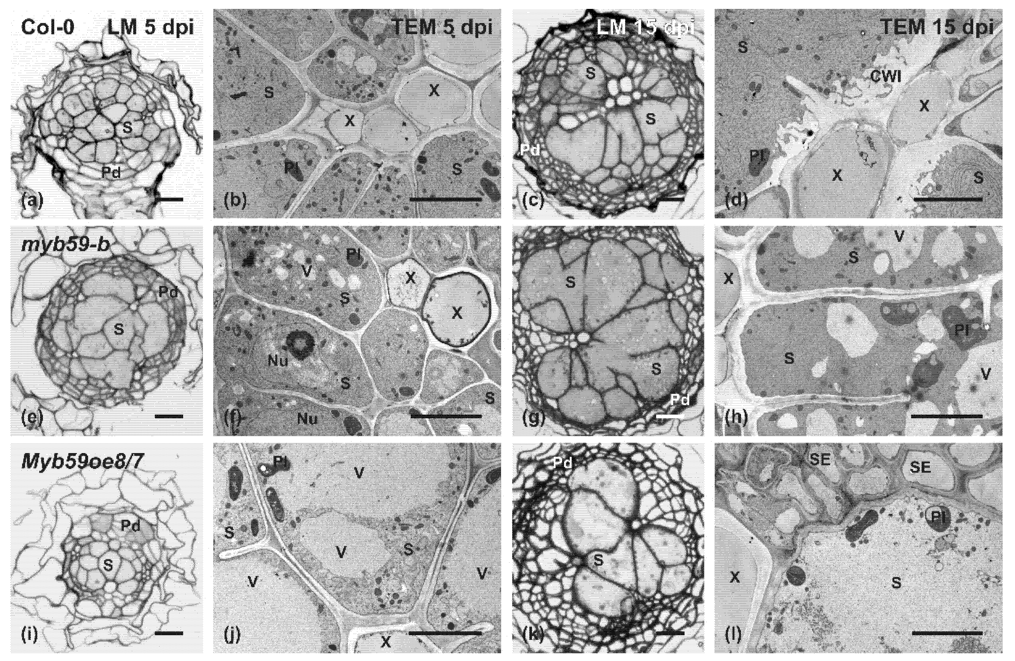
2.4. Cellular and Ultrastructural Differences of Syncytia

Based on the results of nematode developmental tests and AtMYB59 expression analyses, the myb59-b mutant and MYB59oe8/7 overexpressing line were selected for detailed investigations of the anatomical and ultrastructural organization of syncytia induced by H. schachtii (Figure 6). There were no differences in the anatomy or development of uninfected roots. The roots of mutant and AtMYB59 overexpressing lines developed typical primary and secondary states of growth as wild-type plants (data not shown).
In all the analyzed genotypes, syncytia were induced and well-developed at 5 dpi (Figure 6a,e,i), and they were only composed of vascular cylinder cells. The syncytia, at their widest region, were surrounded by dividing pericycle cells forming the periderm. In the MYB59oe8/7 line, the periderm development was the least advanced in comparison with other genotypes at 5 dpi. The surface of the cross-section of the syncytia induced in mutant roots was, at this stage of their development, similar to that of the wild-type plants. In contrast, the syncytia induced in the roots of the MYB59oe8/7 line were smaller (on cross-sections) than the syncytia induced in the roots of the wild-type or myb59-b mutant plants. The difference in their size was mostly a result of the much weaker hypertrophy of syncytial elements. Additionally, fewer cell wall openings were formed between the syncytial elements in the MYB59oe8/7 line. These anatomical differences became more obvious in 15 dpi syncytia (Figure 6c,g,k). At this developmental stage, on cross-sections obtained from the widest region of the syncytium, the hypertrophy of individual syncytial elements and the size and number of cell wall openings were even higher in the syncytia induced in myb59-b mutant roots than in syncytia developed in wild-type plants. Syncytia induced in the roots of MYB59oe8/7 line were still smaller than those in wild-type plant roots due to the low numbers of cells incorporated into the syncytium and the low numbers of cell wall openings between syncytial elements.
Ultrastructural analysis of syncytia showed similarities between syncytia induced in myb59-b mutant and wild-type plants (Figure 6b,d,f,h). These syncytia had an electron-dense cytoplasm with notably smaller vacuoles, enlarged nuclei and nucleoli, and high numbers of plastids, mitochondria and endoplasmic reticulum cisternae. However, in syncytia developed in myb59-b mutant roots, small vacuoles were more numerous than in syncytia formed in wild-type plants at both examined time points (Figure 6f,h). Additionally, at 15 dpi in syncytia induced in myb59-b roots, numerous huge plastids with a highly developed thylakoid systems were observed (Figure 6h). Similar plastids were not found in syncytia induced in other tested lines. In contrast, syncytia developed in the roots of the MYB59oe8/7 line showed less electron-dense cytoplasm and lower numbers of endoplasmic reticulum cisternae at 5 dpi than syncytia induced in wild-type roots (Figure 6j vs. Figure 6b). Additionally, the large part of their volume was occupied by large vacuoles or relatively electron-translucent cytoplasm (Figure 6j). Many plastids contained starch grains that were not found in syncytia induced in wild-type plants and myb59-b mutants (Figure 6j vs. Figure 6b,f). Only the remains of degraded cytoplasm, plastids, mitochondria, and rarely, the debris of endoplasmic reticulum cisternae were present in syncytia induced in the roots of the MYB59oe8/7 line at 15 dpi (Figure 6l).
In all examined lines and at both analyzed stages of syncytia development, direct contact was maintained between the outer cell wall of the syncytium and the conductive elements of the vascular cylinder (Figure 6). Additionally, there were no degenerated cells around developing syncytia. In all analyzed genotypes, the cell wall ingrowths systems (similar to that depicted in Figure 6d), in a more or less expanded shape, were formed on the outer syncytial cell wall adjacent to the xylem vessels. Thus, we concluded that the degeneration of syncytia in MYB59oe8/7 line was not caused by the limited inflow of water and organic compounds from the conductive elements but was evoked by internal triggers.

2.5. cis Regulatory Elements Analysis of AtMYB59 Gene Promoter

Using a New Place bioinformatics tool, we found 57 different classes of cis elements (162 in total) in a 704 bp-long promoter sequence of AtMYB59 gene and 105 (belonging to 42 classes) out of 162 also occurred in the shorter, 507 bp-long, analyzed promoter fragment. Since no difference was discovered in the activity of both promoter fragments in the roots of A. thaliana plants before or after infection by H. schachtii, the cis elements responsible for the downregulation of the AtMYB59 gene expression in syncytia are likely to remain present in the 507 bp fragment. The bioinformatics analysis indicated 27 cis elements that are specific for the 507 bp fragment and absent in the 197 bp fragment by which the 704 bp fragment was reduced. Among these, there were eight ABA- and/or dehydration-responsive cis elements, four resistance- and/or wounding-responsive cis elements, one auxin- and 1 jasmonate-responsive cis element, and 13 other types of cis elements that may be related to the response of plants to abiotic or biotic stresses (Table 1).

3. Discussion

Transcription factors are regulatory proteins that specifically bind to relevant cis acting elements in the promoter region of a gene to activate its expression. The regulation of gene transcription is an important method to modulate plant growth as well as to adjust the plant response to various biotic and abiotic stresses. Transcription factors can be divided into different families according to the specificity of the DNA binding region [15]. AtMYB59, analyzed in this work, encodes putative R2R3-MYB protein. The predicted protein sequence contains two nuclear localization signals (NLS) in the R3 repeat, both required for transport to the nucleus [14,16]. The ability of the AtMYB59 protein to regulate transcriptional activity in plant cells was confirmed; it is involved in the regulation of cell-cycle progression and root growth [16].
In previous studies, it was found that AtMYB59 is differentially regulated in an organ-specific manner as well as after leaves are treated with different phytohormones and subjected to different stresses [14,16]. The transcript accumulation level of AtMYB59 in leaves, inflorescences, or roots was confirmed in this study; however, transcript synthesis after treating roots or leaves with hormones differed. In this research, we focused on the regulation of AtMYB59 expression in roots, since they are infested by the plant parasitic nematode H. schachtii. The upregulation of AtMYB59 transcription in roots after hormones treatment did not occur; however, its upregulation in leaves was induced by ABA application. In a previous work [14], the upregulation of AtMYB59 splice variant 1 transcript synthesis was observed in leaves after JA and SA applications, but not after ABA treatment. Partially similar results were obtained in another study, where plants were treated with a lower concentration of ABA for 6 h [17]. The moderate upregulation of AtMYB59 expression in leaves and the strong downregulation in roots were also reported [17]. All these results confirm that AtMYB59 is differentially regulated upon ABA treatment and that the level of its expression depends on the organ, ABA concentration, and treatment duration. The observed differences may have resulted from the three-times-longer exposure time applied in our study (24 h) than in the previous experiment conducted by Li et al. [14], who treated plants for 8 h. Presumably, the plant’s response to the foliar application of hormones is faster when JA and SA are used, whereas the response to ABA, as shown in our work, can appear after prolonged exposure.
ABA is considered a hormone primarily involved in plant responses to abiotic stress, particularly drought and salinity [18]. However, ABA can also play a pivotal role in plant immunity. A set of studies showed that ABA treatment can modulate the plant defense response to pathogens, including parasites. The application of ABA increased the susceptibility of rice and tomato to root-knot nematodes (Meloidogyne graminicola or M. javanica) infection [19,20].
In this work, we identified several ABA- and drought-responsive elements in the fragment of AtMYB59 promoter. Additionally, AtMYB59 expression was upregulated after ABA treatment, suggesting that ABA plays a role in the induction of AtMYB59 expression. However, the regulation of AtMYB59 gene function by ABA may be liberalized during root infection with parasites, which should be the subject of further investigations.
In opposition to previous research results [14,16,21], where longer AtMYB59 promoter fragments (2078, 2009, and 1600 bp) were exploited, we analyzed two shorter fragments (507 and 704 bp long) to determine their potential role during root infestation by cyst nematodes. GUS activity driven by 2078 bp-long AtMYB59 promoter was detected in seedlings, mature leaves, flowers, siliques and stems [14]. Du et al. [21], using a 2009 bp long AtMYB59 promoter fragment, observed GUS activity in seedling roots (including root hairs). When a 1600 bp-long fragment of AtMYB59 promoter was used, GUS activity was detected in the hypocotyls and roots (including root hairs), vascular tissue, and root tip meristem, as well in the leaf edges and the pedicels of siliques [16]. In this work, transgenic plants containing truncated versions of AtMYB59 promoter showed reduced and transient GUS activity in the cotyledons and the vascular cylinder in confined regions of the roots. This result may indicate that cis regulatory elements responsible for the activation of AtMYB59 promoter in other organs were removed by the severe reduction in promoter length. During root infection by juveniles, GUS activity was completely lost early upon invasion at the site of infection and around the developing syncytium. This result shows that cis elements responsible for the reduction in AtMYB59 expression level after nematode attack and during successive nematode feeding and development were preserved in the truncated sequences of AtMYB59 promoter.
The expression of AtMYB59 in yeast cells resulted in the inhibition of their proliferation and caused the formation of bi-nuclear cells with high aneuploid DNA content in parallel with the elongation of cell shape [16]. AtMYB59 protein probably affects cell proliferation by interfering with DNA replication, chromosome separation, cell division and cell growth [16]. It was shown that AtMYB59 transcript accumulation strongly increased in S or S-to-G2 phases of the cell cycle in comparison with the remaining cell cycle phases when its expression levels were low [16]. Our microscopic examinations showed that the root anatomy of mutant and AtMYB59-overexpressing plants is the same. However, substantial differences concerning syncytium development and organization clearly appeared. Similar to yeast cells, the overexpression of AtMYB59 in plants seems to disturb the regular development of syncytia and surrounding secondary cover tissue (periderm). High levels of AtMYB59 protein apparently lead to a lower number of dividing pericyclic cells necessary to properly develop a periderm surrounding the syncytium or cause serious delays in this process. Additionally, syncytia induced in AtMYB59oe plants were generally smaller, composed of fewer elements that were interconnected by only a few and small cell wall openings. This may indicate that AtMYB59 adversely influences syncytium development, not only by interference in the cell cycle and cell division, but also through obvious changes in the properties of the cell walls, which finally results in the development of smaller and less-effective syncytia, which prematurely deteriorate, thus stopping the development of the females of the beet cyst nematode.
Du et al. [21] showed that AtMYB59 maintains the distribution of K+ and the balance of NO3¯ between roots and shoots by the positive regulation of Nitrate Transporter1.5 (NRT1.5)/Nitrate Transporter/Peptide Transporter Family7.3 (NPF7.3) transcription in response to low K+ stress. It was proven that AtMYB59 protein directly binds to the NPF7.3 gene promoter. Another study showed that AtMYB59, as a negative regulator in calcium (Ca) signaling and homeostasis during Ca deficiency, leads to the regulation of plant growth and stress responses [17].
Considering the aforementioned results and the role of AtMYB59 indicated in our work in the response of A. thaliana to cyst nematode attack, it seems that AtMYB59 may play a broad role from metabolism modulation to the responses to abiotic and biotic stresses.
Sixteen classes of transcription factors seem to be specific to the plant kingdom [22]. Two of them, MYB and WRKY families, are key players in, inter alia, the plant response to biotic stresses [23,24]. In A. thaliana, there are well-evidenced examples of transcription factors that play a role in the plant immune response. Among them are genes such as AtMYB96, AtMYB30, or AtMYB46 [25,26,27,28] participating in the responses to viruses, bacteria, or fungi, and against nematodes such as AtWRKY23 and AtWRKY72. The former is probably the best-characterized transcription factor involved in plant–nematode interactions. Its protein was synthesized during the early stages of nematode feeding site development. The knocking down of AtWRKY23 gene expression resulted in a decreased infection rate of the beet cyst nematode [29]. Another protein, AtWRKY72, is required for full basal defense against M. incognita [30]. It is an orthologue of tomato SlWRKY72a and b, which are transcriptionally upregulated during disease resistance mediated by the Mi1 nematode resistance gene. It was also shown that 28 out of 66 WRKY genes identified in the A. thaliana genome were significantly downregulated; only six were upregulated in roots infected with H. schachtii [31]. Since they were downregulated in syncytia, AtWRKY6, AtWRKY11, AtWRKY17 and AtWRKY33 genes were studied in detail, and it was confirmed that their silencing is essential for successful nematode development [31]. There is an example available showing that MYB and WRKY transcription factors can influence each other: AtMYB44 regulates defense responses by the transcriptional activation of downstream AtWRKY70 by direct binding to a conserved cis regulatory element in its promoter [32,33].
The role of the MYB genes in response to nematode infection is generally poorly understood and examined. It was shown that AtMYB12 was transiently upregulated in 9 dpi syncytia induced by H. schachtii, and the myb12 mutant was less susceptible to this parasite because the average number of females developed in its roots was lower. It was suggested that AtMYB12 is more essential for infection and syncytia development than being involved in plant defense. AtMYB12 controls AtCHS and AtFLS1 (encoding chalcone synthase and flavonol synthase, respectively) and they both may contribute to plant defense against nematodes [34]. Recently, the novel miR858 (microRNA858)–AtMYB83 regulatory system in plant–cyst nematode interactions was reported [35]. It was shown that both interactors were transcriptionally upregulated in the syncytia induced by H. schachtii. Overexpression of miR858 led to a reduced susceptibility of A. thaliana, whereas the decreased expression of miR858 enhanced plant susceptibility to H. schachtii. Similarly, the overexpression of a non-cleavable coding sequence of AtMYB83 significantly increased plant susceptibility, whereas myb83 mutation decreased plant susceptibility [35]. The miR858-resistant variant of AtMYB12 overexpression lines exhibited elevated susceptibility to the nematode. The authors suggested that AtMYB12 may constitute part of the miR858/AtMYB83 regulatory loop modulating the plant response to nematode infection [35]. Although transcription factors are proteins that regulate the transcription of other genes, there are also genes encoding transcription factors or microRNAs, which reversely regulate the transcription of primary transcription factors. Thus, the interaction network appears complex and intertwined, and requires further extensive and detailed research.
AtMYB59 expression is downregulated in infection sites in A. thaliana roots during the entire duration of H. schachtii parasitism. The inhibition of the activity of the truncated fragments of the AtMYB59 promoter in infection sites showed that cis regulatory elements are located relatively closely to the transcription start site; however, further investigation should be conducted to establish the factors influencing the properties of the regulatory sequences and their activity, as well as the putative transcription regulation of AtMYB59 by miRNAs.

4. Materials and Methods

4.1. Plant Material and Culture Conditions

Wild-type ecotype Col-0, T-DNA insertional mutants of AtMYB59 (myb59-a and myb59-b), and transgenic lines overexpressing AtMYB59 (MYB59oe1/4, MYB59oe4/3, and MYB59oe8/7; all in Col-0 genetic background) of Arabidopsis thaliana were used in our experiments. Seeds of myb59-a (At5g59780; GK-627C09) and myb59-b (SALK_137001; with 2 T-DNA insertions, in AtMYB59 locus and in the intergenic region between At4g32480 and At4g32490 loci) were obtained from the Nottingham Arabidopsis Stock Centre (UK). Their seeds were surface-sterilized in 95% (v/v) ethanol for 2 min, then in 5% (v/v) solution of ClNaO (Sigma-Aldrich, St. Louis, MO, USA) for 8 min, and washed three times in excess dH2O. Seeds were placed on KNOP [36] or 1/2MS [37] medium in Ø90 mm Petri dishes. They were stratified for 48 h at 4 °C after being placed on the medium. Plants were cultivated under a 12 h day/12 h night photoperiod (125 µmol m−2 s−1) at 21 °C.

4.2. Gene Construct Preparation

Genomic DNA was isolated from frozen A. thaliana Col-0 leaves using the Genomic Mini AX Plant kit (A&A Biotechnology, Gdańsk, Poland). Genomic sequences of AtMYB59 gene promoter were amplified using primers: F1p: CACC/AGCCACTTATCACACACCCA or F2p: CACC/CCTTCTCTCCCTCACACACAC and Rp: GGAACCTATGGCATTCCTCTT. The two obtained DNA fragments covering the promoter sequence had lengths of 704 and 507 bp. The PCR products were cloned into pENTR™/D-TOPO® vector (Thermo Scientific, Waltham, MA, USA) and sequenced. The promoter fragments were subcloned into a pKGWFS7 vector containing the kanamycin resistance gene (nptII) and GFP and GUS reporter genes (https://gatewayvectors.vib.be, accessed on 14 June 2021) using Gateway® LR Clonase® II Enzyme mix (Thermo Scientific).
Uninfected root segments of 14-day-old A. thaliana plants were used to amplify the coding AtMYB59 DNA sequence (splice variant 2). The following primers pair was used: F: CACC/ATGAAACTTGTGCAAGAAGAATACCG and R: CTAAAGGCGACCACTACCATG. The PCR product was cloned into vector pENTR™/D-TOPO® (Thermo Scientific) and sequenced. The coding sequence was subcloned into pK7WG2D vector under the control of 35S promoter (https://gatewayvectors.vib.be, accessed on 14 June 2021) using Gateway® LR Clonase® II Enzyme mix (Thermo Scientific). The desired gene constructs were transferred into Agrobacterium tumefaciens EHA105 clone by electroporation (MicroPulser; Bio-Rad, Hercules, CA, USA).

4.3. A. thaliana Transformation

Transgenic overexpression lines (MYB59oe) with the coding sequence of AtMYB59 driven by 35S promoter and transgenic reporter lines with promoter sequences of AtMYB59 fused with the GUS coding sequence (pMyb59::GUS) were obtained by the floral dip transformation method [38]. Genotype homozygosity was confirmed using the ratio of kanamycin-resistant to non-resistant T2 plants germinating on kanamycin-containing medium.

4.4. Mutants Genotyping

The homozygosity of T-DNA insertion lines was confirmed in the relevant DNA sequence sites using PCR and the site-specific primers for myb59-a (LP1: AAGAGGAATGCCATAGGTTCC and RP1: AGTGGGTGGTGATTTTTGATG with o8409: ATATTGACCATCATACTCATTGC (for GABI-KAT)) and myb59-b: (LP2: CGCTCTTCTTGTGGAGTCATC and RP2: AAAACAATTCGACCCCTTTTG with SALK_LBb1.3: ATTTTGCCGATTTCGGAAC) under the standard PCR conditions recommended by the SALK collection.

4.5. RNA Extraction and RT-PCR

Total RNA was used for cDNA synthesis and semi-quantitative reverse transcription–polymerase chain reaction (RT-PCR) or quantitative real-time-PCR analyses of AtMYB59 expression. It was extracted from 50 mg of root segments without apical meristems of 14-day-old uninfected A. thaliana plants (0 dpi), root segments containing syncytia at 5 or 15 dpi, young leaves, or flower buds. In other experiments, total RNA was also isolated from: (i) uninfected roots treated for 24 h with jasmonic acid (JA), (-)-methyl jasmonate (MeJA), salicylic acid (SA), or abscisic acid (ABA) solutions (Sigma, Saint Louis, MO, USA), each at 100 µM final concentration applied to the roots/culture media; (ii) leaves of 14-day-old A. thaliana plants after the foliar application of the aforementioned phytohormones. Total RNA was also extracted from the roots of myb59-a and myb59-b mutants and MYB59oe lines of A. thaliana to confirm down- or upregulation of AtMYB59.
Collected tissue samples were immediately frozen in liquid nitrogen and homogenized using a Precellys 24 tissue homogenizer (Bertin Technologies, Montigny-le-Bretonneux, France). Total RNA was extracted using a modified method of Chomczynski and Sacchi [39], including a DNase I treatment step in accordance with the manufacturer’s instructions (Thermo Scientific). The concentration and purity of the RNA were validated on a Nanodrop 2000 (Thermo Scientific), and the RNA integrity was checked by electrophoresis in 1% (w/v) agarose gel.
First-strand cDNA was synthesized from 0.2 μg purified RNA using a RevertAid™ H Minus First Strand cDNA Synthesis Kit (Thermo Scientific) and diluted three times. For semi-qPCR, 1 μL of cDNA preparations was used in each 10 μL reaction with gene-specific primers. The optimal number of PCR cycles was first determined and amplifications were conducted using 27 cycles for the gene expression analysis of infected roots and hormone-treated leaves or 33 cycles for the analysis of leaves, buds and roots. Each cycle consisted of denaturation at 94 °C for 50 s, annealing at 60 °C for 50 s, and elongation at 72 °C for 2 min. An Applied Biosystem 9700 GeneAmp (Applied Biosystems, Foster City, CA, USA) thermal cycler was used. For AtMYB59 relative gene expression, the following primers were used F: GGAGGCTCCAACGGGAAAAT and R: GTTGGAGAAGCCAGAGGAGG. Actin2 (AT3g8780) [40] was used as a reference gene and F: CTTGCACCAAGCAGCATGAA and R: CCCCAGCTTTTTAAGCCTTTGATC primers were used.
For the gene expression analysis of roots treated with phytohormones, quantitative PCR analysis was performed using SybrGreen Master Mix (Roche, Basel, Switzerland) and 1 μL cDNA as a template. Reactions were run on a LightCycler 96 (Roche). The obtained results were calculated using tree reference genes: Actin2 (AT3g18780) F: CTTGCACCAAGCAGCATGAA and R: CCCCAGCTTTTTAAGCCTTTGATC, Actin8 (At1g49240) F: ATGAAGATTAAGGTCGTGGCA and R: TCCGAGTTTGAAGAGGCTAC, and GAPDH (AT1g13440) F: TTGGTGACAACAGGTCAAGCA and R: AAACTTGTCGCTCAATG-CAATC.

4.6. Promoter cis Element Analysis

The promoter sequences of AtMYB59, 704 and 507 bp long, were analyzed using the online database New Place to localize cis regulatory elements [41].

4.7. GUS Activity Assay

Histochemical detection of GUS activity was performed as described by Wiśniewska et al. [42]. GUS activity was examined in the leaves, flowers, siliques, seedlings, non-infected roots, as well as the roots containing 5 and 15 dpi syncytia with associated J2s of H. schachtii.

4.8. Nematode Infection Assay

Cysts of beet cyst nematodes (Heterodera schachtii Schmidt) were collected from white mustard (Sinapis alba cv. Albatros) roots grown in vitro on KNOP medium. They were incubated in 3 mM ZnCl2 (Merck, Darmstadt, Germany) and hatched infective J2s were collected after 6–7 days [36]. J2s were sterilized in 0.05% (w/v) HgCl2 (Poch S.A., Gliwice, Poland) for 2 min, and immediately washed three times in distilled H2O. Fourteen-day-old A. thaliana plants, wild types, overexpression lines (MYB59oe), mutants (myb59-a and -b), and promoter lines (pMyb59::GUS) were grown aseptically and inoculated with 70 J2s per plant under sterile conditions [43]. Inoculated plants were cultivated under the same conditions as described above. The number of infection sites (at 5 dpi) and the number of females and males (at 15 dpi) were counted per plant.

4.9. Anatomic and Ultrastructural Analysis

Uninfected roots and root segments containing 5 and 15 dpi syncytia were dissected and processed for microscopic examinations as described by Różańska et al. [44].

4.10. Statistical Analysis

The significance of differences in the data was tested using Fisher’s multiple range test and one-way ANOVA. The least significant difference (LSD) was calculated at p < 0.05. The RT-PCR experiments were performed in three biological replicates. The nematode infection assay was performed with at least five biological replicates for genotype (n > 30).

Author Contributions

Conceptualization, A.W.; methodology, A.W., E.R. and M.S.; investigation, A.W., K.W., E.R., K.L., K.K. and M.S.; data curation, A.W. and M.S.; writing—original draft preparation, A.W.; writing—review and editing, A.W. and M.S.; supervision of experimental work A.W. and M.S.; project administration, A.W.; funding acquisition, A.W. All authors have read and agreed to the published version of the manuscript.

Funding

This work was financed by the Polish National Science Centre (NCN) on the basis of decision number DEC-2013/09/B/NZ9/02381 (granted to Anita Wiśniewska).

Institutional Review Board Statement

Not applicable.

Informed Consent Statement

Not applicable.

Data Availability Statement

Not applicable.

Conflicts of Interest

The authors declare no conflict of interest.

References

  1. Jones, J.T.; Haegeman, A.; Danchin, E.G.J.; Gaur, H.S.; Helder, J.; Jones, M.G.K.; Kikuchi, T.; Manzanilla-López, R.; Palomares-Rius, J.E.; Wesemael, W.M.L.; et al. Top 10 plant-parasitic nematodes in molecular plant pathology. Mol. Plant Pathol. 2013, 14, 946–961. [Google Scholar] [CrossRef] [PubMed]
  2. Wyss, U. Observations on the feeding behaviour of Heterodera schachtii throughout development, including events during moulting. Fundam. Appl. Nematol. 1992, 15, 75–89. [Google Scholar]
  3. Golinowski, W.; Grundler, F.M.W.; Sobczak, M. Changes in the structure of Arabidopsis thaliana during female development of the plant-parasitic nematode Heterodera schachtii. Protoplasma 1996, 194, 103–116. [Google Scholar] [CrossRef]
  4. Siddique, S.; Radakovic, Z.S.; Carola, M.; Chronis, D.; Novák, O.; Ramireddy, E.; Holbein, J.; Matera, C.; Hütten, M.; Gutbrod, P.; et al. A parasitic nematode releases cytokinin that controls cell division and orchestrates feeding site formation in host plants. Proc. Natl. Acad. Sci. USA 2015, 112, 12669–12674. [Google Scholar] [CrossRef] [PubMed] [Green Version]
  5. Feller, A.; MacHemer, K.; Braun, E.L.; Grotewold, E. Evolutionary and comparative analysis of MYB and bHLH plant transcription factors. Plant J. 2011, 66, 94–116. [Google Scholar] [CrossRef]
  6. Kranz, H.D.; Denekamp, M.; Greco, R.; Jin, H.; Leyva, A.; Meissner, R.C.; Petroni, K.; Urzainqui, A.; Bevan, M.; Martin, C.; et al. Towards functional characterisation of the members of the R2R3-MYB gene family from Arabidopsis thaliana. Plant J. 1998, 16, 263–276. [Google Scholar] [CrossRef]
  7. Cheng, C.-Y.; Krishnakumar, V.; Chan, A.P.; Thibaud-Nissen, F.; Schobel, S.; Town, C.D. Araport11: A complete reannotation of the Arabidopsis thaliana reference genome. Plant J. 2017, 89, 789–804. [Google Scholar] [CrossRef] [Green Version]
  8. Prouse, M.B.; Campbell, M.M. The interaction between MYB proteins and their target DNA binding sites. Biochim. Biophys. Acta Gene Regul. Mech. 2012, 1819, 67–77. [Google Scholar] [CrossRef]
  9. Glazebrook, J. Contrasting mechanisms of defense against biotrophic and necrotrophic pathogens. Annu. Rev. Phytopathol. 2005, 43, 205–227. [Google Scholar] [CrossRef]
  10. Spoel, S.H.; Dong, X. Making sense of hormone crosstalk during plant immune responses. Cell Host Microbe 2008, 3, 348–351. [Google Scholar] [CrossRef] [Green Version]
  11. Zheng, Q.; Putker, V.; Goverse, A. Molecular and cellular mechanisms involved in host-specific resistance to cyst nematodes in crops. Front. Plant Sci. 2021, 12, 641582. [Google Scholar] [CrossRef]
  12. Szakasits, D.; Heinen, P.; Wieczorek, K.; Hofmann, J.; Wagner, F.; Kreil, D.P.; Sykacek, P.; Grundler, F.M.W.; Bohlmann, H. The transcriptome of syncytia induced by the cyst nematode Heterodera schachtii in Arabidopsis roots. Plant J. 2009, 57, 771–784. [Google Scholar] [CrossRef] [PubMed] [Green Version]
  13. Barcala, M.; García, A.; Cabrera, J.; Casson, S.; Lindsey, K.; Favery, B.; García-Casado, G.; Solano, R.; Fenoll, C.; Escobar, C. Early transcriptomic events in microdissected Arabidopsis nematode-induced giant cells. Plant J. 2010, 61, 698–712. [Google Scholar] [CrossRef] [PubMed]
  14. Li, J.; Li, X.; Guo, L.; Lu, F.; Feng, X.; He, K.; Wei, L.; Chen, Z.; Qu, L.-J.; Gu, H. A subgroup of MYB transcription factor genes undergoes highly conserved alternative splicing in Arabidopsis and rice. J. Exp. Bot. 2006, 57, 1263–1273. [Google Scholar] [CrossRef] [PubMed] [Green Version]
  15. Liu, L. The roles of MYB transcription factors on plant defense responses and its molecular mechanism. Heredity 2008, 30, 1265–1271. [Google Scholar] [CrossRef] [PubMed]
  16. Mu, R.L.; Cao, Y.R.; Liu, Y.F.; Lei, G.; Zou, H.F.; Liao, Y.; Wang, H.W.; Zhang, W.K.; Ma, B.; Du, J.Z.; et al. An R2R3-type transcription factor gene AtMYB59 regulates root growth and cell cycle progression in Arabidopsis. Cell Res. 2009, 19, 1291–1304. [Google Scholar] [CrossRef] [PubMed] [Green Version]
  17. Fasani, E.; DalCorso, G.; Costa, A.; Zenoni, S.; Furini, A. The Arabidopsis thaliana transcription factor MYB59 regulates calcium signalling during plant growth and stress response. Plant Mol. Biol. 2019, 99, 517–534. [Google Scholar] [CrossRef]
  18. Chen, K.; Li, G.; Bressan, R.A.; Song, C.; Zhu, J.; Zhao, Y. Abscisic acid dynamics, signaling, and functions in plants. J. Integr. Plant Biol. 2020, 62, 25–54. [Google Scholar] [CrossRef] [Green Version]
  19. Kyndt, T.; Nahar, K.; Haeck, A.; Verbeek, R.; Demeestere, K.; Gheysen, G. Interplay between carotenoids, abscisic acid and jasmonate guides the compatible rice-Meloidogyne graminicola interaction. Front. Plant Sci. 2017, 8. [Google Scholar] [CrossRef] [PubMed]
  20. Moosavi, M.R. The effect of gibberellin and abscisic acid on plant defense responses and on disease severity caused by Meloidogyne javanica on tomato plants. J. Gen. Plant Pathol. 2017, 83, 173–184. [Google Scholar] [CrossRef]
  21. Du, X.Q.; Wang, F.L.; Li, H.; Jing, S.; Yu, M.; Li, J.; Wu, W.H.; Kudla, J.; Wang, Y. The transcription factor MYB59 regulates K+/NO3¯ translocation in the Arabidopsis response to low K+ stress. Plant Cell 2019, 31, 699–714. [Google Scholar] [CrossRef] [PubMed] [Green Version]
  22. Riechmann, J.L. Transcriptional regulation: A genomic overview. Arab. B. 2002, 1, e0085. [Google Scholar] [CrossRef] [Green Version]
  23. Dubos, C.; Stracke, R.; Grotewold, E.; Weisshaar, B.; Martin, C.; Lepiniec, L. MYB transcription factors in Arabidopsis. Trends Plant Sci. 2010, 15, 573–581. [Google Scholar] [CrossRef] [PubMed]
  24. Rushton, P.J.; Somssich, I.E.; Ringler, P.; Shen, Q.J. WRKY transcription factors. Trends Plant Sci. 2010, 15, 247–258. [Google Scholar] [CrossRef]
  25. Geri, C.; Cecchini, E.; Giannakou, M.E.; Covey, S.N.; Milner, J.J. Altered patterns of gene expression in Arabidopsis elicited by cauliflower mosaic virus (CaMV) infection and by a CaMV gene VI transgene. Mol. Plant Microbe Interact. 1999, 12, 377–384. [Google Scholar] [CrossRef] [Green Version]
  26. Seo, P.J.; Park, C.-M. MYB96-mediated abscisic acid signals induce pathogen resistance response by promoting salicylic acid biosynthesis in Arabidopsis. New Phytol. 2010, 186, 471–483. [Google Scholar] [CrossRef] [PubMed]
  27. Vailleau, F.; Daniel, X.; Tronchet, M.; Montillet, J.-L.; Triantaphylides, C.; Roby, D. A R2R3-MYB gene, AtMYB30, acts as a positive regulator of the hypersensitive cell death program in plants in response to pathogen attack. Proc. Natl. Acad. Sci. USA 2002, 99, 10179–10184. [Google Scholar] [CrossRef] [Green Version]
  28. Ramírez, V.; Agorio, A.; Coego, A.; García-Andrade, J.; Hernández, M.J.; Balaguer, B.; Ouwerkerk, P.B.F.; Zarra, I.; Vera, P. MYB46 modulates disease susceptibility to Botrytis cinerea in Arabidopsis. Plant Physiol. 2011, 155, 1920–1935. [Google Scholar] [CrossRef] [Green Version]
  29. Grunewald, W.; Karimi, M.; Wieczorek, K.; Van de Cappelle, E.; Wischnitzki, E.; Grundler, F.; Inzé, D.; Beeckman, T.; Gheysen, G. A role for AtWRKY23 in feeding site establishment of plant-parasitic nematodes. Plant Physiol. 2008, 148, 358–368. [Google Scholar] [CrossRef] [PubMed] [Green Version]
  30. Bhattarai, K.K.; Atamian, H.S.; Kaloshian, I.; Eulgem, T. WRKY72-type transcription factors contribute to basal immunity in tomato and Arabidopsis as well as gene-for-gene resistance mediated by the tomato R gene Mi-1. Plant J. 2010, 63, 229–240. [Google Scholar] [CrossRef]
  31. Ali, M.A.; Wieczorek, K.; Kreil, D.P.; Bohlmann, H. The beet cyst nematode Heterodera schachtii modulates the expression of WRKY transcription factors in syncytia to favour its development in Arabidopsis roots. PLoS ONE 2014, 9, e102360. [Google Scholar] [CrossRef] [Green Version]
  32. Shim, J.S.; Choi, Y.D. Direct regulation of WRKY70 by AtMYB44 in plant defense responses. Plant Signal. Behav. 2013, 8, e24509. [Google Scholar] [CrossRef] [Green Version]
  33. Shim, J.S.; Jung, C.; Lee, S.; Min, K.; Lee, Y.-W.; Choi, Y.; Lee, J.S.; Song, J.T.; Kim, J.-K.; Choi, Y.D. AtMYB44 regulates WRKY70 expression and modulates antagonistic interaction between salicylic acid and jasmonic acid signaling. Plant J. 2013, 73, 483–495. [Google Scholar] [CrossRef]
  34. Hamamouch, N.; Winkel, B.S.J.; Li, C.; Davis, E.L. Modulation of Arabidopsis flavonol biosynthesis genes by cyst and root-knot nematodes. Plants 2020, 9, 253. [Google Scholar] [CrossRef] [PubMed] [Green Version]
  35. Piya, S.; Kihm, C.; Rice, J.H.; Baum, T.J.; Hewezi, T. Cooperative regulatory functions of miR858 and MYB83 during cyst nematode parasitism. Plant Physiol. 2017, 174, 1897–1912. [Google Scholar] [CrossRef] [PubMed] [Green Version]
  36. Sijmons, P.C.; Grundler, F.M.W.; Mende, N.; Burrows, P.R.; Wyss, U. Arabidopsis thaliana as a new model host for plant-parasitic nematodes. Plant J. 1991, 1, 245–254. [Google Scholar] [CrossRef]
  37. Murashige, T.; Skoog, F. A revised medium for rapid growth and bio assays with tobacco tissue cultures. Physiol. Plant 1962, 15, 473–497. [Google Scholar] [CrossRef]
  38. Clough, S.J.; Bent, A.F. Floral dip: A simplified method for Agrobacterium-mediated transformation of Arabidopsis thaliana. Plant J. 1998, 16, 735–743. [Google Scholar] [CrossRef] [Green Version]
  39. Chomczynski, P.; Sacchi, N. The single-step method of RNA isolation by acid guanidinium thiocyanate–phenol–chloroform extraction: Twenty-something years on. Nat. Protoc. 2006, 1, 581–585. [Google Scholar] [CrossRef] [PubMed]
  40. Grundler, F.; Hofmann, J. Identification of reference genes for qRT-PCR studies of gene expression in giant cells and syncytia induced in Arabidopsis thaliana by Meloidogyne incognita and Heterodera schachtii. Nematology 2007, 9, 317–323. [Google Scholar] [CrossRef]
  41. Higo, K.; Ugawa, Y.; Iwamoto, M.; Korenaga, T. Plant cis-acting regulatory DNA elements (PLACE) database: 1999. Nucleic Acids Res. 1999, 27, 297–300. [Google Scholar] [CrossRef] [Green Version]
  42. Wiśniewska, A.; Dąbrowska-Bronk, J.; Szafrański, K.; Fudali, S.; Święcicka, M.; Czarny, M.; Wilkowska, A.; Morgiewicz, K.; Matusiak, J.; Sobczak, M.; et al. Analysis of tomato gene promoters activated in syncytia induced in tomato and potato hairy roots by Globodera rostochiensis. Transgenic Res. 2013, 22, 557–569. [Google Scholar] [CrossRef] [PubMed] [Green Version]
  43. Siddique, S.; Matera, C.; Radakovic, Z.S.; Shamim Hasan, M.; Gutbrod, P.; Rozanska, E.; Sobczak, M.; Torres, M.A.; Grundler, F.M.W. Parasitic worms stimulate host NADPH oxidases to produce reactive oxygen species that limit plant cell death and promote infection. Sci. Signal. 2014, 7, ra33. [Google Scholar] [CrossRef] [PubMed] [Green Version]
  44. Różańska, E.; Czarnocka, W.; Baranowski, Ł.; Mielecki, J.; de Almeida Engler, J.; Sobczak, M. Expression of both Arabidopsis γ-tubulin genes is essential for development of a functional syncytium induced by Heterodera schachtii. Plant Cell Rep. 2018, 37, 1279–1292. [Google Scholar] [CrossRef] [PubMed] [Green Version]
Figure 1. The relative expression levels of AtMYB59 in syncytia induced by H. schachtii in wild-type A. thaliana: (a) relative transcript levels of AtMYB59 in roots at 0, 5 and 15 dpi; and (b) relative transcript levels of AtMYB59 in floral buds, leaves and roots of one-month-old plants. The bars show mean values ± standard deviation. a, b homogenous groups. (p < 0.05; ANOVA; LSD).
Ijms 22 06450 g001
Figure 2. Histochemical staining of GUS activity driven by fragments of AtMYB59 promoter in transgenic A. thaliana plants: (ac,e) control non-inoculated plants; (d,f) infected roots with juveniles and syncytia at 5 dpi; (ad) GUS activity driven by the F1 (704 bp) fragment of the AtMYB59 promoter; (e,f) GUS activity driven by the F2 (507 bp) fragment of the AtMYB59 promoter; (a), three-day-old seedling (slight GUS activity present in cotyledons and strong in roots); (b), seven-day-old seedling (GUS activity present in roots and vascular tissues of cotyledons); (c) GUS activity under control of the F1 promoter fragment in non-inoculated roots; (d) GUS activity under control of the F1 promoter fragment in 5 dpi roots; (e) GUS activity under the control of the F2 promoter fragment in non-inoculated roots; (f) GUS activity under the control of the F2 promoter fragment in 5 dpi roots. GUS activity was evidenced in the vascular tissues of non-inoculated roots, but it disappeared in nematode feeding sites. Abbreviation: S, syncytium. Arrows indicated juveniles of the nematode. Scale bars: 1 mm (a); 10 mm (b); 100 µm (cf).
Ijms 22 06450 g002
Figure 3. The relative expression level of AtMYB59 gene in wild-type A. thaliana after phytohormones treatment: (a) relative transcript levels in roots treated with ABA, SA, JA, or MeJA (100 µM concentration each); and (b) relative transcript levels in leaves treated with ABA (100 µM). The bars show mean values ± standard deviation. a, b homogenous groups (p < 0.05; ANOVA; LSD).
Ijms 22 06450 g003
Figure 4. The relative expression levels of the AtMYB59 gene in roots of wild-type (WT), myb59-a and myb 59-b mutants, and MYB59oe overexpression lines: (a) relative transcript levels in AtMYB59 mutants roots; (b) relative transcript levels in MYB59oe lines. The bars show mean values ± standard deviation. a, b homogenous groups (p < 0.05; ANOVA; LSD).
Ijms 22 06450 g004
Figure 5. Development of H. schachtii juveniles on wild-type, mutant, and Myb59 overexpressing A. thaliana lines at 5 and 15 dpi; (a) developmental test comparing the susceptibility of wild-type and myb59-a and myb59-b mutant plants for infection with beet cyst nematode; (b) developmental test comparing the susceptibility of wild-type and different homozygotic Myb59 overexpressing lines (MYB59oe1/4, MYB59oe4/3 and MYB59oe8/7). The bars show mean values ± standard deviation. a, ab, b, c, d, e homogenous groups (p < 0.05; ANOVA; LSD).
Ijms 22 06450 g005
Figure 6. Development of syncytia induced by H. schachtii in wild-type, mutant and AtMYB59 overexpressing genotypes of A. thaliana: (a,c,e,g,i,k) light microscopy (LM) images of cross-sections stained with toluidine blue; (b,d,f,h,j,l) transmission electron microscopy (TEM) images; sections were obtained from syncytia induced in the roots of wild-type plants (Col-0) (ad), myb59-b T-DNA mutant (eh), and AtMYB59 overexpressing line (MYB59oe8/7) (il) at 5 (a,b,e,f,i,j) and 15 dpi (c,d,g,h,k,l). Abbreviations: CWI, cell wall ingrowths; Pd, periderm; Pl, plastid; S, syncytium; SE, sieve element; X, xylem; V, vacuole. Scale bars, 20 µm (a,c,e,g,i,k) and 5 µm (b,d,f,h,j,l).
Ijms 22 06450 g006
Table 1. Predicted cis elements specific for the 507 bp-long promoter of AtMYB59.
Transcription Factor or cis Element NameSignal SequenceNew Place Ref.NumberDescription
ABRELATERD1ACGTGS0004141ABA- and dehydration responsive
ABRERATCALMACGYGBS0005071ABA- and dehydration responsive
ACGTATERD1ACGTS0004152dehydration responsive
ANAERO1CONSENSUSAAACAAAS0004772anaerobically induced
BIHD1OSTGTCAS0004981resistance responsive*
CATATGGMSAURCATATGS0003702auxin responsive
CCAATBOX1CCAATS0000302heat shock responsive
CIACADIANLELHCCAANNNNATCS0002521circadian related
CMSRE1IBSPOATGGACGGS0005111carbohydrate metabolite signal responsive element 1
DPBFCOREDCDC3ACACNNGS0002922ABA responsive
EBOXBNNAPACANNTGS0001444light responsive, phenylpropanoid biosynthesis related
EECCRCAH1GANTTNCS0004941CO2 responsive
GT1CONSENSUSGRWAAWS0001986light regulated
MYB1ATWAACCAS0004081Dehydration responsive
MYBCORECNGTTRS0001762dehydration responsive, flavonoid biosynthesis related
MYCCONSENSUSATCANNTGS0004074dehydration and cold responsive
NODCON1GMAAAGATS0004611nodule specific
OSE1ROOTNODULEAAAGATS0004673nodule specific
PALBOXAPCCCGTCCS0001371fungal elicitor, wounding and light responsive
POLASIG3AATAATS0000882polyA signal
QARBNEXTAAACGTGTS0002441wounding responsive
RAV1AATCAACAS0003144ABA, drought and cold responsive
T/GBOXATPIN2AACGTGS0004581jasmonate-responsive
TATABOX2TATAAATS0001091TATA box
TATABOX4TATATAAS0001112TATA box
TATAPVTRNALEUTTTATATAS0003401TATA box-like
WUSATAgTTAATGGS0004331root meristem specific
* bolded, resistance/wounding-responsive cis elements.
Publisher’s Note: MDPI stays neutral with regard to jurisdictional claims in published maps and institutional affiliations.

