Article

Visfatin Promotes the Metastatic Potential of Chondrosarcoma Cells by Stimulating AP-1-Dependent MMP-2 Production in the MAPK Pathway

1
Graduate Institute of Acupuncture Science, China Medical University, Taichung 404022, Taiwan
2
Department of Medical Research, China Medical University Hospital, Taichung 404022, Taiwan
3
Department of Pharmacology, School of Medicine, China Medical University, Taichung 404022, Taiwan
4
Graduate Institute of Biomedical Sciences, China Medical University, Taichung 404022, Taiwan
5
Department of Sports Medicine, College of Health Care, China Medical University, Taichung 404022, Taiwan
6
Department of Orthopedic Surgery, China Medical University Hospital, Taichung 404022, Taiwan
7
Division of Hematology and Oncology, Department of Internal Medicine, China Medical University Hospital, Taichung 404022, Taiwan
8
Graduate Institute of Basic Medical Science, China Medical University, Taichung 404022, Taiwan
9
Department of Orthopedic Surgery, China Medical University Beigang Hospital, Yunlin 651012, Taiwan
10
School of Oral Hygiene, College of Oral Medicine, Taipei Medical University, Taipei 110301, Taiwan
11
Department of Medicine, MacKay Medical College, New Taipei City 252005, Taiwan
12
Graduate Institute of Natural Products, College of Pharmacy, Kaohsiung Medical University, Kaohsiung 807378, Taiwan
13
Institute of Biomedical Sciences, Mackay Medical College, Taipei 104217, Taiwan
14
Division of Sports Medicine & Surgery, Department of Orthopedic Surgery, MacKay Memorial Hospital, Taipei 104217, Taiwan
15
Department of Biotechnology, College of Health Science, Asia University, Taichung 413005, Taiwan
16
Chinese Medicine Research Center, China Medical University, Taichung 404022, Taiwan
*
Authors to whom correspondence should be addressed.
Co-first authors: These authors contributed equally to this work.
Int. J. Mol. Sci. 2021, 22(16), 8642; https://0-doi-org.brum.beds.ac.uk/10.3390/ijms22168642
Submission received: 6 July 2021 / Revised: 30 July 2021 / Accepted: 8 August 2021 / Published: 11 August 2021
(This article belongs to the Special Issue Osteoporosis: From Molecular Mechanisms to Therapies 3.0)

Abstract

:
Chondrosarcoma is a malignant bone tumor that is characterized by high metastatic potential and marked resistance to radiation and chemotherapy. The knowledge that adipokines facilitate the initiation, progression, metastasis, and treatment resistance of various tumors has driven several in vitro and in vivo investigations into the effects of adipokines resistin, leptin, and adiponectin upon the development and progression of chondrosarcomas. Another adipokine, visfatin, is known to regulate tumor progression and metastasis, although how this molecule may affect chondrosarcoma metastasis is unclear. Here, we found that visfatin facilitated cellular migration via matrix metalloproteinase-2 (MMP-2) production in human chondrosarcoma cells and overexpression of visfatin enhanced lung metastasis in a mouse model of chondrosarcoma. Visfatin-induced stimulation of MMP-2 synthesis and activation of the AP-1 transcription factor facilitated chondrosarcoma cell migration via the ERK, p38, and JNK signaling pathways. This evidence suggests that visfatin is worth targeting in the treatment of metastatic chondrosarcoma.

1. Introduction

Chondrosarcoma occurs typically in cartilage-enriched bone (e.g., femur, tibia, or pelvis) [1,2] and has a high propensity to metastasize to distant organs [1]. High-grade chondrosarcomas are particularly prone to metastasize to the lungs [3,4], so it is essential to have treatments that prevent this phenomenon.
Metastasis involves the synthesis of proteolytic enzymes such as matrix metalloproteinases (MMPs), which are capable of degrading the extracellular matrix (ECM) and basement tissue [5,6]. Not only have significantly higher levels of MMP-2 production been recorded in human chondrosarcoma specimens than in normal cartilage [7], but increasingly higher levels of MMP-2 expression stimulate the invasion and metastatic potential of chondrosarcoma cells [7,8]. Notably, chemokine (C-C motif) ligand 3 (CCL3)-facilitated increases in MMP-2 synthesis in chondrosarcoma cells encourage their migratory abilities, while inhibition of MMP-2 expression abolishes this effect of CCL3 [7]. Thus, inhibiting MMP-2 expression would likely be a useful means of preventing chondrosarcoma metastasis.
Visfatin is a proinflammatory adipocytokine that is abundantly expressed in visceral fats. It elicits insulomimetic activity and reportedly regulates different cellular functions such as growth, migration, differentiation, and apoptosis in mammalian cells [9,10]. Significantly higher visfatin levels have been identified in patients with different cancers compared with cancer-free individuals [11], which is not surprising in light of the evidence revealing that visfatin expression is critical for several tumor-related processes including proliferation, angiogenesis, metastasis and drug resistance [12,13,14]. Recent evidence has documented a potentially important new role for visfatin in the context of metabolic disease, showing that visfatin upregulates ECM proteins including osteopontin (Opn), collagen type VI, MMP-2, and MMP-9 in 3T3-L1 pre-adipocytes, and induces Opn gene expression via PI3K, JNK, MAPK/ERK, and NOTCH1 [15]. However, no details are available as to the involvement of visfatin in chondrosarcoma metastasis. In this study, we found that visfatin promotes chondrosarcoma metastasis in vitro and in vivo. Visfatin also facilitates the migration of human chondrosarcoma cells through AP-1-dependent MMP-2 production in MAPK signaling pathways.

2. Results

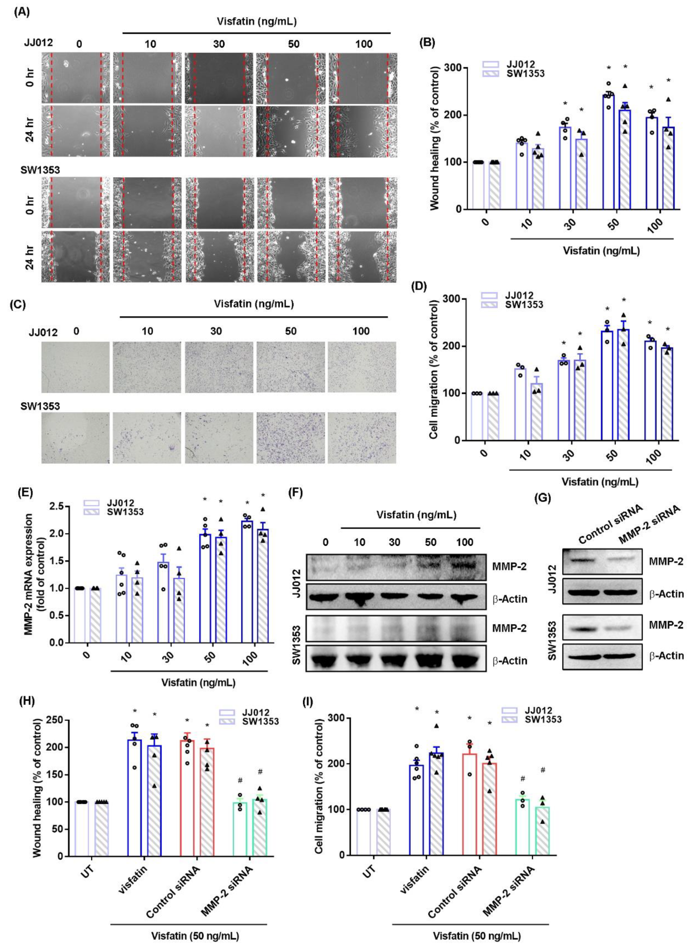
2.1. Visfatin Facilitates MMP-2-Dependent Migration in Chondrosarcoma Cells

Visfatin facilitates the progression and survival of several different types of cancers [12]. MMP-2 reportedly regulates the invasion and metastasis of chondrosarcoma cells [7,16]. We initially found that visfatin treatment (10–100 ng/mL) promoted cellular migration ability of JJ012 and SW1353 cells, according to Transwell and wound healing assay data (Figure 1A–D). We also found that stimulating the cells with visfatin enhanced mRNA expression and protein synthesis of MMP-2 (Figure 1E,F). In addition, we found that mRNA expression and protein synthesis of MMP-2, as well as wound healing and cell migration ability, were similar when we treated chondrosarcoma cells with visfatin 50 or 100 ng/mL, so we chose 50 ng/mL in all further experiments. Transfecting the cells with MMP-2 siRNA diminished visfatin-induced promotion of cellular migration (Figure 1G–I), implying that MMP-2 is critical to the effects of visfatin upon chondrosarcoma cells.

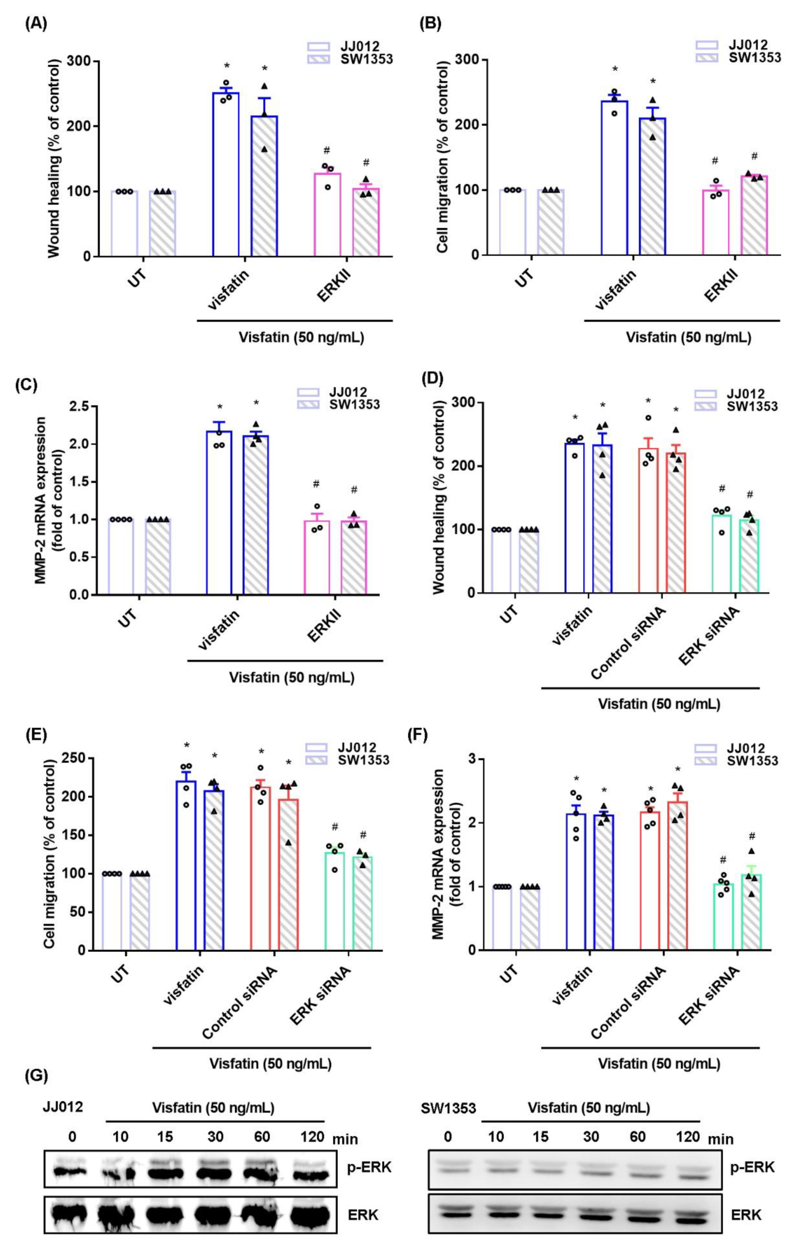
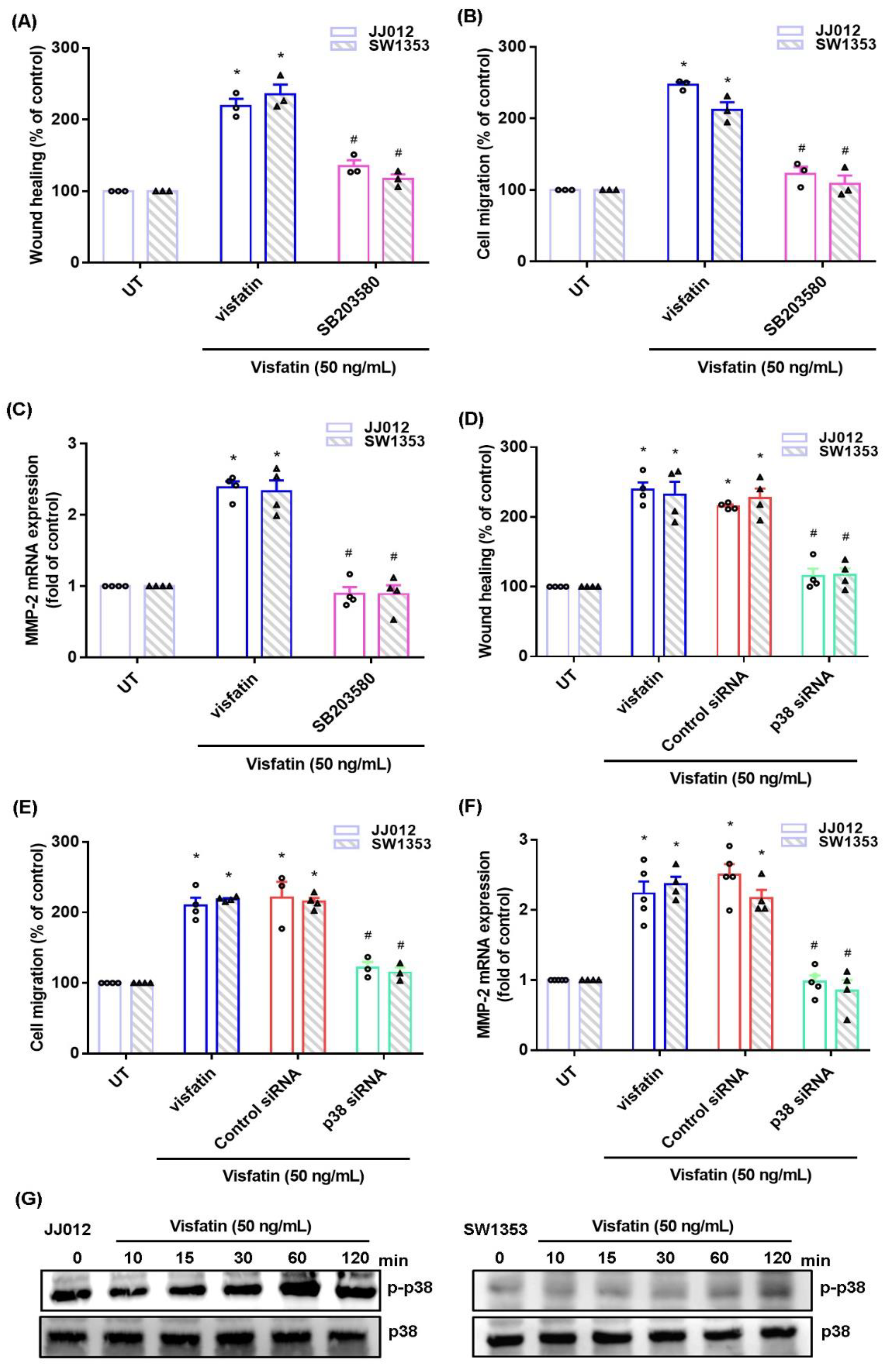
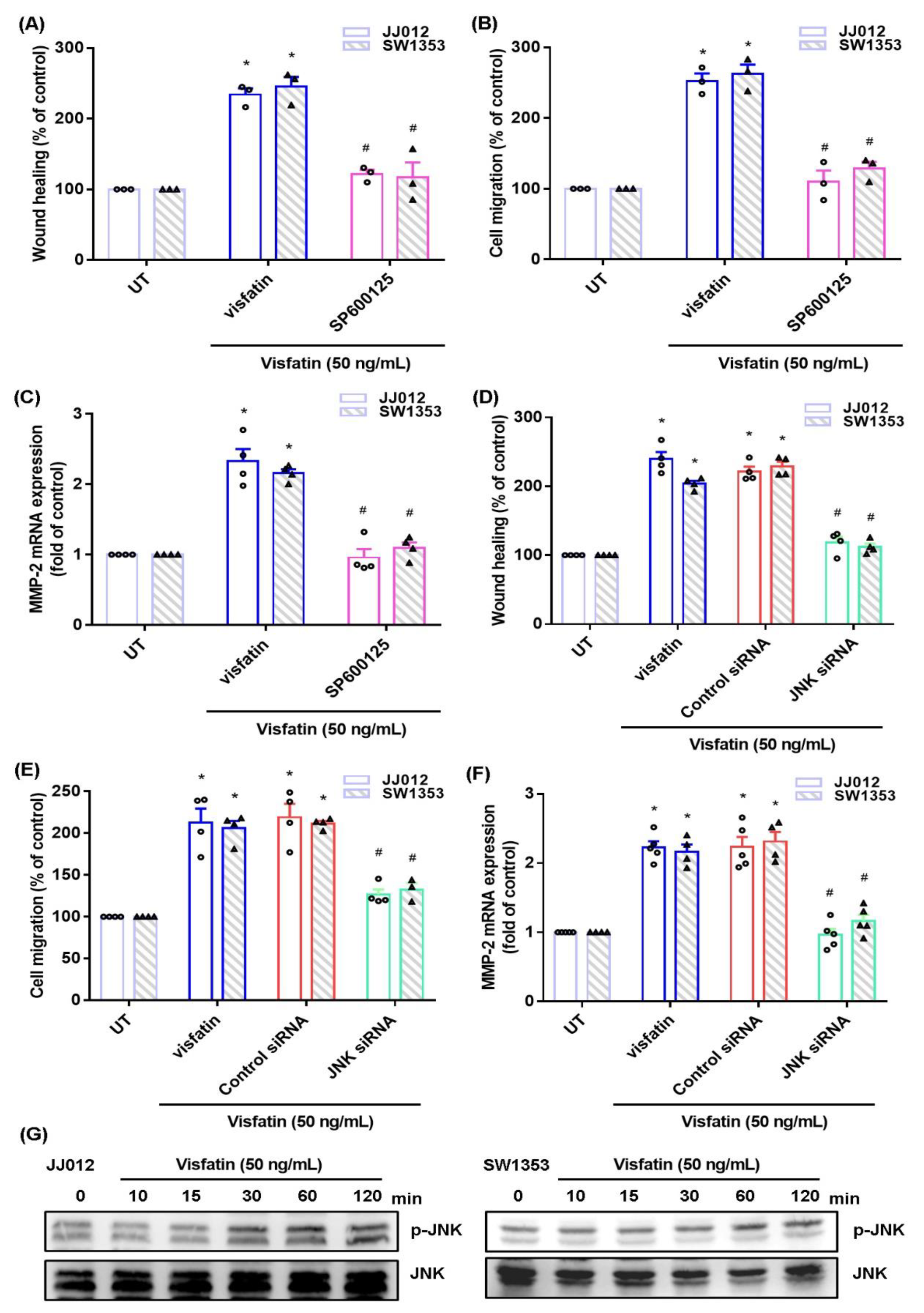
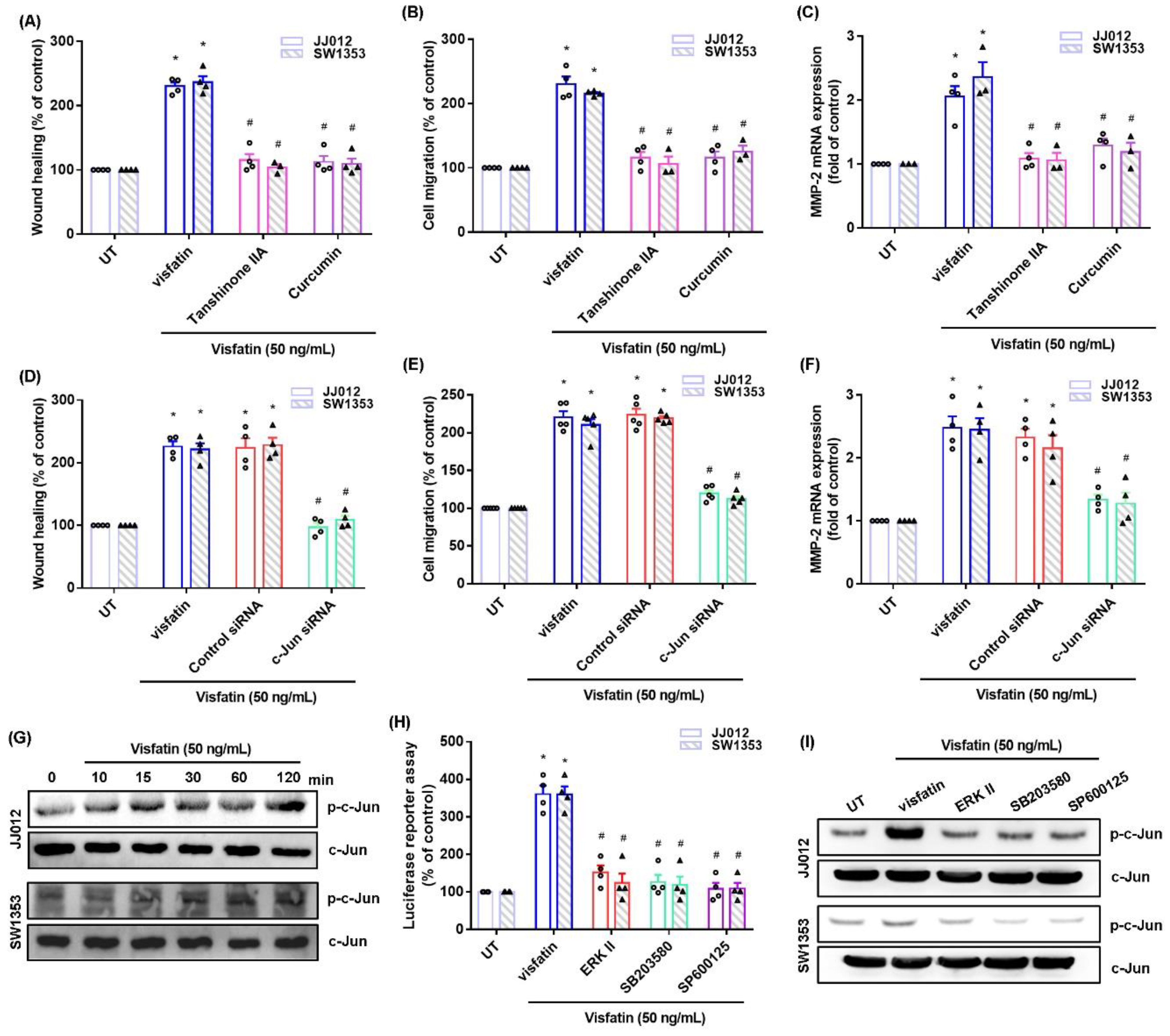
2.2. The MAPK Signaling Pathway Mediates the Effects of Visfatin upon MMP-2 Synthesis and Migration of Human Chondrosarcoma Cells

The MAPK (ERK, p38, and JNK) signaling pathway is critical in chondrosarcoma metastasis [17,18]. Treating cells with ERK, p38, or JNK inhibitor (ERK II, SB203580, or SP600125) significantly antagonized visfatin-induced stimulation of cell migration and MMP-2 production (Figure 2A–C, Figure 3A–C and Figure 4A–C). Similar effects were observed when the cell lines were transfected with ERK, p38 or c-JNK siRNAs (Figure 2D–F, Figure 3D–F and Figure 4D–F). Visfatin stimulation time-dependently promoted ERK, p38, and JNK phosphorylation (Figure 2G, Figure 3G and Figure 4G).

2.3. AP-1-Mediated MMP-2 Expression Regulates Visfatin-Induced Stimulation of Chondrosarcoma Cell Migration

AP-1 transcriptional activity is critical for MMP-2-mediated cancer migratory activities [18,19]. AP-1 inhibitor treatment with tanshinone IIA or curcumin effectively inhibited visfatin-induced cellular migratory activities and lowered MMP-2 expression (Figure 5A–C). The same effects were seen when the cells were transfected with siRNA against c-Jun (Figure 5D–F), whereas visfatin stimulation facilitated the phosphorylation of c-Jun (Figure 5G). ERK, p38, and JNK inhibitors all suppressed visfatin-enhanced promotion of c-Jun phosphorylation and AP-1 luciferase activity (Figure 5H,I). Therefore, visfatin increases chondrosarcoma cell migration through AP-1-dependent MMP-2 production in the MAPK pathway.

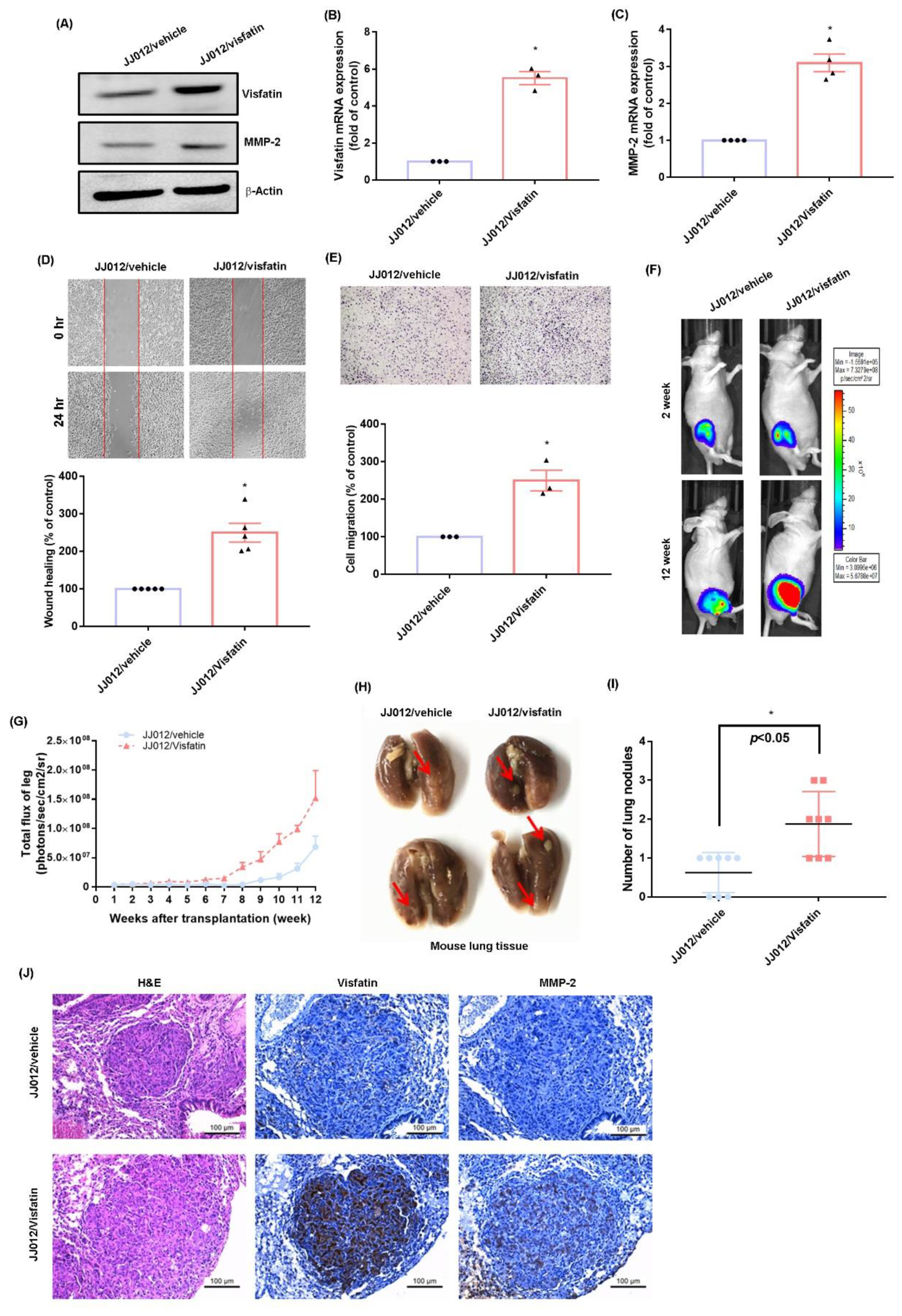
2.4. Overexpression of Visfatin Facilitates the Metastasis of Chondrosarcoma Cells in the Mouse Lung
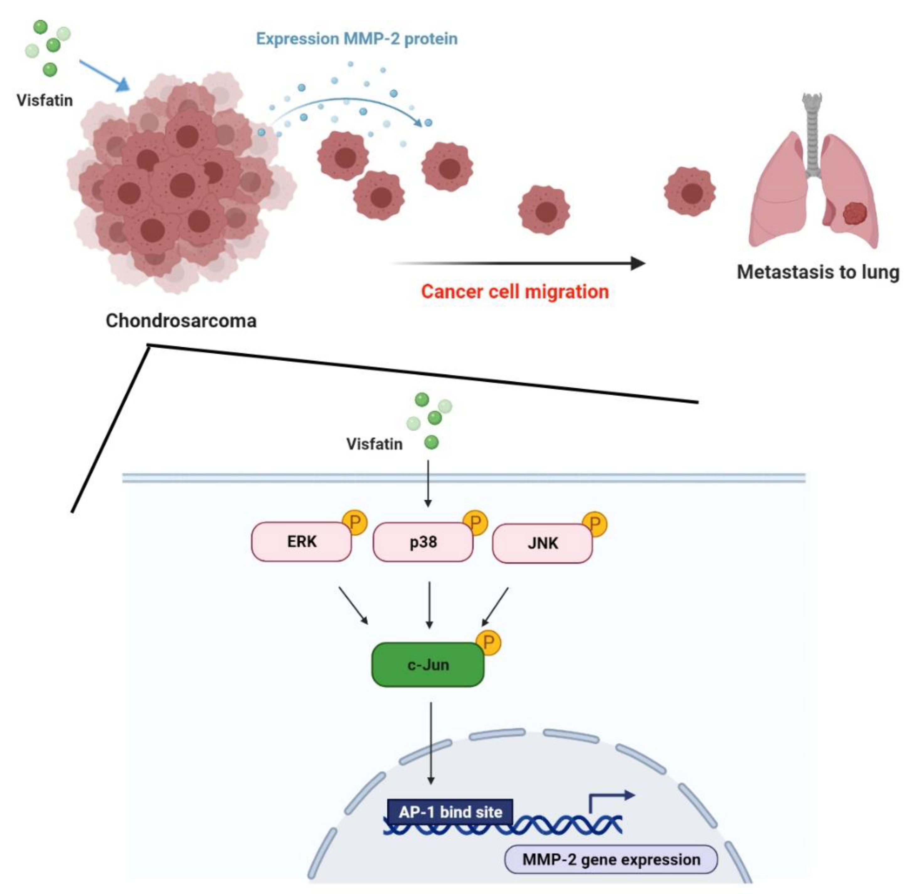
We used the orthotopic in vivo model of chondrosarcoma lung metastasis to examine the stimulatory effects of visfatin in metastatic chondrosarcoma [20]. JJ012/visfatin cells expressed high mRNA and protein levels of both visfatin and MMP-2, and they exhibited a high migratory ability (Figure 6A–E). JJ012/vehicle and JJ012/visfatin cells were orthotopically implanted into the right leg tibia and tumor size was monitored by the IVIS system (Figure 6F,G). Overexpression of visfatin significantly increased tumor growth in the tibia (Figure 6F,G). At 12 weeks, metastasis to the lung was significantly more likely with JJ012/visfatin cells than with JJ012/vehicle cells (Figure 6H,I). IHC results revealed significant increases visfatin and MMP-2 expression in the JJ012/visfatin orthotopic model (Figure 6J), confirming that visfatin facilitates the metastasis of chondrosarcoma to the mouse lung.

3. Discussion

Chondrosarcoma is a malignant bone neoplasm that constitutes almost one-third (~26%) of all bone cancers [21]. Chemotherapy and radiotherapy have very limited effectiveness, so treatment with surgery is therefore the major management modality for chondrosarcoma. This malignancy is notorious for its aggressive clinical course and propensity to metastasize [22]. An effective adjuvant remedy is urgently needed to suppress chondrosarcoma metastasis [1,3].
Visfatin, also known as nicotinamide phosphoribosyltransferase (NAMPT) or pre-B cell colony-enhancing factor (PBEF), is an adipocytokine with several intriguing properties. Visfatin essentially affects the biosynthesis of nicotinamide adenine dinucleotide (NAD) and causes NAD+ (the oxidized form of NAD) accumulation, thereby affecting many NAD-dependent proteins such as sirtuins, PARPs, MARTs, and CD38/157, promoting the synthesis of adenosine triphosphate (ATP) [10]. Such effects ultimately lead to drug resistance in cancer cells with enhanced proliferation and metastasis. Some research has reported that high levels of visfatin expression enhance NAD+ production and increase the proliferation of colorectal cancer cells by activating the Wnt/β-catenin pathway [23]. In gastric cancer, visfatin facilitates cell migratory and invasive abilities, as well as the EMT process, by targeting the SNAIL transcription factor SNAI1 via NF-κB signaling [24]. Thus, visfatin is critical for tumor cell proliferation, migration, and survival [24,25], although the effects of visfatin in chondrosarcoma metastasis are uncertain. Our cellular and preclinical investigations found that visfatin reliably promotes chondrosarcoma metastasis. We also confirmed that visfatin facilitates chondrosarcoma cell migration through AP-1-dependent MMP-2 expression in the MAPK signaling pathway.
Elevated MMP-2 levels are apparent in numerous tumor metastatic processes [26] and MMP-2 reportedly mediates chondrosarcoma metastasis [7,18]. Researchers have therefore proposed the targeting of MMP-2 in the management of tumor metastasis [26,27]. Here, we found that visfatin increases levels of mRNA and protein synthesis in chondrosarcoma cells. Genetic knockdown of MMP-2 reduced the effects of visfatin upon chondrosarcoma cell migratory activity. We also observed that overexpression of visfatin facilitated increases in MMP-2 synthesis and chondrosarcoma metastasis in vitro and in vivo, leading us to believe that visfatin promotes chondrosarcoma metastasis via the upregulation of MMP-2.
Activation of the MAPK pathway is important in the adjustment of different cellular effects [28,29]. This signaling pathway also regulates the expression of MMP-mediated cancer motility [30,31]. Our results show that visfatin promotes the phosphorylation of ERK, p38, and JNK, while their respective pharmacological inhibitors suppress visfatin-induced promotion of MMP-2 expression and chondrosarcoma cell migration. This phenomenon is confirmed by similar effects observed with genetic siRNAs of ERK, p38, and JNK. This evidence reveals that activation of ERK, p38, and JNK signaling controls visfatin-enhanced promotion of MMP-2 synthesis and migration of chondrosarcoma cells.
Numerous transcription factor-binding sites have been reported in the 5′-regulatory region of MMP-2 [32]. AP-1 is an important transcriptional factor that regulates MMP-2 transcriptional ability and tumor metastasis [33]. We found that both AP-1 inhibitors (tanshinone IIA and curcumin) inhibited visfatin-induced facilitation of MMP-2 synthesis and chondrosarcoma cell migration. Confirmation of these effects by genetic inhibition using c-Jun siRNA indicate that AP-1 transcriptional activation is involved in the effects of visfatin upon MMP-2 expression and chondrosarcoma cell migration. We also observed that visfatin enhances c-Jun phosphorylation and AP-1 luciferase activity. The pharmacological inhibitors of ERK, p38, and JNK all antagonized visfatin-mediated activities, suggesting that visfatin promotes AP-1-dependent MMP-2 production and chondrosarcoma migration through ERK, p38, and JNK signaling.

4. Materials and Methods

4.1. Materials

Visfatin, MMP-2, ERK, p38, JNK, c-Jun, and β-actin antibodies were obtained from GeneTex (Hsinchu, Taiwan). The phosphorylated forms of ERK, p38, JNK, and c-Jun antibodies were bought from Cell Signaling (Danvers, MA, USA). Taqman® One-Step PCR Master Mix and qPCR primers and probes were bought from Applied Biosystems (Foster City, CA, USA). Recombinant human visfatin was obtained from PeproTech (Rocky Hill, NJ, USA). All other chemicals not already mentioned were acquired from Sigma–Aldrich (St. Louis, MO, USA).

4.2. Cell Culture

The human chondrosarcoma cell line SW1353 was bought from ATCC (Manassas, VA, USA), while the other chondrosarcoma cell line JJ012 was kindly provided by Dr. Sean P. Scully (University of Miami School of Medicine, Miami, FL, USA). JJ012 cells stably expressing the visfatin complementary DNA (cDNA) clone (JJ012/visfatin cells) were established according to our previous method [20]. Cells were cultured 50%/50% in DMEM/α-MEM, 10% FBS, and antibiotics, then maintained in a humidified incubator at 37 °C in 5% CO2.

4.3. Cell Migration Assay

Chondrosarcoma cells were cultured on the upper chambers of Transwell plates (Costar, NY, USA), while visfatin and pharmaceutical inhibitors were treated to the lower chambers. After 18 h of treatment, migrated cells were fixed with 3.7% formaldehyde and stained with crystal violet, finally counted manually under the microscope [34,35].

4.4. Wound Healing Assay

The confluent chondrosarcoma monolayer was scratched by a fine pipette tip to make extended scratches. Cells were then treated with the conditions as indicated, migratory activity was evaluated by microscopy after 24 h and the rate of wound closure was analyzed [36].

4.5. Western Blot

SDS-PAGE was used to resolve the extracted proteins, which were transferred to PVDF membranes, as described in our previous publications [37,38,39]. Membranes were blocked for 1 h with PBST containing 4% non-fat milk, then treated with antibodies targeting visfatin, MMP-2, and β-actin for 1 h, before being incubated for 1 h with HRP-conjugated secondary antibodies. We visualized the blot membranes using a Fujifilm LAS-4000 imaging system (GE Healthcare, Little Chalfont, UK).

4.6. mRNA and miRNA Quantification

Total RNA was extracted from chondrosarcoma cell lines using TRIzol kit (MDBio, Taipei, Taiwan) and RNA contents were examined by a NanoVue Plus spectrophotometer (GE Healthcare Life Sciences; Pittsburgh, PA, USA). The M-MLV RT kit (Thermo Fisher Scientific; Waltham, MA, USA) and the Mir-X™ miRNA First-Strand Synthesis kit were used to perform reverse transcription of total RNA into cDNA. Real-time PCR analysis was performed using SYBR with MMP-2 or GAPDH primers. GAPDH mRNA expression was used as an internal control. The specific primer sequences for these genes are as follows: MMP-2: 5’-GGCCCTGTCACTCCTGAGAT-3’ (forward), 5’-GGCATCCAGGTTATCGGGG A-3’ (reverse), GAPDH: 5’-AATGGACAACTGGTCGTGGAC-3’ (forward), and 5’-CCCTCCAGGGGATCTGTTTG-3’ (reverse). Quantitative real-time PCR (qPCR) analysis was examined according to our previous reports [40,41].

4.7. Luciferase Assay

Chondrosarcoma cells were transfected with the AP-1 luciferase plasmid (Stratagene; St. Louis, MO, USA) using Lipofectamine 2000, then stimulated with pharmaceutical inhibitors and visfatin for 24 h. Luciferase activity was monitored using a luciferase assay kit [40,42,43].

4.8. Small Interfering RNA (siRNA) Transfection

Knockdown efficiency was confirmed by wound healing, Transwell cell migration, real-time PCR, and Western blot assay, as described in the following sections. Cells were transfected with control, p38, JNK, ERK, c-Jun, or MMP-2 siRNAs, as according to the manufacturers’ recommendations in standard procedures. The siRNAs (ON-TARGETplus SMARTpool) were purchased from GE Dharmacon (Lafayette, CO, USA). Cells were transfected with siRNAs using Lipofectamine 2000 reagent [44].

4.9. Establishment of Stably Transfected Cells

JJ012/visfatin or JJ012/vehicle (control shRNA) plasmids were obtained from MDBio, Inc. (Taipei, Taiwan) and transfected into cancer cells using Lipofectamine 2000 transfection reagent. Twenty-four hours after transfection, stable transfectants were selected in G418 (Geneticin) (Life Technologies, Grand Island, NY, USA) at a concentration of 200 μg/mL. Thereafter, the selection medium was replaced every 3 days. After 2 weeks of selection in G418, clones of resistant cells were isolated.

4.10. Tumor Xenograft Study

JJ012/vehicle or JJ012/visfatin cells (5  ×  106) were orthotopically injected into 4-week-old male BALB/c nude mice (Taipei’s National Laboratory Animal Center), according to a previous protocol [20]. Tumor growth in the tibiae was examined each week by bioluminescence imaging by Xenogen IVIS imaging system 200 (PerkinElmer, MA, USA). After 12 weeks, the mice were sacrificed by CO2 inhalation. The lungs were then excised for further examination. All animal procedures were approved and performed in accordance with the guidelines of the Institutional Animal Care and Use Committee of China Medical University (CMUIACUC-2019-079).

4.11. Immunohistochemistry (IHC) Staining

Mouse lung tissues were rehydrated and treated with primary anti-visfatin or MMP-2 antibodies, then incubated with biotin-labeled secondary antibody. The slides were treated with the ABC kit (Vector Laboratories, CA, USA) and then photographed using the microscope.

4.12. Statistical Analysis

All values are presented as the mean ± standard deviation (SD) Differences between two experimental groups were assessed for significance using the Student’s t-test and considered to be significant if the p value was <0.05.

5. Conclusions

In conclusion, our study has identified that visfatin facilitates the metastatic potential of chondrosarcoma cells via AP-1-dependent MMP-2 production in the ERK, p38, and JNK pathway (Figure 7). We believe that targeting visfatin expression in metastatic chondrosarcoma offers a new way to address this aggressive malignancy.

Author Contributions

Methodology, S.-Y.H., C.-Y.L., and C.-C.Y.; validation, H.-T.C., M.-Y.L., and Y.-C.F.; formal analysis, Y.-W.H.; resources, H.-T.C., M.-Y.L., and Y.-C.F.; data curation, J.-F.L., S.-W.W., and W.-C.C.; writing—original draft preparation, C.-H.T.; visualization, S.-Y.H., C.-Y.L., and C.-C.Y.; funding acquisition, H.-T.C., M.-Y.L., and C.-H.T. All authors have read and agreed to the published version of the manuscript.

Funding

This work was supported by grants from the Ministry of Science and Technology of Taiwan (MOST 107-2320-B-039-019-MY3), China Medical University Hospital (DMR-109-013, DMR-110-017, DMR-110-158, DMR-110-105).

Institutional Review Board Statement

The authors declare that all data supporting the findings of this study are available within the article. All animal procedures were approved and performed in accordance with the guidelines of the Institutional Animal Care and Use Committee of China Medical University (CMUIACUC-2019-079).

Informed Consent Statement

None of the authors of this paper has any financial or personal relationships with other people or organizations that could inappropriately influence this work.

Data Availability Statement

The datasets used and/or analyzed during this study are available from the corresponding authors upon reasonable request.

Acknowledgments

We would like to thank Iona J. MacDonald from China Medical University for her English language revision of this manuscript.

Conflicts of Interest

The authors declare no conflict of interest.

References

  1. Macdonald, I.J.; Lin, C.-Y.; Kuo, S.-J.; Su, C.-M.; Tang, C.-H. An update on current and future treatment options for chondrosarcoma. Expert Rev. Anticancer Ther. 2019, 19, 773–786. [Google Scholar] [CrossRef]
  2. Chen, P.-C.; Cheng, H.-C.; Yang, S.-F.; Lin, C.-W.; Tang, C.-H. The CCN Family Proteins: Modulators of Bone Development and Novel Targets in Bone-Associated Tumors. BioMed Res. Int. 2014, 2014, 1–11. [Google Scholar] [CrossRef]
  3. Chen, J.-C.; Fong, Y.-C.; Tang, C.-H. Novel Strategies for the Treatment of Chondrosarcomas: Targeting Integrins. BioMed Res. Int. 2013, 2013, 1–11. [Google Scholar] [CrossRef]
  4. Ferguson, J.L.; Turner, S.P. Bone Cancer: Diagnosis and Treatment Principles. Am. Fam. Physician 2018, 98, 205–213. [Google Scholar]
  5. Fan, T.M.; Roberts, R.D.; Lizardo, M.M. Understanding and Modeling Metastasis Biology to Improve Therapeutic Strategies for Combating Osteosarcoma Progression. Front. Oncol. 2020, 10, 13. [Google Scholar] [CrossRef] [PubMed] [Green Version]
  6. Steeg, P.S. Targeting metastasis. Nature reviews. Cancer 2016, 16, 201–218. [Google Scholar]
  7. Hsu, C.-J.; Wu, M.-H.; Chen, C.-Y.; Tsai, C.-H.; Hsu, H.-C.; Tang, C.-H. AMP-activated protein kinase activation mediates CCL3-induced cell migration and matrix metalloproteinase-2 expression in human chondrosarcoma. Cell Commun. Signal. 2013, 11, 68. [Google Scholar] [CrossRef] [PubMed] [Green Version]
  8. Tsou, H.-K.; Chen, H.-T.; Hung, Y.-H.; Chang, C.-H.; Li, T.-M.; Fong, Y.-C.; Tang, C.-H. HGF and c-Met Interaction Promotes Migration in Human Chondrosarcoma Cells. PLoS ONE 2013, 8, e53974. [Google Scholar] [CrossRef] [PubMed] [Green Version]
  9. Khaidizar, F.; Bessho, Y.; Nakahata, Y. Nicotinamide Phosphoribosyltransferase as a Key Molecule of the Aging/Senescence Process. Int. J. Mol. Sci. 2021, 22, 3709. [Google Scholar] [CrossRef]
  10. Dakroub, A.; Nasser, S.A.; Younis, N.; Bhagani, H.; Al-Dhaheri, Y.; Pintus, G.; Eid, A.A.; El-Yazbi, A.F.; Eid, A.H. Visfatin: A Possible Role in Cardiovasculo-Metabolic Disorders. Cells 2020, 9, 2444. [Google Scholar] [CrossRef] [PubMed]
  11. Mohammadi, M.; Mianabadi, F.; Mehrad-Majd, H. Circulating visfatin levels and cancers risk: A systematic review and meta-analysis. J. Cell. Physiol. 2018, 234, 5011–5022. [Google Scholar] [CrossRef] [PubMed]
  12. Lin, T.-C. The role of visfatin in cancer proliferation, angiogenesis, metastasis, drug resistance and clinical prognosis. Cancer Manag. Res. 2019, 11, 3481–3491. [Google Scholar] [CrossRef] [PubMed] [Green Version]
  13. Liu, H.; Liu, N.; Zhao, Y.; Zhu, X.; Wang, C.; Liu, Q.; Gao, C.; Zhao, X.; Li, J. Oncogenic USP22 supports gastric cancer growth and metastasis by activating c-Myc/NAMPT/SIRT1-dependent FOXO1 and YAP signaling. Aging 2019, 11, 9643–9660. [Google Scholar] [CrossRef] [PubMed]
  14. Zhang, H.; Zhang, N.; Liu, Y.; Su, P.; Liang, Y.; Li, Y.; Wang, X.; Chen, T.; Song, X.; Sang, Y.; et al. Epigenetic Regulation of NAMPT by NAMPT-AS Drives Metastatic Progression in Triple-Negative Breast Cancer. Cancer Res. 2019, 79, 3347–3359. [Google Scholar] [CrossRef] [Green Version]
  15. Ezzati-Mobaser, S.; Malekpour-Dehkordi, Z.; Nourbakhsh, M.; Tavakoli-Yaraki, M.; Ahmadpour, F.; Golpour, P.; Nourbakhsh, M. The up-regulation of markers of adipose tissue fibrosis by visfatin in pre-adipocytes as well as obese children and adolescents. Cytokine 2020, 134, 155193. [Google Scholar] [CrossRef]
  16. Roomi, M.W.; Kalinovsky, T.; Monterrey, J.; Rath, M.; Niedzwiecki, A. In vitro modulation of MMP-2 and MMP-9 in adult human sarcoma cell lines by cytokines, inducers and inhibitors. Int. J. Oncol. 2013, 43, 1787–1798. [Google Scholar] [CrossRef] [Green Version]
  17. Wu, M.H.; Chen, L.-M.; Hsu, H.-H.; Lin, J.A.; Lin, Y.-M.; Tsai, F.-J.; Tsai, C.-H.; Huang, C.-Y.; Tang, C.-H. Endothelin-1 enhances cell migration through COX-2 up-regulation in human chondrosarcoma. Biochim. Biophys. Acta Gen. Subj. 2013, 1830, 3355–3364. [Google Scholar] [CrossRef]
  18. Hou, C.-H.; Chiang, Y.-C.; Fong, Y.-C.; Tang, C.-H. WISP-1 increases MMP-2 expression and cell motility in human chondrosarcoma cells. Biochem. Pharmacol. 2011, 81, 1286–1295. [Google Scholar] [CrossRef]
  19. Zheng, P.; Chen, X.; Xie, J.; Chen, X.; Lin, S.; Ye, L.; Chen, L.; Lin, J.; Yu, X.; Zheng, M. Capn4 is induced by and required for Epstein-Barr virus latent membrane protein 1 promotion of nasopharyngeal carcinoma metastasis through ERK/AP-1 signaling. Cancer Sci. 2019, 111, 72–83. [Google Scholar] [CrossRef] [Green Version]
  20. Tzeng, H.E.; Tang, C.H.; Wu, S.H.; Chen, H.T.; Fong, Y.C.; Lu, Y.C.; Chen, W.C.; Huang, H.D.; Lin, C.Y.; Wang, S.W. CCN6-mediated MMP-9 activation enhances metastatic potential of human chondrosarcoma. Cell Death Dis. 2018, 9, 955. [Google Scholar] [CrossRef] [Green Version]
  21. Leddy, L.R.; Holmes, R.E. Chondrosarcoma of bone. Cancer Treat. Res. 2014, 162, 117–130. [Google Scholar] [PubMed]
  22. Gelderblom, H.; Hogendoorn, P.C.; Dijkstra, S.D.; van Rijswijk, C.S.; Krol, A.D.; Taminiau, A.H.; Bovee, J.V. The clinical approach towards chondrosarcoma. Oncologist 2008, 13, 320–329. [Google Scholar] [CrossRef] [PubMed]
  23. Ye, C.; Qi, L.; Li, X.; Wang, J.; Yu, J.; Zhou, B.; Guo, C.; Chen, J.; Zheng, S. Targeting the NAD(+) salvage pathway suppresses APC mutation-driven colorectal cancer growth and Wnt/beta-catenin signaling via increasing Axin level. Cell Commun. Signal. CCS 2020, 18, 16. [Google Scholar] [CrossRef] [Green Version]
  24. Cao, D.; Chu, L.; Xu, Z.; Gong, J.; Deng, R.; Wang, B.; Zhou, S. Visfatin facilitates gastric cancer malignancy by targeting snai1 via the NF-kappaB signaling. Hum. Exp. Toxicol. 2021, 09603271211006168. [Google Scholar] [CrossRef]
  25. Li, Y.; Yu, C.; Deng, W. Roles and mechanisms of adipokines in drug resistance of tumor cells. Eur. J. Pharmacol. 2021, 899, 174019. [Google Scholar] [CrossRef] [PubMed]
  26. Yang, J.-S.; Lin, C.-W.; Su, S.-C.; Yang, S.-F. Pharmacodynamic considerations in the use of matrix metalloproteinase inhibitors in cancer treatment. Expert Opin. Drug Metab. Toxicol. 2016, 12, 191–200. [Google Scholar] [CrossRef] [PubMed]
  27. Zhong, Y.; Lu, Y.-T.; Sun, Y.; Shi, Z.-H.; Li, N.-G.; Tang, Y.-P.; Duan, J.-A. Recent opportunities in matrix metalloproteinase inhibitor drug design for cancer. Expert Opin. Drug Discov. 2017, 13, 75–87. [Google Scholar] [CrossRef] [PubMed]
  28. Wang, Y.-H.; Kuo, S.-J.; Liu, S.-C.; Wang, S.-W.; Tsai, C.-H.; Fong, Y.-C.; Tang, C.-H. Apelin Affects the Progression of Osteoarthritis by Regulating VEGF-Dependent Angiogenesis and miR-150-5p Expression in Human Synovial Fibroblasts. Cells 2020, 9, 594. [Google Scholar] [CrossRef] [Green Version]
  29. Chen, C.-Y.; Su, C.-M.; Huang, Y.-L.; Tsai, C.-H.; Fuh, L.-J.; Tang, C.-H. CCN1 Induces Oncostatin M Production in Osteoblasts via Integrin-Dependent Signal Pathways. PLoS ONE 2014, 9, e106632. [Google Scholar] [CrossRef] [PubMed]
  30. Zhu, X.; Wang, K.; Chen, Y. Ophiopogonin D suppresses TGF-beta1-mediated metastatic behavior of MDA-MB-231 breast carcinoma cells via regulating ITGB1/FAK/Src/AKT/beta-catenin/MMP-9 signaling axis. Toxicol. Vitr. 2020, 69, 104973. [Google Scholar] [CrossRef]
  31. Chuang, C.-Y.; Ho, Y.-C.; Lin, C.-W.; Yang, W.-E.; Yu, Y.-L.; Tsai, M.-C.; Yang, S.-F.; Su, S.-C. Salvianolic acid A suppresses MMP-2 expression and restrains cancer cell invasion through ERK signaling in human nasopharyngeal carcinoma. J. Ethnopharmacol. 2020, 252, 112601. [Google Scholar] [CrossRef] [PubMed]
  32. Egeblad, M.; Werb, Z. New functions for the matrix metalloproteinases in cancer progression. Nat. Rev. Cancer 2002, 2, 161–174. [Google Scholar] [CrossRef] [PubMed]
  33. Jie, Z.; Xie, Z.; Zhao, X.; Sun, X.; Yu, H.; Pan, X.; Shen, S.; Qin, A.; Fang, X.; Fan, S. Glabridin inhibits osteosarcoma migration and invasion via blocking the p38- and JNK-mediated CREB-AP1 complexes formation. J. Cell. Physiol. 2018, 234, 4167–4178. [Google Scholar] [CrossRef] [PubMed]
  34. Lee, H.-P.; Wang, S.-W.; Wu, Y.-C.; Lin, L.-W.; Tsai, F.-J.; Yang, J.-S.; Li, T.-M.; Tang, C.-H. Soya-cerebroside inhibits VEGF-facilitated angiogenesis in endothelial progenitor cells. Food Agric. Immunol. 2020, 31, 193–204. [Google Scholar] [CrossRef] [Green Version]
  35. Hou, C.-H.; Lin, F.-L.; Hou, S.-M.; Liu, J.-F. Cyr61 promotes epithelial-mesenchymal transition and tumor metastasis of osteosarcoma by Raf-1/MEK/ERK/Elk-1/TWIST-1 signaling pathway. Mol. Cancer 2014, 13, 1–13. [Google Scholar] [CrossRef] [Green Version]
  36. Liu, J.-F.; Lee, C.-W.; Tsai, M.-H.; Tang, C.-H.; Chen, P.-C.; Lin, L.-W.; Lin, C.-Y.; Lu, C.-H.; Lin, Y.-F.; Yang, S.-H.; et al. Thrombospondin 2 promotes tumor metastasis by inducing matrix metalloproteinase-13 production in lung cancer cells. Biochem. Pharmacol. 2018, 155, 537–546. [Google Scholar] [CrossRef]
  37. Lee, H.-P.; Chen, P.-C.; Wang, S.-W.; Fong, Y.-C.; Tsai, C.-H.; Tsai, F.-J.; Chung, J.-G.; Huang, C.-Y.; Yang, J.-S.; Hsu, Y.-M.; et al. Plumbagin suppresses endothelial progenitor cell-related angiogenesis in vitro and in vivo. J. Funct. Foods 2018, 52, 537–544. [Google Scholar] [CrossRef]
  38. Lee, H.-P.; Wang, S.-W.; Wu, Y.-C.; Tsai, C.-H.; Tsai, F.-J.; Chung, J.-G.; Huang, C.-Y.; Yang, J.-S.; Hsu, Y.-M.; Yin, M.-C.; et al. Glucocerebroside reduces endothelial progenitor cell-induced angiogenesis. Food Agric. Immunol. 2019, 30, 1033–1045. [Google Scholar] [CrossRef] [Green Version]
  39. Liu, S.C.; Tsai, C.H.; Wu, T.Y.; Tsai, C.H.; Tsai, F.J.; Chung, J.G.; Huang, C.Y.; Yang, J.S.; Hsu, Y.M.; Yin, M.C.; et al. Soya-cerebroside reduces IL-1 beta-induced MMP-1 production in chondrocytes and inhibits cartilage degradation: Implications for the treatment of osteoarthritis. Food Agric. Immunol. 2019, 30, 620–632. [Google Scholar] [CrossRef]
  40. Yang, Y.-C.; Chiou, P.-C.; Chen, P.-C.; Liu, P.-Y.; Huang, W.-C.; Chao, C.-C.; Tang, C.-H. Melatonin reduces lung cancer stemness through inhibiting of PLC, ERK, p38, β-catenin, and Twist pathways. Environ. Toxicol. 2018, 34, 203–209. [Google Scholar] [CrossRef]
  41. Wang, M.; Chao, C.-C.; Chen, P.-C.; Liu, P.-I.; Yang, Y.-C.; Su, C.-M.; Huang, W.-C.; Tang, C.-H. Thrombospondin enhances RANKL-dependent osteoclastogenesis and facilitates lung cancer bone metastasis. Biochem. Pharmacol. 2019, 166, 23–32. [Google Scholar] [CrossRef]
  42. Su, C.-M.; Tang, C.-H.; Chi, M.-J.; Lin, C.-Y.; Fong, Y.-C.; Liu, Y.-C.; Chen, W.-C.; Wang, S.-W. Resistin facilitates VEGF-C-associated lymphangiogenesis by inhibiting miR-186 in human chondrosarcoma cells. Biochem. Pharmacol. 2018, 154, 234–242. [Google Scholar] [CrossRef] [PubMed]
  43. Wu, T.J.; Lin, C.Y.; Tsai, C.H.; Huang, Y.L.; Tang, C.H. Glucose suppresses IL-1beta-induced MMP-1 expression through the FAK, MEK, ERK, and AP-1 signaling pathways. Environ. Toxicol. 2018, 33, 1061–1068. [Google Scholar] [CrossRef] [PubMed]
  44. Tsai, C.-H.; Yang, D.-Y.; Lin, C.-Y.; Chen, T.-M.; Tang, C.-H.; Huang, Y.-L. Sphingosine-1-phosphate suppresses chondrosarcoma metastasis by upregulation of tissue inhibitor of metalloproteinase 3 through suppressing miR-101 expression. Mol. Oncol. 2017, 11, 1380–1398. [Google Scholar] [CrossRef] [PubMed]
Figure 1. Visfatin promotes MMP-2-dependent cell migration in human chondrosarcoma. (AD) Cells were incubated with visfatin (10–100 ng/mL) and cell migration was examined by Transwell and wound healing assays. (E,F) Cells were incubated with visfatin (10–100 ng/mL) and levels of MMP-2 mRNA and protein expression were examined by qPCR and Western blot assays. (GI) Cells were transfected with MMP-2 siRNAs then stimulated with visfatin. Cell migration and MMP-2 expression levels were examined by Transwell, wound healing, and Western blot assays. Quantitative results are expressed as the mean ± SD. All experiments were repeated 3 to 5 times. * p < 0.05 compared with the UT group; # p < 0.05 compared with the visfatin-treated group. UT, untreated control.
Ijms 22 08642 g001
Figure 2. The ERK pathway mediates visfatin-induced MMP-2 expression and cell migration. (AF) Cells were pretreated with an ERK inhibitor (ERK II) or transfected with an ERK siRNA, then stimulated with visfatin. Cell migration and levels of MMP-2 expression were examined by Transwell, wound healing, and qPCR. (G) Cells were incubated with visfatin for the indicated time intervals; ERK phosphorylation was examined by Western blot. Quantitative results are expressed as the mean ± SD. All experiments were repeated 3 to 5 times. * p < 0.05 compared with the UT group; # p < 0.05 compared with the visfatin-treated group. UT, untreated control.
Ijms 22 08642 g002
Figure 3. The p38 pathway mediates visfatin-induced MMP-2 expression and cell migration. (AF) Cells were pretreated with a p38 inhibitor (SB253080) or transfected with a p38 siRNA, then stimulated with visfatin. Cell migration and levels of MMP-2 expression were examined by Transwell, wound healing, and qPCR. (G) Cells were incubated with visfatin for the indicated time intervals; p38 phosphorylation was examined by Western blot. Quantitative results are expressed as the mean ± SD. All experiments were repeated 3 to 5 times. * p < 0.05 compared with the UT group; # p < 0.05 compared with the visfatin-treated group. UT, untreated control.
Ijms 22 08642 g003
Figure 4. The JNK pathway mediates visfatin-induced MMP-2 expression and cell migration. (AF) Cells were pretreated with a JNK inhibitor (SP600125) or transfected with a JNK siRNA, then stimulated with visfatin. Cell migration and levels of MMP-2 expression were examined by Transwell, wound healing, and qPCR. (G) Cells were incubated with visfatin for the indicated time intervals; JNK phosphorylation was examined by Western blot. Quantitative results are expressed as the mean ± SD. All experiments were repeated 3 to 5 times. * p < 0.05 compared with the UT group; # p < 0.05 compared with the visfatin-treated group. UT, untreated control.
Ijms 22 08642 g004
Figure 5. Visfatin induces MMP-2 production and cell migration through AP-1 activation in chondrosarcoma cells. (AF) Cells were pretreated with AP-1 inhibitors (tanshinone IIA or curcumin) or transfected with a c-Jun siRNA, then stimulated with visfatin. Cell migration and levels of MMP-2 expression were examined by Transwell, wound healing, and qPCR. (G) Cells were incubated with visfatin for the indicated time intervals; c-Jun phosphorylation was examined by Western blot. (H,I) Cells were treated with indicated inhibitors then stimulated visfatin, the c-Jun phosphorylation and AP-1 luciferase activity was examined. Quantitative results are expressed as the mean ± SD. All experiments were repeated 3 to 5 times. * p < 0.05 compared with the UT group; # p < 0.05 compared with the visfatin-treated group. UT, untreated control.
Ijms 22 08642 g005
Figure 6. Visfatin promotes chondrosarcoma metastasis to lungs in vivo. (AE) MMP-2 levels and migratory ability of JJ012/vehicle and JJ012/visfatin cells were examined by qPCR, Western blot, and Transwell. (F,G) The mice were injected with JJ012/vehicle or JJ012/visfatin cells. Lung metastasis was monitored by bioluminescence imaging at the indicated time intervals, then quantified by photon images. (H,I) After 12 weeks, the mice were humanely sacrificed and the lung tissue was excised, photographed, and quantified. (J) Levels of visfatin and MMP-2 expression in lung tumors were subjected to IHC analysis. Quantitative results are expressed as the mean ± SD. All experiments were repeated 3 to 5 times. * p < 0.05 compared with the JJ012/vehicle group. JJ012/vehicle, control shRNA; JJ012/visfatin, visfatin overexpression.
Ijms 22 08642 g006
Figure 7. Schema illustrating the effects of visfatin in chondrosarcoma metastasis. Visfatin facilitates the metastatic potential of chondrosarcoma cells via AP-1-dependent MMP-2 production in the ERK, p38, and JNK pathways.
Ijms 22 08642 g007
Publisher’s Note: MDPI stays neutral with regard to jurisdictional claims in published maps and institutional affiliations.

