
Therapeutic Strategies for IVD Regeneration through Hyaluronan/SDF-1-Based Hydrogel and Intravenous Administration of MSCs

 , , , and
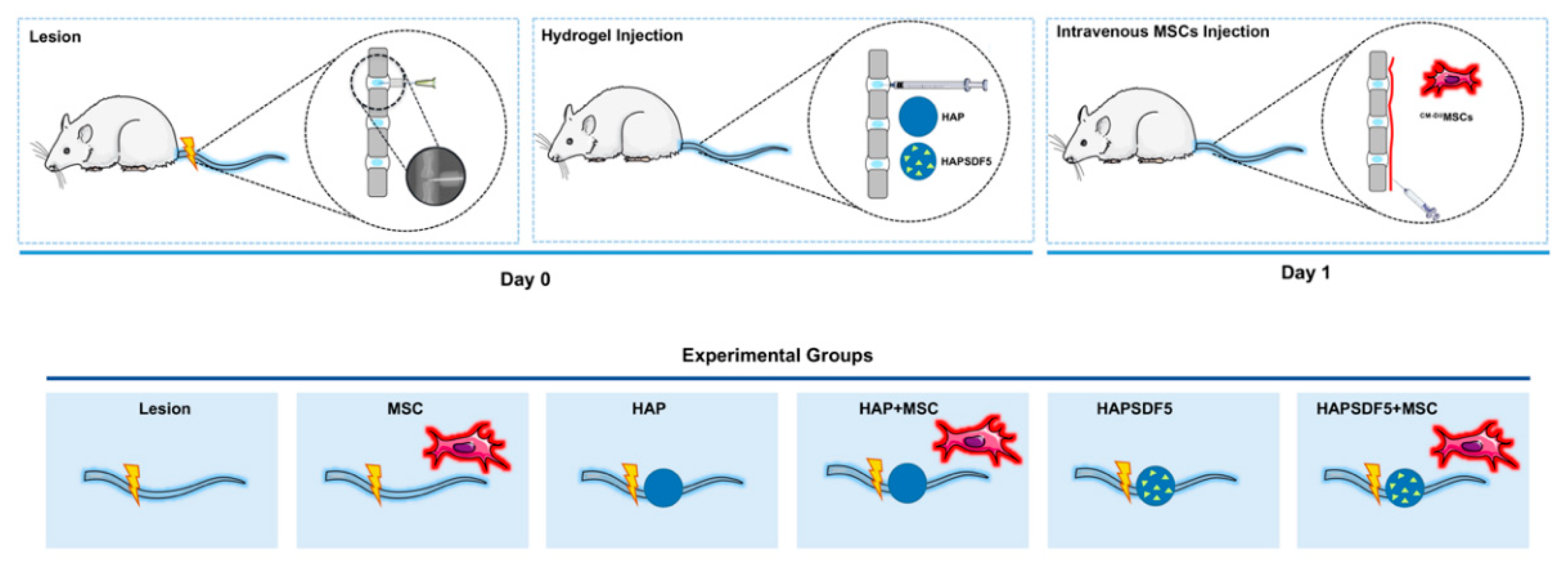
Abstract
:1. Introduction
2. Results
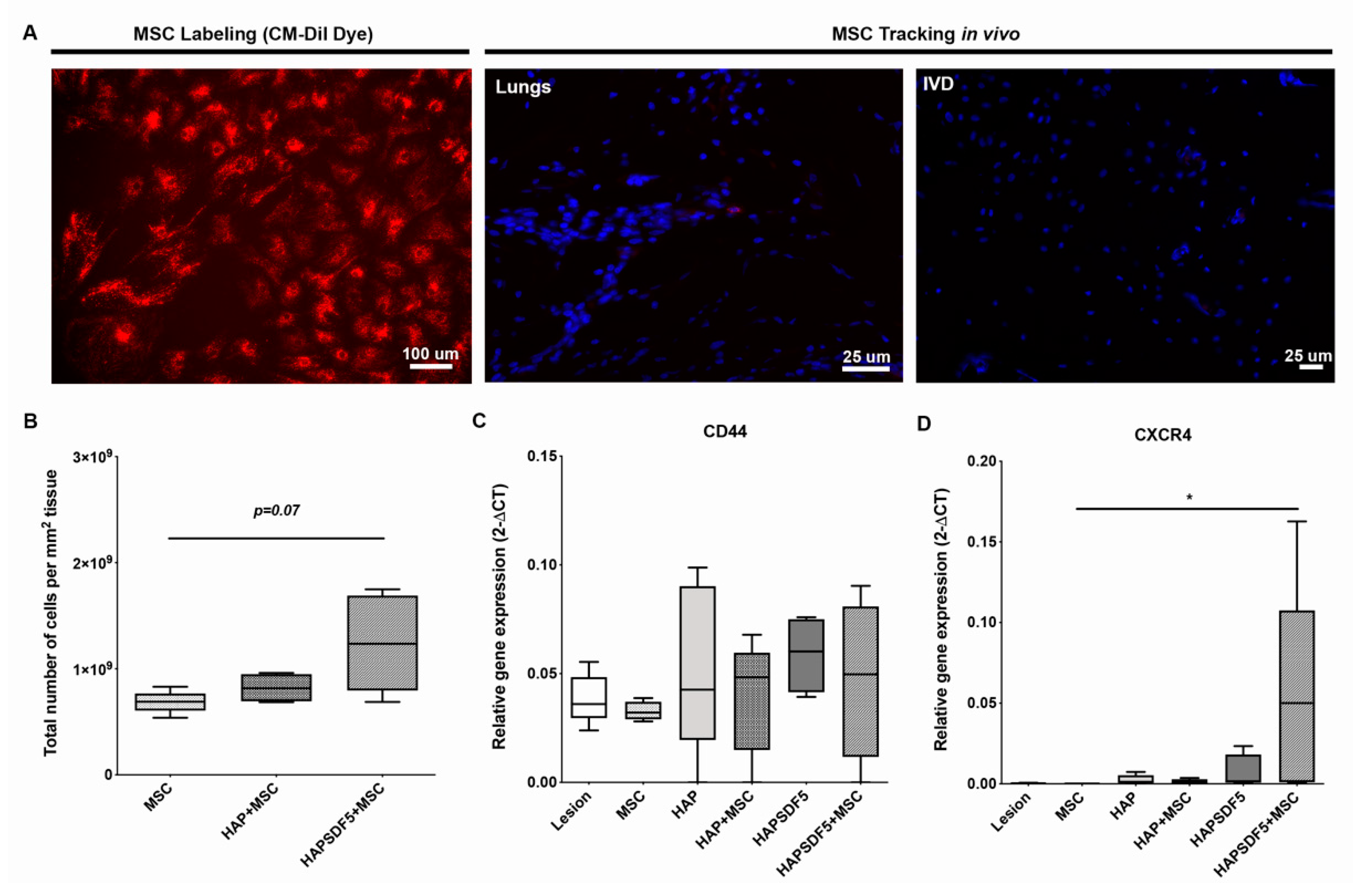
2.1. Potential of HAPSDF5 Intradiscal Injection to Recruit MSCs
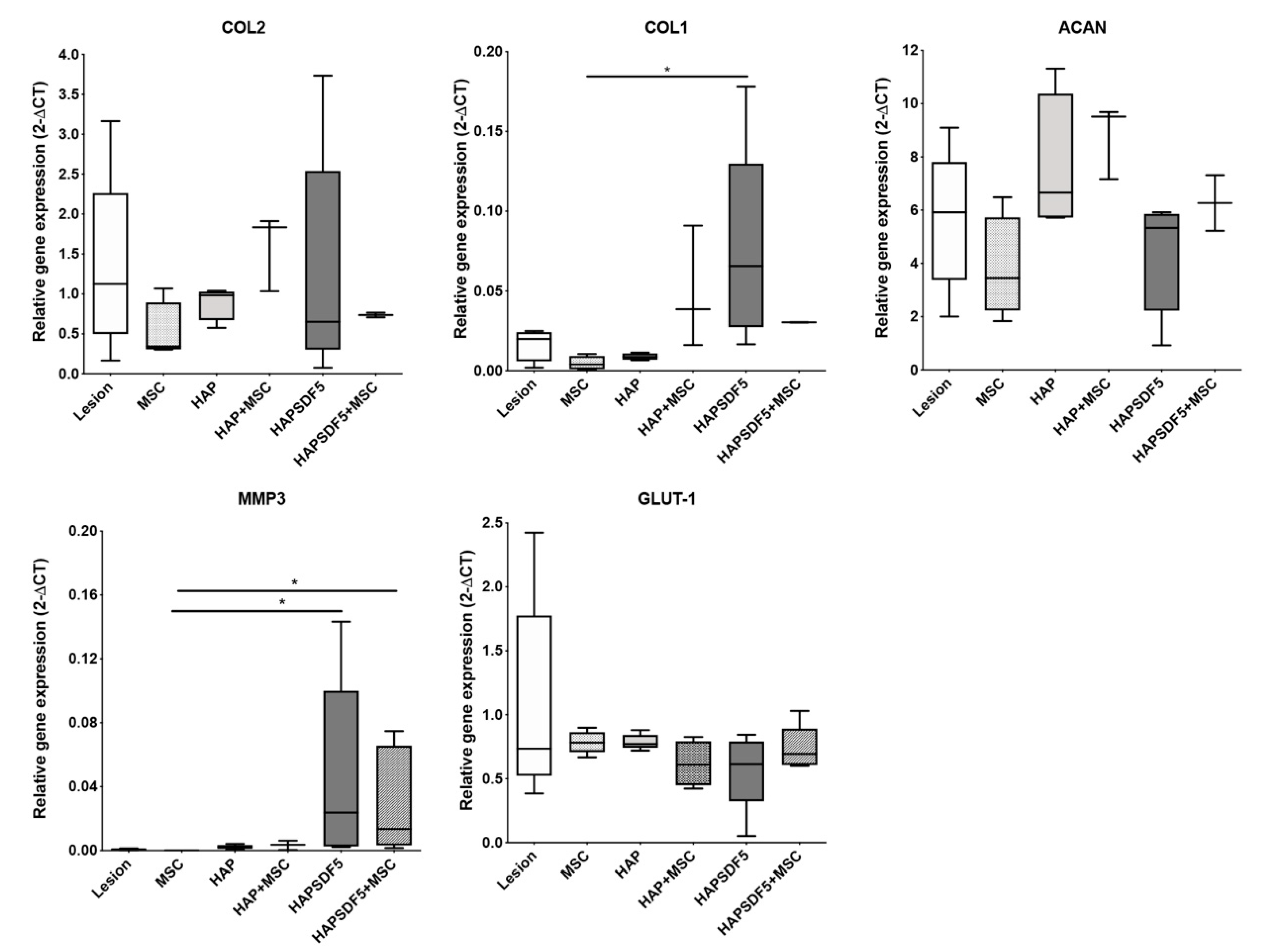
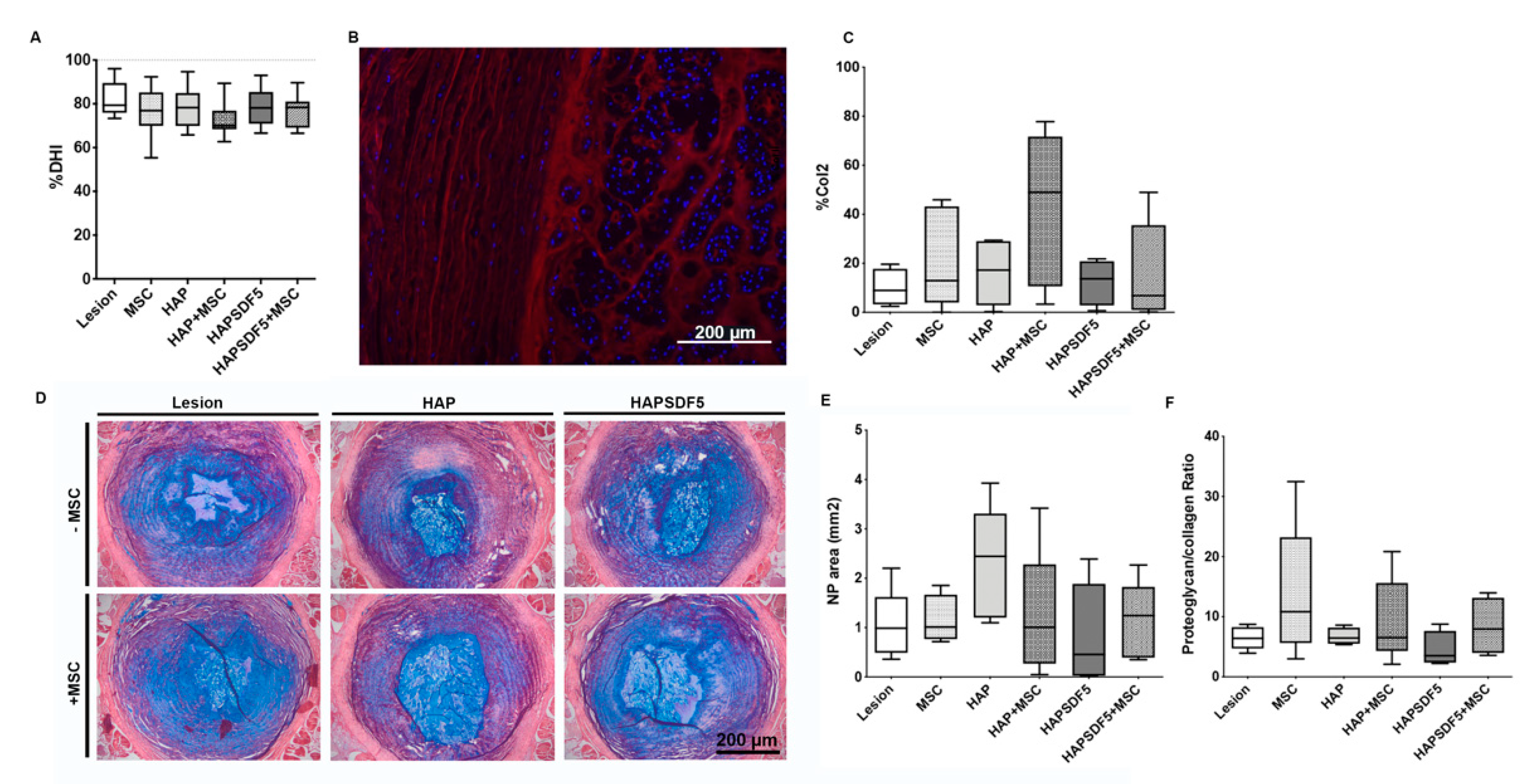
2.2. Potential of HAPSDF5 Intradiscal Injection for IVD Regeneration
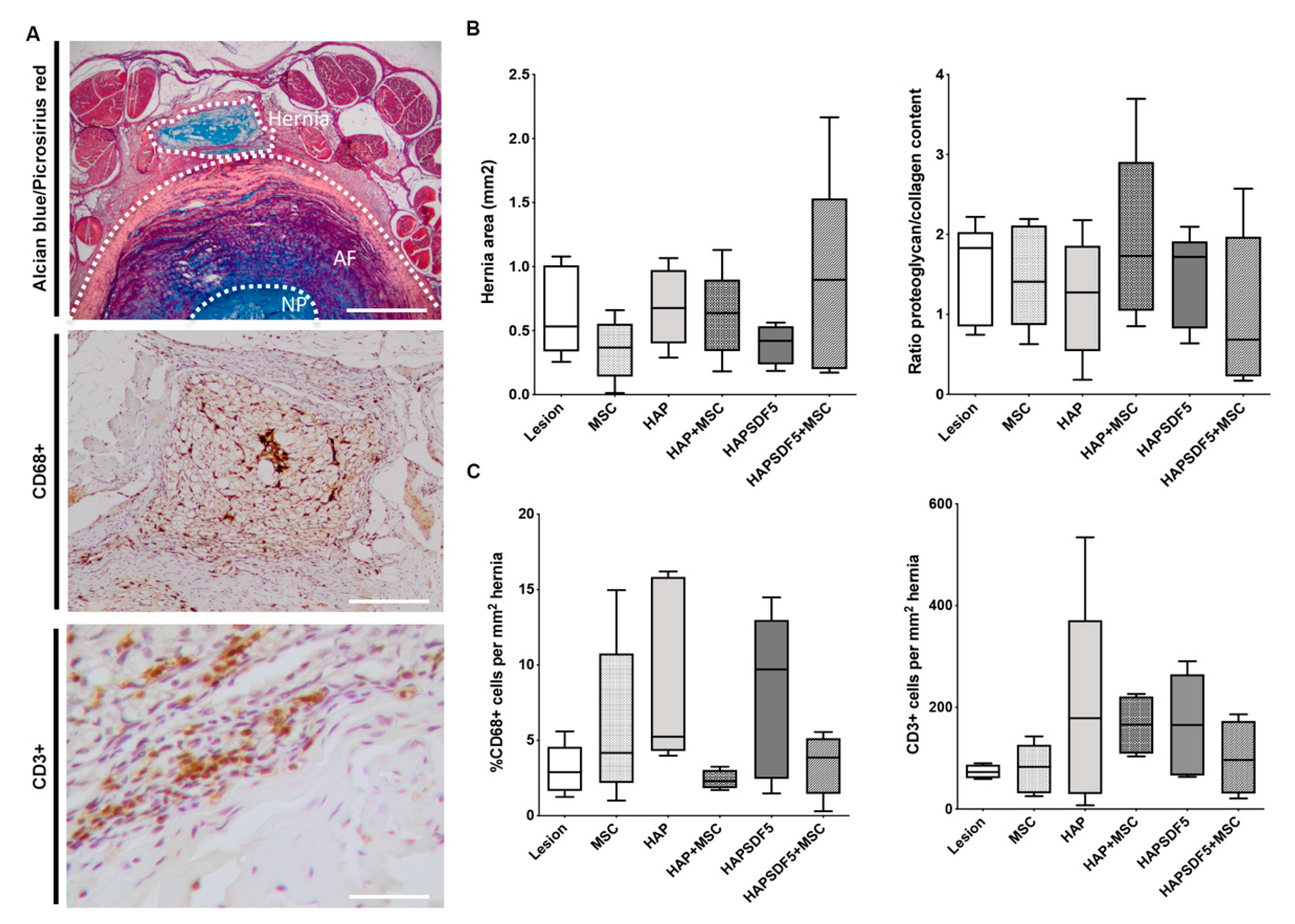
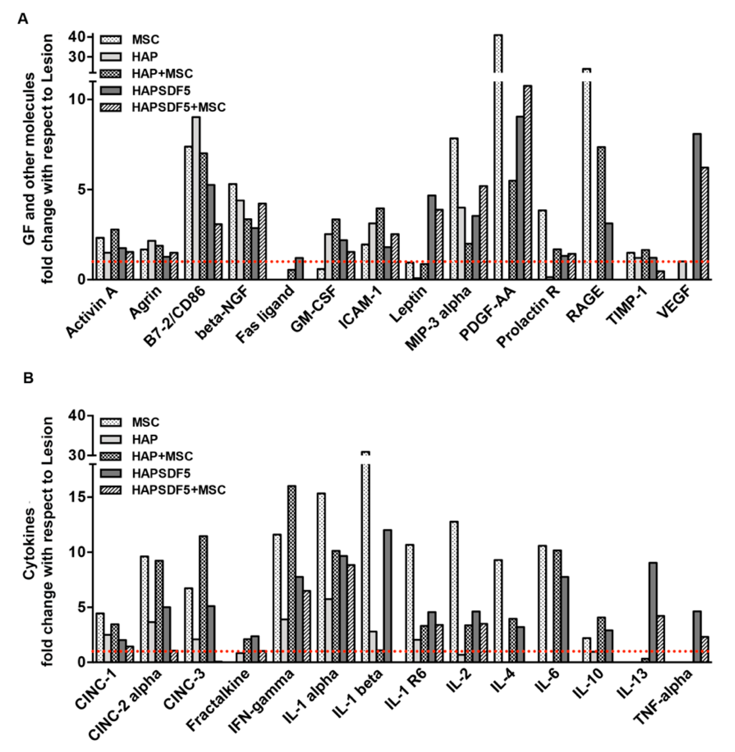
2.3. Influence of Intradiscal HAPSDF5 Delivery in Hernia Formation and Local Immune Response
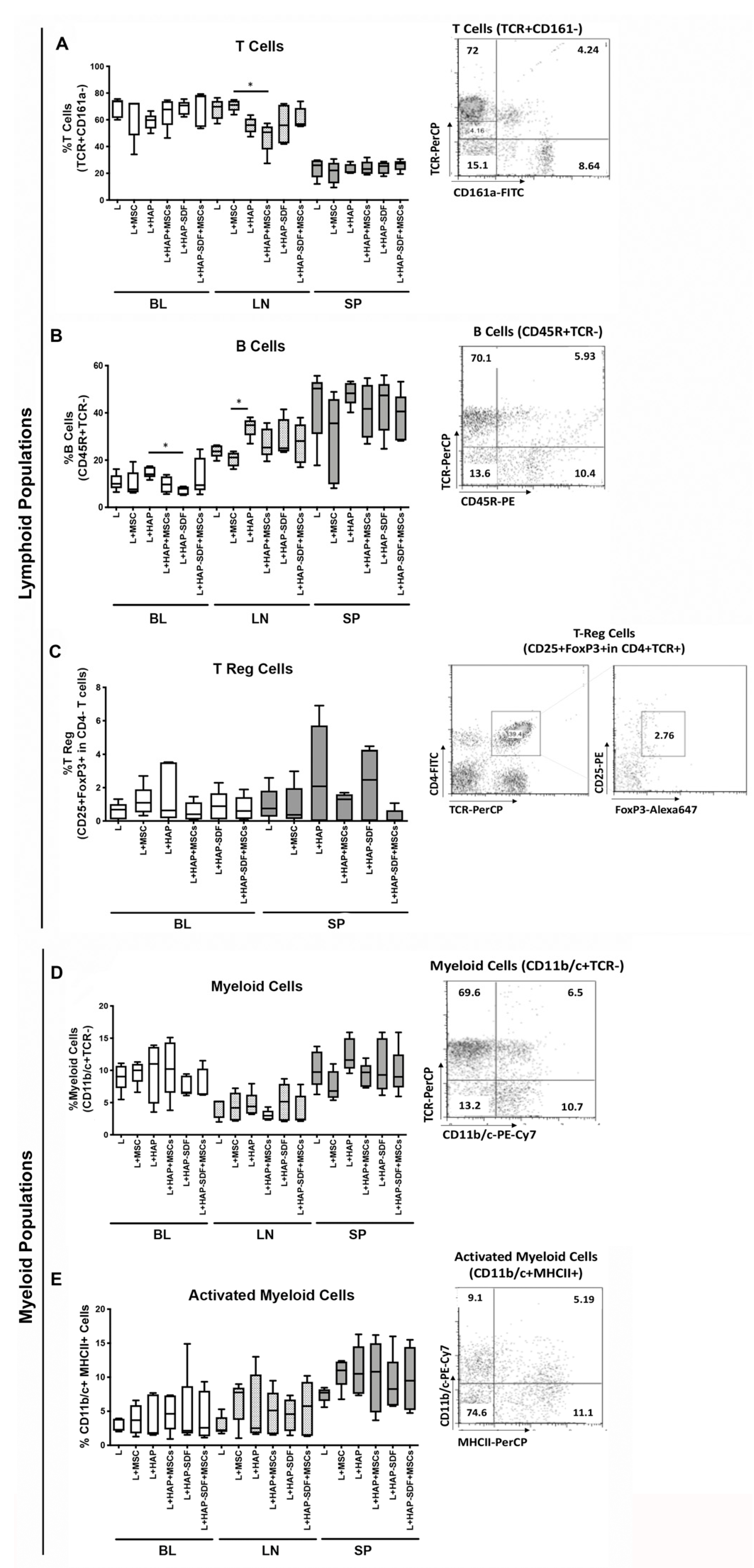
2.4. Systemic Immune Response to Intradiscal HAPSDF5 Delivery
3. Discussion
4. Materials and Methods
4.1. IVD Lesion Model
4.2. Hydrogel Preparation and SDF-1 Incorporation
4.3. Hydrogel Intradiscal Administration
4.4. rMSC Isolation, Labelling and Administration
4.5. Determination of the Disc Height Index
4.6. IVD Collection and Histological Analysis
4.7. Alcian Blue/Picrosirius Red Staining
4.8. Immunofluorescence for Collagen Type II
4.9. Immunohistochemistry for CD68+ and CD3+ Cells
4.10. RNA Isolation and Quantitative Real-Time Reverse Transcription Polymerase Chain Reaction
4.11. Plasma Cytokine Quantification
4.12. Flow Cytometry Analysis of Systemic Immune Cell Populations
4.13. Statistical Analysis
5. Conclusions
Author Contributions
Funding
Institutional Review Board Statement
Acknowledgments
Conflicts of Interest
References
- Gruber, H.E.; Ingram, J.A.; Norton, H.J.; Hanley, E.N., Jr. Senescence in cells of the aging and degenerating in-tervertebral disc: Immunolocalization of senescence-associated beta-galactosidase in human and sand rat discs. Spine 2007, 32, 321–327. [Google Scholar] [CrossRef]
- Roberts, S.; Evans, E.H.; Kletsas, D.; Jaffray, D.C.; Eisenstein, S.M. Senescence in human intervertebral discs. Eur. Spine J. 2006, 15, S312–S316. [Google Scholar] [CrossRef] [Green Version]
- Molinos, M.; Almeida, C.R.; Caldeira, J.; Cunha, C.; Goncalves, R.M.; Barbosa, M.A. Inflammation in intervertebral disc degeneration and regeneration. J. R. Soc. Interface R. Soc. 2015, 12, 20141191. [Google Scholar] [CrossRef] [PubMed]
- Ying, J.; Han, Z.; Pei, S.; Su, L.; Ruan, D. Effects of Stromal Cell-Derived Factor-1alpha Secreted in Degenerative Intervertebral Disc on Activation and Recruitment of Nucleus Pulposus-Derived Stem Cells. Stem Cells Int. 2019, 2019, 9147835. [Google Scholar] [CrossRef] [PubMed] [Green Version]
- Ying, J.-W.; Wen, T.-Y.; Pei, S.-S.; Su, L.-H.; Ruan, D.-K. Stromal cell-derived factor-1alpha promotes recruitment and differentiation of nucleus pulposus-derived stem cells. World J. Stem Cells 2019, 11, 196–211. [Google Scholar] [CrossRef] [PubMed]
- D’Este, M.; Eglin, D.; Alini, M. Lessons to be learned and future directions for intervertebral disc biomaterials. Acta Biomater. 2018, 78, 13–22. [Google Scholar] [CrossRef] [PubMed]
- Henriksson, H.B.; Svala, E.; Skioldebrand, E.; Lindahl, A.; Brisby, H. Support of Concept That Migrating Progenitor Cells from Stem Cell Niches Contribute to Normal Regeneration of the Adult Mammal Intervertebral Disc: A Descriptive Study in the New Zealand White Rabbit. Spine 2012, 37, 722–732. [Google Scholar] [CrossRef] [PubMed]
- Illien-Junger, S.; Pattappa, G.; Peroglio, M.; Benneker, L.M.; Stoddart, M.; Sakai, D.; Mochida, J.; Grad, S.; Alini, M. Homing of Mesenchymal Stem Cells in Induced Degenerative Intervertebral Discs in a Whole Organ Culture System. Spine 2012, 37, 1865–1873. [Google Scholar] [CrossRef] [PubMed]
- Oh, W.; Kim, D.-S.; Yang, Y.S.; Lee, J.K. Immunological properties of umbilical cord blood-derived mesenchymal stromal cells. Cell. Immunol. 2008, 251, 116–123. [Google Scholar] [CrossRef]
- Richardson, S.M.; Kalamegam, G.; Pushparaj, P.N.; Matta, C.; Memic, A.; Khademhosseini, A.; Mobasheri, R.; Poletti, F.L.; Hoyland, J.A.; Mobasheri, A. Mesenchymal stem cells in regenerative medicine: Focus on articular cartilage and intervertebral disc regeneration. Methods 2016, 99, 69–80. [Google Scholar] [CrossRef]
- De Becker, A.; Riet, I.V. Homing and migration of mesenchymal stromal cells: How to improve the efficacy of cell therapy? World J. Stem Cells 2016, 8, 73–87. [Google Scholar] [CrossRef]
- Wang, Y.; Chen, X.; Cao, W.; Shi, Y. Plasticity of mesenchymal stem cells in immunomodulation: Pathological and therapeutic implications. Nat. Immunol. 2014, 15, 1009–1016. [Google Scholar] [CrossRef] [PubMed]
- Katagiri, W.; Osugi, M.; Kawai, T.; Hibi, H. First-in-human study and clinical case reports of the alveolar bone regeneration with the secretome from human mesenchymal stem cells. Head Face Med. 2016, 12, 5. [Google Scholar] [CrossRef] [Green Version]
- Teixeira, F.G.; Carvalho, M.M.; Panchalingam, K.M.; Rodrigues, A.J.; Mendes-Pinheiro, B.; Anjo, S.; Manadas, B.; Behie, L.A.; Sousa, N.; Salgado, A.J. Impact of the Secretome of Human Mesenchymal Stem Cells on Brain Structure and Animal Behavior in a Rat Model of Parkinson’s Disease. Stem Cells Transl. Med. 2016, 6, 634–646. [Google Scholar] [CrossRef]
- Zhao, Q.; Ren, H.; Han, Z. Mesenchymal stem cells: Immunomodulatory capability and clinical potential in immune diseases. J. Cell. Immunother. 2016, 2, 3–20. [Google Scholar] [CrossRef] [Green Version]
- Sakai, D.; Nishimura, K.; Tanaka, M.; Nakajima, D.; Grad, S.; Alini, M.; Kawada, H.; Ando, K.; Mochida, J. Migration of bone marrow-derived cells for endogenous repair in a new tail-looping disc degeneration model in the mouse: A pilot study. Spine J. 2015, 15, 1356–1365. [Google Scholar] [CrossRef] [PubMed] [Green Version]
- Pereira, C.L.; Goncalves, R.M.; Peroglio, M.; Pattappa, G.; D’Este, M.; Eglin, D.; Barbosa, M.A.; Alini, M.; Grad, S. The effect of hyaluronan-based delivery of stromal cell-derived factor-1 on the recruitment of MSCs in degenerating interver-tebral discs. Biomaterials 2014, 35, 8144–8153. [Google Scholar] [CrossRef] [PubMed]
- Pereira, C.L.; Teixeira, G.Q.; Ribeiro-Machado, C.; Caldeira, J.; Costa, M.; Figueiredo, F.; Fernandes, R.; Aguiar, P.; Grad, S.; Barbosa, M.A.; et al. Mesenchymal Stem/Stromal Cells seeded on cartilaginous endplates promote Intervertebral Disc Regeneration through Extracellular Matrix Remodeling. Sci. Rep. 2016, 6, 33836. [Google Scholar] [CrossRef] [Green Version]
- Pereira, C.L.; Teixeira, G.Q.; Ferreira, J.R.; D’Este, M.; Eglin, D.; Alini, M.; Grad, S.; Barbosa, M.A.; Gonçalves, R.M. Stromal Cell Derived Factor-1-Mediated Migration of Mesenchymal Stem Cells Enhances Collagen Type II Ex-pression in Intervertebral Disc. Tissue Eng. Part A 2018, 24, 1818–1830. [Google Scholar] [CrossRef]
- Cunha, C.; Almeida, C.R.; Almeida, M.I.; Silva, A.M.; Molinos, M.; Lamas, S.; Pereira, C.L.; Teixeira, G.Q.; Monteiro, A.T.; Santos, S.G.; et al. Systemic Delivery of Bone Marrow Mesenchymal Stem Cells for In Situ Intervertebral Disc Regen-eration. Stem Cells Transl. Med. 2017, 6, 1029–1039. [Google Scholar] [CrossRef]
- Bleul, C.C.; Fuhlbrigge, R.C.; Casasnovas, J.M.; Aiuti, A.; Springer, T.A. A highly efficacious lymphocyte chemoattractant, stromal cell-derived factor 1 (SDF-1). J. Exp. Med. 1996, 184, 1101–1109. [Google Scholar] [CrossRef] [PubMed] [Green Version]
- Ponte, A.L.; Marais, E.; Gallay, N.; Langonné, A.; Delorme, B.; Herault, O.; Charbord, P.; Domenech, J. The In Vitro Migration Capacity of Human Bone Marrow Mesenchymal Stem Cells: Comparison of Chemokine and Growth Factor Chemotactic Activities. Stem Cells 2007, 25, 1737–1745. [Google Scholar] [CrossRef] [PubMed]
- Penn, M.S.; Pastore, J.; Miller, T.; Aras, R. SDF-1 in myocardial repair. Gene Ther. 2012, 19, 583–587. [Google Scholar] [CrossRef]
- Wang, Y.; Sun, X.; Lv, J.; Zeng, L.; Wei, X.; Wei, L. Stromal Cell-Derived Factor-1 Accelerates Cartilage Defect Repairing by Recruiting Bone Marrow Mesenchymal Stem Cells and Promoting Chondrogenic Differentiation. Tissue Eng. Part A 2017, 23, 1160–1168. [Google Scholar] [CrossRef] [PubMed]
- Zhang, H.; Zhang, L.; Chen, L.; Li, W.; Li, F.; Chen, Q. Stromal Cell-derived Factor-1 and its Receptor CXCR4 are Upregulated Expression in Degenerated Intervertebral Discs. Int. J. Med. Sci. 2014, 11, 240–245. [Google Scholar] [CrossRef] [Green Version]
- Wei, J.-N.; Cai, F.; Wang, F.; Wu, X.-T.; Liu, L.; Hong, X.; Tang, W.-H. Transplantation of CXCR4 Overexpressed Mesenchymal Stem Cells Augments Regeneration in Degenerated Intervertebral Discs. DNA Cell Biol. 2016, 35, 241–248. [Google Scholar] [CrossRef]
- He, Z.; Jia, M.; Yu, Y.; Yuan, C.; Wang, J. Roles of SDF-1/CXCR4 axis in cartilage endplate stem cells mediated promotion of nucleus pulposus cells proliferation. Biochem. Biophys. Res. Commun. 2018, 506, 94–101. [Google Scholar] [CrossRef]
- Zhang, H.; Zhu, T.; Zhang, L.; Wu, Q. Stromal cellderived factor-1 induces matrix metalloproteinase expression in human endplate chondrocytes, cartilage endplate degradation in explant culture, and the amelioration of nucleus pulposus degeneration in vivo. Int. J. Mol. Med. 2018, 41, 969–976. [Google Scholar]
- Zhang, H.; Yu, S.; Zhao, X.; Mao, Z.; Gao, C. Stromal cell-derived factor-1alpha-encapsulated albumin/heparin nanoparticles for induced stem cell migration and intervertebral disc regeneration in vivo. Acta Biomater. 2018, 72, 217–227. [Google Scholar] [CrossRef]
- Mortisen, D.; Peroglio, M.; Alini, M.; Eglin, D. Tailoring Thermoreversible Hyaluronan Hydrogels by “Click” Chemistry and RAFT Polymerization for Cell and Drug Therapy. Biomacromolecules 2010, 11, 1261–1272. [Google Scholar] [CrossRef]
- Peroglio, M.; Eglin, D.; Benneker, L.M.; Alini, M.; Grad, S. Thermoreversible hyaluronan-based hydrogel supports in vitro and ex vivo disc-like differentiation of human mesenchymal stem cells. Spine J. 2013, 13, 1627–1639. [Google Scholar] [CrossRef] [PubMed]
- Rosenzweig, D.H.; Fairag, R.; Mathieu, A.P.; Li, L.; Eglin, D.; D’Este, M.; Steffen, T.; Weber, M.H.; Ouellet, J.A.; Haglund, L. Thermoreversible hyaluronan-hydrogel and autologous nucleus pulposus cell delivery regenerates human inter-vertebral discs in an ex vivo, physiological organ culture model. Eur. Cell Mater. 2018, 36, 200–217. [Google Scholar] [CrossRef] [PubMed]
- D’Este, M.; Sprecher, C.M.; Milz, S.; Nehrbass, D.; Dresing, I.; Zeiter, S.; Alini, M.; Eglin, D. Evaluation of an injectable thermoresponsive hyaluronan hydrogel in a rabbit osteochondral defect model. J. Biomed. Mater. Res. Part A 2016, 104, 1469–1478. [Google Scholar] [CrossRef] [PubMed]
- Cunha, C.; Lamas, S.; Goncalves, R.; Barbosa, M. Joint analysis of IVD herniation and degeneration by rat caudal needle puncture model. J. Orthop. Res. 2017, 35, 258–268. [Google Scholar] [CrossRef] [PubMed] [Green Version]
- Cunha, C.; Silva, A.J.; Pereira, P.; Vaz, R.; Gonçalves, R.M.; Barbosa, M.A. The inflammatory response in the regression of lumbar disc herniation. Arthritis Res. 2018, 20, 251. [Google Scholar] [CrossRef] [Green Version]
- Pattappa, G.; Peroglio, M.; Sakai, D.; Mochida, J.; Benneker, L.M.; Alini, M.; Grad, S. CCL5/RANTES is a key chemoattractant released by degenerative intervertebral discs in organ culture. Eur. Cell Mater. 2014, 27, 124–136. [Google Scholar] [CrossRef]
- Vadalà, G.; Sowa, G.; Hubert, M.; Gilbertson, L.G.; Denaro, V.; Kang, J.D. Mesenchymal stem cells injection in degenerated intervertebral disc: Cell leakage may induce osteophyte formation. J. Tissue Eng. Regen. Med. 2011, 6, 348–355. [Google Scholar] [CrossRef]
- Schrepfer, S.; Deuse, T.; Reichenspurner, H.; Fischbein, M.; Robbins, R.; Pelletier, M. Stem Cell Transplantation: The Lung Barrier. Transplant. Proc. 2007, 39, 573–576. [Google Scholar] [CrossRef]
- Hu, B.; He, R.; Ma, K.; Wang, Z.; Cui, M.; Hu, H.; Rai, S.; Wang, B.; Shao, Z. Intervertebral Disc-Derived Stem/Progenitor Cells as a Promising Cell Source for Intervertebral Disc Regeneration. Stem Cells Int. 2018, 2018, 7412304. [Google Scholar] [CrossRef]
- Vo, N.V.; Hartman, R.A.; Yurube, T.; Jacobs, L.J.; Sowa, G.A.; Kang, J.D. Expression and regulation of metallo-proteinases and their inhibitors in intervertebral disc aging and degeneration. Spine J. 2013, 13, 331–341. [Google Scholar] [CrossRef] [PubMed] [Green Version]
- Sánchez-Martín, L.; Estecha, A.; Samaniego, R.; Sánchez-Ramón, S.; Vega, M.Á.; Sánchez-Mateos, P. The chemokine CXCL12 regulates monocyte-macrophage differentiation and RUNX3 expression. Blood 2011, 117, 88–97. [Google Scholar] [CrossRef] [PubMed] [Green Version]
- Cunha, C.; Teixeira, G.Q.; Ribeiro-Machado, C.; Pereira, C.L.; Ferreira, J.R.; Molinos, M.; Santos, G.S.; Barbosa, M.A.; Raquel, M.G. Modulation of the In Vivo Inflammatory Response by Pro- Versus Anti-Inflammatory Intervertebral Disc Treatments. Int. J. Mol. Sci. 2020, 21, 1730. [Google Scholar] [CrossRef] [Green Version]
- Hu, X.; Wu, R.; Jiang, Z.; Wang, L.; Chen, P.; Zhang, L.; Yang, L.; Wu, Y.; Chen, H.; Chen, H.; et al. Leptin Signaling Is Required for Augmented Therapeutic Properties of Mesenchymal Stem Cells Conferred by Hypoxia Preconditioning. Stem Cells 2014, 32, 2702–2713. [Google Scholar] [CrossRef] [PubMed] [Green Version]
- Ruifrok, A.C.; Johnston, D.A. Quantification of histochemical staining by color deconvolution. Anal. Quant. Cytol. Histol. 2001, 23, 291–299. [Google Scholar] [PubMed]


Publisher’s Note: MDPI stays neutral with regard to jurisdictional claims in published maps and institutional affiliations. |
© 2021 by the authors. Licensee MDPI, Basel, Switzerland. This article is an open access article distributed under the terms and conditions of the Creative Commons Attribution (CC BY) license (https://creativecommons.org/licenses/by/4.0/).
Share and Cite
Cunha, C.; Leite Pereira, C.; Ferreira, J.R.; Ribeiro-Machado, C.; Grad, S.; Santos, S.G.; Gonçalves, R.M. Therapeutic Strategies for IVD Regeneration through Hyaluronan/SDF-1-Based Hydrogel and Intravenous Administration of MSCs. Int. J. Mol. Sci. 2021, 22, 9609. https://0-doi-org.brum.beds.ac.uk/10.3390/ijms22179609
Cunha C, Leite Pereira C, Ferreira JR, Ribeiro-Machado C, Grad S, Santos SG, Gonçalves RM. Therapeutic Strategies for IVD Regeneration through Hyaluronan/SDF-1-Based Hydrogel and Intravenous Administration of MSCs. International Journal of Molecular Sciences. 2021; 22(17):9609. https://0-doi-org.brum.beds.ac.uk/10.3390/ijms22179609
Chicago/Turabian StyleCunha, Carla, Catarina Leite Pereira, Joana R. Ferreira, Cláudia Ribeiro-Machado, Sibylle Grad, Susana G. Santos, and Raquel M. Gonçalves. 2021. "Therapeutic Strategies for IVD Regeneration through Hyaluronan/SDF-1-Based Hydrogel and Intravenous Administration of MSCs" International Journal of Molecular Sciences 22, no. 17: 9609. https://0-doi-org.brum.beds.ac.uk/10.3390/ijms22179609