Article

Synthesis of Finely Controllable Sizes of Au Nanoparticles on a Silica Template and Their Nanozyme Properties

1
Department of Bioscience and Biotechnology, Konkuk University, Seoul 05029, Korea
2
Department of Chemical and Biological Engineering, Hanbat National University, Daejeon 34158, Korea
*
Authors to whom correspondence should be addressed.
Int. J. Mol. Sci. 2021, 22(19), 10382; https://0-doi-org.brum.beds.ac.uk/10.3390/ijms221910382
Submission received: 24 August 2021 / Revised: 16 September 2021 / Accepted: 22 September 2021 / Published: 26 September 2021
(This article belongs to the Special Issue Functional Optical Nano/Micromaterials)

Abstract

:
The precise synthesis of fine-sized nanoparticles is critical for realizing the advantages of nanoparticles for various applications. We developed a technique for preparing finely controllable sizes of gold nanoparticles (Au NPs) on a silica template, using the seed-mediated growth and interval dropping methods. These Au NPs, embedded on silica nanospheres (SiO2@Au NPs), possess peroxidase-like activity as nanozymes and have several advantages over other nanoparticle-based nanozymes. We confirmed their peroxidase activity; in addition, factors affecting the activity were investigated by varying the reaction conditions, such as concentrations of tetramethyl benzidine and H2O2, pH, particle amount, reaction time, and termination time. We found that SiO2@Au NPs are highly stable under long-term storage and reusable for five cycles. Our study, therefore, provides a novel method for controlling the properties of nanoparticles and for developing nanoparticle-based nanozymes.

1. Introduction

Enzymes are biocatalysts that play an important role in living systems. However, they are expensive, difficult to store, laborious to produce, and are easily denatured in external environments from varying temperature, pH, and chemical stressors [1,2]. These drawbacks critically limit their practical uses [3].
To overcome the above limitations, nanozymes have been developed as a new alternative to enzymes [1,4]. Nanozymes are nanomaterials that possess an intrinsic enzyme-like activity and have advantages such as stability in external environments, reasonable costs, and good catalytic activity [1,2,5,6,7,8,9].
Since the discovery of the unique peroxidase-like activity of Fe3O4 magnetic nanoparticles (NPs) by Yan’s group in 2007, numerous researchers around the world have gained an interest in nanozymes made of metal nanomaterials [10]. Metal nanomaterials, including gold (Au) NPs, platinum NPs, iron oxide NPs, cerium oxide NPs, manganese oxide NPs, and copper oxide NPs, began to be used as nanozymes [5,6,10,11,12,13,14,15]. Most of them have enzyme-like activities, such as those of peroxidases, catalases, oxidases, and superoxide oxidases [1,2,3,5,6,10,14,15,16,17,18,19,20,21,22].
Among the various metal nanomaterials, Au NPs have attracted the most attention because of their outstanding catalytic properties and advantages [23,24,25,26,27,28,29,30,31,32,33,34,35]. Au NPs can be synthesized easily and are stable [36,37]. In addition, the physical/chemical properties of Au NPs can be controlled by controlling their size and shape [24,26,33,35]. These NPs are also highly biocompatible and are easy to functionalize [24,30,35,38,39,40,41,42]. However, gold is not cheap, and the catalytic reactions depend on the surface area of the NPs. Fine-sized NPs can be cost-effective, but they are difficult to separate for reuse [1,3,8,9,10,17,43,44]. The development of a structure that uses small amounts of gold to acquire a large surface area, while still being easily separated, might prove to be a highly useful material in the field of nanozymes. A nanostructure where Au NPs are assembled onto a silica (SiO2) sphere core was developed by the Halas group in 1998 [45]. The chemical and optical properties of the Au NP-embedded SiO2 structure can be changed by simply controlling the diameter of the core and layered nanoparticles [46,47,48,49,50,51]. The NP-embedded SiO2 nanostructures are also cost-effective since only small amounts of expensive Au NPs are embedded onto the silica core, and they are easily separated from the reaction solution with the SiO2 core.
Au NP-assembled SiO2 nanostructures have been investigated in various fields [50,52,53,54,55,56] and have been found to have many merits due to the combined properties of Au NPs and of the silica core, simultaneously utilizing the outstanding and unique features of Au NPs and the inert and versatile feature of SiO2 [52,54,55,57]. In addition, the absorbance spectra of the nanostructures can be tuned across the visible and infrared regions by controlling the size of the Au NPs [38,39,58,59]. Due to these properties, the Au NP-assembled SiO2 nanostructures have broad applications. While there are many possible approaches for the control of the size and density of Au NPs on a template surface, a method of synthesis for those nanostructures has not yet been established [46,48,49,50,58,59]. For this reason, the low density and non-uniform morphology of Au NPs on nanostructures remain a considerable challenge [46,48,49,50]. There is, therefore, a need for the development of an improved method for preparing Au-NP-assembled silica nanostructures.
Our group recently developed SiO2@Au nanostructures in which Au NPs were densely immobilized on the surface of a SiO2 nanosphere [60]. For this nanostructure, the SiO2 nanosphere was used as a template, and the Au NPs were uniformly and densely introduced on it, using the seed-mediated growth method. SiO2@Au nanostructures have enhanced separation and re-dispersion properties and are more stable during surface modification than Au NPs. Moreover, they have shown potential as effective nanozymes. In this study, besides introducing dense and uniform Au NPs onto the SiO2 nanospheres, we also developed a facile method to very precisely control the size of Au NPs on the SiO2 surface, and we investigated their optical and catalytic characteristics by controlling the size of the Au NPs. Furthermore, various factors affecting the peroxidase-like activity of SiO2@Au NPs were also studied.

2. Results and Discussion

2.1. Preparation of Size-Controlled Au NPs-Assembled Silica Nanostructures (SiO2@Au NPs)

The SiO2@Au NPs were prepared by using seed-mediated growth synthesis, consisting of two steps: embedding Au seeds on the SiO2 surface, and growth of the Au NPs via the addition of an Au3+ precursor and reductant in intervals [45,52]. First, the Au seeds (~2.5 nm) were prepared by using an Au3+ precursor (HAuCl4) and tetrakis(hydroxymethyl)phosphonium chloride (THPC). Then, the Au seeds were incubated with aminated SiO2 nanospheres (~160 nm) to obtain Au-seeded SiO2 nanospheres, as previously reported [56,57,60,61,62]. On the Au-seeded SiO2 nanospheres, the reduction of the Au3+ precursor was directly conducted by using ascorbic acid (AA), which is a mild reducing agent, in the presence of polyvinylpyrrolidone (PVP) as a stabilizer. In these mildly reducing conditions, the progress of the growth stage is much slower than in strongly reducing conditions, making it easier to control the growth procedure [36]. A low concentration of the Au3+ precursor and AA were added onto the Au-seeded SiO2 in 5 min intervals until the desired concentrations for fine control over the size of the Au NPs are attained. The amounts are indicated in Table 1.
First, numerous small Au NPs (~2.5 nm) were attached throughout the SiO2 surface as seeds for the further growth of Au NPs. It is called SiO2@Au NP without the Au (III) precursor (0 µM), as shown in Figure 1b(i).
Subsequently, various concentrations of the Au3+ precursor were added to the Au-seeded SiO2 nanospheres at intervals; the size of the Au NPs grew larger as the Au3+ concentration was increased in Figure 1b(ii–vi). Moreover, the size of the Au NPs on the SiO2 nanospheres was precisely controlled with high levels of density and uniformity, which were confirmed clearly, as shown in Figure 1. At a high concentration of Au3+ (>200 µM), Au NPs on the SiO2 surface merged with each other and became one larger particle. When the size of the Au NPs increased, the color of the solution changed in the order of pale brown, then pink, purple, dark blue, and finally black (Figure 2a). These changes occurred depending on the size and shape of the NPs, due to their localized surface plasmon resonance (LSPR) [37,46,48]. When nanoparticles are close to one another, the absorption spectra of proximally located nanoparticles red-shift considerably from that of solitary particles [58,59]. Increasing the space between the particles reduces the shift [59]. Mie’s theory accounts for how increasing nanoparticle diameters induces the absorption spectra to red-shift by changing the electric surface charge density of the NPs [58]. The results of our study showed that the growth of Au NPs on SiO2@Au was controlled well by the color change of the particle suspension, transmission electron microscopy (TEM) images, and absorbance according to the abovementioned theories.
The absorption spectra of SiO2 showed that the absorbance band was red-shifted and broadened upon an increase in the concentration of the Au3+ precursor, indicating the formation of larger Au NPs, which change the proximate interparticle distance (Figure 2b,c). In the absorbance band of SiO2@Au-NPs-treated 0 μM Au3+ precursor, no peak was observed due to the exceedingly tiny size of the Au NPs. On the other hand, 50, 100, 150, 200, and 300 Au3+-treated SiO2@Au showed peaks at 543, 571, 593, 619, and 632 nm respectively (Figure 2c). Moreover, the absorbance bandwidth broadened as the size of the Au NPs was increased.

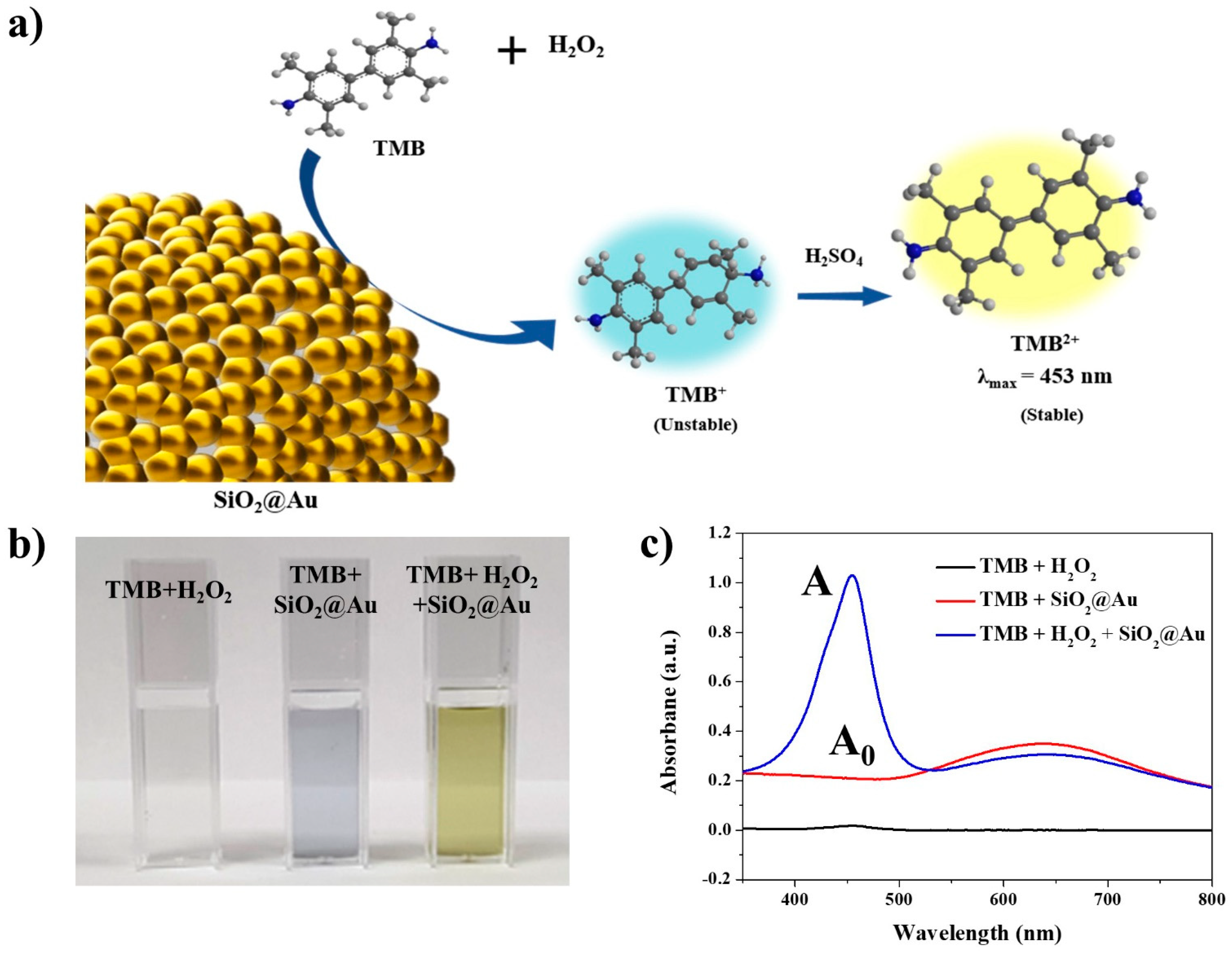
2.2. Verification of the Peroxidase-like Activity of SiO2@Au NPs

The peroxidase-like activity of SiO2@Au NPs was evaluated through oxidation of a 3,3′,5,5′-tetramethylbenzidine (TMB) substrate. The TMB oxidation reaction involves the transfer of two electrons that each produce a clear color change. When the first electron is transferred to form TMB+ via oxidization of TMB, the TMB solution changes from colorless to blue. Since TMB+ is quite unstable in an acidic environment, it further oxidizes to TMB2+ when the second electron is transferred; TMB2+ is stable in acidic conditions, exhibiting a yellow color and a maximum absorption peak at 453 nm (Figure 3a) [63].
To confirm the peroxidase-like activity of the SiO2@Au NPs, TMB + H2O2, TMB + SiO2@Au NPs, and TMB + H2O2 + SiO2@Au NPs were prepared in a pH 4 buffer for the peroxidase assay. The TMB + H2O2 solution was colorless, and an absorbance peak at 453 nm did not appear, as shown in Figure 3b,c. This result indicated that peroxidase-like activity did not occur in the absence of SiO2@Au. Next, the color of the TMB + SiO2@Au NP sample was entirely on account of the SiO2@Au NPs, displaying an absorbance band with a maximum peak at 630 nm. However, a yellow solution and an absorbance band at 453 nm were observed in the TMB + H2O2 + SiO2@Au NPs sample. These results showed that SiO2@Au NPs catalyzed TMB oxidation in the presence of H2O2, indicating that a peroxidase-like reaction occurred due to the peroxidase-mimicking property of SiO2@Au NPs.

2.3. The Peroxidase-like Activity Depends on the Size of the Au NPs of the SiO2@Au NPs

To investigate the correlation between the size of the Au NPs and the peroxidase-like activity of SiO2@Au NPs, various kinds of SiO2@Au NPs with different Au NPs sizes were prepared. The concentrations of the treated Au3+ precursors were 0, 50, 100, 150, 200, and 300 µM each, resulting in the formation of 1.4, 4.3, 6.4, 7.3, 9.5, and 15 nm diameter Au NPs on the SiO2@Au structures, respectively (Table 1 and Figure 2b). Each of these SiO2@Au NPs was subjected to the TMB assay to estimate their peroxidase-like activity. An absorbance peak at 453 nm was observed in the UV–Vis absorption spectra of all samples, indicating that all of the SiO2@Au NPs had peroxidase-like activity, irrespective of the size of the Au NPs (Supplementary Materials Figure S1). However, the SiO2@Au NPs produced without any Au3+ precursor treatment showed relatively very weak peroxidase-like activity. This may be because the size of the Au NPs on the silica core was too small; there were a large number of vacant spaces on the SiO2@Au NPs with the given number of Au NPs, providing an insufficient surface area for the reaction between the Au NPs and reactants. On the other hand, SiO2@Au NPs treated with more than 50 µM Au3+ precursor showed high peroxidase-like activity. The size of the Au NPs was rapidly increased when concentrations of Au3+ precursor exceeded 50 µM, as shown in the TEM images (Figure 1b(ii). As the size of the Au NPs on the SiO2@Au NPs was increased, the surface area which can react with reactants also increased. Therefore, the peroxidase-like activity of the SiO2@Au NPs was increased as the concentrations of the Au3+ precursor were increased (Figure 4a,b). Even though the size of the Au NPs grew as the concentration of the Au3+ precursor increased, severe aggregation occurred immediately after the peroxidase reaction in the SiO2@Au was treated with over 200 µM of the Au3+ precursor. Since good dispersibility is an important factor for generating a constant and stable catalytic activity, 150 µM of Au3+ precursor-treated SiO2@Au NPs, which have high peroxidase-like activity without aggregation, was used in the subsequent experiments [64,65].

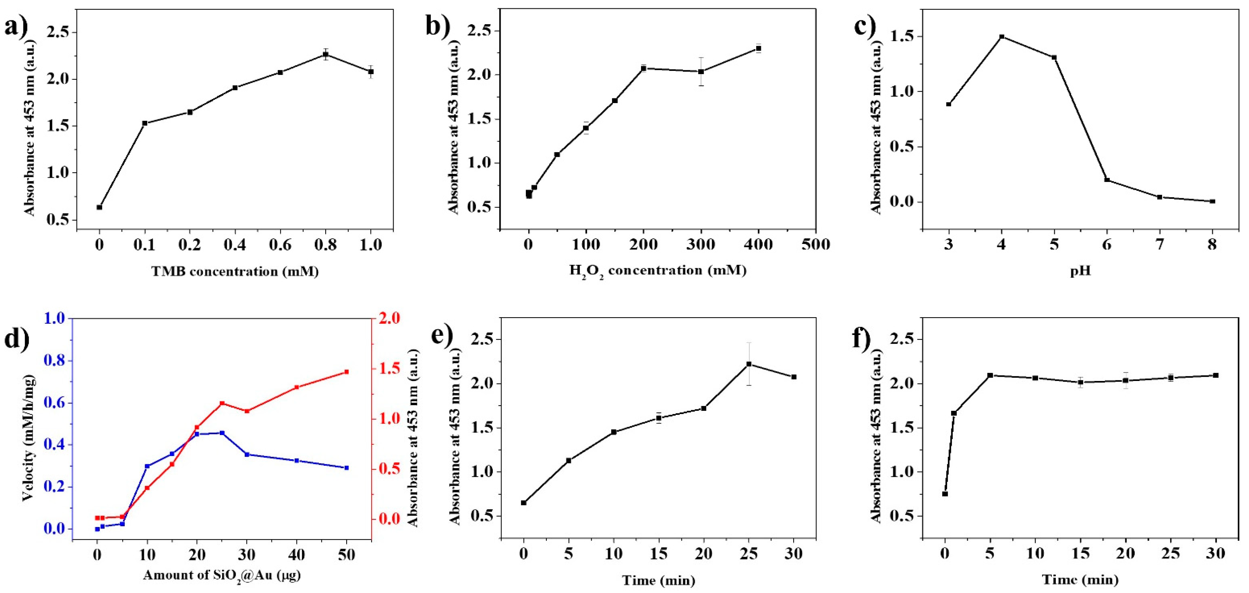
2.4. Effects of Reaction Conditions on the Peroxidase-like Activity of SiO2@Au NPs

It is known that the catalytic activity of nanozymes is affected by reaction conditions such as those associated with an enzyme [10,32,66,67,68,69]. For this reason, the effect of reaction conditions on the peroxidase-like activity on SiO2@Au NPs was investigated. The concentration of TMB and H2O2, pH of the buffer, the number of SiO2@Au NPs, reaction time, and termination time were considered in this study. For confirming the effects of TMB concentrations, the concentrations of TMB varied from 0 to 1.0 mM, while the other conditions were fixed in the peroxidase assay (Supplementary Materials Figure S2). The catalytic activity of SiO2@Au NPs increased until the concentrations of TMB were 0.8 mM; they then decreased at 1.0 mM, because the poor solubility of TMB in an aqueous buffer caused precipitation during the oxidation reaction (Figure 5a) [70]. To calculate the kinetic activities of SiO2@Au toward TMB concentration, various concentration of TMB in the range of 0.1 to 0.4 mM were prepared and then mixed to H2O2, and the absorbance were monitored every 10s. The absorbance at 200 s was used to calculate the Michaelis–Menten constants (Km) and the maximum reaction velocity (Vmax) in our study. The kinetic activities of SiO2@Au toward TMB concentration in the range of 0.1 to 0.4 mM TMB were plotted in Supplementary Materials Figure S3. A linear regression was found in the concentration of TMB from 0.1 to 0.4 mM. Km were obtained by using Linewaerver–Burk plots. The apparent Michaelis constant Km was calculated to be 0.060 mM and the maximum reaction velocity Vmax was 2.3 × 10⁻¹⁰ M−1∙s−1. Km of SiO2@Au was much lower than that of horseradish peroxidase enzyme, indicating that SiO2@Au has a higher affinity for TMB than horseradish peroxidase (Km = 0.438 mM). Moreover, Km of SiO2@Au is lower than those Au NPs (Km = 0.123 mM), glucose-oxidase-conjugated Au-attached magnetic SiO2 microsphere (Km = 0.208 mM), Au-NPs-decorated porous silica microsphere (Km = 0.523 mM), Prussian-blue-decorated latex nanoparticle (Km = 2.19 mM), MnO2 nanoparticles (Km = 0.083 mM), and sulfate-latex-conjugated polyelectrolyte functionalized MnO2 NPs (Km = 0.099 mM) [71,72,73,74,75].
In the case of the concentration of hydrogen peroxide, the catalytic activity of SiO2@Au NPs was increased steeply until reaching 200 mM H2O2 and decreased at a concentration of 300 mM H2O2 (Supplementary Materials Figure S4). Even though the activity again increased at 400 mM H2O2, the rate of increase was lower than 200 mM H2O2 (Figure 5b). Subsequently, various buffers with different pH were subjected to the peroxidase assay (Supplementary Materials Figure S5). The highest activity was shown under pH 4.0, at which H2O2 was more stable and TMB dissolved maximally (Figure 5c) [10,44,70,76,77,78]. The velocity of the catalytic reaction showed the highest value when 20 and 25 µg of SiO2@Au NPs were treated in the sample where the rest of the conditions were fixed (Figure 5d and Supplementary Materials Figure S6). About 25 min was required for the TMB+ oxidation and 5 min for the termination of the TMB+ oxidation to obtain stable results (Figure 5e,f).

2.5. Long-Term Stability and Reusability Test of SiO2@Au as Nanozyme

SiO2@Au NPs are substantially more advantageous over enzymes, owing to their long-term stability and reusability. Denaturation during storage and their on–off usage are the major defects when using enzymes in practice [1,2]. To verify the long-term stability of their peroxidase-like activity, the SiO2@Au NPs were examined by repeating the peroxidase assay every day, at the same time, for 14 days and on the 31st day after they were produced, keeping them under storage at 25 °C in the meantime (Figure 6a). The results showed that the peroxidase-like activity remained highly stable for at least 30 days after the fabrication of the SiO2@Au NPs. In sequence, the reusability of the SiO2@Au NPs as a nanozyme was evaluated through repeated peroxidase assays. Notably, the peroxidase-like activity of stored SiO2@Au until the fourth round was 98% of the first cycle level and mildly reduced at the fifth cycle to 89% of the first round. The reusability of SiO2@Au NPs was significantly high compared to a previous report on an Au NP-embedded silica nanostructure [79]. Moreover, SiO2@Au NPs remained 90% of catalytic activity, while SiO2@Au without the SiO2 core lost 60% of catalytic activity after five cycles of use (Supplementary Materials Figure S7). These results indicate that SiO2@Au NPs excel not only at being highly reusable but also at separating easily from the reaction mixture.

3. Materials and Methods

3.1. Chemicals and Reagents

Tetraethylorthosilicate (TEOS), tetrakis(hydroxymethyl)phosphonium chloride (THPC), chloroauric acid (HAuCl4), 3-aminopropyltriethoxysilane (APTS), AA, PVP (MW 40,000), and TMB were purchased from Sigma-Aldrich (St. Louis, MO, USA). Ammonium hydroxide (NH4OH, 27%), ethyl alcohol (EtOH, 99.9%), sodium hydroxide (NaOH), and sulfuric acid (H2SO4) were purchased from Samchun (Seoul, Korea). Hydrogen peroxide (H2O2) was purchased from Daejung (Siheung, Gyeonggi-do, Korea). Phosphate buffer saline containing 0.1% Tween 20 (PBST, pH 7.4) was purchased from Dynebio (Seongnam, Gyeonggi-do, Korea).

3.2. Characterization

The transmission electron microscope (TEM) images of the samples were taken by using a JEM-F200 multi-purpose electron microscope (JEOL, Akishima, Tokyo, Japan) with a maximum accelerated voltage of 200 kV. The UV–Vis absorption spectra of the sample were measured by an Optizen POP UV/Vis spectrometer (Mecasys, Seoul, Korea). The centrifugation of samples was performed by using a microcentrifuge 1730R (LaboGene, Lyngen, Denmark).

3.3. Synthesis of Gold Nanoparticles (Au NPs) Assembled SiO2 Nanostructure (SiO2@Au NPs)

The SiO2@Au NPs were synthesized according to the previous report [60]. The colloidal Au was prepared by stirring 47.5 mL of water, 0.5 mL of 0.2 M NaOH, 12 μL of THPC, and 1 mL of 50 mM HAuCl4 for 1 h. Silica nanospheres (~160 nm) were prepared by using the modified Stöber method [80]. Briefly, 40 mL of EtOH, 1.6 mL TEOS, and 3 mL of NH4OH were allowed to react with each other for 20 h. The amino group was introduced to the surface of 2 mg SiO2 NPs by treating them with 62 μL of APTS. The aminated SiO2 NPs were incubated with colloidal Au (~2.5 nm) for 12 h. After several cycles of centrifugation of the mixture at 8500 rpm for 10 min, 2 mg of Au-seeded SiO2 NPs were obtained and dispersed in 2 mL of PVP solution (1 mg/mL of PVP in water). Subsequently, 200 μL of Au-seeded SiO2 NPs (1 mg/mL) suspension was added to 9.8 mL of PVP solution. Under stirring, 20 μL of 10 mM HAuCl4 solution (in water, Au3+ precursor) and 40 μL of AA solution, (10 mM AA in water, reducing agent) were added to the mixture in sequence. The reaction mixture was stirred for 5 min. To control the size of the Au NPs on the surface of the Au-seeded SiO2 nanospheres, 10 mM of Au3+ precursor and AA were added. Until the concentrations of Au3+ reached 50, 100, 150, 200, and 300 μM in the various mixtures, the same volumes of Au3+ precursor and AA were repeatedly added every 5 min. The SiO2@Au NPs were washed several times with centrifugation at 8500 rpm for 10 min. The washed SiO2@Au NPs were dispersed in 1 mL of 0.1% PBST solution to obtain a 0.2 mg/mL SiO2@Au NP suspension.

3.4. Peroxidase-like Activity of SiO2@Au

To verify the peroxidase-like activity of SiO2@Au NPs, 100 μL of TMB solution (10 mM in EtOH) and 100 μL of the various SiO2@Au NPs synthesized from 50, 100, 150, 200, and 300 μM Au3+, respectively, were added to 700 μL of pH 4 buffer. Then, freshly prepared 100 μL of H2O2 solution (2 M in pH 4 buffer) was added, and the mixture was incubated for 30 min at room temperature. To terminate the reaction, 500 μL of 1 M H2SO4 was added to each mixture and incubated for 10 min. The absorbance of the mixture at 350–800 nm was measured by using the UV–Vis spectrometer.

3.5. Peroxidase-like Activity of SiO2@Au in Various Reaction Conditions

3.5.1. TMB Concentration

All assays of peroxidase-like activity were carried out in 1.5 mL Eppendorf tubes at room temperature. The TMB solutions were prepared in EtOH at various concentrations (1, 2, 4, 6, 8, and 10 mM, respectively). Then, 100 μL of each TMB solution, 100 μL of 2 M H2O2, and 100 μL of SiO2@Au (0.2 mg/mL) were added to 700 μL of pH 4 buffer. The final concentrations of TMB in the reaction mixture were 0.1, 0.2, 0.4, 0.6, 0.8, and 1.0 mM. After incubating the mixture for 30 min, 500 μL of 1 M H2SO4 was added to terminate the reaction. The absorbance of mixtures was measured by using a UV–Vis spectrometer.

3.5.2. H2O2 Concentration

Various concentrations of H2O2 solutions (1, 2, 3, and 4 M) were prepared. Next, 100 μL of 6 mM TMB solution, 100 μL of each H2O2 solutions, and 100 μL of SiO2@Au (0.2 mg/mL) were added to 700 μL of pH 4 buffer. The final concentrations of H2O2 in the mixtures were 100, 200, 300, and 400 mM, respectively. After incubating the mixtures for 30 min at room temperature, we added 500 μL of 1 M H2SO4 to each to terminate the reaction.

3.5.3. Buffer pH Value

Buffers of pH 3, 4, 5, 6, 7, and 8 were prepared. Next, 700 μL of each prepared buffer was added, followed by adding 100 μL of TMB solution, 100 μL of 2 M H2O2 solution, and 100 μL of SiO2@Au NPs (0.2 mg/mL). The mixtures were incubated for 30 min, and the reaction was terminated by using 500 μL of 1 M H2SO4.

3.5.4. Amount of SiO2@Au NPs

All reagents, including 100 μL of 6 mM TMB solution, 100 μL 2 M H2O2 solution, and 700 μL pH 4 buffer, were added to tubes. Then 1, 5, 10 15, 20, 25, 30, 40, and 50 μg of SiO2@Au were dispersed in 100 μL PBST each and added to the mixtures, followed by incubation and termination.

3.5.5. Reaction Time

To investigate the effect of reaction time on their peroxidase-like activity, the mixtures containing 100 μL of 6 mM TMB solution, 100 μL of SiO2@Au (0.2 mg/mL), and 100 μL of 2 M H2O2 solution were added to 700 μL of pH 4 buffer. Next, the mixtures were incubated for 0, 5, 10, 15, 20, 25, and 30 min respectively and terminated by using 1 M H2SO4.

3.5.6. Termination Time

Mixtures including 700 μL of pH 4 buffer, 100 μL of 6 mM TMB solution, 100 μL of SiO2@Au (0.2 mg/mL), and 100 μL of 2 M H2O2 solution were prepared and incubated for 30 min. After adding 500 μL of 1 M H2SO4, we incubated each sample for 0, 5, 10, 15, 20, 25, and 30 min to terminate the reaction.

3.6. Long-Term Stability of Peroxidase-like Activity

To investigate the long-term stability of peroxidase-like activity of SiO2@Au, the experiment was repeated every day for 2 weeks and on the 31st day after their fabrication. The experimental procedures were conducted as follows: adding 100 μL of 6 M TMB solution, 100 μL of SiO2@Au (0.2 mg/mL), and 100 μL of 2 M H2O2 solution to 700 μL of pH 4 buffer; 30 min reaction time; 10 min for termination, using 500 μL of 1 M H2SO4.

3.7. Reusability as Nanozymes

To investigate the reusability of SiO2@Au, the peroxidase assay was performed according to the mentioned procedures. After the assay was performed, the SiO2@Au NPs were collected by using centrifugation at 10,000 rpm for 10 min, and the absorbance of the supernatant at 453 nm was measured by using a UV–Vis spectrometer. The peroxidase assay was repeated with the collected SiO2@Au NPs.

4. Conclusions

In summary, we successfully synthesized finely controllably sized Au NPs on the SiO2 nanosphere (SiO2@Au), using the seed-mediated growth method and interval dropping method under mild conditions. The effect of the size of Au NPs on the SiO2@Au NPs was confirmed by the TEM images, color changing of its suspension, and UV–Vis absorption spectra. Moreover, we investigated the factors affecting the peroxidase-like activity of SiO2@Au NPs, such as TMB concentration, H2O2 concentration, pH, SiO2@Au NPs amount, reaction time, and termination time. Furthermore, SiO2@Au NPs showed high stability during the 30-day-long storage time at room temperature and outstanding reusability for five cycles. This work is therefore meaningful for utilizing controllable nanoparticles in various fields and provides a better approach to develop nanoparticle-based nanozymes.

Supplementary Materials

Author Contributions

Conceptualization, X.-H.P. and B.-H.J.; methodology, B.S. and X.-H.P.; investigation, B.S. and J.K.; formal analysis, J.K.; software, S.H.L.; writing—original draft preparation, B.S., W.K.; writing—review and editing, X.-H.P. and B.-H.J.; supervisor, X.-H.P. and B.-H.J. All authors have read and agreed to the published version of the manuscript.

Funding

This work was supported by the KU Research Professor Program of Konkuk University. This research was funded by the Ministry of Science and ITC, grant numbers NRF-2019R1G1A1006488 and by the Ministry of Education, grant number NRF-2018R1D1A1B07045708.

Institutional Review Board Statement

Not applicable.

Informed Consent Statement

Not applicable.

Data Availability Statement

Data is contained within the article or supplementary material.

Acknowledgments

The authors are grateful for the financial support from the NRF of Korea. Further, the author gives thanks for the financial support from Konkuk University.

Conflicts of Interest

The authors declare no conflict of interest.

References

  1. Wei, H.; Wang, E. Nanomaterials with enzyme-like characteristics (nanozymes): Next-generation artificial enzymes. Chem. Soc. Rev. 2013, 42, 6060–6093. [Google Scholar] [CrossRef]
  2. Manea, F.; Houillon, F.B.; Pasquato, L.; Scrimin, P. Nanozymes: Gold-Nanoparticle-Based Transphosphorylation Catalysts. Angew. Chem. Int. Ed. 2004, 43, 6165–6169. [Google Scholar] [CrossRef] [PubMed]
  3. Wei, H.; Wang, E. Fe3O4 Magnetic Nanoparticles as Peroxidase Mimetics and Their Applications in H2O2 and Glucose Detection. Anal. Chem. 2008, 80, 2250–2254. [Google Scholar] [CrossRef] [PubMed]
  4. Antuña-Jiménez, D.; Blanco-López, M.C.; Miranda-Ordieres, A.J.; Lobo-Castañón, M.J. Artificial enzyme with magnetic properties and peroxidase activity on indoleamine metabolite tumor marker. Polymers 2014, 55, 1113–1119. [Google Scholar] [CrossRef]
  5. Zhang, K.; Hu, X.; Liu, J.; Yin, J.-J.; Hou, S.; Wen, T.; He, W.; Ji, Y.; Guo, Y.; Wang, Q.; et al. Formation of PdPt Alloy Nanodots on Gold Nanorods: Tuning Oxidase-like Activities via Composition. Langmuir 2011, 27, 2796–2803. [Google Scholar] [CrossRef] [PubMed]
  6. He, W.; Jia, H.; Li, X.; Lei, Y.; Li, J.; Zhao, H.; Mi, L.; Zhang, L.; Zheng, Z. Understanding the formation of CuS concave superstructures with peroxidase-like activity. Nanoscale 2012, 4, 3501–3506. [Google Scholar] [CrossRef]
  7. Ju, H. Sensitive biosensing strategy based on functional nanomaterials. Sci. China Ser. B Chem. 2011, 54, 1202–1217. [Google Scholar] [CrossRef]
  8. Celardo, I.; Pedersen, J.Z.; Traversa, E.; Ghibelli, L. Pharmacological potential of cerium oxide nanoparticles. Nanoscale 2011, 3, 1411–1420. [Google Scholar] [CrossRef]
  9. Kotov, N.A. Inorganic Nanoparticles as Protein Mimics. Science 2010, 330, 188–189. [Google Scholar] [CrossRef]
  10. Gao, L.; Zhuang, J.; Nie, L.; Zhang, J.; Zhang, Y.; Gu, N.; Wang, T.; Feng, J.; Yang, D.; Perrett, S.; et al. Intrinsic peroxidase-like activity of ferromagnetic nanoparticles. Nat. Nanotechnol. 2007, 2, 577–583. [Google Scholar] [CrossRef]
  11. Cortie, M.B.; van der Lingen, E. Catalytic Gold Nano-Particles. Mater. Forum 2002, 26, 1–14. [Google Scholar] [CrossRef] [Green Version]
  12. Bond, G.C. Gold: A relatively new catalyst. Gold Bull. 2001, 34, 117–119. [Google Scholar] [CrossRef] [Green Version]
  13. Asati, A.; Santra, S.; Kaittanis, C.; Nath, S.; Perez, J.M. Oxidase-Like Activity of Polymer-Coated Cerium Oxide Nanoparticles. Angew. Chem. Int. Ed. 2009, 48, 2308–2312. [Google Scholar] [CrossRef] [PubMed]
  14. Chen, W.; Chen, J.; Feng, Y.-B.; Hong, L.; Chen, Q.-Y.; Wu, L.-F.; Lin, X.-H.; Xia, X.-H. Peroxidase-like activity of water-soluble cupric oxide nanoparticles and its analytical application for detection of hydrogen peroxide and glucose. Analyst 2012, 137, 1706–1712. [Google Scholar] [CrossRef] [PubMed]
  15. Wang, J.; Zhao, H.; Song, J.; Zhu, T.; Xu, W. Structure-Activity Relationship of Manganese Oxide Catalysts for the Catalytic Oxidation of (chloro)-VOCs. Catalysts 2019, 9, 726. [Google Scholar] [CrossRef] [Green Version]
  16. Karakoti, A.; Singh, S.; Dowding, J.M.; Seal, S.; Self, W. Redox-active radical scavenging nanomaterials. Chem. Soc. Rev. 2010, 39, 4422–4432. [Google Scholar] [CrossRef] [PubMed]
  17. Jiao, X.; Song, H.; Zhao, H.; Bai, W.; Zhang, L.; Lv, Y. Well-redispersed ceria nanoparticles: Promising peroxidase mimetics for H2O2 and glucose detection. Anal. Methods 2012, 4, 3261–3267. [Google Scholar] [CrossRef]
  18. Zhang, X.-Q.; Gong, S.-W.; Zhang, Y.; Yang, T.; Wang, C.-Y.; Gu, N. Prussian blue modified iron oxide magnetic nanoparticles and their high peroxidase-like activity. J. Mater. Chem. 2010, 20, 5110–5116. [Google Scholar] [CrossRef]
  19. Chaudhari, K.N.; Chaudhari, N.; Yu, J.-S. Peroxidase mimic activity of hematiteiron oxides (α-Fe2O3) with different nanostructures. Catal. Sci. Technol. 2012, 2, 119–124. [Google Scholar] [CrossRef]
  20. Dutta, A.K.; Maji, S.K.; Srivastava, D.N.; Mondal, A.; Biswas, P.; Paul, P.; Adhikary, B. Peroxidase-like activity and amperometric sensing of hydrogen peroxide by Fe2O3 and Prussian Blue-modified Fe2O3 nanoparticles. J. Mol. Catal. A Chem. 2012, 360, 71–77. [Google Scholar] [CrossRef]
  21. Comotti, M.; Della Pina, C.; Falletta, E.; Rossi, M. Aerobic Oxidation of Glucose with Gold Catalyst: Hydrogen Peroxide as Intermediate and Reagent. Adv. Synth. Catal. 2006, 348, 313–316. [Google Scholar] [CrossRef]
  22. Shen, X.; Liu, W.; Gao, X.; Lu, Z.; Wu, X.; Gao, X. Mechanisms of Oxidase and Superoxide Dismutation-like Activities of Gold, Silver, Platinum, and Palladium, and Their Alloys: A General Way to the Activation of Molecular Oxygen. J. Am. Chem. Soc. 2015, 137, 15882–15891. [Google Scholar] [CrossRef] [PubMed]
  23. Medley, C.D.; Smith, J.E.; Tang, Z.; Wu, Y.; Bamrungsap, S.; Tan, W. Gold Nanoparticle-Based Colorimetric Assay for the Direct Detection of Cancerous Cells. Anal. Chem. 2008, 80, 1067–1072. [Google Scholar] [CrossRef]
  24. Popovtzer, R.; Agrawal, A.; Kotov, N.; Popovtzer, A.; Balter, J.; Carey, T.; Kopelman, R. Targeted Gold Nanoparticles Enable Molecular CT Imaging of Cancer. Nano Lett. 2008, 8, 4593–4596. [Google Scholar] [CrossRef] [Green Version]
  25. Fang, S.-B.; Tseng, W.Y.; Lee, H.-C.; Tsai, C.-K.; Huang, J.-T.; Hou, S.-Y. Identification of Salmonella using colony-print and detection with antibody-coated gold nanoparticles. J. Microbiol. Methods 2009, 77, 225–228. [Google Scholar] [CrossRef]
  26. Kim, C.S.; Wilder-Smith, P.; Ahn, Y.-C.; Liaw, L.-H.L.; Chen, Z.; Kwon, Y.J. Enhanced detection of early-stage oral cancer in vivo by optical coherence tomography using multimodal delivery of gold nanoparticles. J. Biomed. Opt. 2009, 14, 034008. [Google Scholar] [CrossRef] [PubMed]
  27. Thaxton, C.S.; Elghanian, R.; Thomas, A.D.; Stoeva, S.I.; Lee, J.-S.; Smith, N.D.; Schaeffer, A.J.; Klocker, H.; Horninger, W.; Bartsch, G.; et al. Nanoparticle-based bio-barcode assay redefines "undetectable" PSA and biochemical recurrence after radical prostatectomy. Proc. Natl. Acad. Sci. USA 2009, 106, 18437–18442. [Google Scholar] [CrossRef] [Green Version]
  28. Zhang, J.; Wang, L.; Zhang, H.; Boey, F.; Song, S.; Fan, C. Aptamer-Based Multicolor Fluorescent Gold Nanoprobes for Multiplex Detection in Homogeneous Solution. Small 2010, 6, 201–204. [Google Scholar] [CrossRef]
  29. Huo, Q.; Colon, J.; Cordero, A.; Bogdanovic, J.; Baker, C.H.; Goodison, S.; Pensky, M.Y. A Facile Nanoparticle Immunoassay for Cancer Biomarker Discovery. J. Nanobiotechnol. 2011, 9, 1–12. [Google Scholar] [CrossRef] [Green Version]
  30. LeDuc, C.; Jung, J.-M.; Carney, R.R.; Stellacci, F.; Lounis, B. Direct Investigation of Intracellular Presence of Gold Nanoparticles via Photothermal Heterodyne Imaging. ACS Nano 2011, 5, 2587–2592. [Google Scholar] [CrossRef]
  31. Von Maltzahn, G.; Park, J.-H.; Lin, K.Y.; Singh, N.; Schwöppe, C.; Mesters, R.; Berdel, W.E.; Ruoslahti, E.; Sailor, M.J.; Bhatia, S.N. Nanoparticles that communicate in vivo to amplify tumour targeting. Nat. Mater. 2011, 10, 545–552. [Google Scholar] [CrossRef] [PubMed] [Green Version]
  32. Wang, H.; Zheng, L.; Peng, C.; Guo, R.; Shen, M.; Shi, X.; Zhang, G. Computed tomography imaging of cancer cells using acetylated dendrimer-entrapped gold nanoparticles. Biomaterials 2011, 32, 2979–2988. [Google Scholar] [CrossRef] [PubMed]
  33. Zhang, Y.; Qian, J.; Wang, D.; Wang, Y.; He, S. Multifunctional Gold Nanorods with Ultrahigh Stability and Tunability for In Vivo Fluorescence Imaging, SERS Detection, and Photodynamic Therapy. Angew. Chem. Int. Ed. 2012, 52, 1148–1151. [Google Scholar] [CrossRef] [PubMed]
  34. Youssef, A.M.; Abdel-Aziz, M.; El-Sayed, S. Chitosan nanocomposite films based on Ag-NP and Au-NP biosynthesis by Bacillus Subtilis as packaging materials. Int. J. Biol. Macromol. 2014, 69, 185–191. [Google Scholar] [CrossRef] [PubMed]
  35. Zhang, Z.; Wang, J.; Nie, X.; Wen, T.; Ji, Y.; Wu, X.; Zhao, Y.; Chen, C. Near Infrared Laser-Induced Targeted Cancer Therapy Using Thermoresponsive Polymer Encapsulated Gold Nanorods. J. Am. Chem. Soc. 2014, 136, 7317–7326. [Google Scholar] [CrossRef]
  36. Grzelczak, M.; Pérez-Juste, J.; Mulvaney, P.; Liz-Marzán, L.M. Shape control in gold nanoparticle synthesis. Chem. Soc. Rev. 2008, 37, 1783–1791. [Google Scholar] [CrossRef]
  37. Daniel, M.-C.; Astruc, D. Gold Nanoparticles: Assembly, Supramolecular Chemistry, Quantum-Size-Related Properties, and Applications toward Biology, Catalysis, and Nanotechnology. Chem. Rev. 2004, 104, 293–346. [Google Scholar] [CrossRef]
  38. Henglein, A. Physicochemical properties of small metal particles in solution: "Microelectrode" reactions, chemisorption, composite metal particles, and the atom-to-metal transition. J. Phys. Chem. 1993, 97, 5457–5471. [Google Scholar] [CrossRef]
  39. Belloni, J. Metal nanocolloids. Curr. Opin. Colloid Interface Sci. 1996, 1, 184–196. [Google Scholar] [CrossRef]
  40. Toshima, N.; Yonezawa, T. Bimetallic nanoparticles—novel materials for chemical and physical applications. New J. Chem. 1998, 22, 1179–1201. [Google Scholar] [CrossRef]
  41. Brust, M.; Kiely, C. Some recent advances in nanostructure preparation from gold and silver particles: A short topical review. Colloids Surf. A Physicochem. Eng. Asp. 2002, 202, 175–186. [Google Scholar] [CrossRef]
  42. Lin, Y.-C.; Yu, B.-Y.; Lin, W.-C.; Lee, S.-H.; Kuo, C.-H.; Shyue, J.-J. Tailoring the surface potential of gold nanoparticles with self-assembled monolayers with mixed functional groups. J. Colloid Interface Sci. 2009, 340, 126–130. [Google Scholar] [CrossRef]
  43. Fan, K.; Cao, C.; Pan, Y.; Lu, D.; Yang, D.; Feng, J.; Song, L.; Liang, M.; Yan, X. Magneto ferritin nanoparticles for targeting and visualizing tumour tissues. Nat. Nanotechnol. 2012, 7, 459–464. [Google Scholar] [CrossRef]
  44. He, W.; Zhou, Y.-T.; Wamer, W.G.; Hu, X.; Wu, X.; Zheng, Z.; Boudreau, M.D.; Yin, J.-J. Intrinsic catalytic activity of Au nanoparticles with respect to hydrogen peroxide decomposition and superoxide scavenging. Biomaterials 2013, 34, 765–773. [Google Scholar] [CrossRef]
  45. Westcott, S.L.; Oldenburg, S.J.; Lee, A.T.R.; Halas, N. Formation and Adsorption of Clusters of Gold Nanoparticles onto Functionalized Silica Nanoparticle Surfaces. Langmuir 1998, 14, 5396–5401. [Google Scholar] [CrossRef]
  46. Prodan, E.; Nordlander, P.; Halas, N.J. Electronic Structure and Optical Properties of Gold Nanoshells. Nano Lett. 2003, 3, 1411–1415. [Google Scholar] [CrossRef]
  47. Wilhelm, P.; Stephan, D. On-line tracking of the coating of nanoscaled silica with titania nanoparticles via zeta-potential measurements. J. Colloid Interface Sci. 2006, 293, 88–92. [Google Scholar] [CrossRef] [PubMed]
  48. Loo, C.; Lin, A.; Hirsch, L.; Lee, M.-H.; Barton, J.; Halas, N.; West, J.; Drezek, R. Nanoshell-Enabled Photonics-Based Imaging and Therapy of Cancer. Technol. Cancer Res. Treat. 2004, 3, 33–40. [Google Scholar] [CrossRef] [PubMed]
  49. Zhang, Y.-F.; Wang, J.-H.; Ma, L.; Nan, F.; Cheng, Z.-Q.; Zhou, L.; Wang, Q.-Q. Growth of silver-coated gold nanoshells with enhanced linear and nonlinear optical responses. J. Nanoparticle Res. 2015, 17, 1–10. [Google Scholar] [CrossRef]
  50. Lu, L.; Zhang, H.; Sun, G.; Xi, A.S.; Wang, H.; Li, X.; Wang, A.X.; Zhao, B. Aggregation-Based Fabrication and Assembly of Roughened Composite Metallic Nanoshells: Application in Surface-Enhanced Raman Scattering. Langmuir 2003, 19, 9490–9493. [Google Scholar] [CrossRef]
  51. Gawande, M.B.; Goswami, A.; Asefa, T.; Guo, H.; Biradar, A.V.; Peng, D.-L.; Zboril, R.; Varma, R.S. Core–shell nanoparticles: Synthesis and applications in catalysis and electrocatalysis. Chem. Soc. Rev. 2015, 44, 7540–7590. [Google Scholar] [CrossRef]
  52. Xue, J.; Wang, C.; Ma, Z. A facile method to prepare a series of SiO2@Au core/shell structured nanoparticles. Mater. Chem. Phys. 2007, 105, 419–425. [Google Scholar] [CrossRef]
  53. Brito-Silva, A.M.; Sobral-Filho, R.G.; Barbosa-Silva, R.; de Araújo, C.B.; Galembeck, A.; Brolo, A.G. Improved Synthesis of Gold and Silver Nanoshells. Langmuir 2013, 29, 4366–4372. [Google Scholar] [CrossRef] [PubMed]
  54. Tharion, J.; Satija, J.; Mukherji, S. Glucose mediated synthesis of gold nanoshells: A facile and eco-friendly approach conferring high colloidal stability. RSC Adv. 2014, 4, 3984–3991. [Google Scholar] [CrossRef]
  55. Garcia-Soto, M.J.; González-Ortega, O. Synthesis of silica-core gold nanoshells and some modifications/variations. Gold Bull. 2016, 49, 111–131. [Google Scholar] [CrossRef]
  56. Shim, S.; Pham, X.-H.; Cha, M.G.; Lee, Y.-S.; Jeong, D.H.; Jun, B.-H. Size effect of gold on Ag-coated Au nanoparticle-embedded silica nanospheres. RSC Adv. 2016, 6, 48644–48650. [Google Scholar] [CrossRef]
  57. Pham, X.-H.; Hahm, E.; Kang, E.; Na Ha, Y.; Lee, S.H.; Rho, W.-Y.; Lee, Y.-S.; Jeong, D.H.; Jun, B.-H. Gold-silver bimetallic nanoparticles with a Raman labeling chemical assembled on silica nanoparticles as an internal-standard-containing nanoprobe. J. Alloy. Compd. 2019, 779, 360–366. [Google Scholar] [CrossRef]
  58. Su, K.-H.; Wei, A.Q.-H.; Zhang, X.; Mock, J.J.; Smith, A.D.R.; Schultz, S. Interparticle Coupling Effects on Plasmon Resonances of Nanogold Particles. Nano Lett. 2003, 3, 1087–1090. [Google Scholar] [CrossRef]
  59. Jain, P.; El-Sayed, M.A. Plasmonic coupling in noble metal nanostructures. Chem. Phys. Lett. 2010, 487, 153–164. [Google Scholar] [CrossRef]
  60. Seong, B.; Bock, S.; Hahm, E.; Huynh, K.-H.; Kim, J.; Lee, S.H.; Pham, X.-H.; Jun, B.-H. Synthesis of Densely Immobilized Gold-Assembled Silica Nanostructures. Int. J. Mol. Sci. 2021, 22, 2543. [Google Scholar] [CrossRef]
  61. Pham, X.-H.; Hahm, E.; Kang, E.; Son, B.S.; Ha, Y.; Kim, H.-M.; Jeong, D.H.; Jun, B.-H. Control of Silver Coating on Raman Label Incorporated Gold Nanoparticles Assembled Silica Nanoparticles. Int. J. Mol. Sci. 2019, 20, 1258. [Google Scholar] [CrossRef] [Green Version]
  62. Pham, X.-H.; Hahm, E.; Huynh, K.-H.; Son, B.S.; Kim, H.-M.; Jeong, D.H.; Jun, B.-H. 4-Mercaptobenzoic Acid Labeled Gold-Silver-Alloy-Embedded Silica Nanoparticles as an Internal Standard Containing Nanostructures for Sensitive Quantitative Thiram Detection. Int. J. Mol. Sci. 2019, 20, 4841. [Google Scholar] [CrossRef] [Green Version]
  63. Link, S.; El-Sayed, M.A. Spectral Properties and Relaxation Dynamics of Surface Plasmon Electronic Oscillations in Gold and Silver Nanodots and Nanorods. J. Phys. Chem. B 1999, 103, 8410–8426. [Google Scholar] [CrossRef]
  64. Josephy, P.D.; Eling, T.; Mason, R.P. The horseradish peroxidase-catalyzed oxidation of 3,5,3’,5’-tetramethylbenzidine. Free radical and charge-transfer complex intermediates. J. Biol. Chem. 1982, 257, 3669–3675. [Google Scholar] [CrossRef]
  65. Li, B.L.; Luo, H.Q.; Lei, J.L.; Li, N.B. Hemin-functionalized MoS2 nanosheets: Enhanced peroxidase-like catalytic activity with a steady state in aqueous solution. RSC Adv. 2014, 4, 24256–24262. [Google Scholar] [CrossRef]
  66. Ma, M.; Zhang, Y.; Gu, N. Peroxidase-like catalytic activity of cubic Pt nanocrystals. Colloids Surf. A Physicochem. Eng. Asp. 2011, 373, 6–10. [Google Scholar] [CrossRef]
  67. Asati, A.; Kaittanis, C.; Santra, S.; Perez, J.M. pH-Tunable Oxidase-Like Activity of Cerium Oxide Nanoparticles Achieving Sensitive Fluorigenic Detection of Cancer Biomarkers at Neutral pH. Anal. Chem. 2011, 83, 2547–2553. [Google Scholar] [CrossRef] [Green Version]
  68. Ge, C.; Fang, G.; Shen, X.; Chong, Y.; Wamer, W.G.; Gao, X.; Chai, Z.; Chen, C.; Yin, J.-J. Facet Energy versus Enzyme-like Activities: The Unexpected Protection of Palladium Nanocrystals against Oxidative Damage. ACS Nano 2016, 10, 10436–10445. [Google Scholar] [CrossRef] [PubMed]
  69. Lin, L.; Song, X.; Chen, Y.; Rong, M.; Zhao, T.; Wang, Y.; Jiang, Y.; Chen, X. Intrinsic peroxidase-like catalytic activity of nitrogen-doped graphene quantum dots and their application in the colorimetric detection of H2O2 and glucose. Anal. Chim. Acta 2015, 869, 89–95. [Google Scholar] [CrossRef]
  70. Shah, V.; Shah, S.; Shah, H.; Rispoli, F.J.; McDonnell, K.T.; Workeneh, S.; Karakoti, A.; Kumar, A.; Seal, S. Antibacterial Activity of Polymer Coated Cerium Oxide Nanoparticles. PLoS ONE 2012, 7, e47827. [Google Scholar] [CrossRef] [Green Version]
  71. Frey, A.; Meckelein, B.; Externest, D.; Schmidt, M. A stable and highly sensitive 3,3′,5,5′-tetramethylbenzidine-based substrate reagent for enzyme-linked immunosorbent assays. J. Immunol. Methods 2000, 233, 47–56. [Google Scholar] [CrossRef]
  72. Jiang, B.; Duan, D.; Gao, L.; Zhou, M.; Fan, K.; Tang, Y.; Xi, J.; Bi, Y.; Tong, Z.; Gao, G.F.; et al. Standardized assays for determining the catalytic activity and kinetics of peroxidase-like nanozymes. Nat. Protoc. 2018, 13, 1506–1520. [Google Scholar] [CrossRef] [PubMed]
  73. Gökçal, B.; Hamaloğlu, K.Ö.; Kip, Ç.; Güngör, S.Y.; Büber, E.; Tuncel, A. Glutathione detection in human serum using gold nanoparticle decorated, monodisperse porous silica microspheres in the magnetic form. Anal. Methods 2020, 12, 5219–5228. [Google Scholar] [CrossRef] [PubMed]
  74. Gökçal, B.; Kip, Ç.; Tuncel, A. One-pot, direct glucose detection in human whole blood without using a dilution factor by a magnetic nanozyme with dual enzymatic activity. J. Alloy. Compd. 2020, 843, 156012. [Google Scholar] [CrossRef]
  75. Alsharif, N.B.; Bere, K.; Sáringer, S.; Samu, G.F.; Takács, D.; Hornok, V.; Szilagyi, I. Design of hybrid biocatalysts by controlled heteroaggregation of manganese oxide and sulfate latex particles to combat reactive oxygen species. J. Mater. Chem. B 2021, 9, 4929–4940. [Google Scholar] [CrossRef]
  76. Alsharif, N.B.; Samu, G.F.; Sáringer, S.; Muráth, S.; Szilagyi, I. A colloid approach to decorate latex particles with Prussian blue nanozymes. J. Mol. Liq. 2020, 309, 113066. [Google Scholar] [CrossRef]
  77. Tian, J.; Liu, S.; Luo, Y.; Sun, X. Fe(III)-based coordination polymer nanoparticles: Peroxidase-like catalytic activity and their application to hydrogen peroxide and glucose detection. Catal. Sci. Technol. 2012, 2, 432–436. [Google Scholar] [CrossRef]
  78. Song, Y.; Qu, K.; Zhao, C.; Ren, J.; Qu, X. Graphene Oxide: Intrinsic Peroxidase Catalytic Activity and Its Application to Glucose Detection. Adv. Mater. 2010, 22, 2206–2210. [Google Scholar] [CrossRef]
  79. Lipinski, B. Hydroxyl Radical and Its Scavengers in Health and Disease. Oxidative Med. Cell. Longev. 2011, 2011, 1–9. [Google Scholar] [CrossRef] [Green Version]
  80. You, L.; Mao, Y.; Ge, J. Synthesis of Stable SiO2@Au-Nanoring Colloids as Recyclable Catalysts: Galvanic Replacement Taking Place on the Surface. J. Phys. Chem. C 2012, 116, 10753–10759. [Google Scholar] [CrossRef]
Figure 1. (a) Typical scheme of synthesis process of SiO2@Au nanoparticles. (b) The transmission electronic microscopy (TEM) images of gold-embedded silica nanospheres (SiO2@Au NPs) fabricated in various concentrations of Au3+: (i) 0 μM, (ii) 50 μM, (iii) 100 μM, (iv) 150 μM, (v) 200 μM, and (vi) 300 μM.
Ijms 22 10382 g001
Figure 2. (a) Optical images and (b) size of Au NPs, and (c) UV–Vis absorption spectra of SiO2@Au NPs fabricated in various concentrations of Au3+ precursor: (i) 0 μM, (ii) 50 μM, (iii) 100 μM, (iv) 150 μM, (v) 200 μM, and (vi) 300 μM.
Ijms 22 10382 g002
Figure 3. (a) Illustration of the peroxidase-like activity of the SiO2@Au NPs. (b) Optical image and (c) UV–Vis absorption spectra of tetramethylbenzidine (TMB) + H2O2, TMB + SiO2@Au NPs, and TMB + H2O2 + SiO2@Au NPs after termination with 1 M H2SO4 in the mixture of 1 mM TMB and 200 mM H2O2. The peak which appears at 453 nm wavelength originated from the oxidized TMB substrate.
Ijms 22 10382 g003
Figure 4. (a) UV–Vis absorption spectra of SiO2@Au NPs in the presence of TMB without H2O2 (A0) and with H2O2 (A). (b) Absorbance plots of 1 mM TMB and 200 mM H2O2 in the presence of various SiO2@Au NPs fabricated in different Au3+ concentrations in the range of 0 to 300 µM.
Ijms 22 10382 g004
Figure 5. Effects of different reaction conditions on the peroxidase-like activity of SiO2@Au NPs in a mixture of TMB and H2O2: (a) TMB concentration, (b) H2O2 concentration, (c) pH value of the solution, (d) the velocity and amount of SiO2@Au, (e) reaction time, and (f) termination time.
Ijms 22 10382 g005
Figure 6. (a) Long-term stability of the peroxidase-like activity and (b) reusability of SiO2@Au NPs in a mixture of 0.6 mM TMB, 200 mM H2O2, and pH 4 buffer. The SiO2@Au NPs were stored in PBST (0.1%) at room temperature.
Ijms 22 10382 g006
Table 1. Amount of material controlling TEM) images of gold-embedded silica nanospheres (SiO2@Au NPs) and its effect. The sizes of Au NPs were measured by using TEM images.
SampleAu-Seeded SiO2 (mg)Au3+ (μM)Ascorbic Acid (μM)Au Size (nm)λmax (nm)λmax (a.u.)Suspension Color
I200001.41--Pale brown
Ii200501004.335430.27Pink
Iii2001002006.425710.54Purple
Iv2001503007.335930.87Dark blue
V2002004009.566191.20Dark blue
Vi20030060015.276321.27Dark blue
Sample numbers correspond to those mentioned in Figure 1.
Publisher’s Note: MDPI stays neutral with regard to jurisdictional claims in published maps and institutional affiliations.

