
Molecular Basis, Diagnostic Challenges and Therapeutic Approaches of Bartter and Gitelman Syndromes: A Primer for Clinicians


Abstract
:1. Introduction
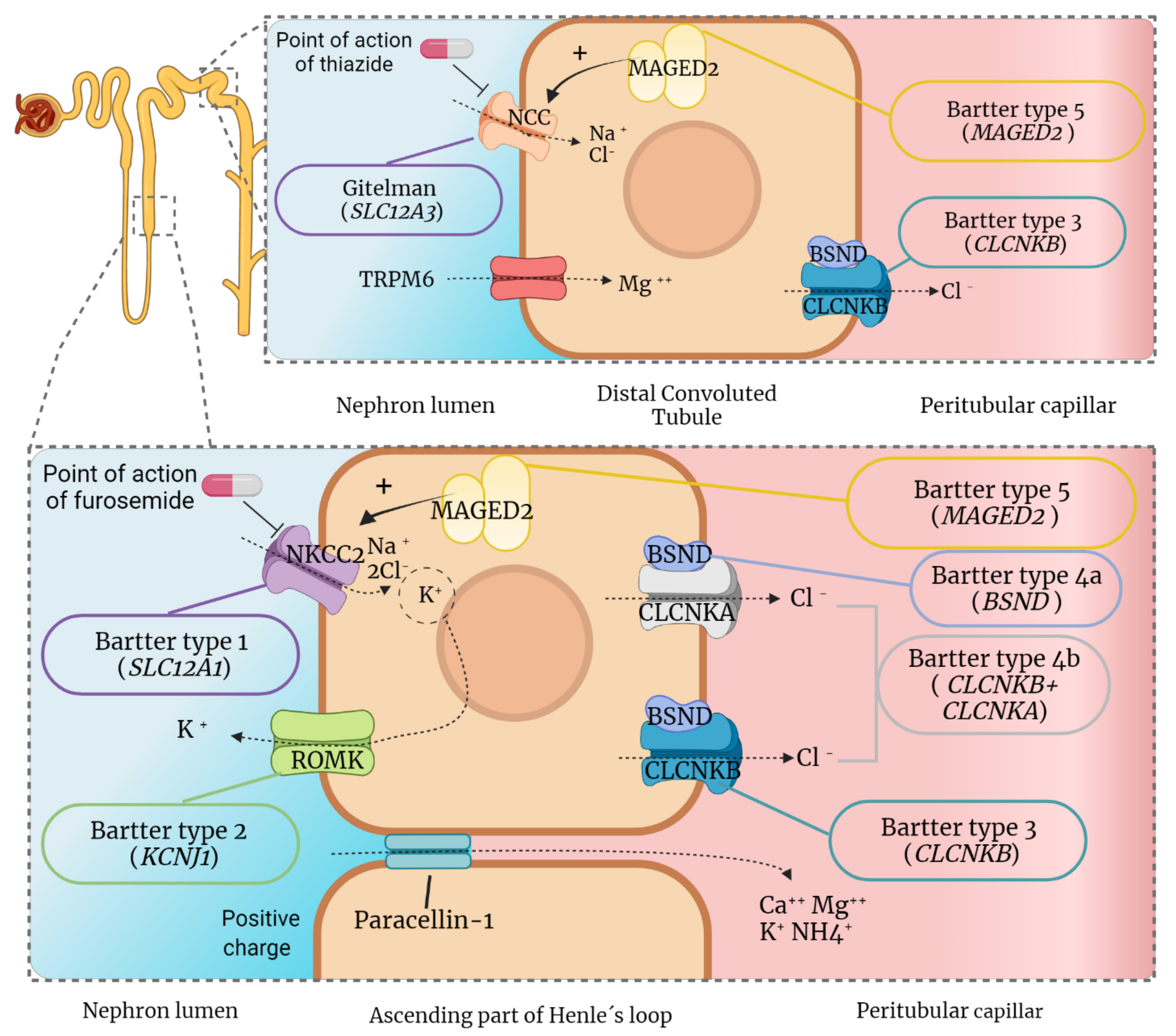
2. Renal Physiology of Electrolytes
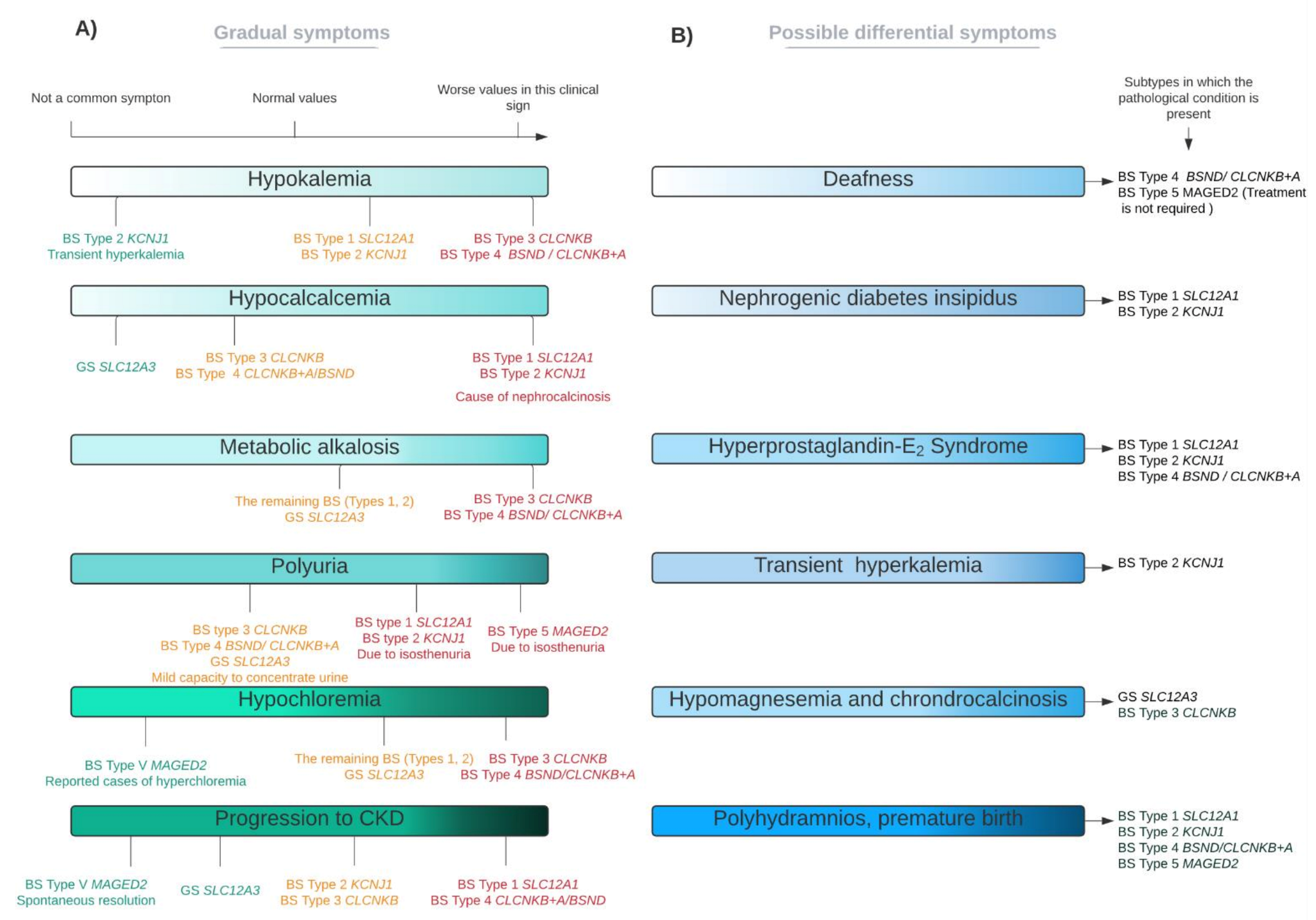
3. Molecular Basis and Clinical Features of the Diseases
3.1. Molecular Basis of Gitelman Syndrome and Clinical Consequences
3.2. Molecular Basis of Bartter Syndrome and Clinical Consequences
- Bartter syndrome types 1 and 2
- Bartter syndrome types 3 and 4
3.3. Long-Term Outcomes in GS and BS
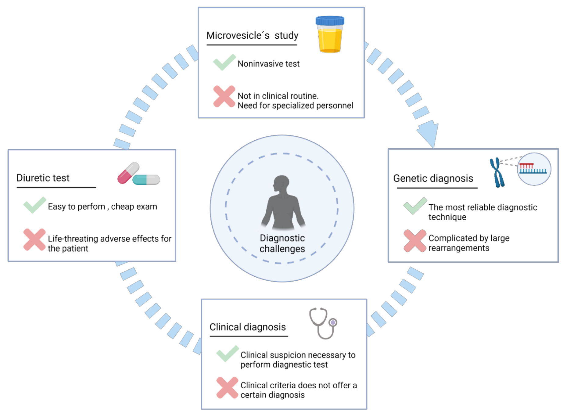
4. Diagnostic Approaches
4.1. Clinical Diagnosis
4.2. Diuretics Tests
4.3. Analysis of Urinary Microvesicles
4.4. Genetic Diagnosis
Heterozygous Carriers for Gitelman Mutations
5. Therapeutic Approaches
5.1. Current Pharmacological Treatments
5.1.1. Oral Salt Supplementation
5.1.2. Non-Steroidal Anti-Inflammatory Drugs (NSAIDs)
5.1.3. Potassium Sparing Diuretics
5.1.4. Renin-Angiotensin-Aldosterone System Inhibitors
5.1.5. Growth Hormone (GH)
5.1.6. Overview of the Effectiveness of These Treatments
5.2. Future Therapeutic Perspectives
6. Conclusions
Author Contributions
Funding
Institutional Review Board Statement
Informed Consent Statement
Data Availability Statement
Acknowledgments
Conflicts of Interest
Abbreviations
| ACE2 | angiotensin converting enzyme 2 |
| ADH | antidiuretic hormone |
| BS | Bartter syndrome |
| BSND | barttin |
| cAMP | cyclic adenosine monophosphate |
| CASR | extracellular calcium-sensing receptor |
| CLCNKA | chloride channel protein ClC-Ka |
| CLCNKB | chloride channel protein ClC-Kb |
| CNVs | copy number variants |
| DCT | distal convoluted tubule |
| ERAD | endoplasmic reticulum-associated degradation |
| GS | Gitelman syndrome |
| MLPA® | Multiplex ligation-dependent amplification probe |
| MVBs | multivesicular bodies |
| NGS | next generation sequencing |
| PGE2 | prostaglandin E2 |
| RAAS | renin-angiotensin-aldosterone system |
| NKCC2 | solute carrier family 12 member 1 |
| NCC | solute carrier family 12 member 3 |
| ROMK | ATP-sensitive inward rectifier potassium channel 1 |
| SNPs | single nucleotide polymorfism |
| TAL | ascending loop of Henle |
| TRPM6 | transient receptor potential cation channel subfamily M member 6 |
| TRPV5 | transient receptor potential cation channel subfamily V |
| UTR | non-translated regions |
| VUS | variants of uncertain significance |
References
- Arakawa, H.; Kubo, H.; Washio, I.; Staub, A.Y.; Nedachi, S.; Ishiguro, N.; Nakanishi, T.; Tamai, I. Rat Kidney Slices for Evaluation of Apical Membrane Transporters in Proximal Tubular Cells. J. Pharm. Sci. 2019, 108, 2798–2804. [Google Scholar] [CrossRef] [PubMed]
- Downie, M.L.; Lopez Garcia, S.C.; Kleta, R.; Bockenhauer, D. Inherited Tubulopathies of the Kidney. Clin. J. Am. Soc. Nephrol. 2020, 16, CJN14481119. [Google Scholar] [CrossRef] [PubMed] [Green Version]
- Mitchell, J.E.; Pomeroy, C.; Seppala, M.; Huber, M. Pseudo-Bartter’s syndrome, diuretic abuse, idiopathic edema, and eating disorders. Int. J. Eat. Disord. 1988, 7, 225–237. [Google Scholar] [CrossRef]
- Bartter and Gitelman Syndromes—UpToDate. Available online: https://www.uptodate.com/contents/inherited-hypokalemic-salt-losing-tubulopathies-pathophysiology-and-overview-of-clinical-manifestations (accessed on 14 May 2021).
- Hureaux, M.; Ashton, E.; Dahan, K.; Houillier, P.; Blanchard, A.; Cormier, C.; Koumakis, E.; Iancu, D.; Belge, H.; Hilbert, P.; et al. High-throughput sequencing contributes to the diagnosis of tubulopathies and familial hypercalcemia hypocalciuria in adults. Kidney Int. 2019, 96, 1408–1416. [Google Scholar] [CrossRef]
- Bao, M.; Cai, J.; Yang, X.; Ma, W. Genetic screening for Bartter syndrome and Gitelman syndrome pathogenic genes among individuals with hypertension and hypokalemia. Clin. Exp. Hypertens. 2019, 41, 381–388. [Google Scholar] [CrossRef]
- Orphanet: Bartter Syndrome. Available online: https://www.orpha.net/consor/cgi-bin/OC_Exp.php?Lng=GB&Expert=112 (accessed on 26 May 2021).
- Frederic, C. Hyperplasia of the juxtaglomerular complex with hyperaldosteronism and hypokalemic alkalosis. A new syndrome. Am. J. Med. 1962, 33, 811–828. [Google Scholar] [CrossRef]
- Simon, D.B.; Karet, F.E.; Hamdan, J.M.; Di Pietro, A.; Sanjad, S.A.; Lifton, R.P. Bartter’s syndrome, hypokalaemic alkalosis with hypercalciuria, is caused by mutations in the Na-K-2CI cotransporter NKCC2. Nat. Genet. 1996, 13, 183–188. [Google Scholar] [CrossRef]
- Simon, D.B.; Karet, F.E.; Rodriguez-Soriano, J.; Hamdan, J.H.; DiPietro, A.; Trachtman, H.; Sanjad, S.A.; Lifton, R.P. Genetic heterogeneity of Bartter’s syndrome revealed by mutations in the K+ channel, ROMK. Nat. Genet. 1996, 14, 152–156. [Google Scholar] [CrossRef]
- Simon, D.B.; Bindra, R.S.; Mansfield, T.A.; Nelson-Williams, C.; Mendonca, E.; Stone, R.; Schurman, S.; Nayir, A.; Alpay, H.; Bakkaloglu, A.; et al. Mutations in the chloride channel gene, CLCNKB, cause Bartter’s syndrome type III. Nat. Genet. 1997, 17, 171–178. [Google Scholar] [CrossRef]
- Birkenhäger, R.; Otto, E.; Schürmann, M.J.; Vollmer, M.; Ruf, E.M.; Maier-Lutz, I.; Beekmann, F.; Fekete, A.; Omran, H.; Feldmann, D.; et al. Mutation of BSND causes Bartter syndrome with sensorineural deafness and kidney failure. Nat. Genet. 2001, 29, 310–314. [Google Scholar] [CrossRef]
- Laghmani, K.; Beck, B.B.; Yang, S.-S.; Seaayfan, E.; Wenzel, A.; Reusch, B.; Vitzthum, H.; Priem, D.; Demaretz, S.; Bergmann, K.; et al. Polyhydramnios, Transient Antenatal Bartter’s Syndrome, and MAGED2 Mutations. N. Engl. J. Med. 2016, 374, 1853–1863. [Google Scholar] [CrossRef]
- Schlingmann, K.P.; Konrad, M.; Jeck, N.; Waldegger, P.; Reinalter, S.C.; Holder, M.; Seyberth, H.W.; Waldegger, S. Salt Wasting and Deafness Resulting from Mutations in Two Chloride Channels. N. Engl. J. Med. 2004, 350, 1314–1319. [Google Scholar] [CrossRef]
- Bichet, D.G.; Fujiwara, T.M. Reabsorption of Sodium Chloride—Lessons from the Chloride Channels. N. Engl. J. Med. 2004, 350, 1281–1283. [Google Scholar] [CrossRef]
- Gitelman, H.J.; Graham, J.B.; Welt, L.G. A familial disorder characterized by hypokalemia and hypomagnesemia. Ann. N. Y. Acad. Sci. 1969, 162, 856–864. [Google Scholar] [CrossRef]
- Simon, D.B.; Nelson-Williams, C.; Bia, M.J.; Ellison, D.; Karet, F.E.; Molina, A.M.; Vaara, I.; Iwata, F.; Cushner, H.M.; Koolen, M.; et al. Gitelman’s variant of Bartter’s syndrome, inherited hypokalaemic alkalosis, is caused by mutations in the thiazide-sensitive Na-Cl cotransporter. Nat. Genet. 1996, 12, 24–30. [Google Scholar] [CrossRef]
- Punzi, L.; Calo, L.; Schiavon, F.; Pianon, M.; Rosada, M.; Todesco, S. Chondrocalcinosis is a feature of Gitelman’s variant of Bartter’s syndrome: A new look at the hypomagnesemia associated with calcium pyrophosphate dihydrate crystal deposition disease. Rev. Rhum. 1998, 65, 571–574. [Google Scholar]
- George, A.L.; Neilson, E.G. Biología celular y molecular de los riñones. In Harrison. Principios de Medicina Interna, 19th ed.; Kasper, D., Fauci, A., Hauser, S., Longo, D., Jameson, J.L., Loscalzo, J., Eds.; McGraw-Hill Education: New York, NY, USA, 2019. [Google Scholar]
- Kleta, R.; Bockenhauer, D. Salt-losing tubulopathies in children: What’s new, what’s controversial? J. Am. Soc. Nephrol. 2018, 29, 727–739. [Google Scholar] [CrossRef] [Green Version]
- Fox, S.I. Fisiología de los riñones. In Fisiología Humana, 14th ed.; McGraw-Hill Education: New York, NY, USA, 2017. [Google Scholar]
- Eaton, D.C.; Pooler, J.P. Mecanismos de transporte tubular. In Fisiología Médica. Un Enfoque por Aparatos y Sistemas; Raff, H., Levitzky, M., Eds.; McGraw-Hill Education: New York, NY, USA, 2015. [Google Scholar]
- Seyberth, H.W.; Schlingmann, K.P. Bartter- and Gitelman-like syndromes: Salt-losing tubulopathies with loop or DCT defects. Pediatr. Nephrol. 2011, 26, 1789–1802. [Google Scholar] [CrossRef] [Green Version]
- Beck Laurence, H.J.; Salant, D.J. Enfermedades tubulointersticiales del riñón. In Harrison. Principios de Medicina Interna, 20th ed.; Jameson, J.L., Fauci, A.S., Kasper, D.L., Hauser, S.L., Longo, D.L., Loscalzo, J., Eds.; McGraw-Hill Education: New York, NY, USA, 2018. [Google Scholar]
- Besouw, M.T.P.; Kleta, R.; Bockenhauer, D. Bartter and Gitelman syndromes: Questions of class. Pediatr. Nephrol. 2020, 35, 1815–1824. [Google Scholar] [CrossRef] [Green Version]
- Velázquez, H.; Náray-Fejes-Tóth, A.; Silva, T.; Andújar, E.; Reilly, R.F.; Desir, G.V.; Ellison, D.H. Rabbit distal convoluted tubule coexpresses NaCl cotransporter and 11β- hydroxysteroid dehydrogenase II mRNA. Kidney Int. 1998, 54, 464–472. [Google Scholar] [CrossRef] [Green Version]
- Ellison, D.H.; Terker, A.S.; Gamba, G. Potassium and its discontents: New insight, new treatments. J. Am. Soc. Nephrol. 2016, 27, 981–989. [Google Scholar] [CrossRef] [Green Version]
- Maeoka, Y.; McCormick, J.A. NaCl cotransporter activity and Mg2+ handling by the distal convoluted tubule. Am. J. Physiol.—Ren. Physiol. 2020, 319, F1043–F1053. [Google Scholar] [CrossRef]
- Kunchaparty, S.; Palcso, M.; Berkman, J.; Velázquez, H.; Desir, G.V.; Bernstein, P.; Reilly, R.F.; Ellison, D.H. Defective processing and expression of thiazide-sensitive Na-Cl cotransporter as a cause of Gitelman’s syndrome. Am. J. Physiol.—Ren. Physiol. 1999, 277. [Google Scholar] [CrossRef]
- Kurtz, I. Molecular pathogenesis of Bartter’s and Gitelman’s syndromes. Kidney Int. 1998, 54, 1396–1410. [Google Scholar] [CrossRef] [Green Version]
- Peters, M.; Jeck, N.; Reinalter, S.; Leonhardt, A.; Tönshoff, B.; Klaus, G.; Konrad, M.; Seyberth, H.W. Clinical presentation of genetically defined patients with hypokalemic salt-losing tubulopathies. Am. J. Med. 2002, 112, 183–190. [Google Scholar] [CrossRef]
- Nijenhuis, T.; Vallon, V.; Van Der Kemp, A.W.C.M.; Loffing, J.; Hoenderop, J.G.J.; Bindels, R.J.M. Enhanced passive Ca2+ reabsorption and reduced Mg2+ channel abundance explains thiazide-induced hypocalciuria and hypomagnesemia. J. Clin. Investig. 2005, 115, 1651–1658. [Google Scholar] [CrossRef] [Green Version]
- Blanchard, A.; Bockenhauer, D.; Bolignano, D.; Calò, L.A.; Cosyns, E.; Devuyst, O.; Ellison, D.H.; Karet Frankl, F.E.; Knoers, N.V.A.M.; Konrad, M.; et al. Gitelman syndrome: Consensus and guidance from a Kidney Disease: Improving Global Outcomes (KDIGO) Controversies Conference. In Proceedings of the Kidney International; Elsevier B.V.: Amsterdam, The Netherlands, 2017; Volume 91, pp. 24–33. [Google Scholar]
- Seys, E.; Andrini, O.; Keck, M.; Mansour-Hendili, L.; Courand, P.Y.; Simian, C.; Deschenes, G.; Kwon, T.; Bertholet-Thomas, A.; Bobrie, G.; et al. Clinical and genetic spectrum of bartter syndrome type 3. J. Am. Soc. Nephrol. 2017, 28, 2540–2552. [Google Scholar] [CrossRef] [Green Version]
- Dai, L.J.; Ritchie, G.; Kerstan, D.; Kang, H.S.; Cole, D.E.C.; Quamme, G.A. Magnesium transport in the renal distal convoluted tubule. Physiol. Rev. 2001, 81, 51–84. [Google Scholar] [CrossRef]
- Franken, G.A.C.; Adella, A.; Bindels, R.J.M.; de Baaij, J.H.F. Mechanisms coupling sodium and magnesium reabsorption in the distal convoluted tubule of the kidney. Acta Physiol. 2021, 231, e13528. [Google Scholar] [CrossRef]
- Ea, H.K.; Blanchard, A.; Dougados, M.; Roux, C. Chondrocalcinosis secondary to hypomagnesemia in Gitelman’s syndrome. J. Rheumatol. 2005, 32, 1840–1842. [Google Scholar] [PubMed]
- Leone, F.A.; Rezende, L.A.; Ciancaglini, P.; Pizauro, J.M. Allosteric modulation of pyrophosphatase activity of rat osseous plate alkaline phosphatase by magnesium ions. Int. J. Biochem. Cell Biol. 1998, 30, 89–97. [Google Scholar] [CrossRef]
- Calò, L.; Punzi, L.; Semplicini, A. Hypomagnesemia and chondrocalcinosis in Bartter’s and Gitelman’s syndrome: Review of the pathogenetic mechanisms. Am. J. Nephrol. 2000, 20, 347–350. [Google Scholar] [CrossRef] [PubMed]
- Pollak, M.R.; Friedman, D.J. The genetic architecture of kidney disease. Clin. J. Am. Soc. Nephrol. 2020, 15, 268–275. [Google Scholar] [CrossRef] [PubMed] [Green Version]
- Stewart, D.; Iancu, D.; Ashton, E.; Courtney, A.E.; Connor, A.; Walsh, S.B. Transplantation of a Gitelman Syndrome Kidney Ameliorates Hypertension: A Case Report. Am. J. Kidney Dis. 2019, 73, 421–424. [Google Scholar] [CrossRef]
- Ji, W.; Foo, J.N.; O’Roak, B.J.; Zhao, H.; Larson, M.G.; Simon, D.B.; Newton-Cheh, C.; State, M.W.; Levy, D.; Lifton, R.P. Rare independent mutations in renal salt handling genes contribute to blood pressure variation. Nat. Genet. 2008, 40, 592–599. [Google Scholar] [CrossRef] [Green Version]
- Monette, M.Y.; Rinehart, J.; Lifton, R.P.; Forbush, B. Rare mutations in the human NA-K-CL cotransporter (NKCC2) associated with lower blood pressure exhibit impaired processing and transport function. Am. J. Physiol.—Ren. Physiol. 2011, 300, 840–847. [Google Scholar] [CrossRef] [Green Version]
- Balavoine, A.S.; Bataille, P.; Vanhille, P.; Azar, R.; Noël, C.; Asseman, P.; Soudan, B.; Wémeau, J.L.; Vantyghem, M.C. Phenotype-genotype correlation and follow-up in adult patients with hypokalaemia of renal origin suggesting Gitelman syndrome. Eur. J. Endocrinol. 2011, 165, 665–673. [Google Scholar] [CrossRef] [Green Version]
- Calò, L.A.; Davis, P.A. Are the clinical presentations (Phenotypes) of gitelman’s and bartter’s syndromes gene mutations driven by their effects on intracellular ph, their “ph” enotype? Int. J. Mol. Sci. 2020, 21, 5660. [Google Scholar] [CrossRef]
- Calò, L.A.; Davis, P.A.; Rossi, G.P. Understanding themechanisms of angiotensin II signaling involved in hypertension and its long-term sequelae: Insights from Bartter’s and Gitelman’s syndromes, humanmodels of endogenous angiotensin II signaling antagonism. J. Hypertens. 2014, 32, 2109–2119. [Google Scholar] [CrossRef]
- Melander, O.; Orho-Melander, M.; Bengtsson, K.; Lindblad, U.; Råstam, L.; Groop, L.; Hulthén, U.L. Genetic variants of thiazide-sensitive NaCl-cotransporter in Gitelman’s syndrome and primary hypertension. Hypertension 2000, 36, 389–394. [Google Scholar] [CrossRef] [Green Version]
- Berry, M.R.; Robinson, C.; Karet Frankl, F.E. Unexpected clinical sequelae of Gitelman syndrome: Hypertension in adulthood is common and females have higher potassium requirements. Nephrol. Dial. Transplant. 2013, 28, 1533–1542. [Google Scholar] [CrossRef] [Green Version]
- Evans, R.D.R.; Antonelou, M.; Sathiananthamoorthy, S.; Rega, M.; Henderson, S.; Ceron-Gutierrez, L.; Barcenas-Morales, G.; Müller, C.A.; Doffinger, R.; Walsh, S.B.; et al. Inherited salt-losing tubulopathies are associated with immunodeficiency due to impaired IL-17 responses. Nat. Commun. 2020, 11, 1–19. [Google Scholar] [CrossRef]
- Fujimura, J.; Nozu, K.; Yamamura, T.; Minamikawa, S.; Nakanishi, K.; Horinouchi, T.; Nagano, C.; Sakakibara, N.; Nakanishi, K.; Shima, Y.; et al. Clinical and Genetic Characteristics in Patients With Gitelman Syndrome. Kidney Int. Rep. 2019, 4, 119–125. [Google Scholar] [CrossRef] [Green Version]
- Blanchard, A.; Vallet, M.; Dubourg, L.; Hureaux, M.; Allard, J.; Haymann, J.P.; de la Faille, R.; Arnoux, A.; Dinut, A.; Bergerot, D.; et al. Resistance to insulin in patients with gitelman syndrome and a subtle intermediate phenotype in heterozygous carriers: A cross-sectional study. J. Am. Soc. Nephrol. 2019, 30, 1534–1545. [Google Scholar] [CrossRef]
- Ren, H.; Qin, L.; Wang, W.; Ma, J.; Zhang, W.; Shen, P.Y.; Shi, H.; Li, X.; Chen, N. Abnormal glucose metabolism and insulin sensitivity in Chinese patients with Gitelman syndrome. Am. J. Nephrol. 2013, 37, 152–157. [Google Scholar] [CrossRef]
- Han, Y.; Zhao, X.; Wang, S.; Wang, C.; Tian, D.; Lang, Y.; Bottillo, I.; Wang, X.; Shao, L. Eleven novel SLC12A1 variants and an exonic mutation cause exon skipping in Bartter syndrome type I. Endocrine 2020, 64, 708–718. [Google Scholar] [CrossRef]
- Brochard, K.; Boyer, O.; Blanchard, A.; Loirat, C.; Niaudet, P.; MacHer, M.A.; Deschenes, G.; Bensman, A.; Decramer, S.; Cochat, P.; et al. Phenotype-genotype correlation in antenatal and neonatal variants of Bartter syndrome. Nephrol. Dial. Transplant. 2009, 24, 1455–1464. [Google Scholar] [CrossRef] [Green Version]
- Mourani, C.C.; Sanjad, S.A.; Akatcherian, C.Y. Bartter syndrome in a neonate: Early treatment with indomethacin. Pediatr. Nephrol. 2000, 14, 143–145. [Google Scholar] [CrossRef]
- Finer, G.; Shalev, H.; Birk, O.S.; Galron, D.; Jeck, N.; Sinai-Treiman, L.; Landau, D. Transient neonatal hyperkalemia in the antenatal (ROMK defective) Bartter syndrome. J. Pediatr. 2003, 142, 318–323. [Google Scholar] [CrossRef]
- Gamba, G.; Friedman, P.A. Thick ascending limb: The Na+:K+:2Cl− co-transporter, NKCC2, and the calcium-sensing receptor, CaSR. Pflugers Arch. Eur. J. Physiol. 2009, 458, 61–76. [Google Scholar] [CrossRef] [Green Version]
- Mutig, K.; Kahl, T.; Saritas, T.; Godes, M.; Persson, P.; Bates, J.; Raffi, H.; Rampoldi, L.; Uchida, S.; Hille, C.; et al. Activation of the bumetanide-sensitive Na +,K +, 2Cl -Cotransporter (NKCC2) is facilitated by Tamm-Horsfall protein in a chloride-sensitive manner. J. Biol. Chem. 2011, 286, 30200–30210. [Google Scholar] [CrossRef] [Green Version]
- Schiano, G.; Glaudemans, B.; Olinger, E.; Goelz, N.; Müller, M.; Loffing-Cueni, D.; Deschenes, G.; Loffing, J.; Devuyst, O. The Urinary Excretion of Uromodulin is Regulated by the Potassium Channel ROMK. Sci. Rep. 2019, 9, 1–12. [Google Scholar] [CrossRef]
- Marcoux, A.A.; Tremblay, L.E.; Slimani, S.; Fiola, M.J.; Mac-Way, F.; Garneau, A.P.; Isenring, P. Molecular characteristics and physiological roles of Na+–K+–Cl− cotransporter 2. J. Cell. Physiol. 2021, 236, 1712–1729. [Google Scholar] [CrossRef]
- Grill, A.; Schießl, I.M.; Gess, B.; Fremter, K.; Hammer, A.; Castrop, H. Salt-losing nephropathy in mice with a null mutation of the Clcnk2 gene. Acta Physiol. 2016, 218, 198–211. [Google Scholar] [CrossRef]
- Hennings, J.C.; Andrini, O.; Picard, N.; Paulais, M.; Huebner, A.K.; Cayuqueo, I.K.L.; Bignon, Y.; Keck, M.; Cornière, N.; Böhm, D.; et al. The ClC-K2 chloride channel is critical for salt handling in the distal nephron. J. Am. Soc. Nephrol. 2017, 28, 209–217. [Google Scholar] [CrossRef]
- Pérez-Rius, C.; Castellanos, A.; Gaitán-Peñas, H.; Navarro, A.; Artuch, R.; Barrallo-Gimeno, A.; Estévez, R. Role of zebrafish ClC-K/barttin channels in apical kidney chloride reabsorption. J. Physiol. 2019, 597, 3969–3983. [Google Scholar] [CrossRef]
- Nozu, K.; Inagaki, T.; Fu, X.J.; Nozu, Y.; Kaito, H.; Kanda, K.; Sekine, T.; Igarashi, T.; Nakanishi, K.; Yoshikawa, N.; et al. Molecular analysis of digenic inheritance in Bartter syndrome with sensorineural deafness. J. Med. Genet. 2008, 45, 182–186. [Google Scholar] [CrossRef]
- Estévez, R.; Boettger, T.; Stein, V.; Birkenhäger, R.; Otto, E.; Hildebrandt, F.; Jentsch, T.J. Barttin is a Cl− channel β-subunit crucial for renal Cl− reabsorption and inner ear K+ secretion. Nature 2001, 414, 558–561. [Google Scholar] [CrossRef] [PubMed]
- Matsumura, Y.; Uchida, S.; Kondo, Y.; Miyazaki, H.; Ko, S.B.H.; Hayama, A.; Morimoto, T.; Liu, W.; Arisawa, M.; Sasaki, S.; et al. Overt nephrogenic diabetes insipidus in mice lacking the CLC-K1 chloride channel. Nat. Genet. 1999, 21, 95–98. [Google Scholar] [CrossRef] [PubMed]
- Kruegel, J.; Rubel, D.; Gross, O. Alport syndrome—Insights from basic and clinical research. Nat. Rev. Nephrol. 2013, 9, 170–178. [Google Scholar] [CrossRef] [PubMed]
- Rost, S.; Bach, E.; Neuner, C.; Nanda, I.; Dysek, S.; Bittner, R.E.; Keller, A.; Bartsch, O.; Mlynski, R.; Haaf, T.; et al. Novel form of X-linked nonsyndromic hearing loss with cochlear malformation caused by a mutation in the type IV collagen gene COL4A6. Eur. J. Hum. Genet. 2014, 22, 208–215. [Google Scholar] [CrossRef] [Green Version]
- Villard, E.; Perret, C.; Gary, F.; Proust, C.; Dilanian, G.; Hengstenberg, C.; Ruppert, V.; Arbustini, E.; Wichter, T.; Germain, M.; et al. A genome-wide association study identifies two loci associated with heart failure due to dilated cardiomyopathy. Eur. Heart J. 2011, 32, 1065–1076. [Google Scholar] [CrossRef] [Green Version]
- Quigley, R.; Saland, J.M. Transient antenatal Bartter’s Syndrome and X-linked polyhydramnios: Insights from the genetics of a rare condition. Kidney Int. 2016, 90, 721–723. [Google Scholar] [CrossRef]
- Allison, S.J. Renal physiology: MAGED2 mutations in transient antenatal Bartter syndrome. Nat. Rev. Nephrol. 2016, 12, 377. [Google Scholar]
- Bakhos-douaihy, D.; Seaayfan, E.; Demaretz, S.; Komhoff, M.; Laghmani, K. Differential effects of stch and stress—Inducible hsp70 on the stability and maturation of nkcc2. Int. J. Mol. Sci. 2021, 7, 2207. [Google Scholar] [CrossRef]
- Donnelly, B.F.; Needham, P.G.; Snyder, A.C.; Roy, A.; Khadem, S.; Brodsky, J.L.; Subramanya, A.R. Hsp70 and Hsp90 multichaperone complexes sequentially regulate thiazide-sensitive cotransporter endoplasmic reticulum-associated degradation and biogenesis. J. Biol. Chem. 2013, 288, 13124–13135. [Google Scholar] [CrossRef] [Green Version]
- Needham, P.G.; Mikoluk, K.; Dhakarwal, P.; Khadem, S.; Snyder, A.C.; Subramanya, A.R.; Brodsky, J.L. The thiazide-sensitive NaCl cotransporter is targeted for chaperone-dependent endoplasmic reticulum-associated degradation. J. Biol. Chem. 2011, 286, 43611–43621. [Google Scholar] [CrossRef] [Green Version]
- Rosenbaek, L.L.; Rizzo, F.; Wu, Q.; Rojas-Vega, L.; Gamba, G.; MacAulay, N.; Staub, O.; Fenton, R.A. The thiazide sensitive sodium chloride co-transporter NCC is modulated by site-specific ubiquitylation. Sci. Rep. 2017, 7. [Google Scholar] [CrossRef]
- Legrand, A.; Treard, C.; Roncelin, I.; Dreux, S.; Bertholet-Thomas, A.; Broux, F.; Bruno, D.; Decramer, S.; Deschenes, G.; Djeddi, D.; et al. Prevalence of novel MAGED2 mutations in antenatal bartter syndrome. Clin. J. Am. Soc. Nephrol. 2018, 13, 242–250. [Google Scholar] [CrossRef] [Green Version]
- Loffing, J.; Vallon, V.; Loffing-Cueni, D.; Aregger, F.; Richter, K.; Pietri, L.; Bloch-Faure, M.; Hoenderop, J.G.J.; Shull, G.E.; Meneton, P.; et al. Altered renal distal tubule structure and renal Na+ and Ca 2+ handling in a mouse model for Gitelman’s syndrome. J. Am. Soc. Nephrol. 2004, 15, 2276–2288. [Google Scholar] [CrossRef] [Green Version]
- Wang, L.; Zhang, C.; Su, X.; Lin, D.H.; Wang, W. Caveolin-1 deficiency inhibits the basolateral K+ channels in the distal convoluted tubule and impairs renal K+ and Mg2+ transport. J. Am. Soc. Nephrol. 2015, 26, 2678–2690. [Google Scholar] [CrossRef] [Green Version]
- Giebisch, G. Renal Potassium Channels: Function, Regulation, and Structure. In Proceedings of the Kidney International; Blackwell Publishing Inc.: Hoboken, NJ, USA, 2001; Volume 60, pp. 436–445. [Google Scholar]
- Waldegger, S.; Jentsch, T.J. Functional and structural analysis of ClC-K chloride channels involved in renal disease. J. Biol. Chem. 2000, 275, 24527–24533. [Google Scholar] [CrossRef] [Green Version]
- Li, D.; Tian, L.; Hou, C.; Kim, C.E.; Hakonarson, H.; Levine, M.A. Association of mutations in SLC12A1 encoding the NKCC2 cotransporter with neonatal primary hyperparathyroidism. J. Clin. Endocrinol. Metab. 2016, 101, 2196–2200. [Google Scholar] [CrossRef] [Green Version]
- Wongsaengsak, S.; Vidmar, A.P.; Addala, A.; Kamil, E.S.; Sequeira, P.; Fass, B.; Pitukcheewanont, P. A novel SLC12A1 gene mutation associated with hyperparathyroidism, hypercalcemia, nephrogenic diabetes insipidus, and nephrocalcinosis in four patients. Bone 2017, 97, 121–125. [Google Scholar] [CrossRef]
- Chen, Q.; Wang, X.; Min, J.; Wang, L.; Mou, L. Kidney stones and moderate proteinuria as the rare manifestations of Gitelman syndrome. BMC Nephrol. 2021, 22, 12. [Google Scholar] [CrossRef]
- Demoulin, N.; Aydin, S.; Cosyns, J.P.; Dahan, K.; Cornet, G.; Auberger, I.; Loffing, J.; Devuyst, O. Gitelman syndrome and glomerular proteinuria: A link between loss of sodium-chloride cotransporter and podocyte dysfunction? Nephrol. Dial. Transplant. 2014, 29, iv117–iv120. [Google Scholar] [CrossRef] [Green Version]
- Walsh, P.R.; Tse, Y.; Ashton, E.; Iancu, D.; Jenkins, L.; Bienias, M.; Kleta, R.; Van’t Hoff, W.; Bockenhauer, D. Clinical and diagnostic features of Bartter and Gitelman syndromes. Clin. Kidney J. 2018, 11, 302–309. [Google Scholar] [CrossRef] [Green Version]
- Bettinelli, A.; Borsa, N.; Bellantuono, R.; Syrèn, M.L.; Calabrese, R.; Edefonti, A.; Komninos, J.; Santostefano, M.; Beccaria, L.; Pela, I.; et al. Patients With Biallelic Mutations in the Chloride Channel Gene CLCNKB: Long-Term Management and Outcome. Am. J. Kidney Dis. 2007, 49, 91–98. [Google Scholar] [CrossRef]
- Stokman, M.F.; Renkema, K.Y.; Giles, R.H.; Schaefer, F.; Knoers, N.V.A.M.; van Eerde, A.M. The expanding phenotypic spectra of kidney diseases: Insights from genetic studies. Nat. Rev. Nephrol. 2016, 12, 472–483. [Google Scholar] [CrossRef]
- Watanabe, S.; Fukumoto, S.; Chang, H.; Takeuchi, Y.; Hasegawa, Y.; Okazaki, R.; Chikatsu, N.; Fujita, T. Association between activating mutations of calcium-sensing receptor and Bartter’s syndrome. Lancet 2002, 360, 692–694. [Google Scholar] [CrossRef]
- Carmosino, M.; Gerbino, A.; Hendy, G.N.; Torretta, S.; Rizzo, F.; Debellis, L.; Procino, G.; Svelto, M. NKCC2 activity is inhibited by the Bartter’s syndrome type 5 gain-of-function CaR-A843E mutant in renal cells. Biol. Cell 2015, 107, 98–110. [Google Scholar] [CrossRef] [PubMed]
- Vargas-Poussou, R.; Huang, C.; Hulin, P.; Houillier, P.; Jeunemaître, X.; Paillard, M.; Planelles, G.; Déchaux, M.; Miller, R.T.; Antignac, C. Functional characterization of a calcium-sensing receptor mutation in severe autosomal dominant hypocalcemia with a Bartter-like syndrome. J. Am. Soc. Nephrol. 2002, 13, 2259–2266. [Google Scholar] [CrossRef] [PubMed] [Green Version]
- Huang, C.; Sindic, A.; Hill, C.E.; Hujer, K.M.; Chan, K.W.; Sassen, M.; Wu, Z.; Kurachi, Y.; Nielsen, S.; Romero, M.F.; et al. Interaction of the Ca2+-sensing receptor with the inwardly rectifying potassium channels Kir4.1 and Kir4.2 results in inhibition of channel function. Am. J. Physiol.—Ren. Physiol. 2007, 292, F1073–F1081. [Google Scholar] [CrossRef] [Green Version]
- Vezzoli, G.; Arcidiacono, T.; Paloschi, V.; Terranegra, A.; Biasion, R.; Weber, G.; Mora, S.; Syren, M.L.; Coviello, D.; Cusi, D.; et al. Autosomal dominant hypocalcemia with mild type 5 Bartter syndrome. J. Nephrol. 2006, 19, 525–528. [Google Scholar] [PubMed]
- Matsunoshita, N.; Nozu, K.; Shono, A.; Nozu, Y.; Fu, X.J.; Morisada, N.; Kamiyoshi, N.; Ohtsubo, H.; Ninchoji, T.; Minamikawa, S.; et al. Differential diagnosis of Bartter syndrome, Gitelman syndrome, and pseudo-Bartter/Gitelman syndrome based on clinical characteristics. Genet. Med. 2016, 18, 180–188. [Google Scholar] [CrossRef] [Green Version]
- Alfandary, H.; Landau, D. Future considerations based on the information from Barrter’s and Gitelman’s syndromes. Curr. Opin. Nephrol. Hypertens. 2017, 26, 9–13. [Google Scholar] [CrossRef]
- Jain, G.; Ong, S.; Warnock, D.G. Genetic disorders of potassium homeostasis. Semin. Nephrol. 2013, 33, 300–309. [Google Scholar] [CrossRef]
- Kamel, K.S.; Halperin, M.L. Use of Urine Electrolytes and Urine Osmolalityin Clinical Diagnosis of Fluid, Electrolytes, and Acid-Base Disorders. Kidney Int. Rep. 2021, 6, 1211–1224. [Google Scholar] [CrossRef]
- Persu, A.; Lafontaine, J.J.; Devuyst, O. Chronic hypokalaemia in young women—It is not always abuse of diuretics. Nephrol. Dial. Transplant. 1999, 14, 1021–1025. [Google Scholar] [CrossRef] [Green Version]
- Adalat, S.; Woolf, A.S.; Johnstone, K.A.; Wirsing, A.; Harries, L.W.; Long, D.A.; Hennekam, R.C.; Ledermann, S.E.; Rees, L.; Van’t Hoff, W.; et al. HNF1B mutations associate with hypomagnesemia and renal magnesium wasting. J. Am. Soc. Nephrol. 2009, 20, 1123–1131. [Google Scholar] [CrossRef] [Green Version]
- Verhave, J.C.; Bech, A.P.; Wetzels, J.F.M.; Nijenhuis, T. Hepatocyte nuclear factor 1β-associated kidney disease: More than renal cysts and diabetes. J. Am. Soc. Nephrol. 2016, 27, 345–353. [Google Scholar] [CrossRef] [Green Version]
- Viering, D.H.H.M.; de Baaij, J.H.F.; Walsh, S.B.; Kleta, R.; Bockenhauer, D. Genetic causes of hypomagnesemia, a clinical overview. Pediatr. Nephrol. 2017, 32, 1123–1135. [Google Scholar] [CrossRef] [Green Version]
- Bamgbola, O.F.; Ahmed, Y. Differential diagnosis of perinatal Bartter, Bartter and Gitelman syndromes. Clin. Kidney J. 2021, 14, 36–48. [Google Scholar] [CrossRef]
- Kintu, B.; Brightwell, A. Episodic seasonal pseudo-bartter syndrome in cystic fibrosis. Paediatr. Respir. Rev. 2014, 15, 19–21. [Google Scholar] [CrossRef]
- Kim, Y.K.; Song, H.C.; Kim, Y.-S.; Choi, E.J. Acquired Gitelman Syndrome. Electrolytes Blood Press. 2009, 7, 5. [Google Scholar] [CrossRef] [Green Version]
- Barathidasan, G.S.; Krishnamurthy, S.; Karunakar, P.; Rajendran, R.; Ramya, K.; Dhandapany, G.; Ramamoorthy, J.G.; Ganesh, R.N. Systemic lupus erythematosus complicated by a Gitelman-like syndrome in an 8-year-old girl. CEN Case Rep. 2020, 9, 129–132. [Google Scholar] [CrossRef]
- Matsunoshita, N.; Nozu, K.; Yoshikane, M.; Kawaguchi, A.; Fujita, N.; Morisada, N.; Ishimori, S.; Yamamura, T.; Minamikawa, S.; Horinouchi, T.; et al. Congenital chloride diarrhea needs to be distinguished from Bartter and Gitelman syndrome. J. Hum. Genet. 2018, 63, 887–892. [Google Scholar] [CrossRef]
- Kurteva, E.; Lindley, K.J.; Hill, S.M.; Köglmeier, J. Mucosal Abnormalities in Children With Congenital Chloride Diarrhea—An Underestimated Phenotypic Feature? Front. Pediatr. 2020, 8. [Google Scholar] [CrossRef]
- Nozu, K.; Yamamura, T.; Horinouchi, T.; Nagano, C.; Sakakibara, N.; Ishikura, K.; Hamada, R.; Morisada, N.; Iijima, K. Inherited salt-losing tubulopathy: An old condition but a new category of tubulopathy. Pediatr. Int. 2020, 62, 428–437. [Google Scholar] [CrossRef]
- Nesbit, M.A.; Hannan, F.M.; Howles, S.A.; Babinsky, V.N.; Head, R.A.; Cranston, T.; Rust, N.; Hobbs, M.R.; Heath, H.; Thakker, R.V. Mutations Affecting G-Protein Subunit α 11 in Hypercalcemia and Hypocalcemia. N. Engl. J. Med. 2013, 368, 2476–2486. [Google Scholar] [CrossRef] [Green Version]
- Okazaki, R.; Chikatsu, N.; Nakatsu, M.; Takeuchi, Y.; Ajima, M.; Miki, J.; Fujita, T.; Arai, M.; Totsuka, Y.; Tanaka, K.; et al. A Novel Activating Mutation in Calcium-Sensing Receptor Gene Associated with a Family of Autosomal Dominant Hypocalcemia 1. J. Clin. Endocrinol. Metab. 1999, 84, 363–366. [Google Scholar] [CrossRef] [PubMed]
- Devuyst, O.; Olinger, E.; Weber, S.; Eckardt, K.U.; Kmoch, S.; Rampoldi, L.; Bleyer, A.J. Autosomal dominant tubulointerstitial kidney disease. Nat. Rev. Dis. Prim. 2019, 5, 1–20. [Google Scholar] [CrossRef] [PubMed] [Green Version]
- Schaeffer, C.; Olinger, E. Clinical and genetic spectra of kidney disease caused by REN mutations. Kidney Int. 2020, 98, 1397–1400. [Google Scholar] [CrossRef] [PubMed]
- Bockenhauer, D.; Bichet, D.G. Inherited secondary nephrogenic diabetes insipidus: Concentrating on humans. Am. J. Physiol.—Ren. Physiol. 2013, 304, 1037–1042. [Google Scholar] [CrossRef] [PubMed] [Green Version]
- Bockenhauer, D.; Van’T Hoff, W.; Dattani, M.; Lehnhardt, A.; Subtirelu, M.; Hildebrandt, F.; Bichet, D.G. Secondary nephrogenic diabetes insipidus as a complication of inherited renal diseases. Nephron—Physiol. 2010, 116. [Google Scholar] [CrossRef] [PubMed] [Green Version]
- D’Alessandri-Silva, C.; Carpenter, M.; Ayoob, R.; Barcia, J.; Chishti, A.; Constantinescu, A.; Dell, K.M.; Goodwin, J.; Hashmat, S.; Iragorri, S.; et al. Diagnosis, Treatment, and Outcomes in Children With Congenital Nephrogenic Diabetes Insipidus: A Pediatric Nephrology Research Consortium Study. Front. Pediatr. 2020, 7, 550. [Google Scholar] [CrossRef] [Green Version]
- Kennedy, J.D.; Dinwiddie, R.; Daman-Willems, C.; Dillon, M.J.; Matthew, D.J. Pseudo-Bartter’s syndrome in cystic fibrosis. Arch. Dis. Child. 1990, 65, 786–787. [Google Scholar] [CrossRef] [Green Version]
- Okamoto, T.; Tajima, T.; Hirayama, T.; Sasaki, S. A patient with Dent disease and features of Bartter syndrome caused by a novel mutation of CLCN5. Eur. J. Pediatr. 2012, 171, 401–404. [Google Scholar] [CrossRef]
- Chen, Y.S.; Fang, H.C.; Chou, K.J.; Lee, P.T.; Hsu, C.Y.; Huang, W.C.; Chung, H.M.; Chen, C.L. Gentamicin-Induced Bartter-like Syndrome. Am. J. Kidney Dis. 2009, 54, 1158–1161. [Google Scholar] [CrossRef]
- Chrispal, A.; Boorugu, H.; Prabhakar, A.; Moses, V. Amikacin-induced type 5 Bartter-like syndrome with severe hypocalcemia. J. Postgrad. Med. 2009, 55, 208. [Google Scholar] [CrossRef]
- Medication Guides. Arikayce. Available online: https://www.accessdata.fda.gov/scripts/cder/daf/index.cfm?event=medguide.page (accessed on 13 May 2021).
- Cinotti, R.; Lascarrou, J.B.; Azais, M.A.; Colin, G.; Quenot, J.P.; Mahé, P.J.; Roquilly, A.; Gaultier, A.; Asehnoune, K.; Reignier, J. Diuretics decrease fluid balance in patients on invasive mechanical ventilation: The randomized-controlled single blind, IRIHS study. Crit. Care 2021, 25, 1–9. [Google Scholar] [CrossRef]
- Blowey, D.L. Diuretics in the treatment of hypertension. Pediatr. Nephrol. 2016, 31, 2223–2233. [Google Scholar] [CrossRef]
- Nozu, K.; Iijima, K.; Kanda, K.; Nakanishi, K.; Yoshikawa, N.; Satomura, K.; Kaito, H.; Hashimura, Y.; Ninchoji, T.; Komatsu, H.; et al. The pharmacological characteristics of molecular-based inherited salt-losing tubulopathies. J. Clin. Endocrinol. Metab. 2010, 95. [Google Scholar] [CrossRef] [Green Version]
- Colussi, G.; Bettinelli, A.; Tedeschi, S.; De Ferrari, M.E.; Syrén, M.L.; Borsa, N.; Mattiello, C.; Casari, G.; Bianchetti, M.G. A thiazide test for the diagnosis of renal tubular hypokalemic disorders. Clin. J. Am. Soc. Nephrol. 2007, 2, 454–460. [Google Scholar] [CrossRef] [Green Version]
- A Translational Approach to Gitelman Syndrome—Full Text View—ClinicalTrials.gov. Available online: https://clinicaltrials.gov/ct2/show/NCT00822107 (accessed on 26 May 2021).
- Konrad, M.; Nijenhuis, T.; Ariceta, G.; Bertholet-Thomas, A.; Calo, L.A.; Capasso, G.; Emma, F.; Schlingmann, K.P.; Singh, M.; Trepiccione, F.; et al. Diagnosis and management of Bartter syndrome: Executive summary of the consensus and recommendations from the European Rare Kidney Disease Reference Network Working Group for Tubular Disorders. Kidney Int. 2021, 99, 324–335. [Google Scholar] [CrossRef]
- Corbetta, S.; Raimondo, F.; Tedeschi, S.; Syrèn, M.L.; Rebora, P.; Savoia, A.; Baldi, L.; Bettinelli, A.; Pitto, M. Urinary exosomes in the diagnosis of Gitelman and Bartter syndromes. Nephrol. Dial. Transplant. 2015, 30, 621–630. [Google Scholar] [CrossRef] [Green Version]
- Pisitkun, T.; Shen, R.F.; Knepper, M.A. Identification and proteomic profiling of exosomes in human urine. Proc. Natl. Acad. Sci. USA 2004, 101, 13368–13373. [Google Scholar] [CrossRef] [Green Version]
- Williams, T.L.; Bastos, C.; Faria, N.; Karet Frankl, F.E. Making urinary extracellular vesicles a clinically tractable source of biomarkers for inherited tubulopathies using a small volume precipitation method: Proof of concept. J. Nephrol. 2020, 33, 383–386. [Google Scholar] [CrossRef] [Green Version]
- Snoek, R.; Stokman, M.F.; Lichtenbelt, K.D.; van Tilborg, T.C.; Simcox, C.E.; Paulussen, A.D.C.; Dreesen, J.C.M.F.; van Reekum, F.; Lely, A.T.; Knoers, N.V.A.M.; et al. Preimplantation Genetic Testing for Monogenic Kidney Disease. Clin. J. Am. Soc. Nephrol. 2020, CJN.03550320. [Google Scholar] [CrossRef]
- Lim, M.; Gannon, D. Diagnosis and outpatient management of Gitelman syndrome from the first trimester of pregnancy. BMJ Case Rep. 2021, 14, e241756. [Google Scholar] [CrossRef]
- Wu, W.F.; Pan, M. The outcome of two pregnancies in a patient with Gitelman syndrome: Case report and review of the literature. J. Matern. Neonatal Med. 2020, 33, 4171–4173. [Google Scholar] [CrossRef]
- Han, Y.; Lin, Y.; Sun, Q.; Wang, S.; Gao, Y.; Shao, L. Mutation spectrum of Chinese patients with bartter syndrome. Oncotarget 2017, 8, 101614–101622. [Google Scholar] [CrossRef] [Green Version]
- Feldmann, D.; Alessandri, J.L.; Deschênes, G. Large deletion of the 5’ end of the ROMK1 gene causes antenatal Bartter syndrome. J. Am. Soc. Nephrol. 1998, 9, 2357–2359. [Google Scholar] [CrossRef]
- García Castaño, A.; Pérez de Nanclares, G.; Madariaga, L.; Aguirre, M.; Madrid, A.; Nadal, I.; Navarro, M.; Lucas, E.; Fijo, J.; Espino, M.; et al. Genetics of Type III Bartter Syndrome in Spain, Proposed Diagnostic Algorithm. PLoS ONE 2013, 8, e74673. [Google Scholar] [CrossRef] [Green Version]
- Castaño, A.G.; De Nanclares, G.P.; Madariaga, L.; Aguirre, M.; Madrid, Á.; Chocrón, S.; Nadal, I.; Navarro, M.; Lucas, E.; Fijo, J.; et al. Poor phenotype-genotype association in a large series of patients with Type III Bartter syndrome. PLoS ONE 2017, 12, e173581. [Google Scholar] [CrossRef]
- Ma, J.; Ren, H.; Lin, L.; Zhang, C.; Wang, Z.; Xie, J.; Shen, P.Y.; Zhang, W.; Wang, W.; Chen, X.N.; et al. Genetic Features of Chinese Patients with Gitelman Syndrome: Sixteen Novel SLC12A3 Mutations Identified in a New Cohort. Am. J. Nephrol. 2016, 44, 113–121. [Google Scholar] [CrossRef]
- Vargas-Poussou, R.; Dahan, K.; Kahila, D.; Venisse, A.; Riveira-Munoz, E.; Debaix, H.; Grisart, B.; Bridoux, F.; Unwin, R.; Moulin, B.; et al. Spectrum of mutations in Gitelman syndrome. J. Am. Soc. Nephrol. 2011, 22, 693–703. [Google Scholar] [CrossRef]
- Shen, Q.; Chen, J.; Yu, M.; Lin, Z.; Nan, X.; Dong, B.; Fang, X.; Chen, J.; Ding, G.; Zhang, A.; et al. Multi-centre study of the clinical features and gene variant spectrum of Gitelman syndrome in Chinese children. Clin. Genet. 2021, 99, 558–564. [Google Scholar] [CrossRef]
- Zhang, L.; Peng, X.; Zhao, B.; Zhu, Z.; Wang, Y.; Tian, D.; Yan, Z.; Yao, L.; Liu, J.; Qiu, L.; et al. Clinical and laboratory features of female Gitelman syndrome and the pregnancy outcomes in a Chinese cohort. Nephrology 2020, 25, 749–757. [Google Scholar] [CrossRef]
- Mighton, C.; Shickh, S.; Uleryk, E.; Pechlivanoglou, P.; Bombard, Y. Clinical and psychological outcomes of receiving a variant of uncertain significance from multigene panel testing or genomic sequencing: A systematic review and meta-analysis. Genet. Med. 2021, 23, 22–33. [Google Scholar] [CrossRef]
- Lo, Y.F.; Nozu, K.; Iijima, K.; Morishita, T.; Huang, C.C.; Yang, S.S.; Sytwu, H.K.; Fang, Y.W.; Tseng, M.H.; Lin, S.H. Recurrent deep intronic mutations in the SLC12A3 gene responsible for Gitelman’s syndrome. Clin. J. Am. Soc. Nephrol. 2011, 6, 630–639. [Google Scholar] [CrossRef] [Green Version]
- Zhang, L.; Huang, K.; Wang, S.; Fu, H.; Wang, J.; Shen, H.; Lu, Z.; Chen, J.; Bao, Y.; Feng, C.; et al. Clinical and Genetic Features in 31 Serial Chinese Children With Gitelman Syndrome. Front. Pediatr. 2021, 9, 299. [Google Scholar] [CrossRef]
- García-Nieto, V.M.; Claverie-Martín, F.; Perdomo-Ramírez, A.; Cárdoba-Lanus, E.; Ramos-Trujillo, E.; Mura-Escorche, G.; Tejera-Carreño, P.; Luis-Yanes, M.I. Considerations about the molecular basis of some kidney tubule disorders in relation to inbreeding and population displacement. Nefrologia 2020, 40, 126–132. [Google Scholar] [CrossRef] [PubMed]
- Le Gall, E.C.; Audrézet, M.-P.P.; Rousseau, A.; Hourmant, M.; Renaudineau, E.; Charasse, C.; Morin, M.-P.P.; Moal, M.-C.C.; Dantal, J.; Wehbe, B.; et al. No Title. J. Am. Soc. Nephrol. 2016, 27, 942–951. [Google Scholar] [CrossRef] [Green Version]
- Le Gall, E.C.; Audrézet, M.P.; Chen, J.M.; Hourmant, M.; Morin, M.P.; Perrichot, R.; Charasse, C.; Whebe, B.; Renaudineau, E.; Jousset, P.; et al. Type of PKD1 mutation influences renal outcome in ADPKD. J. Am. Soc. Nephrol. 2013, 24, 1006–1013. [Google Scholar] [CrossRef] [PubMed] [Green Version]
- Wan, X.; Perry, J.; Zhang, H.; Jin, F.; Ryan, K.A.; Van Hout, C.; Reid, J.; Overton, J.; Baras, A.; Han, Z.; et al. Heterozygosity for a Pathogenic Variant in SLC12A3 That Causes Autosomal Recessive Gitelman Syndrome Is Associated with Lower Serum Potassium. J. Am. Soc. Nephrol. 2021, 32, ASN2020071030. [Google Scholar] [CrossRef]
- Hsu, Y.J.; Yang, S.S.; Chu, N.F.; Sytwu, H.K.; Cheng, C.J.; Lin, S.H. Heterozygous mutations of the sodium chloride cotransporter in Chinese children: Prevalence and association with blood pressure. Nephrol. Dial. Transplant. 2009, 24, 1170–1175. [Google Scholar] [CrossRef] [Green Version]
- Fava, C.; Montagnana, M.; Rosberg, L.; Burri, P.; Almgren, P.; Jönsson, A.; Wanby, P.; Lippi, G.; Minuz, P.; Hulthèn, L.U.; et al. Subjects heterozygous for genetic loss of function of the thiazide-sensitive cotransporter have reduced blood pressure. Hum. Mol. Genet. 2008, 17, 413–418. [Google Scholar] [CrossRef]
- Cruz, D.N.; Simon, D.B.; Nelson-Williams, C.; Farhi, A.; Finberg, K.; Burleson, L.; Gill, J.R.; Lifton, R.P. Mutations in the Na-Cl cotransporter reduce blood pressure in humans. Hypertension 2001, 37, 1458–1464. [Google Scholar] [CrossRef] [Green Version]
- Knoers, N.V.A.M. Gitelman syndrome. Adv. Chronic Kidney Dis. 2006, 13, 148–154. [Google Scholar] [CrossRef]
- Ranade, V.V.; Somberg, J.C. Bioavailability and pharmacokinetics of magnesium after administration of magnesium salts to humans. Am. J. Ther. 2001, 8, 345–357. [Google Scholar] [CrossRef]
- Tarnawski, A.S.; Jones, M.K. Inhibition of angiogenesis by NSAIDs: Molecular mechanisms and clinical implications. J. Mol. Med. 2003, 81, 627–636. [Google Scholar] [CrossRef]
- Suleyman, H.; Cadirci, E.; Albayrak, A.; Halici, Z. Nimesulide is a Selective COX-2 Inhibitory, Atypical Non-Steroidal Anti-Inflammatory Drug. Curr. Med. Chem. 2008, 15, 278–283. [Google Scholar] [CrossRef]
- Fulchiero, R.; Seo-Mayer, P. Bartter Syndrome and Gitelman Syndrome. Pediatr. Clin. N. Am. 2019, 66, 121–134. [Google Scholar] [CrossRef]
- Mackie, F.E.; Hodson, E.M.; Roy, L.P.; Knight, J.F. Neonatal Bartter syndrome—Use of indomethacin in the newborn period and prevention of growth failure. Pediatr. Nephrol. 1996, 10, 756–758. [Google Scholar] [CrossRef]
- Gasongo, G.; Greenbaum, L.A.; Niel, O.; Kwon, T.; Macher, M.A.; Maisin, A.; Baudouin, V.; Dossier, C.; Deschênes, G.; Hogan, J. Effect of nonsteroidal anti-inflammatory drugs in children with Bartter syndrome. Pediatr. Nephrol. 2019, 34, 679–684. [Google Scholar] [CrossRef]
- Larkins, N.; Wallis, M.; McGillivray, B.; Mammen, C. A severe phenotype of Gitelman syndrome with increased prostaglandin excretion and favorable response to indomethacin. Clin. Kidney J. 2014, 7, 306–310. [Google Scholar] [CrossRef] [Green Version]
- Blanchard, A.; Vargas-Poussou, R.; Vallet, M.; Caumont-Prim, A.; Allard, J.; Desport, E.; Dubourg, L.; Monge, M.; Bergerot, D.; Baron, S.; et al. Indomethacin, amiloride, or eplerenone for treating hypokalemia in Gitelman syndrome. J. Am. Soc. Nephrol. 2015, 26, 468–475. [Google Scholar] [CrossRef] [Green Version]
- Brater, D.C. Anti-inflammatory agents and renal function. Semin. Arthritis Rheum. 2002, 32, 33–42. [Google Scholar] [CrossRef]
- Kleinknecht, D. Interstitial nephritis, the nephrotic syndrome, and chronic renal failure secondary to nonsteroidal anti-inflammatory drugs. Semin. Nephrol. 1995, 15, 228–235. [Google Scholar]
- Marlicz, W.; Łoniewski, I.; Grimes, D.S.; Quigley, E.M. Nonsteroidal anti-inflammatory drugs, proton pump inhibitors, and gastrointestinal injury: Contrasting interactions in the stomach and small Intestine. Mayo Clin. Proc. 2014, 89, 1699–1709. [Google Scholar] [CrossRef] [Green Version]
- The Hospital for Sick Children: Indomethacin, Oral Suspension. Available online: https://www.sickkids.ca/en/care-services/for-health-care-providers/pharmacy/ (accessed on 18 October 2021).
- Dembo, G.; Park, S.B.; Kharasch, E.D. Central nervous system concentrations of cyclooxygenase-2 inhibitors in humans. Anesthesiology 2005, 102, 409–415. [Google Scholar] [CrossRef]
- Solomon, D.H.; Schneeweiss, S.; Glynn, R.J.; Kiyota, Y.; Levin, R.; Mogun, H.; Avorn, J. Relationship between Selective Cyclooxygenase-2 Inhibitors and Acute Myocardial Infarction in Older Adults. Circulation 2004, 109, 2068–2073. [Google Scholar] [CrossRef] [Green Version]
- Baigent, C.; Bhala, N.; Emberson, J.; Merhi, A.; Abramson, S.; Arber, N.; Baron, J.A.; Bombardier, C.; Cannon, C.; Farkouh, M.E.; et al. Vascular and upper gastrointestinal effects of non-steroidal anti-inflammatory drugs: Meta-analyses of individual participant data from randomised trials. Lancet 2013, 382, 769–779. [Google Scholar] [CrossRef] [Green Version]
- Jackson, E.K. Fármacos que afectan la función excretora renal. In Goodman & Gilman: Las Bases Farmacológicas, De La Terapéutic, 13th ed.; Brunton, L.L., Chabner, B.A., Knollmann, B.C., Eds.; McGraw-Hill Education: New York, NY, USA, 2019. [Google Scholar]
- Perazella, M.A. Trimethoprim is a potassium-sparing diuretic like amiloride and causes hyperkalemia in high-risk patients. Am. J. Ther. 1997, 4, 343–348. [Google Scholar] [CrossRef]
- Plumb, L.A.; van’t Hoff, W.; Kleta, R.; Reid, C.; Ashton, E.; Samuels, M.; Bockenhauer, D. Renal apnoea: Extreme disturbance of homoeostasis in a child with Bartter syndrome type IV. Lancet 2016, 388, 631–632. [Google Scholar] [CrossRef] [Green Version]
- Schepkens, H.; Lameire, N. Gitelman’s syndrome: An overlooked cause of chronic hypokalemia and hypomagnesemia in adults. Acta Clin. Belg. 2001, 56, 248–254. [Google Scholar] [CrossRef]
- Koudsi, L.; Nikolova, S.; Mishra, V. Management of a severe case of Gitelman syndrome with poor response to standard treatment. BMJ Case Rep. 2016, 1–3. [Google Scholar] [CrossRef] [Green Version]
- Griffing, G.T.; Komanicky, P.; Aurecchis, S.A.; Sindler, B.H.; Melby, J.C. Amiloride in Bartter’s syndrome. Clin. Pharmacol. Ther. 1982, 31, 713–718. [Google Scholar] [CrossRef]
- Griffing, G.T.; Melby, J.C. The therapeutic use of a new potassium-sparing diuretic, amiloride, and a converting enzyme inhibitor, mk-421, in preventing hypokalemia associated with primary and secondary hyperaldosteronism. Clin. Exp. Hypertens. 1983, A5, 779–801. [Google Scholar] [CrossRef]
- Griffing, G.T.; Aurecchia, S.A.; Sindler, B.H.; Melby, J.C. The Effect of Amiloride on the Renin—Aldosterone System in Primary Hyperaldosteronism and Bartter’s Syndrome. J. Clin. Pharmacol. 1982, 22, 505–512. [Google Scholar] [CrossRef]
- Luqman, A.; Kazmi, A.; Wall, B.M. Bartter’s syndrome in pregnancy: Review of potassium homeostasis in gestation. Am. J. Med. Sci. 2009, 338, 500–504. [Google Scholar] [CrossRef] [PubMed]
- Deruelle, P.; Dufour, P.; Magnenant, E.; Courouble, N.; Puech, F. Maternal Bartter’s syndrome in pregnancy treated by amiloride. Eur. J. Obstet. Gynecol. Reprod. Biol. 2004, 115, 106–107. [Google Scholar] [CrossRef] [PubMed]
- Federal Register: Content and Format of Labeling for Human Prescription Drug and Biological Products. Requirements for Pregnancy and Lactation Labeling. Available online: https://www.federalregister.gov/documents/2008/05/29/E8-11806/content-and-format-of-labeling-for-human-prescription-drug-and-biological-products-requirements-for (accessed on 24 May 2021).
- Santos, F.; Gil-Peña, H.; Blázquez, C.; Coto, E. Gitelman syndrome: A review of clinical features, genetic diagnosis and therapeutic management. Expert Opin. Orphan Drugs 2016, 4, 1005–1009. [Google Scholar] [CrossRef]
- Morales, J.M.; Ruilope, L.M.; Praga, M.; Coto, A.; Alcazar, J.M.; Prieto, C.; Nieto, J.; Rodicio, J.L. Long-term enalapril therapy in Bartter’s syndrome. Nephron 1988, 48, 327. [Google Scholar] [CrossRef]
- Hené, R.J.; Koomans, H.A.; Mees, E.J.D.; Stolpe, A.; Verhoef, G.E.G.; Boer, P. Correction of Hypokalemia in Bartter’s Syndrome by Enalapril. Am. J. Kidney Dis. 1987, 9, 200–205. [Google Scholar] [CrossRef]
- Nascimento, C.L.P.; Garcia, C.L.; Schvartsman, B.G.S.; Vaisbich, M.H. Treatment of Bartter syndrome. Unsolved issue. J. Pediatr. 2014, 90, 512–517. [Google Scholar] [CrossRef] [Green Version]
- Jest, P.; Pedersen, K.E.; Klitgaard, N.A.; Thomsen, N.; Kjaer, K.; Simonsen, E. Angiotensin-converting enzyme inhibition as a therapeutic principle in Bartter’s syndrome. Eur. J. Clin. Pharmacol. 1991, 41, 303–305. [Google Scholar] [CrossRef]
- Álvarez-Nava, F.; Lanes, R. GH/IGF-1 signaling and current knowledge of epigenetics; A review and considerations on possible therapeutic options. Int. J. Mol. Sci. 2017, 18, 1624. [Google Scholar] [CrossRef]
- Ficha Tecnica Genotonorm Kabipen 12 mg Polvo Y Disolvente Para Solucion Inyectable. Available online: https://cima.aemps.es/cima/dochtml/ft/60117/FT_60117.html (accessed on 4 May 2021).
- Cook, D.M.; Rose, S.R. A review of guidelines for use of growth hormone in pediatric and transition patients. Pituitary 2012, 15, 301–310. [Google Scholar] [CrossRef]
- Drube, J.; Wan, M.; Bonthuis, M.; Wühl, E.; Bacchetta, J.; Santos, F.; Grenda, R.; Edefonti, A.; Harambat, J.; Shroff, R.; et al. Clinical practice recommendations for growth hormone treatment in children with chronic kidney disease. Nat. Rev. Nephrol. 2019, 15, 577–589. [Google Scholar] [CrossRef] [Green Version]
- Collett-Solberg, P.F.; Ambler, G.; Backeljauw, P.F.; Bidlingmaier, M.; Biller, B.M.K.; Boguszewski, M.C.S.; Cheung, P.T.; Choong, C.S.Y.; Cohen, L.E.; Cohen, P.; et al. Diagnosis, Genetics, and Therapy of Short Stature in Children: A Growth Hormone Research Society International Perspective. Horm. Res. Paediatr. 2019, 92, 1–14. [Google Scholar] [CrossRef]
- Yuen, K.C.J.; Biller, B.M.K.; Radovick, S.; Carmichael, J.D.; Jasim, S.; Pantalone, K.M.; Hoffman, A.R. American Association of Clinical endocrinologists and American College of Endocrinology guidelines for management of growth hormone deficiency in adults and patients transitioning from pediatric to adult care. Endocr. Pract. 2019, 25, 1191–1232. [Google Scholar] [CrossRef]
- Touyz, R.M. Magnesium in clinical medicine. Front. Biosci. 2004, 9, 1278–1293. [Google Scholar] [CrossRef]
- Sackett, D.L.; Haynes, R.B.; Tugwell, P. Clinical Epidemiology: A Basic Science for Clinical Medicine; Little, Brown & Co.: Boston, MA, USA, 1985; p. 370. ISBN 0-316-76595-3. [Google Scholar]
- Ariceta, G.; Rodríguez-Soriano, J. Inherited Renal Tubulopathies Associated With Metabolic Alkalosis: Effects on Blood Pressure. Semin. Nephrol. 2006, 26, 422–433. [Google Scholar] [CrossRef]
- Shalev, H.; Ohali, M.; Kachko, L.; Landau, D. The neonatal variant of Bartter syndrome and deafness: Preservation of renal function. Pediatrics 2003, 112, 628–633. [Google Scholar] [CrossRef]
- Jeck, N.; Reinalter, S.C.; Henne, T.; Marg, W.; Mallmann, R.; Pasel, K.; Vollmer, M.; Klaus, G.; Leonhardt, A.; Seyberth, H.W.; et al. Hypokalemic salt-losing tubulopathy with chronic renal failure and sensorineural deafness. Pediatrics 2001, 108, e5. [Google Scholar] [CrossRef] [Green Version]
- ClinicalTrials.gov. Bartter Syndrome. Available online: https://clinicaltrials.gov/ct2/results?cond=bartter&term=&cntry=&state=&city=&dist= (accessed on 27 May 2021).
- ClinicalTrials.gov. Gitelman Syndrome. Available online: https://clinicaltrials.gov/ct2/results?cond=gitelman&draw=2&rank=3#rowId2 (accessed on 27 May 2021).
- Diamox® (Acetazolamide Extended-Release Capsules). Available online: https://www.accessdata.fda.gov/drugsatfda_docs/label/2005/12945s037,038lbl.pdf (accessed on 27 May 2021).
- Mazaheri, M.; Assadi, F.; Sadeghi-Bojd, S. Adjunctive acetazolamide therapy for the treatment of Bartter syndrome. Int. Urol. Nephrol. 2020, 52, 121–128. [Google Scholar] [CrossRef]
- Blanchard, A.; Tabard, S.B.; Lamaziere, A.; Bergerot, D.; Zhygalina, V.; Lorthioir, A.; Jacques, A.; Hourton, D.; Azizi, M.; Crambert, G. Adrenal adaptation in potassium-depleted men: Role of progesterone? Nephrol. Dial. Transplant. 2020, 35, 1901–1908. [Google Scholar] [CrossRef]


| Bartter Subtype | OMIM | Inheritance | Causative Gene | Related Protein | UniProt Code |
|---|---|---|---|---|---|
| Type 1 | 601,678 | AR | SLC12A1 | NKCC2 (Solute carrier family 12 member 1) | Q13621 |
| Type 2 | 600,359 | AR | KCNJ1 | ROMK (ATP-sensitive inward rectifier potassium channel 1) | P48048 |
| Type 3 | 607,364 | AR | CLCNKB | CLCNKB (Chloride channel protein ClC-Kb) | P51801 |
| Type 4a | 602,522 | AR | BSND | BSND (Barttin) | Q8WZ55 |
| Type 4b | 613,090 | AR DR | CLCNKB + CLCNKA | CLCNKB (Chloride channel protein ClC-Kb) + CLCNKA (Chloride channel protein ClC-Ka) | P51801 + P51800 |
| Type 5 | 300,470 | XLR | MAGED2 | MAGED2 (Melanoma-associated antigen D2) | Q9UNF1 |
| Gitelman-Like Diseases | Clinical Similarities to Gitelman | Clinical Differences to Gitelman | References |
|---|---|---|---|
| Abuse of thiazide diuretics | Hypochloremic metabolic alkalosis Hyperaldostheronism Hypokalemia Hypomagnesemia | The symptoms will be ruled out after the withdrawal. Hyperchloruric response as indicative parameter | [3,96,97] |
| ADTKD-HNF1-β | Hypomagnesemia Hypokalaemia Hyperparathiroidism Young adult presentation (but not mandatory at this ages) | Dominant inheritance pattern Extrarenal manifestations: MODY type 3 Renal cysts | [98,99,100] |
| Cystic fibrosis | Hypochloremic metabolic alkalosis Hypokalemia Hypochloremia | Levels of chloride in response to treatment Similarities are present with hot weather | [101,102] |
| Autoinmune diseases; SLE 1, Sjögren syndrome, autoimmune thyroiditis | Muscular weakness Hypokalemia Hypomagenesemia Hypocalciuria Cramping | Presence of auto-antibodies against NCC 2 | [103,104] |
| Congenital Chloride Diarrhea | Hyponatremia Hypochloremia | Low chloride in urine but high in stool Watery stool, similar to urine | [105,106,107] |
| Bartter-Like Diseases | Clinical Similarities to Bartter | Clinical Differences to Bartter | References |
|---|---|---|---|
| Abuse of loop diuretics | Hypochloremic metabolic alkalosis Hyperaldostheronism Hypokalemia Hypercalciuria Ototoxicity Hyperuricemiaç | The symptoms will be ruled out after the withdrawal. Hyperchloruric response as indicative parameter | [3,96,97] |
| Autosomal dominant hypocalcemia due to CASR | Hypokalemia Metabolic alkalosis Hyperreninemia, hyperaldosteronism Hypocalcemia with hypercalciuria Nephrocalcinosis | Dominant inheritance pattern Low concentration of PTH 3 | [88,108,109] |
| ADTKD-REN | Early presentation in life (childhood) Hyperuricemia Hypokalemia | Dominant inheritance pattern Hypoaldosteronism Hyporeninism | [110,111] |
| Nephrogenic diabetes insipidus | Polyuria Failure to thrive Hypokalemia, hypercalciuria Nephrocalcinosis | Hypernatremia, hyperchloremia No response to desmopressin | [112,113,114] |
| Cystic fibrosis | Hypochloremic metabolic alkalosis Failure to thrive Hypokalemia Hypochloremia | Levels of chloride in response to treatment Similarities are present in hot weather | [101,102,115] |
| Dent’s disease | Hypokalemic metabolic alkalosis Hypercalciuria Hyperreninism Hyperaldostheronism | Proximal characteristic Proteinuria with low molecular weights | [87,116] |
| Congenital Chloride Diarrhea | Polyhydramnios Premature delivery Hypokalemia Loss of Na+ and Cl− in urine | Low chloride in urine but high in stool Watery stool, similar to urine | [105,106,107] |
| Aminoglycosides antibiotics: gentamicin and amikacin | Metabolic alkalosis Hypocalcemia Hypomagensemia Polyuria Hearing loss (amikacin) | Slow recovery after the finish of the treatment | [96,117,118,119] |
| Therapeutic Approaches | Gitelman Syndrome | Bartter Syndrome |
|---|---|---|
| Supplemental electrolyte drugs | Mandatory, especially with magnesium loss | Mandatory |
| NSAIDs | Possible | Indomethacin as principal treatment in BS |
| Potassium Sparing Diuretics | Possible, but not recommendable | Possible, but not recommendable |
| Inhibitors of RAAS axis | Poorly described, but possible | Possible, especially with nephrotic damage from NSAIDs |
| Growth Hormone | Possible, poor evidence of efficacy | Possible, poor evidence of efficacy |
Publisher’s Note: MDPI stays neutral with regard to jurisdictional claims in published maps and institutional affiliations. |
© 2021 by the authors. Licensee MDPI, Basel, Switzerland. This article is an open access article distributed under the terms and conditions of the Creative Commons Attribution (CC BY) license (https://creativecommons.org/licenses/by/4.0/).
Share and Cite
Nuñez-Gonzalez, L.; Carrera, N.; Garcia-Gonzalez, M.A. Molecular Basis, Diagnostic Challenges and Therapeutic Approaches of Bartter and Gitelman Syndromes: A Primer for Clinicians. Int. J. Mol. Sci. 2021, 22, 11414. https://0-doi-org.brum.beds.ac.uk/10.3390/ijms222111414
Nuñez-Gonzalez L, Carrera N, Garcia-Gonzalez MA. Molecular Basis, Diagnostic Challenges and Therapeutic Approaches of Bartter and Gitelman Syndromes: A Primer for Clinicians. International Journal of Molecular Sciences. 2021; 22(21):11414. https://0-doi-org.brum.beds.ac.uk/10.3390/ijms222111414
Chicago/Turabian StyleNuñez-Gonzalez, Laura, Noa Carrera, and Miguel A. Garcia-Gonzalez. 2021. "Molecular Basis, Diagnostic Challenges and Therapeutic Approaches of Bartter and Gitelman Syndromes: A Primer for Clinicians" International Journal of Molecular Sciences 22, no. 21: 11414. https://0-doi-org.brum.beds.ac.uk/10.3390/ijms222111414