
Comparative Assessment of Silver Nanocomposites’ Biological Effects on the Natural and Synthetic Matrix

 ,
,  and
and 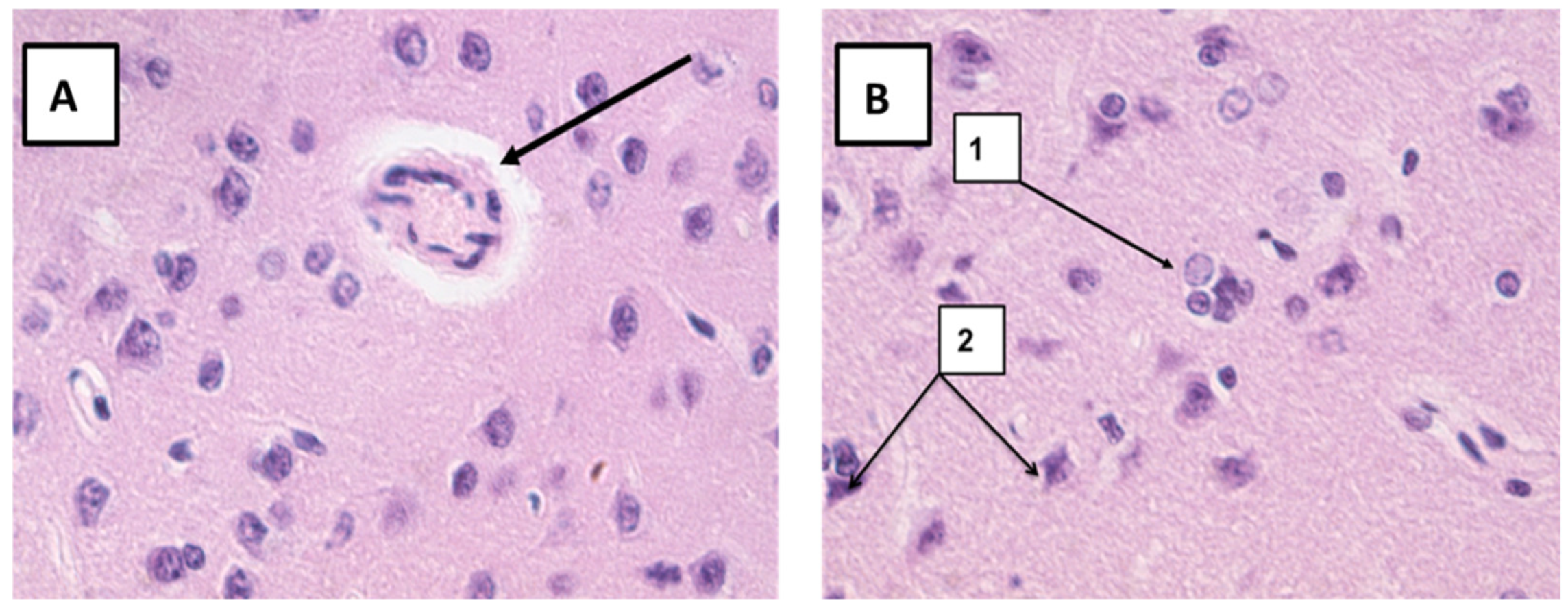
Abstract
:1. Introduction
2. Results
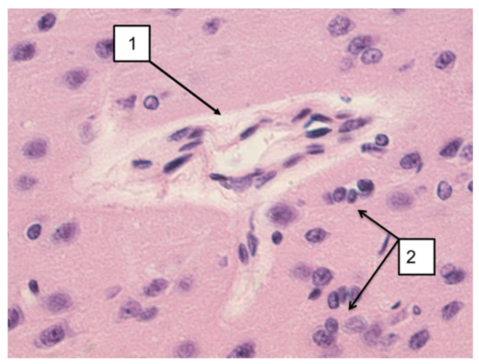
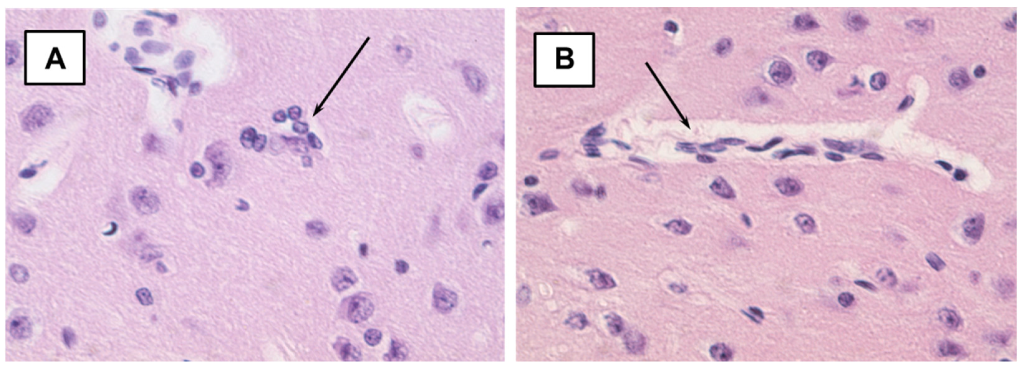
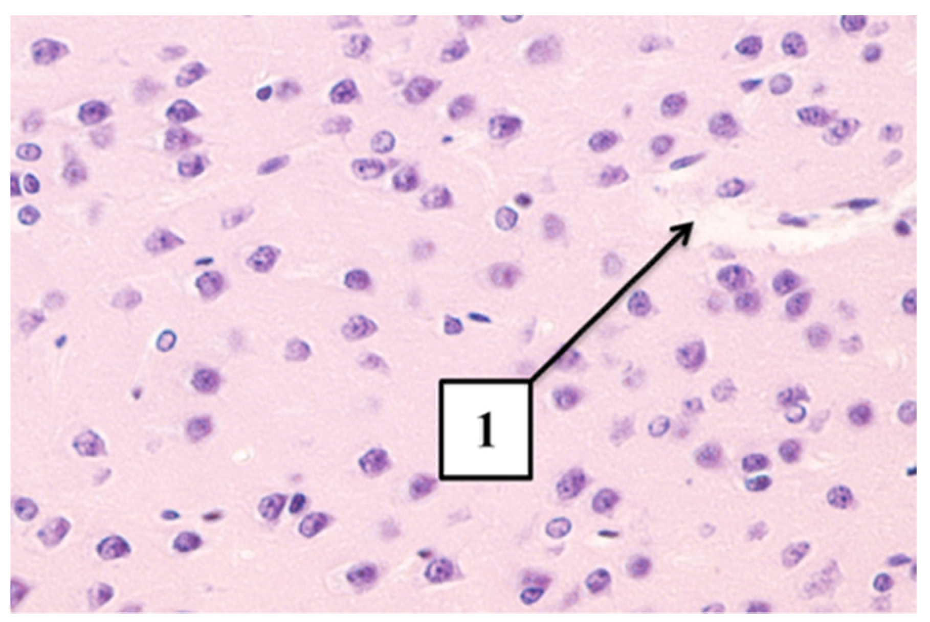
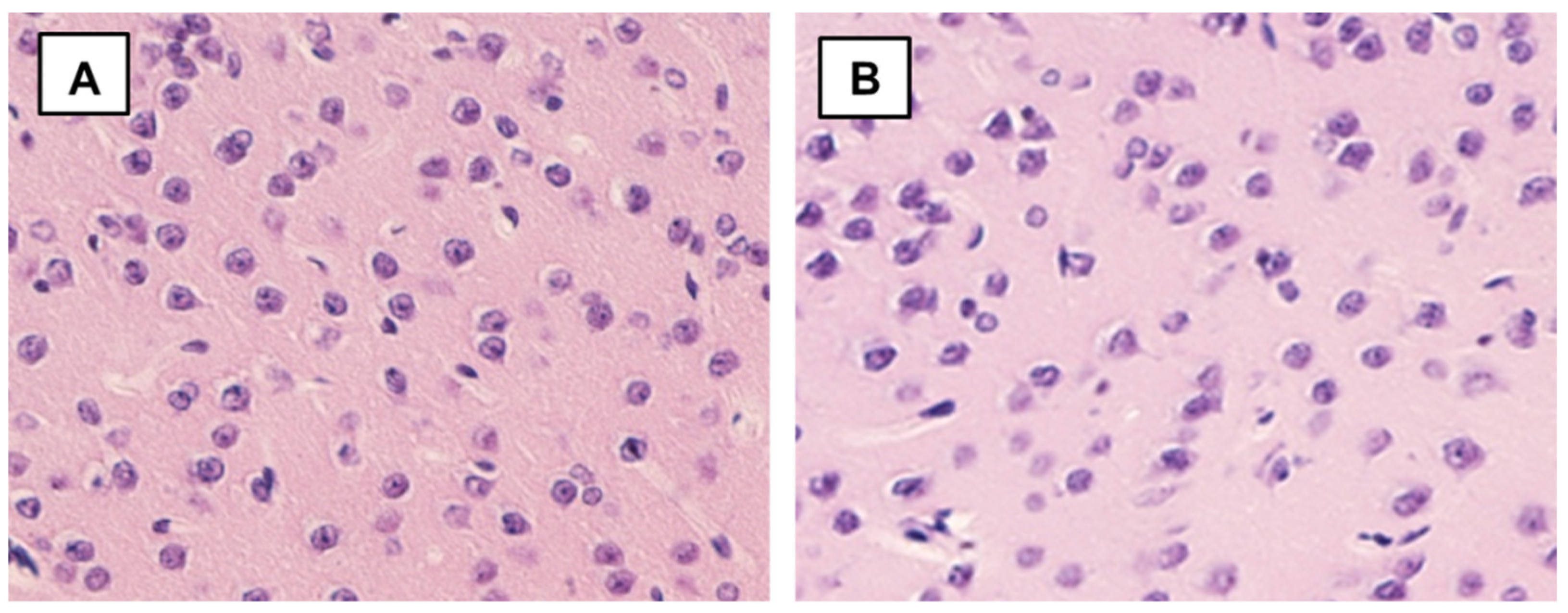
2.1. Histological Results
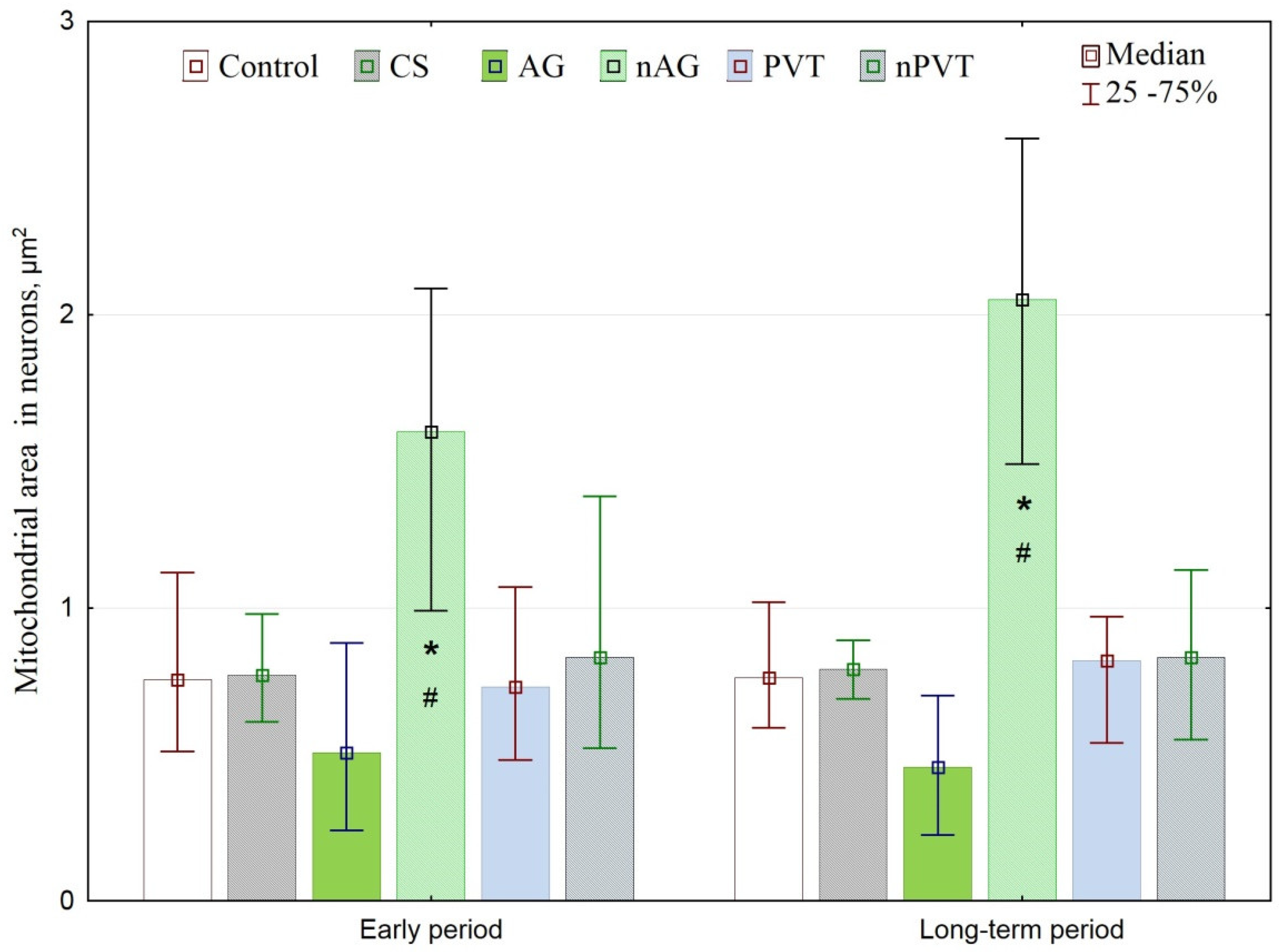
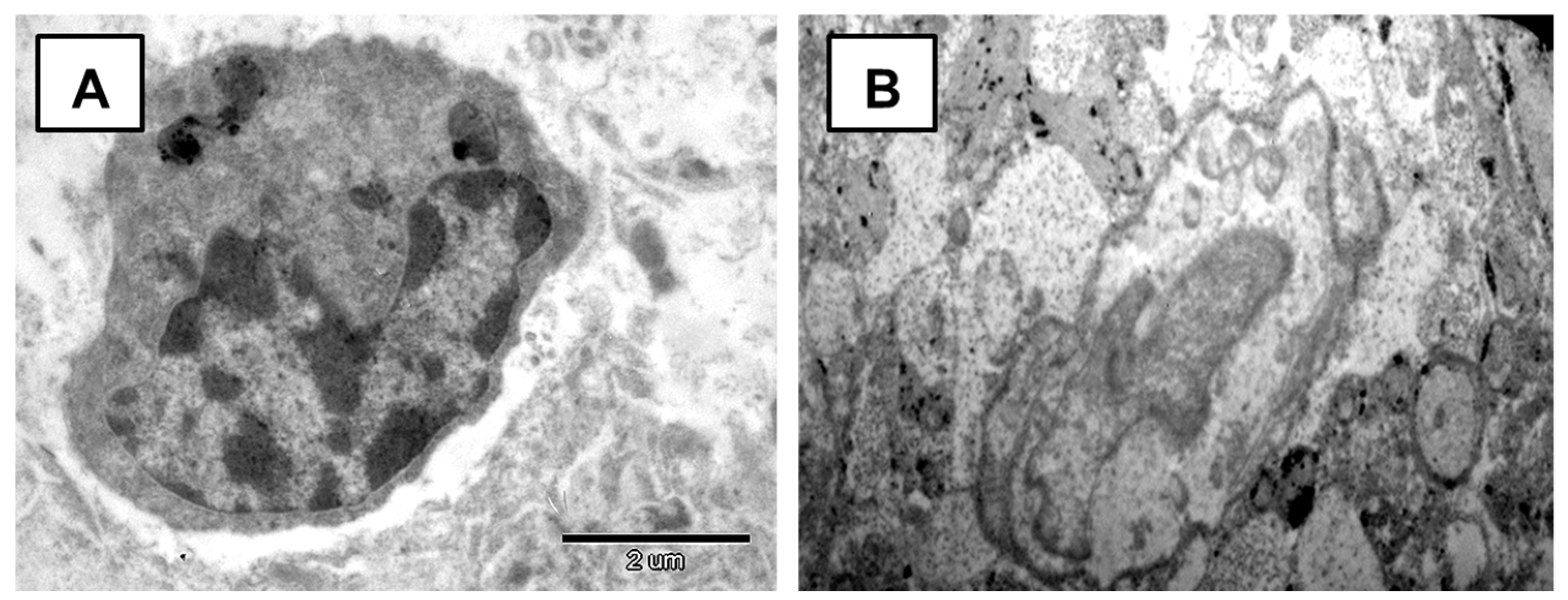
2.2. Results of Ultrastructural Analysis
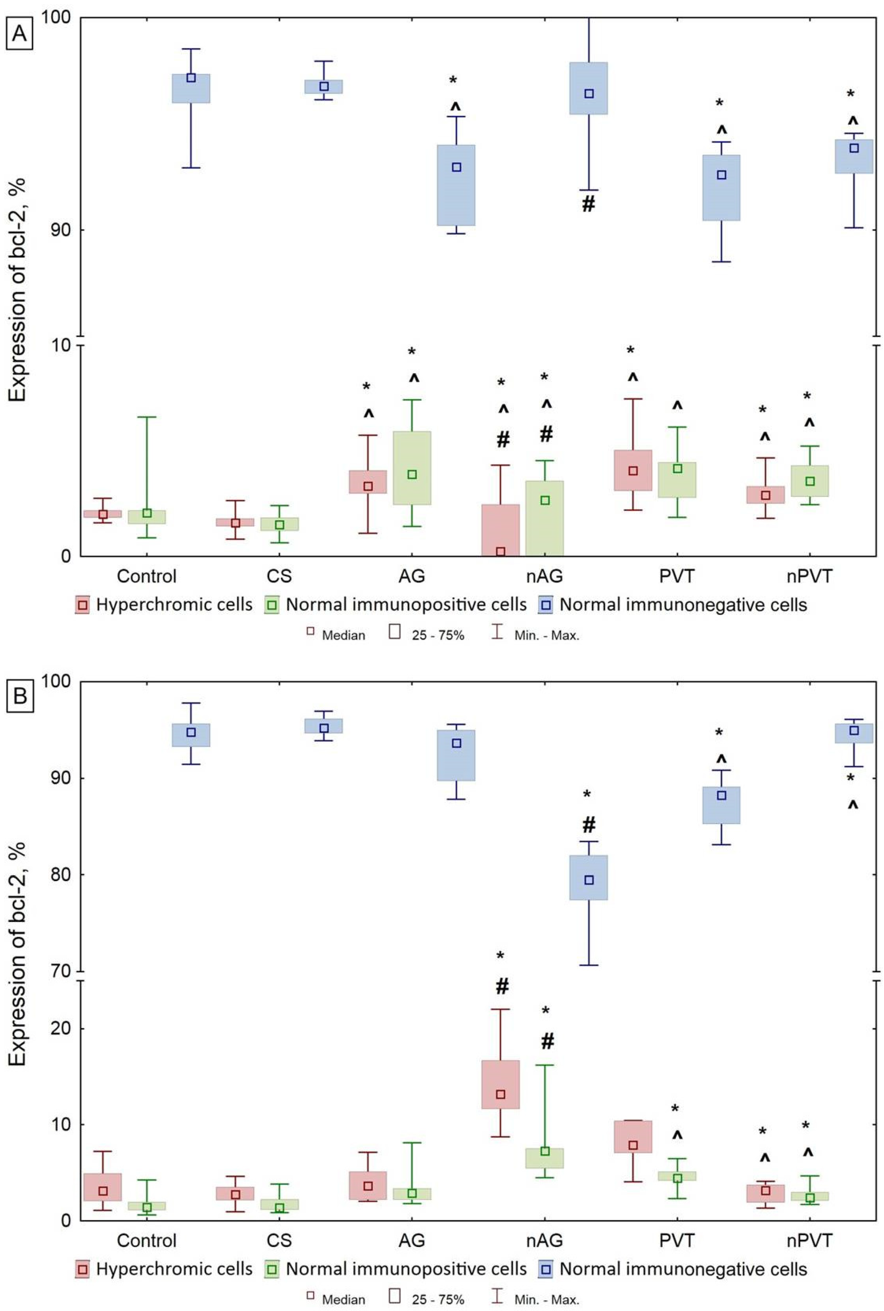
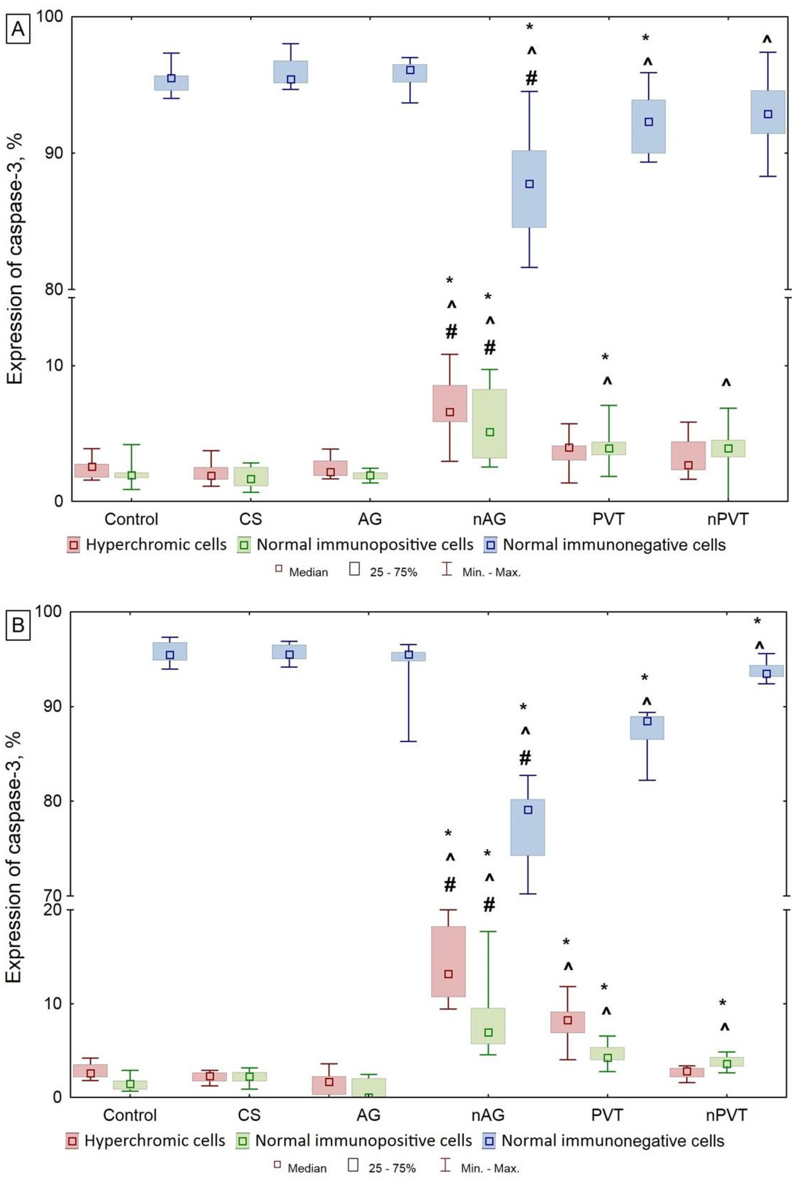
2.3. Results of the Immunohistochemical Investigation
3. Discussion
4. Materials and Methods
4.1. Silver Nanocomposites’ Preparation and Characterization
4.2. Animals and Experimental Design
4.3. Histological Investigation
4.4. Electron Microscopy Investigation
4.5. Immunohistochemical Investigation
4.6. Statistical Analyses
5. Conclusions
Author Contributions
Funding
Institutional Review Board Statement
Informed Consent Statement
Data Availability Statement
Conflicts of Interest
References
- Hoet, P.H.M.; Brüske-Hohlfeld, I.; Salata, O.V. Nanoparticles—Known and unknown health risks. J. Nanobiotech. 2004, 2, 1–15. [Google Scholar] [CrossRef] [PubMed] [Green Version]
- Yang, Z.; Liu, Z.W.; Allaker, R.P.; Reip, P.; Oxford, J.; Ahmad, Z.; Ren, G. A review of nanoparticle functionality and toxicity on the central nervous system. J. R. Soc. Interface 2010, 7, 411–422. [Google Scholar] [CrossRef] [PubMed]
- Ai, J.; Biazar, E.; Jafarpour, M.; Montazeri, M.; Majdi, A.; Aminifard, S.; Zafari, M.; Akbari, H.R.; Rad, H.G. Nanotoxicology and nanoparticle safety in biomedical designs. Int. J. Nanomed. 2011, 6, 1117–1127. [Google Scholar]
- Powers, K.W.; Palazuelos, M.; Moudgil, B.M.; Roberts, S.M. Characterization of the size, shape, and state of dispersion of nanoparticles for toxicological studies. Nanotoxicology 2007, 1, 42–51. [Google Scholar] [CrossRef]
- Lansdown, A.B.G. A pharmacological and toxicological profile of silver as an antimicrobial agent in medical devices. Adv. Pharmacol. Sci. 2010, 2010, 1–16. [Google Scholar] [CrossRef] [PubMed] [Green Version]
- Stensberg, M.C.; Wei, Q.; McLamore, E.S.; Porterfield, D.M.; Wei, A.; Sepúlveda, M.S. Toxicological studies on silver nanoparticles: Challenges and opportunities in assessment, monitoring and imaging. Nanomedicine 2011, 6, 879–898. [Google Scholar] [CrossRef] [PubMed] [Green Version]
- Singh, S.K.; Goswami, K.; Sharma, R.D.; Reddy, M.V.; Dash, D. Novel microfilaricidal activity of nanosilver. Int. J. Nanomed. 2012, 7, 1023–1030. [Google Scholar]
- Ganenko, T.V.; Vereshchagina, S.A.; Korjakina, L.B.; Sukhov, B.G.; Trofimov, B.A.; Kostyro, Y.A.; Fadeeva, T.V. Silver Nanocomposite Based on Sulfated Arabinogalactan with Antimicrobial and Antithrombotic Activity and a Method for Its Preparation. Available online: https://findpatent.ru/patent/246/2462254.html (accessed on 28 October 2021).
- Pozdnyakov, A.S. Polyfunctional (co) Polymers of 1-Vinyl-1,2,4-triazole and Nanocomposites Based on Them. Ph.D. Thesis, Siberian Branch of the Russian Academy of Sciences, Siberia, Russia, November 2011. Available online: https://files.isu.ru/ru/science/boards/him6/Pozdnyakov_AS.pdf (accessed on 28 October 2021).
- Prozorova, G.F.; Korzhova, S.A.; Kon’kova, T.V.; Ermakova, T.G.; Pozdnyakov, A.S.; Sapozhnikov, A.N.; Proydakova, O.A.; Sukhov, B.G.; Arsentyev, K.Y.; Likhoshvai, E.V.; et al. Synthesis and properties of silver and gold nanocomposites on a poly-1-vinyl-1,2,4-triazole matrix. J. Struct. Chem. 2012, 51, 105–108. [Google Scholar] [CrossRef]
- Dzhioev, Y.; Yurinova, G.V.; Sukhov, B.G.; Popkova, S.M.; Ganenko, T.V.; Pogodaeva, N.N.; Vasilieva, D.K.; Rakova, E.B.; Safronova, L.A.; Podgorsky, V.S.; et al. Hemicelluloses and their nanobiocomposites are promising nanostructured synbiotics. Acta Biomed. Sci. 2012, 5–1, 210–212. [Google Scholar]
- Shurygina, I.A.; Sukhov, B.G.; Fadeeva, T.V.; Umanets, V.A.; Shurygin, M.G.; Ganenko, T.V.; Kostyro, Y.A.; Grigoriev, E.G.; Trofimov, B.A. Bactericidal action of Ag(0)-antithrombotic sulfated arabinogalactan nanocomposite: Coevolution of initial nanocomposite and living microbial cell to a novel nonliving nanocomposite. Nanomed. Nanotech. Biol. Med. 2011, 7, 827–833. [Google Scholar] [CrossRef]
- Stebounova, L.V.; Morgan, H.; Grassian, V.H.; Brenner, S. Health and safety implications of occupational exposure to engineered nanomaterials. WIREs Nanomed. Nanobiotech. 2012, 4, 310–321. [Google Scholar] [CrossRef]
- Ordzhonikidze, C.G.; Ramaiyya, L.K.; Egorova, E.M.; Rubanovich, A.V. Genotoxic effects of silver nanoparticles on mice in vivo. Acta Nat. 2009, 3, 99–101. [Google Scholar]
- Guzman, M.; Dille, J.; Godet, S. Synthesis and antibacterial activity of silver nanoparticles against gram-positive and gram-negative bacteria. Nanomed. Nanotech. Biol. Med. 2012, 8, 37–45. [Google Scholar] [CrossRef]
- Hussain, S.M.; Schlager, J.J. Safety evaluation of silver nanoparticles: Inhalation model for chronic exposure. Toxicol. Sci. 2009, 108, 223–224. [Google Scholar] [CrossRef] [PubMed] [Green Version]
- Nallathamby, P.D.; Xu, X.-H.N. Study of cytotoxic and therapeutic effects of stable and purified silver nanoparticles on tumor cells. Nanoscale 2010, 2, 942–952. [Google Scholar] [CrossRef] [PubMed]
- Sharma, H.S.; Sharma, A. Nanoparticles aggravate heat stress induced cognitive deficits, blood-brain barrier disruption, edema formation and brain pathology. Prog. Brain. Res. 2007, 162, 245–273. [Google Scholar] [PubMed]
- Sharma, H.S.; Hussain, S.; Schlager, J.; Ali, S.F.; Sharma, A. Influence of nanoparticles on blood-brain barrier permeability and brain edema formation in rats. Acta Neurochir. Suppl. 2010, 106, 359–364. [Google Scholar]
- Sahoo, S.K.; Parveen, S.; Panda, J.J. The present and future of nanotechnology in human health care. Nanomedicine 2007, 3, 20–31. [Google Scholar] [CrossRef]
- Tang, J.; Xiong, L.; Wang, S.; Wang, J.; Liu, L.; Li, J.; Yuan, F.; Xi, T. Distribution, translocation and accumulation of silver nanoparticles in rats. J. Nanosci. Nanotechnol. 2009, 9, 4924–4932. [Google Scholar] [CrossRef]
- Hadrup, N.; Loeschner, K.; Mortensen, A.; Sharma, A.K.; Qvortrup, K.; Larsen, E.H.; Lam, H.R. The similar neurotoxic effects of nanoparticulate and ionic silver in vivo and in vitro. Neurotoxicology 2012, 33, 416–423. [Google Scholar] [CrossRef]
- Titov, E.A.; Sosedova, L.M.; Novikov, M.A. Analysis of the toxicity of gadolinium nanocomposites. Nanotechnol. Russ. 2019, 14, 144–148. [Google Scholar] [CrossRef]
- Titov, E.A.; Sosedova, L.M.; Kapustina, E.A.; Yakimova, N.L.; Novikov, M.A.; Lisetskaya, L.G.; Lizarev, A.V. Analysis of the Toxicity of a Cu2O Nanocomposite Encapsulated in a Polymer Matrix of Arabinogalactan. Nanobiotechnol. Rep. 2021, 16, 537–542. [Google Scholar] [CrossRef]
- Frohlich, E. Cellular targets and mechanisms in the cytotoxic action of non-biodegradable engineered nanoparticles. Curr. Drug Metab. 2013, 14, 976–988. [Google Scholar] [CrossRef] [PubMed]
- Xue, Y.; Wu, J.; Sun, J. Four types of inogranic nanoparticles stimulate the inflammatory reaction in brain neurogeia and damage neurons in vitro. Toxicol Lett. 2012, 214, 91–98. [Google Scholar] [CrossRef]
- Nel, A.; Sia, T.; Madler, L.; Li, N. Toxic potential of materials at the nanolevel. Science 2006, 311, 622–627. [Google Scholar] [CrossRef] [PubMed] [Green Version]
- Chen, H.H.; Josephson, L.; Sosnovik, D.E. Imaging of apoptosis in the heart with nanoparticle technology. WIREs Nanomed. Nanobiotech. 2011, 3, 86–99. [Google Scholar] [CrossRef] [Green Version]
- Titov, E.A.; Sosedova, L.M.; Novikov, M.A. Analysis of toxicity of iron oxide nanocomposite encapsulated in a polymer matrix of arabinogalactan. Gig. Sanit. 2021, 3, 285–289. [Google Scholar] [CrossRef]
- Alarifi, S.; Huma, A.; Alkahtani, S.; Alessia, M. Regulation of apoptosis through bcl-2/bax proteins expression and DNA damage by nano-sized gadolinium oxide. Int. J. Nanomed. 2017, 12, 4541–4551. [Google Scholar] [CrossRef] [Green Version]
- Wu, J.; Wang, C.; Sun, J.; Xue, Y. Neurotoxicity of silica nanoparticles: Brain localization and dopaminergic neurons damage pathways. ACS Nano 2011, 5, 4476–4489. [Google Scholar] [CrossRef]
- Deng, X.; Luan, Q.; Chen, W.; Wang, Y.; Wu, M.; Zhang, H.; Jiao, Z. Nanosized zinc oxide particles induce neural stem cell apoptosis. Nanotechnology 2009, 20, 650–658. [Google Scholar] [CrossRef]
- Zhang, Q.L.; Li, M.Q.; Ji, J.W.; Gao, F.P.; Bai, R.; Chen, C.Y.; Wang, Z.W.; Zhang, C.; Niu, Q. In vivo toxicology of nano-alumina on mice neurobehavioral profiles and potential mechanisms. Int. J. Immunopatol. Pharmacol. 2011, 24, 23–29. [Google Scholar]
- Novikov, M.A.; Titov, E.A.; Sosedova, L.M.; Ostroukhova, L.A.; Trofimova, N.N.; Babkin, V.A. Biochemical and morphological changes in white rats after intragastric injection of a synthetic nanobiocomposite based on silver nanoparticles and arabinogalactan. Pharm. Chem. J. 2014, 48, 387–390. [Google Scholar] [CrossRef]
- Prozorova, G.F.; Pozdnyakov, A.S.; Korzhova, S.A.; Ermakova, T.G.; Novikov, M.A.; Titov, E.A.; Sosedova, L.M. Toxicity evaluation of polyvinyltriazole and a related silver-containing nanocomposite. Russ. Chem. Bull. 2014, 63, 2126–2129. [Google Scholar] [CrossRef]
- Korzhevsky, D.E. A Summary of the Basics of Histological Techniques for Physicians and Laboratory Assistants-Histologists; Kroph: Saint Petersburg, Russia, 2008; 48p. [Google Scholar]


Publisher’s Note: MDPI stays neutral with regard to jurisdictional claims in published maps and institutional affiliations. |
© 2021 by the authors. Licensee MDPI, Basel, Switzerland. This article is an open access article distributed under the terms and conditions of the Creative Commons Attribution (CC BY) license (https://creativecommons.org/licenses/by/4.0/).
Share and Cite
Novikov, M.A.; Titov, E.A.; Sosedova, L.M.; Rukavishnikov, V.S.; Vokina, V.A.; Lakhman, O.L. Comparative Assessment of Silver Nanocomposites’ Biological Effects on the Natural and Synthetic Matrix. Int. J. Mol. Sci. 2021, 22, 13257. https://0-doi-org.brum.beds.ac.uk/10.3390/ijms222413257
Novikov MA, Titov EA, Sosedova LM, Rukavishnikov VS, Vokina VA, Lakhman OL. Comparative Assessment of Silver Nanocomposites’ Biological Effects on the Natural and Synthetic Matrix. International Journal of Molecular Sciences. 2021; 22(24):13257. https://0-doi-org.brum.beds.ac.uk/10.3390/ijms222413257
Chicago/Turabian StyleNovikov, Mikhail A., Eugeniy A. Titov, Larisa M. Sosedova, Viktor S. Rukavishnikov, Vera A. Vokina, and Oleg L. Lakhman. 2021. "Comparative Assessment of Silver Nanocomposites’ Biological Effects on the Natural and Synthetic Matrix" International Journal of Molecular Sciences 22, no. 24: 13257. https://0-doi-org.brum.beds.ac.uk/10.3390/ijms222413257