
SALL4 Oncogenic Function in Cancers: Mechanisms and Therapeutic Relevance


Zhejiang Provincial Key Laboratory of Silkworm Bioreactor and Biomedicine, College of Life Sciences and Medicine, Zhejiang Sci-Tech University, Hangzhou 310018, China
*
Author to whom correspondence should be addressed.
Int. J. Mol. Sci. 2022, 23(4), 2053; https://0-doi-org.brum.beds.ac.uk/10.3390/ijms23042053
Submission received: 30 December 2021
/
Revised: 7 February 2022
/
Accepted: 8 February 2022
/
Published: 12 February 2022
(This article belongs to the Collection Feature Papers in Molecular Oncology)
Abstract
:SALL4, a member of the SALL family, is an embryonic stem cell regulator involved in self-renewal and pluripotency. Recently, SALL4 overexpression was found in malignant cancers, including lung cancer, hepatocellular carcinoma, breast cancer, gastric cancer, colorectal cancer, osteosarcoma, acute myeloid leukemia, ovarian cancer, and glioma. This review updates recent advances of our knowledge of the biology of SALL4 with a focus on its mechanisms and regulatory functions in tumors and human hematopoiesis. SALL4 overexpression promotes proliferation, development, invasion, and migration in cancers through activation of the Wnt/β-catenin, PI3K/AKT, and Notch signaling pathways; expression of mitochondrial oxidative phosphorylation genes; and inhibition of the expression of the Bcl-2 family, caspase-related proteins, and death receptors. Additionally, SALL4 regulates tumor progression correlated with the immune microenvironment involved in the TNF family and gene expression through epigenetic mechanisms, consequently affecting hematopoiesis. Therefore, SALL4 plays a critical oncogenic role in gene transcription and tumor growth. However, there are still some scientific hypotheses to be tested regarding whether SALL4 is a therapeutic target, such as different tumor microenvironments and drug resistance. Thus, an in-depth understanding and study of the functions and mechanisms of SALL4 in cancer may help develop novel strategies for cancer therapy.
1. Introduction
In the past 20 years, molecular targeted therapy and biological cellular immunotherapy have become popular therapeutic methods for tumors [1]. To improve the efficacy of therapy and the patients’ prognosis, ongoing development of novel targets is necessary to develop new drugs and combination therapeutic strategies.
Spalt-like transcription factors (SALLs) are relevant to the occurrence, progression, and prognosis of various tumors, such as cervical cancer and renal cancer [2,3,4,5,6]. SALL1, SALL2, and SALL3 are generally downregulated, while SALL4 is highly expressed in tumors [2]. Hypermethylation of SALL1 and SALL2 are correlated with reduced disease-free survival (DFS) in patients with early neck squamous cell carcinoma (HNSCC) [4] and in patients with oral cancer [7], respectively. SALL3 plays a potential tumor suppressor role in cervical cancer [8,9], whereas SALL4 is an oncogene in this cancer [10]. Increasing evidence indicates that SALL4 could be a potential target for tumor treatments and a clinically diagnosed idiosyncratic biomarker in different tumors [2,6]. Functions of SALLs in tumors are noted in Table 1.
Oncofetal SALL4, a zinc finger transcription factor [10,18], is an embryonic stem cell regulator involved in self-renewal and pluripotency [21]. SALL4 located in chromosome 20.q13.2 translates into two isoforms, SALL4A and SALL4B [10,22], which are a result of different internal splicing patterns in exon 2 [6,22,23]. SALL4A has four zinc finger clusters (ZFCs), three ZFCs of which contain either a pair or a trio of C2H2-type zinc fingers [22]. Recent studies have found the patterns of SALL4 to bind DNA-specific genes and regulate their expression, involved in ZFC, both in normal embryonic stem cells (ESCs) and SALL4-dependent tumors. SALL4 can bind to short AT-rich motifs via C2H2 ZFC4 in ESCs, then preventing differentiation [24]. The C-terminal ZFC of SALL4 is responsible for DNA binding, and SALL4 negatively regulates expression of a family of histone 3 lysine 9-specific demethylases (KDMs) in aggressive liver cancer cells [25]. Normally, except in germ cells, SALL4 is silenced in most adult tissues [25]. However, SALL4 was found to be overexpressed in various tumors, such as gastric cancer (GC) [26], lung cancer [15], endometrial cancer [27], hepatocellular carcinoma (HCC) [28], and acute myeloid leukemia (AML) [29]. Additionally, a recent study found that downregulation of SALL4 triggered pluripotency loss, leading to derepression of a set of AT-rich genes that promoted neuronal differentiation [24]. SALL4 is a potential target for the diagnosis and treatment of cancer [18,30]. It participates in the regulation of cell growth, cell cycle, and apoptosis via the expression of articulation-related genes, and it exerts biological effects as a transcription factor in the nucleus, where it plays a crucial role in the occurrence and development of various malignant tumors. Thus, understanding the functions and mechanisms of SALL4 can provide novel insight into how SALL4 can be targeted in tumor therapies.
2. Mechanisms of Action of SALL4 in Tumors
Since there are many abbreviations mentioned in this review, we list all the abbreviations in Table 2.
2.1. SALL4 Activates the Wnt/β-Catenin Signaling Pathway
Various studies have shown that SALL4 significantly dysregulates the Wnt/β-catenin signaling pathway in cancers. The expression of SALL4 was found to be positively correlated with that of tribbles pseudokinase 3 (TRIB3) in GC [21], which activated Wnt/β-catenin signaling by interacting with the β-catenin–TCF4 complex [31]. In addition, TRIB3 activated the Wnt-β-catenin signaling pathway via the C-terminal and N-terminal regions of the kinase-like domain in TRIB3, directly binding to β-catenin in colorectal cancer [32], indicating that activation of Wnt/β-catenin signaling by SALL4 may require TRIB3 expression. Moreover, TRIB3 knockdown decreased β-catenin expression and resulted in downregulation of c-Myc, axis inhibition protein (AXIN2) and cyclin D1 [31]. In addition, in cervical cancer, SALL4 recognizes and binds to the CTNNB1 promoter region as a transcription activator and accelerates the expression of β-catenin to upregulate downstream target genes, including c-Myc and CCND1 [10]. Then, the activation of Wnt/β-catenin signaling promotes cell proliferation and tumor growth in cervical cancer cells [10]. SALL4 also regulates the tumorigenicity of breast cancers by modulating the Wnt3a/β-catenin signaling pathway [16]. Furthermore, upregulation of the mRNA and protein of SALL4, Wnt3a, and β-catenin is associated with tumor differentiation in HCC [19].
The SALL4A and SALL4B isoforms have been found to bind to the β-catenin protein in SALL4B transgenic mice, and the interactions of these factors activate the Wnt/β-catenin pathway synergistically, which plays essential roles in controlling leukemia stem cell (LSC) self-renewal [33,34,35,36,37]. Downregulation of SALL4 contributed to a decrease in B-cell-specific Moloney murine leukemia virus integration site 1 (Bmi-1) expression and inactivated the Wnt3a/β-catenin signaling pathway in intrahepatic cholangiocarcinoma (ICC) [38]. SALL4 expression was positively correlated with lymph node metastasis and tumor–node–metastasis (TNM) stages in colorectal cancer, and β-catenin was expressed markedly higher in colorectal cancer than in normal tissue [39]. Patients with colorectal cancer with coexpression of SALL4 and β-catenin showed advanced lymph node metastasis and TNM stage. Colocalization of SALL4 and β-catenin was also found in the nucleus and cytoplasm [39]. These findings indicated that the interaction of SALL4 with β-catenin plays crucial functional roles in promoting lymph node metastasis and advanced clinical stage. Another study found that SALL4 modulated the stemness of esophageal squamous cell carcinoma (ESCC) via the Wnt/β-catenin signaling pathway and epithelial–mesenchymal transition (EMT). SALL4 promotes intrahepatic cholangiocarcinoma cell proliferation by activating Wnt/β-catenin signaling [19]. The regulation of Wnt/β-catenin signaling pathway by SALL4 is shown in Figure 1.
2.2. SALL4 Inhibits the Expression of PTEN and Activates the PI3K/AKT Signaling Pathway
Phosphatase and tension homolog (PTEN), a tumor suppressor, is expressed at low levels in various cancers [40,41,42]. The well-known PI3K/AKT signaling pathway functions as a key regulator in tumorigenesis [43]. PTEN, as an antagonist of PI3K [44], inhibits AKT activation [45]. High SALL4 expression resulted in proliferative, invasive, and anti-apoptotic effects in lung cancer through activation of PI3K/AKT/mTOR signaling [46]. Other studies found crosstalk between PTEN and SALL4 [47,48,49]. SALL4 silencing resulted in PTEN upregulation, which inhibited PI3K/AKT signaling, indicating that PTEN is a critical functional downstream target of SALL4 in tumor development [50].
Furthermore, SALL4 functions as an oncogene associated with other factors, such as Bmi-1 [51], miR-188-5p [52], and deacetylase [53]. As shown in Figure 2, overexpression of Bmi-1 was found to be positively correlated with SALL4 in AML [54]. Downregulation of SALL4 inhibited Bmi-1 expression, while overexpression of SALL4 induced Bmi-1 expression in transgenic mice [51]. SALL4 negatively regulates PTEN expression and positively regulates Bmi-1 expression by binding their promoter, and then activates the PI3K/AKT signaling pathway in hematological tumors [38]. An additional report showed that SALL4 knockdown increased PTEN expression, inhibited downstream intermediate phosphorylation of AKT and GSK3β, and decreased vascular endothelial growth factor A (VEGFA) expression in clear cell renal cell carcinoma (ccRCC), providing convincing evidence that SALL4 is functionally critical in tumor progression and may be regarded as a drug target [55]. A recent study found that miR-188-5p promoted GC cell proliferation and migration by suppressing PTEN expression and transcriptional upregulation of SALL4 [52]. Additionally, positive SALL4 and high histone deacetylase (HDAC) 1/2 expression were correlated with low PTEN expression and a poor prognosis in HCC patients [53].
2.3. SALL4 Activates the Notch Signaling Pathway
The Notch signaling pathway plays a critical role in stem cell self-renewal and cell differentiation, and Notch is also involved in the progression and self-renewal capacity of various tumors [56,57]. Ectopic expression of Notch3 increased the expression of the embryonic stem cell marker SALL4 [58].
Sex-determining region Y (SRY)-Box 2 (SOX2), a partner of SALL4, is a transcription factor regulating cell fate decisions and stem cell self-renewal and pluripotency in ESCs [59]. Concomitant overexpression of SALL4 and SOX2 is relevant to invasion and metastasis in ESCC [60]. However, the mechanisms by which SOX2 and SALL4 promote self-renewal associated with the Notch pathway are still unclear. As shown in Figure 3, current evidence shows that co-overexpression of SOX2 and SALL4 results in upregulation of Notch ligand such as delta-like 1 (DLL1); receptors Notch1/2/3, which induce expression of helix-loop-helix transcriptional repressor target genes hairy/enhancer of split related to YRPW motif family member 2 (HEY2); HES1 and c-Myc; and transcriptional coactivators such as mastermind-like transcriptional coactivator 1 (MAML1) in ESCCs. The expression patterns of the Notch pathway genes are similar to those of the mRNA levels of SOX2 and SALL4 [57]. These studies suggest correlations between SOX2/SALL4 expression and the Notch signaling pathway.
Intriguingly, transgenic activation of a mutated β-catenin allele was found in murine osteoblasts [61,62] and it induced myelodysplastic syndrome (MDS) and AML at very early ages via dysregulated Notch ligand JAG1 [63,64,65]. Since SALL4B transgenic mice also develop MDS/AML and SALL4B-overexpressing bone marrow (BM) cells do not induce leukemia formation in transplanted mice [29], an interesting question is whether/how SALL4B potentially activates β-catenin signaling, which synergistically dysregulates the hematopoietic stem/progenitor cell (HSC/HPC) osteoblastic niche and thus promotes leukemogenesis. Additional detailed transgenic studies are required to address this possibility.
Furthermore, myeloid ecotropic viral insertion site 1 (MEIS1) has been reported as a homeobox transcription factor involved in cell fate decisions and stem cell properties [66,67]. Expression of MEIS1 was associated with SALL4 in poorly differentiated cancer cells. SALL4 induced MEIS1 expression by binding to the promoter region of MEIS1 [68]. MEIS1 dysregulation may alter the function of the Notch pathway [69,70]. MEIS1 has a close correlation with the stemness state and the Notch signaling pathway involved in SALL4 [71].
Overall, SALL4 can interact with other transcription factors to regulate the Notch pathway or directly interact with Notch and then impact the progression and self-renewal capacity of various cancer cells.
2.4. SALL4 Regulates Expression of Bcl-2 and Bax
Apoptosis is regulated by the balance of B-cell lymphoma 2 (Bcl-2) and Bcl-like-protein 4 (Bax) [72]. Overexpression of antiapoptotic Bcl-2 inhibits cell apoptosis and leads to an imbalance between enhanced proliferation and weakened programmed death through downregulation of Bax [73,74]. SALL4 overexpression in MDS and AML cells results in an increase in Bcl-2 expression and cell survival [75], whereas SALL4 knockdown markedly inhibits Bcl-2 expression in prostate cancer [76], breast cancer [16,77], colorectal cancer (CRC) [78], and nasopharyngeal carcinoma (NPC) [79], and it induces the expression of pro-apoptotic Bax in prostate cancer [76] and NPC [79].
2.5. SALL4 Inhibits Caspase-Related and Death-Receptor Pathways
SALL4 not only regulates cell growth, such as the Wnt/β-catenin and PTEN/PI3K/AKT pathways [21,50], but also affects cell apoptosis via the caspase-related and death-receptor pathways. A study showed that SALL4 regulated the expression of apoptosis genes and apoptotic process-related genes such as CYC3 and CUL3 [80]. SALL4 knockdown in AML cells resulted in upregulation of apoptotic genes, including CARD9, CARD11, TNF, ATF3, TP53, PTEN, and LTA, but downregulation of antiapoptotic genes, such as Bmi-1, Bcl-2, DAD1, BIRC4, BIRC7, and TEGT [80]. Furthermore, a previous study showed that Bmi-1 was one of the major downstream targets of SALL4 in leukemia [51]. SALL4 restored Bmi-1 knockdown-induced cell cycle arrest and apoptosis [80].
SALL4 influences tumor survival by regulating multiple caspase members. SALL4 knockdown induced the expression of caspase-3 and caspase-8 in adult acute B cell lymphoblastic leukemia (B-ALL) [81]. Moreover, various microRNAs (miRNAs) play tumor suppressor roles, such as miRNA-98 and miRNA-33b in HCC and miRNA-219 in glioma by inhibiting SALL4 expression and suppressing the proliferation and metastasis of tumor cells [82,83,84,85]. miR-103, miR-195, and miR-15b have identical 5′ miRNA sequences and share common binding sites in the 3′-untranslated region (UTR) of SALL4, which inhibits SALL4 expression and activates caspase-3 and caspase-7 in glioma cells [86]. Furthermore, miRNA-107 suppressed glioma cell growth by directly targeting SALL4, resulting in the increased expression of Fas-associated by death domain (FADD) and the activation of caspase-8 and caspases-3/7, inducing cell apoptosis [14]. The correlations between SALL4 and Bcl-2, Bax, caspase-related pathway, and death-receptor pathway are shown in Figure 4.
2.6. MiRNAs Regulate the Interaction of SALL4 and TNF Family
Cancer progression is closely related to the modulation of the tumor microenvironment [87]. Tumor necrosis factor alpha (TNF-α), a proinflammatory cytokine, has been reported to be important in the development and progression of HCC by activating the transcription factor nuclear factor κB (NF-κB) [88]. In the tumor microenvironment, it is possible that the antitumor immune response can be switched to a protumor response [89]. During tumor progression, T-cell-mediated antitumor response is significantly impaired, characterized by low secretion of interleukin-2 (IL-2), TNF-α, and interferon-γ (IFN-γ) and a high expression of inhibitory receptors such as PD-1 [90]. As shown in Figure 5, miR-497 negatively regulates SALL4 expression and then inhibits metastasis and self-renewal in HCC. TNF-α, as an inflammatory factor, suppresses miR-497 expression by NF-κB-mediated negative transcriptional regulation and upregulates SALL4 expression, promoting self-renewal and metastasis phenotypes in HCC [91]. Furthermore, SALL4 could bind to the promoter of miR-146a-5p and directly regulate its expression in exosomes. HCC-derived exosomes could remodel macrophages by activating NF-κB signaling and inducing proinflammatory factors.
Additionally, SALL4 promotes the expression of miR-146a-5p by binding its promoter, while silencing SALL4 inhibits the expression of inhibitory receptors and reverses T-cell exhaustion (Figure 5). HCC cell-secreted miR-146a-5p can be delivered by exosomes into macrophages, switch the cytokine profile, and attenuate antigen presentation of macrophages through inhibiting the expression of IFN-γ and TNF-α and upregulating the expression of inhibitory receptors such as programmed death ligand 1 (PD-1) and cytotoxic-T-lymphocyte-antigen-4 (CTLA-4), promoting the production of alternatively activated M2-polarized tumor-associated macrophages [87] (Figure 5).
2.7. SALL4 Regulates Gene Expression through Epigenetic Mechanisms
SALL4 regulates gene expression through epigenetic mechanisms. As shown in Figure 6A, SALL4 has been found to interact with histone methyltransferase mixed lineage leukemia (MLL) and co-occupy the homeobox A9 (HOXA9) promoter region with MLL. Moreover, overexpression of SALL4 enhances markers of histone activation, such as H3K4 and H3K79 methylation, as well as RNA polymerase II (POLII) binding in the same promoter region, which increases HOXA9 expression in AML. The data demonstrated that the SALL4/MLL/HOXA9 pathway was a crucial regulator of leukemic cell survival [92]. In addition, SALL4 interacts directly with DNA methyltransferases (DNMTs), indicating that SALL4 can inhibit transcription by recruiting DNA methyltransferases [93]. SALL4 also interacted with histone lysine-specific demethylase 1 (LSD1) to inhibit gene transcription in stem cells [94]. Furthermore, SALL4 co-occupies target genes with the polycomb repressive complex (PRC), indicating that SALL4 may inhibit gene transcription by inducing PRC components such as Bmi-1 or by interacting with PRC members, including RNF2, PhcI, EZH2, EED, SUZ12, and RbAp46/48, as well as HPC proteins [6,95].
Regulatory malignancies of SALL4 are involved in multiple chromatin modification regulators [29]. As previously reported, epigenetic factors that interact with SALL4 include six types: the nucleosome remodeling deacetylase (NuRD) complex, which includes HDAC1 and HDAC2 [49,96]; DNA methyltransferases-1 (DNMT-1), DNMT-3A, DNMT-3B, DNMT-3 L, and methyl-CpG-binding domain 2 protein (MBD2) [18]; H3K4 methyltransferase MLL1 [92,97]; H3K36 methyltransferase Wolf–Hirschhorn syndrome candidate gene-1 (WHSC1) [98,99]; H3K79 methyltransferase disruptor of telomeric silencing 1-like (DOT1 L) [68,100]; and lysine-specific histone demethylase LSD1/KDM1A [49,94,101] (Figure 6B). Interestingly, SALL4 seems to interact with these epigenetic factors at different sites. For instance, while the amino-terminal 174 amino acid sequence of SALL4 is critical for the SALL4–DNMT1 or SALL4–HDAC interaction, it is less relevant to the SALL4–LSD1 interaction. This is important in designing drugs to block protein–protein interaction-based anti-SALL4 strategies. Notably, clinical epigenetic remedies inhibiting SALL4-interacting epigenetic factors such as DOT1 L, HDAC, and LSD1 have been shown to be effective in treating leukemia [102,103,104,105,106] Figure 6B). Cutaneous melanoma is the most aggressive skin cancer owing to its high invasiveness [107], and melanoma cells rely on developmental programs during tumor initiation and progression [18]. Diener et al. found that SALL4 negatively regulated invasiveness by interacting with HDAC2 and directly binding to invasiveness genes, including NGFR, ETS1, FN1, VEGFR-1, and PDGFC (Figure 6C). SALL4 knockdown with HDAC inhibition promoted an invasive phenotype, while inhibition of histone acetylation partially reversed the invasiveness program induced by SALL4 depletion [18]. Thus, SALL4 was identified as a regulator of melanoma phenotype switching from tumor formation (SALL4 overexpression) to invasiveness (SALL4 loss). These epigenetic factors are not only significant regulators of normal hematopoiesis but are also targets for dysregulation in malignancies, especially in hematological tumors such as leukemia [108,109,110,111].
SALL4 is one of the significant factors required for the enhancement and maintenance of HSC/HPCs [29]. Overexpression of SALL4 in mobilized peripheral blood CD34+ cells promoted ex vivo expansion efficiency by over 10,000-fold for CD34+/CD38− and CD34+/CD38+ cells with appropriate cytokines [112]. Similarly, the SALL4B isoform expanded BM-derived CD34+ nonhuman primate HSCs. SALL4 lentiviral transduction caused a sixfold change in the total cell count of CD133+ HSCs compared to the control group [113,114]. Additionally, Milanovich et al. thought that SALL4 might be important for murine hematopoiesis [115].
SALL4 interacts with acetylase to induce phenotypic conversion. SALL4 is well known as an ESC regulator [116], and SALL4 knockdown induces a phenotype switch and an invasive phenotype via an HDAC2-mediated mechanism [18] (Figure 6C). SALL4 expression is highly correlated with DNA promoter methylation. SALL4, HDAC1, and DNMT proteins co-occupy the same promoter sites of SALL4, and their own transcription may be downregulated via recruitment of both DNA methylation and histone deacetylation enzymes [93,117]. The mRNA expression of DNA methyltransferase 3 alpha (DNMT3A) in the SALL4 knockdown group was markedly downregulated, indicating that DNMT3A may contribute to the regulatory effects of SALL4 [117].
Overall, the function of SALL4 involves many epigenetic mechanisms, and it inhibits or activates gene transcription through epigenetics. SALL4 not only plays a critical role in normal hematopoiesis through such an epigenetic mechanism but is also a common target in leukemia and many malignant tumors.
2.8. SALL4 Induces Mitochondrial Oxidative Phosphorylation during Tumorigenesis
A recent study found that SALL4 induced mitochondrial oxidative phosphorylation (OXPHOS) during tumorigenesis [118] (Figure 7). A phenomenon commonly called the Warburg effect usually occurs in during tumorigenesis [119]. Cancer cells usually rewire their bioenergetic metabolism by changing from mitochondrial OXPHOS to aerobic glycolysis, despite the presence of oxygen. SALL4-positive cells resist this common trend in HCC cell metabolism by driving mitochondrial OXPHOS instead. SALL4 induction of OXPHOS and mitochondrial ATP leads to the allosteric suppression of basal glycolysis rates [118]. Thus, SALL4-positive cancer cells possess a latent capacity for the Warburg effect, but this latent capacity is suppressed in the steady state. Additionally, SALL4 overexpression increases the transcription of mitochondrial OXPHOS genes, and SALL4 silencing leads to downregulation of OXPHOS genes, such as ATP5D, ATP5E, ATP5G2, and NDUFA3, as well as a functional reduction in mitochondrial oxygen consumption and OXPHOS [118] (Figure 7).
Although there are several transcription factors that act as oncogenes, few drugs have been developed to inhibit their activity in tumors [120,121,122]. However, a recent study showed that SALL4 increased OXPHOS, mitochondrial membrane potential, oxygen consumption rate, and utilization of oxidative phosphorylation-related metabolites to generate ATP [123]. The ATP synthase inhibitor oligomycin reduced the viability of HCC and lung cancer cells with high SALL4 expression. These results indicate that inhibitors of OXPHOS may be used for the treatment of HCC with high levels of SALL4.
Overall, SALL4 drives tumorigenesis by binding to and transcriptionally activating OXPHOS genes or expressing high levels of SALL4 by blocking OXPHOS to reduce tumor growth, providing novel therapies for HCC. However, an extension of the understanding of cancer metabolic reprogramming is necessary to push the development of innovative therapies.
2.9. Brief Summary of Mechanisms of SALL4 in Cancers
Specific epigenetic signatures, gene expression patterns, and signaling pathways contribute to the establishment and maintenance of stemness state networks. For one thing, SALL4 is an underlying biomarker that activates signaling pathways and oncoproteins that promote tumor progression including Wnt/β-catenin (Figure 1), PI3K/AKT (Figure 2), Notch signaling pathway (Figure 3), and OXPHOS (Figure 7), as well as Bcl-2, TNF-α, and IFN-γ (Figure 5). Moreover, SALL4 regulates gene expression through epigenetic mechanisms, then affecting tumor progression (Figure 6). For example, SALL4/MLL/HOXA9 pathway is a crucial regulator of leukemic cell survival and SALL4 plays a critical role in promoting melanoma invasion by interacting with HDAC2. For another, SALL4 can also inhibit apoptotic proteins, miRNAs, or pathways to promote tumor development, such as miRNAs, PTEN, Bax, caspase-related pathway, and so on (Figure 4). Overall, as an oncogene, SALL4 plays a pivotal role in carcinogenesis in different types of normal and tumor tissues.
3. Function of SALL4 in Tumors
3.1. Lung Cancer
Lung cancer is one of the most malignant tumors causing cancer-related deaths, with its mortality ranking high among malignancies in China and worldwide; lung cancer is classified into non-small cell lung cancer (NSCLC) and small cell lung cancer (SCLC), and the former represents approximately 85% of all lung cancer cases [124,125,126]. The therapies for lung cancer are chemotherapy, resection, and radiotherapy, but the prognosis is still unsatisfactory, with an overall 5-year survival rate of only 15% for all stages [124,127,128]. Additionally, immunotherapy and molecular targeted therapy have been applied in lung cancer clinical treatment to increase the survival rates [15,129]. A previous study showed that SALL4 expression is abnormally high in lung cancer [130], indicating that a deep understanding of the function underlying the progression and prognosis of lung cancer involved in SALL4 is critical for promoting an early diagnosis and effective therapy. Downregulation of SALL4 inhibits cell proliferation, clonal formation, migration, and invasion of lung cancer while promoting cell arrest in the G0/G1 phase by inhibiting the expression of cell cycle-related proteins such as cyclin B, cyclin D1, and cyclin E [131].
SALL4 regulates the progression of lung cancer through several mechanisms, and it may have a close correlation with drug resistance. SALL4 was upregulated in lung cancer with EGFR mutations [132]. Activation of EGFR could induce SALL4 overexpression by the extracellular signal-regulated kinase 1/2 (ERK1/2) signaling pathway, while SALL4 knockdown could not only inhibit spheroid formation and the expression of CD44, a type of lung cancer stem cell marker, but also increase the sensitivity of EGFR-mutated cells to EGFR inhibitor erlotinib [15]. Furthermore, knockdown of SALL4 can restore cisplatin sensitivity in acquired resistant lung cancer cells via the AKT/mTOR signaling pathway [46], and the antineoplastic drug entinostat can target SALL4-positive lung cancer [133]. SALL4 can also interact with miRNAs in lung cancer, including miR-250 and miR-3619 [134]. Upregulation of SALL4 and homeobox A11 antisense (HOXA11-AS) inhibited miR-3619-5p expression, whereas HOXA11-AS knockdown led to pro-apoptotic and anti-proliferative, -migration, -invasion, and glycolysis effects. Moreover, miR-3619-5p attenuates the inhibitory effects of HOXA11-AS knockdown on the progression of NSCLC cells by directly binding to HOXA11-AS. SALL4 overexpression reversed the antitumor effect of miR-3619-5p by decreasing miR-3619-5p expression. HOXA11-AS silencing inhibited tumor growth via upregulation of miR-3619-5p and downregulation of SALL4 [135].
3.2. Hepatocellular Carcinoma
Liver cancer, the sixth most common cancer, is the second leading cause of cancer mortality worldwide due to limited therapeutic interventions [136]. Liver cancer includes hepatoblastoma (HB) and HCC; the former is a common type of liver cancer reported in infants under 3 years of age, and the latter is the most common type of liver cancer documented, affecting 85–90% of liver cancer patients [118,137,138,139]. SALL4 is reactivated in HCC, where 30–50% of tumors show high SALL4 expression [140]. Since HCC patients can only receive palliative treatments, including chemotherapy and pain relief [141], there are still some challenges for improving HCC therapy. Thus, a better understanding of the function of SALL4 may help patients acquire a novel therapeutic strategy.
SALL4 regulates HCC progression by interacting with miRNAs. miR-15a inhibited cell proliferation, migration, and invasion in HCC by downregulating SALL4 [142]. Furthermore, miR-497 directly targeted SALL4 to inhibit SALL4 expression and the self-renewal and metastasis of HCC [91]. Meanwhile, another study showed that TNF-α inhibited miR-497 expression through NF-κB-mediated negative transcriptional regulation as well as upregulation of SALL4, promoting the self-renewal and metastasis phenotypes of HCC cells [91]. SALL4 mediates miR-146-5p expression in exosomes by binding its promoter. Suppression of the SALL4 and miR-146a-5p interaction decreases the expression of inhibitory receptors on T cells and reverses apoptosis, delaying HCC progression [87].
Additionally, SALL4 can be regarded as a therapeutic target for HCC patients. SALL4 can not only activate the Wnt/β-catenin signaling pathway but also mediate PTEN silencing together with HDAC1/2 and then promote HCC development, leading to a poor prognosis [19,53]. SALL4 can activate the transcription of genes that regulate OXPHOS to increase oxygen consumption, mitochondrial membrane potential, and ATP generation in HCC. Thus, targeting OXPHOS might be used for the treatment of liver tumors with high SALL4 expression [118]. Demethylation of CpGs located within octamer-binding transcription factor 4 (OCT4) and activator of transcription 3 (STAT3) cis-acting elements downstream of the SALL4 transcriptional start site TSS (SALL4TSS) enables OCT4 and STAT3 binding, recruitment of BRG1, and enhanced RNA polymerase II elongation and SALL4 transcription, inducing SALL4 re-expression in HCC associated with hepatitis B or C virus infection [143].
In addition, SALL4 can also be a diagnostic marker for liver cancer. Evaluation of SALL4 and LIN28, a type of RNA-binding protein, increased the accuracy of distinguishing classic gastric hepatoid carcinomas (GHCs) from HCCs [144]. SALL4 serves as a diagnostic marker of subtyping of HB for the cytology and the prognostication of patients [145].
3.3. Breast Cancer
Breast cancer (BC) represents a leading cause of cancer-related death in the female population around the world, causing 14% of all cancer-related deaths [146,147,148]. Current therapeutic methods, including immunotherapy, molecular targeted therapy, cell therapy, radiotherapy, chemotherapy, and surgery, increase patient survival rates [149]. Nevertheless, challenges such as immune escape, drug resistance, and poor apoptosis urgently need to be resolved [150].
With increasing attention given to targeting SALL4 in the treatment of BC, it has been found that SALL4 knockdown results in cell cycle arrest and reversal of chemoresistance in BC cells by downregulating BC resistance proteins [151]. SALL4 knockdown reduced cell proliferation, migration, and invasion in BC cells [152] and induced cell cycle arrest in G0/G1 phase and apoptosis, which might be mediated by downregulating the Wnt/β-catenin pathway [16]. Moreover, SALL4 promotes tumor migration and mammosphere formation in vitro and tumorigenicity in vivo in BC by inducing mesenchymal markers such as vimentin by directly binding to its promoter [153]. Some studies have shown that SALL4 is an important survival marker in static lymph nodes relative to the primary site in BC [153,154]. Tripartite motif-containing 21 (TRIM21), identified as an E3 ubiquitin-protein ligase, is involved in nuclear SALL4 degradation. TRIM21 localizes in the nucleus in BC cells, and TRIM21 knockdown increases the SALL4B-EGFP probe by TRIM21 targeting Lys-190, suggesting that SALL4 is polyubiquitinated at Lys-190 and degraded in BC [155].
Basal-like BC is an aggressive cancer with no effective treatment thus far [156]. CD44, a membrane protein, is a stemness factor in cancer, and its splicing variants are involved in cancer stemness [157,158]. SALL4 modulates CD44 alternative splicing by regulating a splicing factor for CD44 (KHDRBS3), enhancing anoikis resistance in basal-like BC [159]. Furthermore, SALL4 was highly expressed in tamoxifen-resistant (TAMR) patients and had a positive correlation with Oct4, Nanog, and SOX2 stemness markers, suggesting that SALL4 overexpression contributed to TAMR and poor survival rates in tamoxifen-treated BC patients [154]. Thus, targeting SALL4 could be a novel strategy for clinical treatment of BC.
Overall, SALL4 is involved in the positive regulation of BC cell progression. The development of therapies targeting SALL4 may address the problem of drug resistance and provide new therapeutic strategies for aggressive BC.
3.4. Gastric Cancer
Gastric cancer (GC) ranks third in tumor-related mortality worldwide [160]. Although recent therapeutic advancements have decreased the mortality and incidence of GC, the prognosis is still poor, and the 5-year survival rate is lower than 30% [161,162,163]. Overexpressed SALL4 has been found to be an indicator of a poor prognosis in GC [164,165]. SALL4 regulates GC progression by several mechanisms. (1) SALL4 upregulation activates oncogenic Wnt signaling to promote GC tumorigenesis by interacting with the transcriptional mechanisms of TRIB3 [21]. (2) SALL4 exerts its oncogenic activities in GC cells by activating DANCR and the β-catenin pathway [166]. DANCR is a lncRNA that is required for the suppression of epidermal cell differentiation, which is associated with advanced tumor progression and a poor prognosis [167,168]. (3) SALL4 overexpression leads to EMT and metastasis in GC by activating the TGF-β/SMAD signaling pathway [20]. (4) SALL4 promotes GC progression via hexokinase II (HK-2)-mediated glycolysis [26]. However, HK-2 knockdown reversed the promoting role of SALL4 in GC cell proliferation, migration, and invasion [20,166,169], indicating that SALL4 drives GC progression by upregulating HK-2 [26].
MiR-188-5p promoted GC proliferation and migration, and inhibited tumor suppressor PTEN expression by transcriptional upregulation of SALL4. SALL4 knockdown eliminated the proliferative effect of miR-188-5p and increased PTEN expression [52]. Additionally, overexpression of miR-16 suppressed cell viability and migration via SALL4 downregulation [170].
3.5. Colorectal Cancer
Colorectal cancer (CRC) is one of the most common and lethal cancers, leading to approximately 8% of all tumor-related deaths [127,171]. Not only does CRC have a high mortality rate and a poor prognosis if there is metastasis to the lymph nodes [172], but it also has two challenges, therapeutic resistance and tumor relapse [32]. SALL4 is overexpressed in the majority of CRCs and plays an oncogenic role in CRC progression, maintenance, and metastasis [173]. Thus, understanding the exact underlying mechanisms and exploring new diagnostic and therapeutic markers for CRC is urgently needed. SALL4 silencing markedly reduced the proliferation, invasiveness, and drug resistance of CRC via inhibition of Gli1 [174]. Moreover, SALL4 and β-catenin colocalized and interacted with each other in promoting lymph node metastasis and an advanced clinical stage [39]. Additionally, SALL4 mRNA was highly increased in the blood of CRC patients, indicating that SALL4 can be a potential biomarker for early detection of CRC [175]. However, there are still few studies on treating CRC by targeting SALL4.
3.6. Osteosarcoma
Osteosarcoma (OS) is one of the most common primary high-grade bone malignancies, frequently occurring in the metaphysis of long bones, mainly in adolescents and young adults [125]. The estimated incidence of this tumor is 5 per million annually in China [176]. Unfortunately, OS patients still suffer from a low survival rate because of metastatic lesions and drug resistance [177,178,179]. Thus, SALL4 as a therapeutic candidate target in OS has been considered. SALL4 knockdown not only inhibited OS cell proliferation, invasion, and migration in vitro and suppressed OS growth and metastasis in vivo but also inhibited Wnt/β-catenin signaling [180]. Additionally, SALL4 expression was upregulated by sponging miR-107, contributing to OS growth and invasion [17]. These studies show that SALL4 regulates the progression of OS cells and may facilitate the identification of new predictive indicators and targets for OS treatment. However, there are still few studies on SALL4 as a target in the treatment of OS, and more in-depth studies on the molecular mechanism and clinical applications are needed.
3.7. Normal Hematopoietic Function and Leukemia
Leukemia is a type of malignancy in heterogeneous HSCs that is characterized by aberrant accumulation of undifferentiated blasts capable of unrestrained proliferation in the bone marrow and it interferes with the production of normal blood cells [181]. Leukemia is mainly divided into four subtypes, namely, chronic myeloid leukemia (CML), chronic lymphoblastic leukemia (CLL), acute lymphoblastic leukemia (ALL), and AML, the latter being one of the most lethal cancers [182].
AML has a poor prognosis [183] and is a clonal disorder arising from the differentiation arrest of myeloid precursors and the malignant proliferation of bone marrow-derived, self-renewing stem or progenitor cells in the BM and blood [184]. Clinical sequelae generally include discomfort and fatigue, infection, bleeding and/or bruises, and often bone pain [185]. Acute promyelocytic leukemia (APL) is a type of AML [186], and a heterocyclic compound, indole, is considered an attractive candidate for cancer therapy [187]. A previous study has found that indole inhibits the expression of SALL4 mRNA in APL cells and that SALL4 can be regarded as a target in APL [187]. SALL4 is positively related to Bmi-1, which is expressed at markedly higher levels in AML patients [188]. SALL4 mRNA is highly expressed in AML, while it is little expressed in normal hematopoiesis [33].
Additionally, SALL4 expression was higher among AML patients with CD34-positive expression than among those with CD34-negative expression, which may have a close correlation with a high capacity for self-renewal [183]. SALL4 has different effects on both proapoptotic and antiapoptotic pathways in normal and leukemic cells [189]. In the process of normal hematopoiesis, SALL4 is preferentially expressed in human CD34-positive hematopoietic stem/progenitors and downregulated in CD34-negative cells during hematopoietic differentiation [33]. Aberrantly, SALL4 was expressed in primary leukemia, AML, and precursor B-cell lymphoblastic leukemia [184]. Furthermore, SALL4 expression correlates with disease progression in human CML, and its expression in AML patients correlates with treatment status [190]. SALL4 expression patterns in AML and CML patients during different disease progression phases and its relationship to patient survival and risk stratification have been explored.
3.8. Ovarian-Related Diseases
Ovarian cancer (OC) is the fifth cause of cancer-related death among women, and around 21,410 new OC patients and the mortality rate of 13,770 female patients of OC have been reported in 2021 according to the American Cancer Society [191]. Epithelial ovarian carcinoma (EOC) is the most common OC, a serous ovarian carcinoma (SOC), accounting for 95% of ovarian malignancies, which is diagnosed at advanced stages of disease in about 70% cases due to non-specific sign or symptoms of ovarian tumors [192,193]. In clinical diagnosis, there are some common biomarkers for screening high-risk OC females, including carcinoembryonic antigen (CEA), ova1, human epididymis protein 4 (HE4), overa, and risk of ovarian malignancy algorithm (ROMA) [194]. However, these biomarkers have low accuracy and sensitivity [195]. Thus, optimal biomarkers with high sensitivity and accuracy for OC prognosis are needed.
The mRNA expression of SALL4 and aldehyde dehydrogenase 1 (ALDH1) has been found to different in SOC tissues. Specifically, SALL4 was upregulated while ALDH1A1 was downregulated [196]. Clinical study in OC patients showed the same patterns of protein expression of SALL4 and ALDH1A1 [197,198]. The analysis of expression of the two markers has been determined, but the combination or interaction between these two markers has remained elusive. Moreover, co-expression of SALL4 and ALDH1A1 is associated with more aggressive tumor behavior, advanced disease, and poor disease-specific survival (DSS), or progression-free survival (PFS) in SOC cases, and higher co-expression of SALL4/ALDH1A1 was found as an independent prognostic factor for PFS [196]. The application of SALL4/ALDH1A1 may be the sensitive or accurate biomarkers for OC prognosis, but further investigations with more patients are needed to verify above results.
Recently, SALL4 was reported as a target of gene therapy in premature ovarian insufficiency (POI) [199]. POI is a severe female disorder characterized by primary or secondary amenorrhea before 40 years of age [200]. SALL4 is involved in cell growth and development and it is a reasonable target for gene therapy. It is worth mentioning that POI may be seen in patients with a mutated SALL4 gene [201]. Wang et al. observed that compared to wild-type SALL4, all three SALL4 missense variants (1790A>G, 541G>A, and 2279C>T) identified in POI subjects significantly increased the SALL4 protein expression, leading to enhanced SALL4 regulatory activity [202]. These missense variants maybe increase protein stability, with posttranslational regulation, suggesting the essential genetic involvement of SALL4 in the etiology of POI. However, caution must be taken, since SALL4 is known to function as an oncogene in various death-related tumors [20,134,142,152].
3.9. Glioma
Glioma is the most common highly malignant primary brain tumor, representing more than 80% of cancers in the brain [203,204]. Although there are some improvements in the currently available therapeutic interventions, the prognosis of patients is still poor. Identification of the new targets that lead to glioma is critical for anticancer drug development [205].
Previously, miRNAs have been reported as oncogenes or tumor suppressors in the glioma cells [14]. MiR-30a-5p has been found to promote glioma cell growth by targeting septin 7 [206], but miR-124 inhibits the migration and invasion of glioma cells by directly suppressing the expression of Rho-associated protein kinase 1 [207]. After that, SALL4 was gradually identified as a novel target of miRNAs. For example, SALL4 was negatively regulated by miR-219 and miR-181b in glioma cells, suggesting that miR-219 and miR-181b act as a suppressive role in glioma growth and metastasis via targeting SALL4 [85,208]. Although the function of SALL4 in glioma was determined, the regulatory mechanism of miRNA/SALL4 in glioma remained elusive. Aiming at investigating the specific mechanism, Chen et al. explored the correlation between miRNAs and SALL4, finding that miR-103, miR-195, and miR-15b all had the same 5′ “seed” miRNA portion and shared common binding sites in the SALL4 3′-UTR. In addition, the caspase-3/7 expression in glioma cells overexpressing these miRNAs was rescued during SALL4 upregulation [86], indicating that miR-103, miR-195, and miR-15b inhibit glioma cell growth, migration, and invasion through post-transcriptional downregulation of SALL4. Moreover, high methylation decreased miR-98 expression, further promoting migration and invasion in glioma via targeting SALL4 [209]. SALL4 knockdown also induced cell cycle arrest, enhanced early apoptosis, and inhibited invasion in glioma, which was associated with a markedly low expression of the core transcription factors, including POU class 5 homeobox 1; SOX2; and Nanog homeobox in glioblastoma multiforme (GBM), a subtype of glioma [210]. Additionally, SALL4 acts as an oncoprotein by suppression of PTEN expression and activation of PI3K/AKT signaling pathway, thereby facilitating proliferation of glioma cells [50]. Overall, SALL4 serves a crucial role in the glioma pathophysiology and may be a potential approach to the treatment of glioma by regulating miRNAs expression and PTEN/PT3K/AKT signaling pathway.
4. Analysis and Prospects in the Future
This review summarizes the mechanisms and function of SALL4 in lung cancer [131], ovarian cancer [197], cervical cancer [211], renal cancer [2], colon adenocarcinoma [175], breast cancer [155], osteosarcoma [180], and so on, indicating that SALL4 is a promising and potential biological marker for cancer diagnosis and a therapeutic target. SALL4 participates in tumor growth, invasion, and migration in diverse cancers and is involved in the Wnt/β-catenin, Notch, PTEN/PI3K/AKT pathways, TNF family, and some caspase-related proteins. SALL4 can be used not only as a diagnostic biomarker or therapeutic target in cancer but also as a drug resistance marker. Furthermore, SALL4 plays a critical role in both normal hematopoiesis and many malignant tumors through epigenetic mechanisms and activation of OXPHOS genes, providing novel therapies for cancer.
However, there were some limitations of our data source, and additional studies are required to establish best practices for minimizing unwanted batch effects while preserving biological signals. For example, SALL4 expression is related to p53 and the IAP family, but the specific mechanism has not been determined. The pathological role of SALL4 in cancer seems to depend on the cell type and microenvironment. Therefore, it is necessary to establish a mouse model in which the SALL4 subtype is conditionally activated or knocked out in some cell types [6]. Nevertheless, additional studies to exploit the potential of SALL4 for cancer therapy are still valuable and promising.
Author Contributions
Study concept and design: W.-B.O.; data acquisition: B.S., L.X., W.B., W.-B.O.; analysis and interpretation of data: B.S., L.X., W.-B.O.; drafting of the manuscript: B.S., W.-B.O.; technical or material support: W.-B.O.; study supervision: W.-B.O. All authors have read and agreed to the published version of the manuscript.
Funding
This research was supported by the Fundamental Research Funds of Zhejiang Sci-Tech University (2020Y002); the National Natural Science Foundation of China (81728012); the Open Funds of State Key Laboratory of Oncology in South China (HN2021-04); and the National Undergraduate Training Program for Innovation and Entrepreneurship, Zhejiang Xinmiao Talents Program, China.
Institutional Review Board Statement
Not applicable.
Informed Consent Statement
Not applicable.
Data Availability Statement
Not applicable.
Conflicts of Interest
The authors declare no conflict of interest.
References
- Herbst, R.S.; Morgensztern, D.; Boshoff, C. The biology and management of non-small cell lung cancer. Nature 2018, 553, 446–454. [Google Scholar] [CrossRef] [PubMed]
- Ma, T.; Shi, S.; Jiang, H.; Chen, X.; Xu, D.; Ding, X.; Zhang, H.; Xi, Y. A pan-cancer study of spalt-like transcription factors 1/2/3/4 as therapeutic targets. Arch. Biochem. Biophys. 2021, 711, 109016. [Google Scholar] [CrossRef] [PubMed]
- Misawa, K.; Mochizuki, D.; Imai, A.; Misawa, Y.; Endo, S.; Mima, M.; Kawasaki, H.; Carey, T.E.; Kanazawa, T. Epigenetic silencing of SALL3 is an independent predictor of poor survival in head and neck cancer. Clin. Epigenetics 2017, 9, 64. [Google Scholar] [CrossRef] [PubMed]
- Misawa, K.; Misawa, Y.; Imai, A.; Mochizuki, D.; Endo, S.; Mima, M.; Ishikawa, R.; Kawasaki, H.; Yamatodani, T.; Kanazawa, T. Epigenetic modification of SALL1 as a novel biomarker for the prognosis of early stage head and neck cancer. J. Cancer 2018, 9, 941–949. [Google Scholar] [CrossRef] [PubMed] [Green Version]
- Hermosilla, V.E.; Hepp, M.I.; Escobar, D.; Farkas, C.; Riffo, E.N.; Castro, A.F.; Pincheira, R. Developmental SALL2 transcription factor: A new player in cancer. Carcinogenesis 2017, 38, 680–690. [Google Scholar] [CrossRef] [PubMed]
- Zhang, X.; Yuan, X.; Zhu, W.; Qian, H.; Xu, W. SALL4: An emerging cancer biomarker and target. Cancer Lett. 2015, 357, 55–62. [Google Scholar] [CrossRef] [PubMed]
- Imai, A.; Mochizuki, D.; Misawa, Y.; Nakagawa, T.; Endo, S.; Mima, M.; Yamada, S.; Kawasaki, H.; Kanazawa, T.; Misawa, K. SALL2 Is a Novel Prognostic Methylation Marker in Patients with Oral Squamous Carcinomas: Associations with SALL1 and SALL3 Methylation Status. DNA Cell Biol. 2019, 38, 678–687. [Google Scholar] [CrossRef]
- Wei, X.; Zhang, S.; Cao, D.; Zhao, M.; Zhang, Q.; Zhao, J.; Yang, T.; Pei, M.; Wang, L.; Li, Y.; et al. Aberrant Hypermethylation of SALL3 with HPV Involvement Contributes to the Carcinogenesis of Cervical Cancer. PLoS ONE 2015, 10, e0145700. [Google Scholar] [CrossRef] [PubMed]
- Zhao, J.; Wei, X.; Yang, T.; Zhao, M.Y.; Pei, M.L.; Yang, X.F. Correlation between Methylation of SALL3 and Cervical Cancer. Zhongguo Yi Xue Ke Xue Yuan Xue Bao. Acta Acad. Med. Sin. 2019, 41, 609–614. [Google Scholar] [CrossRef]
- Chen, M.; Li, L.; Zheng, P.S. SALL4 promotes the tumorigenicity of cervical cancer cells through activation of the Wnt/β-catenin pathway via CTNNB1. Cancer Sci. 2019, 110, 2794–2805. [Google Scholar] [CrossRef] [Green Version]
- Chi, D.; Zhang, W.; Jia, Y.; Cong, D.; Hu, S. Spalt-Like Transcription Factor 1 (SALL1) Gene Expression Inhibits Cell Proliferation and Cell Migration of Human Glioma Cells Through the Wnt/β-Catenin Signaling Pathway. Med. Sci. Monit. Basic Res. 2019, 25, 128–138. [Google Scholar] [CrossRef] [PubMed]
- Yamamoto, C.; Fukuda, N.; Matsumoto, T.; Higuchi, T.; Ueno, T.; Matsumoto, K. Zinc-finger transcriptional factor Sall1 induces angiogenesis by activation of the gene for VEGF-A. Hypertens. Res. Off. J. Jpn. Soc. Hypertens. 2010, 33, 143–148. [Google Scholar] [CrossRef] [PubMed] [Green Version]
- Sung, C.K.; Yim, H. Roles of SALL2 in tumorigenesis. Arch. Pharmacal. Res. 2017, 40, 146–151. [Google Scholar] [CrossRef] [PubMed]
- He, J.; Zhang, W.; Zhou, Q.; Zhao, T.; Song, Y.; Chai, L.; Li, Y. Low-expression of microRNA-107 inhibits cell apoptosis in glioma by upregulation of SALL4. Int. J. Biochem. Cell Biol. 2013, 45, 1962–1973. [Google Scholar] [CrossRef] [PubMed]
- Du, W.; Ni, L.; Liu, B.; Wei, Y.; Lv, Y.; Qiang, S.; Dong, J.; Liu, X. Upregulation of SALL4 by EGFR activation regulates the stemness of CD44-positive lung cancer. Oncogenesis 2018, 7, 36. [Google Scholar] [CrossRef] [PubMed]
- Liu, C.; Yao, F.; Mao, X.; Li, W.; Chen, H. Effect of SALL4 on the Proliferation, Invasion and Apoptosis of Breast Cancer Cells. Technol. Cancer Res. Treat. 2020, 19, 1533033820980074. [Google Scholar] [CrossRef] [PubMed]
- Wang, Y.; Liu, N.; Li, M.Y.; Du, M.F. Long non-coding RNA ZEB2-AS1 regulates osteosarcoma progression by acting as a molecular sponge of miR-107 to modulate SALL4 expression. Am. J. Transl. Res. 2021, 13, 1140–1154. [Google Scholar]
- Diener, J.; Baggiolini, A.; Pernebrink, M.; Dalcher, D.; Lerra, L.; Cheng, P.F.; Varum, S.; Hausel, J.; Stierli, S.; Treier, M.; et al. Epigenetic control of melanoma cell invasiveness by the stem cell factor SALL4. Nat. Commun. 2021, 12, 5056. [Google Scholar] [CrossRef] [PubMed]
- Wang, J.; Huang, J.; Ma, Q.; Liu, G. Association between quantitative parameters of CEUS and Sall4/Wnt/beta-catenin signaling in patients with hepatocellular carcinoma. Cancer Manag Res. 2019, 11, 3339–3347. [Google Scholar] [CrossRef] [PubMed] [Green Version]
- Zhang, X.; Zhang, P.; Shao, M.; Zang, X.; Zhang, J.; Mao, F.; Qian, H.; Xu, W. SALL4 activates TGF-β/SMAD signaling pathway to induce EMT and promote gastric cancer metastasis. Cancer Manag Res. 2018, 10, 4459–4470. [Google Scholar] [CrossRef] [PubMed] [Green Version]
- Yang, Y.; Wang, X.; Liu, Y.; Hu, Y.; Li, Z.; Li, Z.; Bu, Z.; Wu, X.; Zhang, L.; Ji, J. Up-Regulation of SALL4 Is Associated With Survival and Progression via Putative WNT Pathway in Gastric Cancer. Front. Cell Dev. Biol. 2021, 9, 600344. [Google Scholar] [CrossRef] [PubMed]
- Al-Baradie, R.; Yamada, K.; St Hilaire, C.; Chan, W.M.; Andrews, C.; McIntosh, N.; Nakano, M.; Martonyi, E.J.; Raymond, W.R.; Okumura, S.; et al. Duane radial ray syndrome (Okihiro syndrome) maps to 20q13 and results from mutations in SALL4, a new member of the SAL family. Am. J. Hum. Genet. 2002, 71, 1195–1199. [Google Scholar] [CrossRef] [PubMed] [Green Version]
- Uez, N.; Lickert, H.; Kohlhase, J.; de Angelis, M.H.; Kühn, R.; Wurst, W.; Floss, T. Sall4 isoforms act during proximal-distal and anterior-posterior axis formation in the mouse embryo. Genesis 2008, 46, 463–477. [Google Scholar] [CrossRef] [PubMed]
- Pantier, R.; Chhatbar, K.; Quante, T.; Skourti-Stathaki, K.; Cholewa-Waclaw, J.; Alston, G.; Alexander-Howden, B.; Lee, H.Y.; Cook, A.G.; Spruijt, C.G.; et al. SALL4 controls cell fate in response to DNA base composition. Mol. Cell 2021, 81, 845–858 e848. [Google Scholar] [CrossRef] [PubMed]
- Kong, N.R.; Bassal, M.A.; Tan, H.K.; Kurland, J.V.; Yong, K.J.; Young, J.J.; Yang, Y.; Li, F.; Lee, J.D.; Liu, Y.; et al. Zinc Finger Protein SALL4 Functions through an AT-Rich Motif to Regulate Gene Expression. Cell Rep. 2021, 34, 108574. [Google Scholar] [CrossRef] [PubMed]
- Shao, M.; Zhang, J.; Zhang, J.; Shi, H.; Zhang, Y.; Ji, R.; Mao, F.; Qian, H.; Xu, W.; Zhang, X. SALL4 promotes gastric cancer progression via hexokinase II mediated glycolysis. Cancer Cell Int. 2020, 20, 188. [Google Scholar] [CrossRef]
- Li, A.; Jiao, Y.; Yong, K.J.; Wang, F.; Gao, C.; Yan, B.; Srivastava, S.; Lim, G.S.; Tang, P.; Yang, H.; et al. SALL4 is a new target in endometrial cancer. Oncogene 2015, 34, 63–72. [Google Scholar] [CrossRef] [Green Version]
- Zeng, S.S.; Yamashita, T.; Kondo, M.; Nio, K.; Hayashi, T.; Hara, Y.; Nomura, Y.; Yoshida, M.; Hayashi, T.; Oishi, N.; et al. The transcription factor SALL4 regulates stemness of EpCAM-positive hepatocellular carcinoma. J. Hepatol. 2014, 60, 127–134. [Google Scholar] [CrossRef] [Green Version]
- Yang, J. SALL4 as a transcriptional and epigenetic regulator in normal and leukemic hematopoiesis. Biomark. Res. 2018, 6, 1. [Google Scholar] [CrossRef] [PubMed] [Green Version]
- Yong, K.J.; Chai, L.; Tenen, D.G. Oncofetal gene SALL4 in aggressive hepatocellular carcinoma. N. Engl. J. Med. 2013, 369, 1171–1172. [Google Scholar] [CrossRef] [Green Version]
- Zhang, X.; Zhong, N.; Li, X.; Chen, M.B. TRIB3 promotes lung cancer progression by activating β-catenin signaling. Eur. J. Pharmacol. 2019, 863, 172697. [Google Scholar] [CrossRef] [PubMed]
- Hua, F.; Shang, S.; Yang, Y.W.; Zhang, H.Z.; Xu, T.L.; Yu, J.J.; Zhou, D.D.; Cui, B.; Li, K.; Lv, X.X.; et al. TRIB3 Interacts With β-Catenin and TCF4 to Increase Stem Cell Features of Colorectal Cancer Stem Cells and Tumorigenesis. Gastroenterology 2019, 156, 708–721.e715. [Google Scholar] [CrossRef] [PubMed] [Green Version]
- Ma, Y.; Cui, W.; Yang, J.; Qu, J.; Di, C.; Amin, H.M.; Lai, R.; Ritz, J.; Krause, D.S.; Chai, L. SALL4, a novel oncogene, is constitutively expressed in human acute myeloid leukemia (AML) and induces AML in transgenic mice. Blood 2006, 108, 2726–2735. [Google Scholar] [CrossRef] [PubMed] [Green Version]
- Reya, T.; Clevers, H. Wnt signalling in stem cells and cancer. Nature 2005, 434, 843–850. [Google Scholar] [CrossRef]
- Wang, Y.; Krivtsov, A.V.; Sinha, A.U.; North, T.E.; Goessling, W.; Feng, Z.; Zon, L.I.; Armstrong, S.A. The Wnt/beta-catenin pathway is required for the development of leukemia stem cells in AML. Science 2010, 327, 1650–1653. [Google Scholar] [CrossRef] [Green Version]
- Heidel, F.H.; Mar, B.G.; Armstrong, S.A. Self-renewal related signaling in myeloid leukemia stem cells. Int. J. Hematol. 2011, 94, 109–117. [Google Scholar] [CrossRef] [Green Version]
- Ng, S.W.; Mitchell, A.; Kennedy, J.A.; Chen, W.C.; McLeod, J.; Ibrahimova, N.; Arruda, A.; Popescu, A.; Gupta, V.; Schimmer, A.D.; et al. A 17-gene stemness score for rapid determination of risk in acute leukaemia. Nature 2016, 540, 433–437. [Google Scholar] [CrossRef] [PubMed]
- Zhu, L.; Huang, F.; Deng, G.; Nie, W.; Huang, W.; Xu, H.; Zheng, S.; Yi, Z.; Wan, T. Knockdown of Sall4 inhibits intrahepatic cholangiocarcinoma cell migration and invasion in ICC-9810 cells. OncoTargets Ther. 2016, 9, 5297–5305. [Google Scholar] [CrossRef] [Green Version]
- Hao, L.; Zhao, Y.; Wang, Z.; Yin, H.; Zhang, X.; He, T.; Song, S.; Sun, S.; Wang, B.; Li, Z.; et al. Expression and clinical significance of SALL4 and β-catenin in colorectal cancer. J. Mol. Histol. 2016, 47, 117–128. [Google Scholar] [CrossRef]
- Liu, S.; Ma, X.; Ai, Q.; Huang, Q.; Shi, T.; Zhu, M.; Wang, B.; Zhang, X. NOTCH1 functions as an oncogene by regulating the PTEN/PI3K/AKT pathway in clear cell renal cell carcinoma. Urol. Oncol. 2013, 31, 938–948. [Google Scholar] [CrossRef]
- Ji, Y.; Zheng, M.; Ye, S.; Chen, J.; Chen, Y. PTEN and Ki67 expression is associated with clinicopathologic features of non-small cell lung cancer. J. Biomed. Res. 2014, 28, 462–467. [Google Scholar] [CrossRef] [PubMed] [Green Version]
- Lin, P.C.; Lin, J.K.; Lin, H.H.; Lan, Y.T.; Lin, C.C.; Yang, S.H.; Chen, W.S.; Liang, W.Y.; Jiang, J.K.; Chang, S.C. A comprehensive analysis of phosphatase and tensin homolog deleted on chromosome 10 (PTEN) loss in colorectal cancer. World J. Surg. Oncol. 2015, 13, 186. [Google Scholar] [CrossRef] [PubMed] [Green Version]
- Dasari, V.R.; Kaur, K.; Velpula, K.K.; Gujrati, M.; Fassett, D.; Klopfenstein, J.D.; Dinh, D.H.; Rao, J.S. Upregulation of PTEN in glioma cells by cord blood mesenchymal stem cells inhibits migration via downregulation of the PI3K/Akt pathway. PLoS ONE 2010, 5, e10350. [Google Scholar] [CrossRef] [PubMed]
- Ahmed, M.W.; Kayani, M.A.; Shabbir, G.; Ali, S.M.; Shinwari, W.U.; Mahjabeen, I. Expression of PTEN and its correlation with proliferation marker Ki-67 in head and neck cancer. Int. J. Biol. Markers 2016, 31, e193–203. [Google Scholar] [CrossRef] [PubMed]
- Zhang, L.L.; Mu, G.G.; Ding, Q.S.; Li, Y.X.; Shi, Y.B.; Dai, J.F.; Yu, H.G. Phosphatase and Tensin Homolog (PTEN) Represses Colon Cancer Progression through Inhibiting Paxillin Transcription via PI3K/AKT/NF-κB Pathway. J. Biol. Chem. 2015, 290, 15018–15029. [Google Scholar] [CrossRef] [PubMed] [Green Version]
- Jiang, G.; Liu, C.T. Knockdown of SALL4 overcomes cisplatin-resistance through AKT/mTOR signaling in lung cancer cells. Int. J. Clin. Exp. Pathol. 2018, 11, 634–641. [Google Scholar] [PubMed]
- Nicoletti, N.F.; Erig, T.C.; Zanin, R.F.; Pereira, T.C.; Bogo, M.R.; Campos, M.M.; Morrone, F.B. Mechanisms involved in kinin-induced glioma cells proliferation: The role of ERK1/2 and PI3K/Akt pathways. J. Neuro-Oncol. 2014, 120, 235–244. [Google Scholar] [CrossRef] [PubMed]
- Yang, J.; Yang, Q.; Yu, J.; Li, X.; Yu, S.; Zhang, X. SPOCK1 promotes the proliferation, migration and invasion of glioma cells through PI3K/AKT and Wnt/β-catenin signaling pathways. Oncol. Rep. 2016, 35, 3566–3576. [Google Scholar] [CrossRef] [Green Version]
- Lu, J.; Jeong, H.W.; Kong, N.; Yang, Y.; Carroll, J.; Luo, H.R.; Silberstein, L.E.; Yupoma; Chai, L. Stem cell factor SALL4 represses the transcriptions of PTEN and SALL1 through an epigenetic repressor complex. PLoS ONE 2009, 4, e5577. [Google Scholar] [CrossRef]
- Liu, C.; Wu, H.; Li, Y.; Shen, L.; Yu, R.; Yin, H.; Sun, T.; Sun, C.; Zhou, Y.; Du, Z. SALL4 suppresses PTEN expression to promote glioma cell proliferation via PI3K/AKT signaling pathway. J. Neuro-Oncol. 2017, 135, 263–272. [Google Scholar] [CrossRef] [PubMed] [Green Version]
- Yang, J.; Chai, L.; Liu, F.; Fink, L.M.; Lin, P.; Silberstein, L.E.; Amin, H.M.; Ward, D.C.; Ma, Y. Bmi-1 is a target gene for SALL4 in hematopoietic and leukemic cells. Proc. Natl. Acad. Sci. USA 2007, 104, 10494–10499. [Google Scholar] [CrossRef] [PubMed] [Green Version]
- Wang, M.; Qiu, R.; Gong, Z.; Zhao, X.; Wang, T.; Zhou, L.; Lu, W.; Shen, B.; Zhu, W.; Xu, W. miR-188-5p emerges as an oncomiRNA to promote gastric cancer cell proliferation and migration via upregulation of SALL4. J. Cell Biochem. 2019, 120, 15027–15037. [Google Scholar] [CrossRef] [PubMed]
- Wang, H.; Kohashi, K.; Yoshizumi, T.; Okumura, Y.; Tanaka, Y.; Shimokawa, M.; Iwasaki, T.; Aishima, S.; Maehara, Y.; Oda, Y. Coexpression of SALL4 with HDAC1 and/or HDAC2 is associated with underexpression of PTEN and poor prognosis in patients with hepatocellular carcinoma. Hum. Pathol. 2017, 64, 69–75. [Google Scholar] [CrossRef] [PubMed]
- Shen, Q.; Liu, S.; Hu, J.; Chen, S.; Yang, L.; Li, B.; Wu, X.; Ma, Y.; Yang, J.; Ma, Y.; et al. The differential expression pattern of the BMI-1, SALL4 and ABCA3 genes in myeloid leukemia. Cancer Cell Int. 2012, 12, 42. [Google Scholar] [CrossRef] [PubMed] [Green Version]
- Sun, J.; Tang, Q.; Gao, Y.; Zhang, W.; Zhao, Z.; Yang, F.; Hu, X.; Zhang, D.; Wang, Y.; Zhang, H.; et al. VHL mutation-mediated SALL4 overexpression promotes tumorigenesis and vascularization of clear cell renal cell carcinoma via Akt/GSK-3β signaling. J. Exp. Clin. Cancer Res. CR 2020, 39, 104. [Google Scholar] [CrossRef] [PubMed]
- Oishi, N.; Yamashita, T.; Kaneko, S. Molecular biology of liver cancer stem cells. Liver Cancer 2014, 3, 71–84. [Google Scholar] [CrossRef]
- Forghanifard, M.M.; Kasebi, P.; Abbaszadegan, M.R. SOX2/SALL4 stemness axis modulates Notch signaling genes to maintain self-renewal capacity of esophageal squamous cell carcinoma. Mol. Cell. Biochem. 2021, 476, 921–929. [Google Scholar] [CrossRef] [PubMed]
- Park, J.T.; Chen, X.; Tropè, C.G.; Davidson, B.; Shih Ie, M.; Wang, T.L. Notch3 overexpression is related to the recurrence of ovarian cancer and confers resistance to carboplatin. Am. J. Pathol. 2010, 177, 1087–1094. [Google Scholar] [CrossRef]
- Qi, H.; Pei, D. The magic of four: Induction of pluripotent stem cells from somatic cells by Oct4, Sox2, Myc and Klf4. Cell Res. 2007, 17, 578–580. [Google Scholar] [CrossRef] [Green Version]
- Forghanifard, M.M.; Ardalan Khales, S.; Javdani-Mallak, A.; Rad, A.; Farshchian, M.; Abbaszadegan, M.R. Stemness state regulators SALL4 and SOX2 are involved in progression and invasiveness of esophageal squamous cell carcinoma. Med. Oncol. 2014, 31, 922. [Google Scholar] [CrossRef] [PubMed]
- Yin, T.; Li, L. The stem cell niches in bone. J. Clin. Investig. 2006, 116, 1195–1201. [Google Scholar] [CrossRef] [PubMed]
- Lévesque, J.P.; Helwani, F.M.; Winkler, I.G. The endosteal ’osteoblastic’ niche and its role in hematopoietic stem cell homing and mobilization. Leukemia 2010, 24, 1979–1992. [Google Scholar] [CrossRef] [PubMed] [Green Version]
- Kode, A.; Manavalan, J.S.; Mosialou, I.; Bhagat, G.; Rathinam, C.V.; Luo, N.; Khiabanian, H.; Lee, A.; Murty, V.V.; Friedman, R.; et al. Leukaemogenesis induced by an activating β-catenin mutation in osteoblasts. Nature 2014, 506, 240–244. [Google Scholar] [CrossRef] [Green Version]
- Weber, J.M.; Calvi, L.M. Notch signaling and the bone marrow hematopoietic stem cell niche. Bone 2010, 46, 281–285. [Google Scholar] [CrossRef] [Green Version]
- Pajcini, K.V.; Speck, N.A.; Pear, W.S. Notch signaling in mammalian hematopoietic stem cells. Leukemia 2011, 25, 1525–1532. [Google Scholar] [CrossRef] [PubMed] [Green Version]
- Zhu, J.; Cui, L.; Xu, A.; Yin, X.; Li, F.; Gao, J. MEIS1 inhibits clear cell renal cell carcinoma cells proliferation and in vitro invasion or migration. BMC Cancer 2017, 17, 176. [Google Scholar] [CrossRef] [Green Version]
- Song, F.; Wang, H.; Wang, Y. Myeloid ecotropic viral integration site 1 inhibits cell proliferation, invasion or migration in human gastric cancer. Oncotarget 2017, 8, 90050–90060. [Google Scholar] [CrossRef] [Green Version]
- Yang, L.; Liu, L.; Gao, H.; Pinnamaneni, J.P.; Sanagasetti, D.; Singh, V.P.; Wang, K.; Mathison, M.; Zhang, Q.; Chen, F.; et al. The stem cell factor SALL4 is an essential transcriptional regulator in mixed lineage leukemia-rearranged leukemogenesis. J. Hematol. Oncol. 2017, 10, 159. [Google Scholar] [CrossRef] [Green Version]
- Cillo, C.; Cantile, M.; Faiella, A.; Boncinelli, E. Homeobox genes in normal and malignant cells. J. Cell. Physiol. 2001, 188, 161–169. [Google Scholar] [CrossRef]
- Seifert, A.; Werheid, D.F.; Knapp, S.M.; Tobiasch, E. Role of Hox genes in stem cell differentiation. World J. Stem Cells 2015, 7, 583–595. [Google Scholar] [CrossRef]
- Mahmoudian, R.A.; Forghanifard, M.M. Crosstalk between MEIS1 and markers of different cell signaling pathways in esophageal squamous cell carcinoma. Mol. Biol. Rep. 2020, 47, 3439–3448. [Google Scholar] [CrossRef] [PubMed]
- Chipuk, J.E.; Moldoveanu, T.; Llambi, F.; Parsons, M.J.; Green, D.R. The BCL-2 family reunion. Mol. Cell 2010, 37, 299–310. [Google Scholar] [CrossRef]
- Czabotar, P.E.; Lessene, G.; Strasser, A.; Adams, J.M. Control of apoptosis by the BCL-2 protein family: Implications for physiology and therapy. Nat. Rev. Mol. Cell Biol. 2014, 15, 49–63. [Google Scholar] [CrossRef] [PubMed]
- Akl, H.; Vervloessem, T.; Kiviluoto, S.; Bittremieux, M.; Parys, J.B.; De Smedt, H.; Bultynck, G. A dual role for the anti-apoptotic Bcl-2 protein in cancer: Mitochondria versus endoplasmic reticulum. Biochim. Biophys. Acta 2014, 1843, 2240–2252. [Google Scholar] [CrossRef] [Green Version]
- Wang, F.; Gao, C.; Lu, J.; Tatetsu, H.; Williams, D.A.; Müller, L.U.; Cui, W.; Chai, L. Leukemic survival factor SALL4 contributes to defective DNA damage repair. Oncogene 2016, 35, 6087–6095. [Google Scholar] [CrossRef] [Green Version]
- Liu, K.F.; Shan, Y.X. Effects of siRNA-mediated silencing of Sal-like 4 expression on proliferation and apoptosis of prostate cancer C4-2 cells. Genet. Mol. Res. GMR 2016, 15. [Google Scholar] [CrossRef]
- Hesari, A.; Anoshiravani, A.A.; Talebi, S.; Noruzi, S.; Mohammadi, R.; Salarinia, R.; Zare, R.; Ghasemi, F. Knockdown of sal-like 4 expression by small interfering RNA induces apoptosis in breast cancer cells. J. Cell Biochem. 2019, 120, 9392–9399. [Google Scholar] [CrossRef] [PubMed]
- Hesari, A.; Rajab, S.; Rezaei, M.; Basam, M.; Golmohamadi, S.; Ghasemi, F. Knockdown of Sal-like 4 expression by siRNA induces apoptosis in colorectal cancer. J. Cell Biochem. 2019. [Google Scholar] [CrossRef]
- Nie, X.; Guo, E.; Wu, C.; Liu, D.; Sun, W.; Zhang, L.; Long, G.; Mei, Q.; Wu, K.; Xiong, H.; et al. SALL4 induces radioresistance in nasopharyngeal carcinoma via the ATM/Chk2/p53 pathway. Cancer Med. 2019, 8, 1779–1792. [Google Scholar] [CrossRef]
- Yang, J.; Chai, L.; Gao, C.; Fowles, T.C.; Alipio, Z.; Dang, H.; Xu, D.; Fink, L.M.; Ward, D.C.; Ma, Y. SALL4 is a key regulator of survival and apoptosis in human leukemic cells. Blood 2008, 112, 805–813. [Google Scholar] [CrossRef] [Green Version]
- Ueno, S.; Lu, J.; He, J.; Li, A.; Zhang, X.; Ritz, J.; Silberstein, L.E.; Chai, L. Aberrant expression of SALL4 in acute B cell lymphoblastic leukemia: Mechanism, function, and implication for a potential novel therapeutic target. Exp. Hematol. 2014, 42, 307–316. [Google Scholar] [CrossRef] [PubMed] [Green Version]
- Cheng, J.; Deng, R.; Zhang, P.; Wu, C.; Wu, K.; Shi, L.; Liu, X.; Bai, J.; Deng, M.; Shuai, X.; et al. miR-219-5p plays a tumor suppressive role in colon cancer by targeting oncogene Sall4. Oncol. Rep. 2015, 34, 1923–1932. [Google Scholar] [CrossRef] [PubMed]
- Zhou, W.; Zou, B.; Liu, L.; Cui, K.; Gao, J.; Yuan, S.; Cong, N. MicroRNA-98 acts as a tumor suppressor in hepatocellular carcinoma via targeting SALL4. Oncotarget 2016, 7, 74059–74073. [Google Scholar] [CrossRef]
- Tian, Q.; Xiao, Y.; Wu, Y.; Liu, Y.; Song, Z.; Gao, W.; Zhang, J.; Yang, J.; Zhang, Y.; Guo, T.; et al. MicroRNA-33b suppresses the proliferation and metastasis of hepatocellular carcinoma cells through the inhibition of Sal-like protein 4 expression. Int. J. Mol. Med. 2016, 38, 1587–1595. [Google Scholar] [CrossRef] [Green Version]
- Jiang, B.; Li, M.; Ji, F.; Nie, Y. MicroRNA-219 exerts a tumor suppressive role in glioma via targeting Sal-like protein 4. Exp. Ther. Med. 2017, 14, 6213–6221. [Google Scholar] [CrossRef] [Green Version]
- Chen, L.P.; Zhang, N.N.; Ren, X.Q.; He, J.; Li, Y. miR-103/miR-195/miR-15b Regulate SALL4 and Inhibit Proliferation and Migration in Glioma. Molecules 2018, 23, 2938. [Google Scholar] [CrossRef] [PubMed] [Green Version]
- Yin, C.; Han, Q.; Xu, D.; Zheng, B.; Zhao, X.; Zhang, J. SALL4-mediated upregulation of exosomal miR-146a-5p drives T-cell exhaustion by M2 tumor-associated macrophages in HCC. Oncoimmunology 2019, 8, 1601479. [Google Scholar] [CrossRef] [PubMed]
- Naugler, W.E.; Sakurai, T.; Kim, S.; Maeda, S.; Kim, K.; Elsharkawy, A.M.; Karin, M. Gender disparity in liver cancer due to sex differences in MyD88-dependent IL-6 production. Science 2007, 317, 121–124. [Google Scholar] [CrossRef] [Green Version]
- Scharping, N.E.; Delgoffe, G.M. Tumor Microenvironment Metabolism: A New Checkpoint for Anti-Tumor Immunity. Vaccines 2016, 4, 46. [Google Scholar] [CrossRef] [PubMed] [Green Version]
- Kao, C.; Oestreich, K.J.; Paley, M.A.; Crawford, A.; Angelosanto, J.M.; Ali, M.A.; Intlekofer, A.M.; Boss, J.M.; Reiner, S.L.; Weinmann, A.S.; et al. Transcription factor T-bet represses expression of the inhibitory receptor PD-1 and sustains virus-specific CD8+ T cell responses during chronic infection. Nat. Immunol. 2011, 12, 663–671. [Google Scholar] [CrossRef] [PubMed] [Green Version]
- Zhao, B.; Wang, Y.; Tan, X.; Ke, K.; Zheng, X.; Wang, F.; Lan, S.; Liao, N.; Cai, Z.; Shi, Y.; et al. Inflammatory Micro-environment Contributes to Stemness Properties and Metastatic Potential of HCC via the NF-κB/miR-497/SALL4 Axis. Mol. Oncolytics 2019, 15, 79–90. [Google Scholar] [CrossRef] [PubMed] [Green Version]
- Li, A.; Yang, Y.; Gao, C.; Lu, J.; Jeong, H.W.; Liu, B.H.; Tang, P.; Yao, X.; Neuberg, D.; Huang, G.; et al. A SALL4/MLL/HOXA9 pathway in murine and human myeloid leukemogenesis. J. Clin. Investig. 2013, 123, 4195–4207. [Google Scholar] [CrossRef] [PubMed]
- Yang, J.; Corsello, T.R.; Ma, Y. Stem cell gene SALL4 suppresses transcription through recruitment of DNA methyltransferases. J. Biol. Chem. 2012, 287, 1996–2005. [Google Scholar] [CrossRef] [PubMed] [Green Version]
- Liu, L.; Souto, J.; Liao, W.; Jiang, Y.; Li, Y.; Nishinakamura, R.; Huang, S.; Rosengart, T.; Yang, V.W.; Schuster, M.; et al. Histone lysine-specific demethylase 1 (LSD1) protein is involved in Sal-like protein 4 (SALL4)-mediated transcriptional repression in hematopoietic stem cells. J. Biol. Chem. 2013, 288, 34719–34728. [Google Scholar] [CrossRef] [PubMed] [Green Version]
- Yang, J.; Chai, L.; Fowles, T.C.; Alipio, Z.; Xu, D.; Fink, L.M.; Ward, D.C.; Ma, Y. Genome-wide analysis reveals Sall4 to be a major regulator of pluripotency in murine-embryonic stem cells. Proc. Natl. Acad. Sci. USA 2008, 105, 19756–19761. [Google Scholar] [CrossRef] [PubMed] [Green Version]
- Gao, C.; Dimitrov, T.; Yong, K.J.; Tatetsu, H.; Jeong, H.W.; Luo, H.R.; Bradner, J.E.; Tenen, D.G.; Chai, L. Targeting transcription factor SALL4 in acute myeloid leukemia by interrupting its interaction with an epigenetic complex. Blood 2013, 121, 1413–1421. [Google Scholar] [CrossRef] [PubMed] [Green Version]
- Dou, Y.; Milne, T.A.; Ruthenburg, A.J.; Lee, S.; Lee, J.W.; Verdine, G.L.; Allis, C.D.; Roeder, R.G. Regulation of MLL1 H3K4 methyltransferase activity by its core components. Nat. Struct. Mol. Biol. 2006, 13, 713–719. [Google Scholar] [CrossRef]
- Campos-Sanchez, E.; Deleyto-Seldas, N.; Dominguez, V.; Carrillo-de-Santa-Pau, E.; Ura, K.; Rocha, P.P.; Kim, J.; Aljoufi, A.; Esteve-Codina, A.; Dabad, M.; et al. Wolf-Hirschhorn Syndrome Candidate 1 Is Necessary for Correct Hematopoietic and B Cell Development. Cell Rep. 2017, 19, 1586–1601. [Google Scholar] [CrossRef] [PubMed] [Green Version]
- Nimura, K.; Ura, K.; Shiratori, H.; Ikawa, M.; Okabe, M.; Schwartz, R.J.; Kaneda, Y. A histone H3 lysine 36 trimethyltransferase links Nkx2-5 to Wolf-Hirschhorn syndrome. Nature 2009, 460, 287–291. [Google Scholar] [CrossRef] [PubMed]
- Nguyen, A.T.; Zhang, Y. The diverse functions of Dot1 and H3K79 methylation. Genes Dev. 2011, 25, 1345–1358. [Google Scholar] [CrossRef] [Green Version]
- Liu, L.; Liu, L.; Leung, L.H.; Cooney, A.J.; Chen, C.; Rosengart, T.K.; Ma, Y.; Yang, J. Knockdown of SALL4 Protein Enhances All-trans Retinoic Acid-induced Cellular Differentiation in Acute Myeloid Leukemia Cells. J. Biol. Chem. 2015, 290, 10599–10609. [Google Scholar] [CrossRef] [PubMed] [Green Version]
- Wouters, B.J.; Delwel, R. Epigenetics and approaches to targeted epigenetic therapy in acute myeloid leukemia. Blood 2016, 127, 42–52. [Google Scholar] [CrossRef] [PubMed] [Green Version]
- Gallipoli, P.; Giotopoulos, G.; Huntly, B.J. Epigenetic regulators as promising therapeutic targets in acute myeloid leukemia. Ther. Adv. Hematol. 2015, 6, 103–119. [Google Scholar] [CrossRef] [PubMed] [Green Version]
- Bernt, K.M.; Armstrong, S.A. Targeting epigenetic programs in MLL-rearranged leukemias. Hematol. Am. Soc. Hematology. Educ. Program 2011, 2011, 354–360. [Google Scholar] [CrossRef] [PubMed] [Green Version]
- Saygin, C.; Carraway, H.E. Emerging therapies for acute myeloid leukemia. J. Hematol. Oncol. 2017, 10, 93. [Google Scholar] [CrossRef] [PubMed] [Green Version]
- Song, Y.; Wu, F.; Wu, J. Targeting histone methylation for cancer therapy: Enzymes, inhibitors, biological activity and perspectives. J. Hematol. Oncol. 2016, 9, 49. [Google Scholar] [CrossRef] [PubMed] [Green Version]
- Shain, A.H.; Bastian, B.C. From melanocytes to melanomas. Nat. Rev. Cancer 2016, 16, 345–358. [Google Scholar] [CrossRef]
- Rice, K.L.; Hormaeche, I.; Licht, J.D. Epigenetic regulation of normal and malignant hematopoiesis. Oncogene 2007, 26, 6697–6714. [Google Scholar] [CrossRef] [PubMed] [Green Version]
- Goyama, S.; Kitamura, T. Epigenetics in normal and malignant hematopoiesis: An overview and update 2017. Cancer Sci. 2017, 108, 553–562. [Google Scholar] [CrossRef] [PubMed] [Green Version]
- Ding, L.W.; Sun, Q.Y.; Tan, K.T.; Chien, W.; Mayakonda, A.; Yeoh, A.E.J.; Kawamata, N.; Nagata, Y.; Xiao, J.F.; Loh, X.Y.; et al. Mutational Landscape of Pediatric Acute Lymphoblastic Leukemia. Cancer Res. 2017, 77, 390–400. [Google Scholar] [CrossRef] [PubMed] [Green Version]
- Feng, Z.; Yao, Y.; Zhou, C.; Chen, F.; Wu, F.; Wei, L.; Liu, W.; Dong, S.; Redell, M.; Mo, Q.; et al. Pharmacological inhibition of LSD1 for the treatment of MLL-rearranged leukemia. J. Hematol. Oncol. 2016, 9, 24. [Google Scholar] [CrossRef] [Green Version]
- Aguila, J.R.; Liao, W.; Yang, J.; Avila, C.; Hagag, N.; Senzel, L.; Ma, Y. SALL4 is a robust stimulator for the expansion of hematopoietic stem cells. Blood 2011, 118, 576–585. [Google Scholar] [CrossRef] [Green Version]
- Shen, B.; Zhang, Y.; Dai, W.; Ma, Y.; Jiang, Y. Ex-vivo expansion of nonhuman primate CD34(+) cells by stem cell factor Sall4B. Stem Cell Res. Ther. 2016, 7, 152. [Google Scholar] [CrossRef] [PubMed] [Green Version]
- Mossahebi-Mohammadi, M.; Atashi, A.; Kaviani, S.; Soleimani, M. Efficient Expansion of SALL4-Transduced Umbilical Cord Blood Derived CD133+Hematopoietic Stem Cells. Acta Med. Iran. 2017, 55, 290–296. [Google Scholar] [PubMed]
- Milanovich, S.; Peterson, J.; Allred, J.; Stelloh, C.; Rajasekaran, K.; Fisher, J.; Duncan, S.A.; Malarkannan, S.; Rao, S. Sall4 overexpression blocks murine hematopoiesis in a dose-dependent manner. Exp. Hematol. 2015, 43, 53–64.e51-58. [Google Scholar] [CrossRef] [Green Version]
- Zhang, J.; Tam, W.L.; Tong, G.Q.; Wu, Q.; Chan, H.Y.; Soh, B.S.; Lou, Y.; Yang, J.; Ma, Y.; Chai, L.; et al. Sall4 modulates embryonic stem cell pluripotency and early embryonic development by the transcriptional regulation of Pou5f1. Nat. Cell Biol. 2006, 8, 1114–1123. [Google Scholar] [CrossRef] [PubMed]
- Misawa, K.; Misawa, Y.; Mima, M.; Yamada, S.; Imai, A.; Mochizuki, D.; Nakagawa, T.; Kurokawa, T.; Endo, S.; Kawasaki, H.; et al. Overexpression of Sal-like protein 4 in head and neck cancer: Epigenetic effects and clinical correlations. Cell. Oncol. (Dordr. ) 2020, 43, 631–641. [Google Scholar] [CrossRef]
- Tan, J.L.; Li, F.; Yeo, J.Z.; Yong, K.J.; Bassal, M.A.; Ng, G.H.; Lee, M.Y.; Leong, C.Y.; Tan, H.K.; Wu, C.S.; et al. New High-Throughput Screening Identifies Compounds That Reduce Viability Specifically in Liver Cancer Cells That Express High Levels of SALL4 by Inhibiting Oxidative Phosphorylation. Gastroenterology 2019, 157, 1615–1629.e1617. [Google Scholar] [CrossRef] [Green Version]
- Xie, J.; Ruan, S.; Zhu, Z.; Wang, M.; Cao, Y.; Ou, M.; Yu, P.; Shi, J. Database mining analysis revealed the role of the putative H(+)/sugar transporter solute carrier family 45 in skin cutaneous melanoma. Channels 2021, 15, 496–506. [Google Scholar] [CrossRef] [PubMed]
- Patel, M.N.; Halling-Brown, M.D.; Tym, J.E.; Workman, P.; Al-Lazikani, B. Objective assessment of cancer genes for drug discovery. Nat. Rev. Drug Discov. 2013, 12, 35–50. [Google Scholar] [CrossRef] [PubMed]
- Zheng, W.; Thorne, N.; McKew, J.C. Phenotypic screens as a renewed approach for drug discovery. Drug Discov. Today 2013, 18, 1067–1073. [Google Scholar] [CrossRef] [PubMed] [Green Version]
- Wilding, J.L.; Bodmer, W.F. Cancer cell lines for drug discovery and development. Cancer Res. 2014, 74, 2377–2384. [Google Scholar] [CrossRef] [PubMed] [Green Version]
- Sun, C.; Lan, P.; Han, Q.; Huang, M.; Zhang, Z.; Xu, G.; Song, J.; Wang, J.; Wei, H.; Zhang, J.; et al. Oncofetal gene SALL4 reactivation by hepatitis B virus counteracts miR-200c in PD-L1-induced T cell exhaustion. Nat. Commun. 2018, 9, 1241. [Google Scholar] [CrossRef] [PubMed] [Green Version]
- Hirsch, F.R.; Scagliotti, G.V.; Mulshine, J.L.; Kwon, R.; Curran, W.J., Jr.; Wu, Y.L.; Paz-Ares, L. Lung cancer: Current therapies and new targeted treatments. Lancet (Lond. Engl. ) 2017, 389, 299–311. [Google Scholar] [CrossRef]
- Jemal, A.; Siegel, R.; Xu, J.; Ward, E. Cancer statistics, 2010. CA A Cancer J. Clin. 2010, 60, 277–300. [Google Scholar] [CrossRef] [PubMed]
- DeSantis, C.E.; Lin, C.C.; Mariotto, A.B.; Siegel, R.L.; Stein, K.D.; Kramer, J.L.; Alteri, R.; Robbins, A.S.; Jemal, A. Cancer treatment and survivorship statistics, 2014. CA A Cancer J. Clin. 2014, 64, 252–271. [Google Scholar] [CrossRef] [PubMed]
- Jemal, A.; Bray, F.; Center, M.M.; Ferlay, J.; Ward, E.; Forman, D. Global cancer statistics. CA A Cancer J. Clin. 2011, 61, 69–90. [Google Scholar] [CrossRef] [PubMed] [Green Version]
- Rodriguez-Canales, J.; Parra-Cuentas, E.; Wistuba, I.I. Diagnosis and Molecular Classification of Lung Cancer. Cancer Treat. Res. 2016, 170, 25–46. [Google Scholar] [CrossRef]
- Bagchi, S.; Yuan, R.; Engleman, E.G. Immune Checkpoint Inhibitors for the Treatment of Cancer: Clinical Impact and Mechanisms of Response and Resistance. Annu. Rev. Pathol. 2021, 16, 223–249. [Google Scholar] [CrossRef] [PubMed]
- Kobayashi, D.; Kuribayashi, K.; Tanaka, M.; Watanabe, N. Overexpression of SALL4 in lung cancer and its importance in cell proliferation. Oncol. Rep. 2011, 26, 965–970. [Google Scholar] [CrossRef] [PubMed] [Green Version]
- Li, J.; Zhang, Y.; Tao, X.; You, Q.; Tao, Z.; Zhang, Y.; He, Z.; Ou, J. Knockdown of SALL4 inhibits the proliferation, migration, and invasion of human lung cancer cells in vivo and in vitro. Ann. Transl. Med. 2020, 8, 1678. [Google Scholar] [CrossRef] [PubMed]
- Jia, X.; Qian, R.; Zhang, B.; Zhao, S. The expression of SALL4 is significantly associated with EGFR, but not KRAS or EML4-ALK mutations in lung cancer. J. Thorac. Dis. 2016, 8, 2682–2688. [Google Scholar] [CrossRef] [PubMed] [Green Version]
- Yong, K.J.; Li, A.; Ou, W.B.; Hong, C.K.; Zhao, W.; Wang, F.; Tatetsu, H.; Yan, B.; Qi, L.; Fletcher, J.A.; et al. Targeting SALL4 by entinostat in lung cancer. Oncotarget 2016, 7, 75425–75440. [Google Scholar] [CrossRef] [Green Version]
- Liu, J.; Sauer, M.A.; Hussein, S.G.; Yang, J.; Tenen, D.G.; Chai, L. SALL4 and microRNA: The Role of Let-7. Genes 2021, 12, 1301. [Google Scholar] [CrossRef]
- Xia, H.; Niu, Q.; Ding, Y.; Zhang, Z.; Yuan, J.; Jin, W. Long noncoding HOXA11-AS knockdown suppresses the progression of non-small cell lung cancer by regulating miR-3619-5p/SALL4 axis. J. Mol. Histol. 2021, 52, 729–740. [Google Scholar] [CrossRef] [PubMed]
- Ferlay, J.; Soerjomataram, I.; Dikshit, R.; Eser, S.; Mathers, C.; Rebelo, M.; Parkin, D.M.; Forman, D.; Bray, F. Cancer incidence and mortality worldwide: Sources, methods and major patterns in GLOBOCAN 2012. Int. J. Cancer 2015, 136, E359–E386. [Google Scholar] [CrossRef]
- Sun, X.; Zhuo, X.B.; Hu, Y.P.; Zheng, X.; Zhao, Q.J. A novel matrine derivative WM622 inhibits hepatocellular carcinoma by inhibiting PI3K/AKT signaling pathways. Mol. Cell. Biochem. 2018, 449, 47–54. [Google Scholar] [CrossRef]
- Beck, M.; Schirmacher, P.; Singer, S. Alterations of the nuclear transport system in hepatocellular carcinoma—New basis for therapeutic strategies. J. Hepatol. 2017, 67, 1051–1061. [Google Scholar] [CrossRef] [Green Version]
- Wang, Q.; Liang, N.; Yang, T.; Li, Y.; Li, J.; Huang, Q.; Wu, C.; Sun, L.; Zhou, X.; Cheng, X.; et al. DNMT1-mediated methylation of BEX1 regulates stemness and tumorigenicity in liver cancer. J. Hepatol. 2021. [Google Scholar] [CrossRef]
- Yong, K.J.; Gao, C.; Lim, J.S.; Yan, B.; Yang, H.; Dimitrov, T.; Kawasaki, A.; Ong, C.W.; Wong, K.F.; Lee, S.; et al. Oncofetal gene SALL4 in aggressive hepatocellular carcinoma. N. Engl. J. Med. 2013, 368, 2266–2276. [Google Scholar] [CrossRef] [Green Version]
- Fitzmorris, P.; Shoreibah, M.; Anand, B.S.; Singal, A.K. Management of hepatocellular carcinoma. J. Cancer Res. Clin. Oncol. 2015, 141, 861–876. [Google Scholar] [CrossRef] [PubMed]
- Ma, Y.S.; Liu, J.B.; Lin, L.; Zhang, H.; Wu, J.J.; Shi, Y.; Jia, C.Y.; Zhang, D.D.; Yu, F.; Wang, H.M.; et al. Exosomal microRNA-15a from mesenchymal stem cells impedes hepatocellular carcinoma progression via downregulation of SALL4. Cell Death Discov. 2021, 7, 224. [Google Scholar] [CrossRef] [PubMed]
- Fan, H.; Cui, Z.; Zhang, H.; Mani, S.K.; Diab, A.; Lefrancois, L.; Fares, N.; Merle, P.; Andrisani, O. DNA demethylation induces SALL4 gene re-expression in subgroups of hepatocellular carcinoma associated with Hepatitis B or C virus infection. Oncogene 2017, 36, 2435–2445. [Google Scholar] [CrossRef] [PubMed] [Green Version]
- Zhao, M.; Sun, L.; Lai, J.Z.; Shi, H.; Mei, K.; He, X.; Jin, X.; Lai, J.; Cao, D. Expression of RNA-binding protein LIN28 in classic gastric hepatoid carcinomas, gastric fetal type gastrointestinal adenocarcinomas, and hepatocellular carcinomas: An immunohistochemical study with comparison to SALL4, alpha-fetoprotein, glypican-3, and Hep Par1. Pathol. Res. Pract. 2018, 214, 1707–1712. [Google Scholar] [CrossRef]
- Nakra, T.; Roy, M.; Yadav, R.; Agarwala, S.; Jassim, M.; Khanna, G.; Das, P.; Jain, D.; Mathur, S.R.; Iyer, V.K. Cytomorphology of hepatoblastoma with histological correlation and role of SALL4 immunocytochemistry in its diagnosis, subtyping, and prognostication. Cancer Cytopathol. 2020, 128, 190–200. [Google Scholar] [CrossRef]
- Azamjah, N.; Soltan-Zadeh, Y.; Zayeri, F. Global Trend of Breast Cancer Mortality Rate: A 25-Year Study. Asian Pac. J. Cancer Prev. 2019, 20, 2015–2020. [Google Scholar] [CrossRef]
- Parrella, P. Epigenetic Signatures in Breast Cancer: Clinical Perspective. Breast Care 2010, 5, 66–73. [Google Scholar] [CrossRef] [PubMed]
- Siegel, R.; DeSantis, C.; Virgo, K.; Stein, K.; Mariotto, A.; Smith, T.; Cooper, D.; Gansler, T.; Lerro, C.; Fedewa, S.; et al. Cancer treatment and survivorship statistics, 2012. CA A Cancer J. Clin. 2012, 62, 220–241. [Google Scholar] [CrossRef] [Green Version]
- Singh, S.K.; Singh, S.; Lillard, J.W., Jr.; Singh, R. Drug delivery approaches for breast cancer. Int. J. Nanomed. 2017, 12, 6205–6218. [Google Scholar] [CrossRef] [Green Version]
- Xue, H.Y.; Wong, H.L. Targeting megalin to enhance delivery of anti-clusterin small-interfering RNA nanomedicine to chemo-treated breast cancer. Eur. J. Pharm. Biopharm. Off. J. Arb. Fur Pharm. Verfahr. E.V 2012, 81, 24–32. [Google Scholar] [CrossRef]
- Chen, Y.Y.; Li, Z.Z.; Ye, Y.Y.; Xu, F.; Niu, R.J.; Zhang, H.C.; Zhang, Y.J.; Liu, Y.B.; Han, B.S. Knockdown of SALL4 inhibits the proliferation and reverses the resistance of MCF-7/ADR cells to doxorubicin hydrochloride. BMC Mol. Biol. 2016, 17, 6. [Google Scholar] [CrossRef] [Green Version]
- Noruzi, S.; Vatanchian, M.; Azimian, A.; Niroomand, A.; Salarinia, R.; Oroojalian, F. Silencing SALL-4 Gene by Transfecting Small Interfering RNA with Targeted Aminoglycoside-Carboxyalkyl Polyethylenimine Nano-Polyplexes Reduced Migration of MCF-7 Breast Cancer Cells. Avicenna J. Med. Biotechnol. 2021, 13, 2–8. [Google Scholar] [CrossRef]
- Chen, T.; Tsang, J.Y.S.; Su, X.C.; Li, P.; Sun, W.Q.; Wong, I.L.K.; Choy, K.Y.; Yang, Q.; Tse, G.M.K.; Chan, T.H.; et al. SALL4 promotes tumor progression in breast cancer by targeting EMT. Mol. Carcinog. 2020, 59, 1209–1226. [Google Scholar] [CrossRef] [PubMed]
- Boustan, A.; Mosaffa, F.; Jahangiri, R.; Heidarian-Miri, H.; Dahmardeh-Ghalehno, A.; Jamialahmadi, K. Role of SALL4 and Nodal in the prognosis and tamoxifen resistance of estrogen receptor-positive breast cancer. Mol. Biol. Res. Commun. 2021, 10, 109–119. [Google Scholar] [CrossRef]
- Itou, J.; Li, W.; Ito, S.; Tanaka, S.; Matsumoto, Y.; Sato, F.; Toi, M. Sal-like 4 protein levels in breast cancer cells are post-translationally down-regulated by tripartite motif-containing 21. J. Biol. Chem. 2018, 293, 6556–6564. [Google Scholar] [CrossRef] [Green Version]
- Lo, P.K.; Wolfson, B.; Zhou, Q. Cancer stem cells and early stage basal-like breast cancer. World J. Obstet. Gynecol. 2016, 5, 150–161. [Google Scholar] [CrossRef] [PubMed]
- Yan, Y.; Zuo, X.; Wei, D. Concise Review: Emerging Role of CD44 in Cancer Stem Cells: A Promising Biomarker and Therapeutic Target. Stem Cells Transl. Med. 2015, 4, 1033–1043. [Google Scholar] [CrossRef]
- Louderbough, J.M.; Schroeder, J.A. Understanding the dual nature of CD44 in breast cancer progression. Mol. Cancer Res. MCR 2011, 9, 1573–1586. [Google Scholar] [CrossRef] [Green Version]
- Matsumoto, Y.; Itou, J.; Sato, F.; Toi, M. SALL4—KHDRBS3 network enhances stemness by modulating CD44 splicing in basal-like breast cancer. Cancer Med. 2018, 7, 454–462. [Google Scholar] [CrossRef]
- Bray, F.; Ferlay, J.; Soerjomataram, I.; Siegel, R.L.; Torre, L.A.; Jemal, A. Global cancer statistics 2018: GLOBOCAN estimates of incidence and mortality worldwide for 36 cancers in 185 countries. CA A Cancer J. Clin. 2018, 68, 394–424. [Google Scholar] [CrossRef] [PubMed] [Green Version]
- Kudou, K.; Saeki, H.; Nakashima, Y.; Sasaki, S.; Jogo, T.; Hirose, K.; Hu, Q.; Tsuda, Y.; Kimura, K.; Nakanishi, R.; et al. Postoperative development of sarcopenia is a strong predictor of a poor prognosis in patients with adenocarcinoma of the esophagogastric junction and upper gastric cancer. Am. J. Surg. 2019, 217, 757–763. [Google Scholar] [CrossRef] [PubMed]
- Araki, I.; Washio, M.; Yamashita, K.; Hosoda, K.; Ema, A.; Mieno, H.; Moriya, H.; Katada, N.; Kikuchi, S.; Watanabe, M. Robust vascular invasion concurrent with intense EGFR immunostaining can predict recurrence in patients with stage IB node-negative gastric cancer. Surg. Today 2018, 48, 478–485. [Google Scholar] [CrossRef]
- Cats, A.; Jansen, E.P.M.; van Grieken, N.C.T.; Sikorska, K.; Lind, P.; Nordsmark, M.; Meershoek-Klein Kranenbarg, E.; Boot, H.; Trip, A.K.; Swellengrebel, H.A.M.; et al. Chemotherapy versus chemoradiotherapy after surgery and preoperative chemotherapy for resectable gastric cancer (CRITICS): An international, open-label, randomised phase 3 trial. Lancet. Oncol. 2018, 19, 616–628. [Google Scholar] [CrossRef]
- Zhang, L.; Xu, Z.; Xu, X.; Zhang, B.; Wu, H.; Wang, M.; Zhang, X.; Yang, T.; Cai, J.; Yan, Y.; et al. SALL4, a novel marker for human gastric carcinogenesis and metastasis. Oncogene 2014, 33, 5491–5500. [Google Scholar] [CrossRef] [PubMed] [Green Version]
- Yanagihara, N.; Kobayashi, D.; Kuribayashi, K.; Tanaka, M.; Hasegawa, T.; Watanabe, N. Significance of SALL4 as a drug-resistant factor in lung cancer. Int. J. Oncol. 2015, 46, 1527–1534. [Google Scholar] [CrossRef] [Green Version]
- Pan, L.; Liang, W.; Gu, J.; Zang, X.; Huang, Z.; Shi, H.; Chen, J.; Fu, M.; Zhang, P.; Xiao, X.; et al. Long noncoding RNA DANCR is activated by SALL4 and promotes the proliferation and invasion of gastric cancer cells. Oncotarget 2018, 9, 1915–1930. [Google Scholar] [CrossRef] [Green Version]
- Kretz, M.; Webster, D.E.; Flockhart, R.J.; Lee, C.S.; Zehnder, A.; Lopez-Pajares, V.; Qu, K.; Zheng, G.X.; Chow, J.; Kim, G.E.; et al. Suppression of progenitor differentiation requires the long noncoding RNA ANCR. Genes Dev. 2012, 26, 338–343. [Google Scholar] [CrossRef] [Green Version]
- Liu, Y.; Zhang, M.; Liang, L.; Li, J.; Chen, Y.X. Over-expression of lncRNA DANCR is associated with advanced tumor progression and poor prognosis in patients with colorectal cancer. Int. J. Clin. Exp. Pathol. 2015, 8, 11480–11484. [Google Scholar]
- Yuan, X.; Zhang, X.; Zhang, W.; Liang, W.; Zhang, P.; Shi, H.; Zhang, B.; Shao, M.; Yan, Y.; Qian, H.; et al. SALL4 promotes gastric cancer progression through activating CD44 expression. Oncogenesis 2016, 5, e268. [Google Scholar] [CrossRef]
- Jiang, X.; Wang, Z. miR-16 targets SALL4 to repress the proliferation and migration of gastric cancer. Oncol. Lett. 2018, 16, 3005–3012. [Google Scholar] [CrossRef]
- Sato, T.; Stange, D.E.; Ferrante, M.; Vries, R.G.; Van Es, J.H.; Van den Brink, S.; Van Houdt, W.J.; Pronk, A.; Van Gorp, J.; Siersema, P.D.; et al. Long-term expansion of epithelial organoids from human colon, adenoma, adenocarcinoma, and Barrett’s epithelium. Gastroenterology 2011, 141, 1762–1772. [Google Scholar] [CrossRef] [PubMed]
- Wolpin, B.M.; Mayer, R.J. Systemic treatment of colorectal cancer. Gastroenterology 2008, 134, 1296–1310. [Google Scholar] [CrossRef] [PubMed] [Green Version]
- Forghanifard, M.M.; Moghbeli, M.; Raeisossadati, R.; Tavassoli, A.; Mallak, A.J.; Boroumand-Noughabi, S.; Abbaszadegan, M.R. Role of SALL4 in the progression and metastasis of colorectal cancer. J. Biomed. Sci. 2013, 20, 6. [Google Scholar] [CrossRef] [PubMed] [Green Version]
- Cheng, J.; Deng, R.; Wu, C.; Zhang, P.; Wu, K.; Shi, L.; Liu, X.; Bai, J.; Deng, M.; Gao, J.; et al. Inhibition of SALL4 suppresses carcinogenesis of colorectal cancer via regulating Gli1 expression. Int. J. Clin. Exp. Pathol. 2015, 8, 10092–10101. [Google Scholar] [PubMed]
- Ardalan Khales, S.; Abbaszadegan, M.R.; Abdollahi, A.; Raeisossadati, R.; Tousi, M.F.; Forghanifard, M.M. SALL4 as a new biomarker for early colorectal cancers. J. Cancer Res. Clin. Oncol. 2015, 141, 229–235. [Google Scholar] [CrossRef] [PubMed]
- Chen, W.; Zheng, R.; Baade, P.D.; Zhang, S.; Zeng, H.; Bray, F.; Jemal, A.; Yu, X.Q.; He, J. Cancer statistics in China, 2015. CA A Cancer J. Clin. 2016, 66, 115–132. [Google Scholar] [CrossRef] [PubMed] [Green Version]
- Bacci, G.; Briccoli, A.; Rocca, M.; Ferrari, S.; Donati, D.; Longhi, A.; Bertoni, F.; Bacchini, P.; Giacomini, S.; Forni, C.; et al. Neoadjuvant chemotherapy for osteosarcoma of the extremities with metastases at presentation: Recent experience at the Rizzoli Institute in 57 patients treated with cisplatin, doxorubicin, and a high dose of methotrexate and ifosfamide. Ann. Oncol. Off. J. Eur. Soc. Med. Oncol. 2003, 14, 1126–1134. [Google Scholar] [CrossRef]
- Kager, L.; Zoubek, A.; Pötschger, U.; Kastner, U.; Flege, S.; Kempf-Bielack, B.; Branscheid, D.; Kotz, R.; Salzer-Kuntschik, M.; Winkelmann, W.; et al. Primary metastatic osteosarcoma: Presentation and outcome of patients treated on neoadjuvant Cooperative Osteosarcoma Study Group protocols. J. Clin. Oncol. Off. J. Am. Soc. Clin. Oncol. 2003, 21, 2011–2018. [Google Scholar] [CrossRef] [PubMed]
- Meyers, P.A.; Heller, G.; Healey, J.H.; Huvos, A.; Applewhite, A.; Sun, M.; LaQuaglia, M. Osteogenic sarcoma with clinically detectable metastasis at initial presentation. J. Clin. Oncol. Off. J. Am. Soc. Clin. Oncol. 1993, 11, 449–453. [Google Scholar] [CrossRef] [PubMed]
- Zhang, D.; Jiang, F.; Wang, X.; Li, G. Knockdown of SALL4 Inhibits Proliferation, Migration, and Invasion in Osteosarcoma Cells. Oncol. Res. 2017, 25, 763–771. [Google Scholar] [CrossRef]
- Wang, F.; Lv, H.; Zhao, B.; Zhou, L.; Wang, S.; Luo, J.; Liu, J.; Shang, P. Iron and leukemia: New insights for future treatments. J. Exp. Clin. Cancer Res. CR 2019, 38, 406. [Google Scholar] [CrossRef] [Green Version]
- Siegel, R.L.; Miller, K.D.; Jemal, A. Cancer statistics, 2019. CA A Cancer J. Clin. 2019, 69, 7–34. [Google Scholar] [CrossRef] [Green Version]
- Farawela, H.M.; Zawam, H.M.; Al-Wakeel, H.A.; El-Nagdy, M.H.; El-Refaey, F.A.; Abdel-Rahman, H.A. Expression pattern and prognostic implication of SALL4 gene in myeloid leukemias: A case-control study. Scand. J. Clin. Lab. Invest. 2019, 79, 65–70. [Google Scholar] [CrossRef]
- Ibraheem, F.M.; Badawy, R.; Ayoub, M.A.; Hassan, N.M.; Mostafa, M.N. SALL4 Gene Expression in Acute Myeloid Leukemia. Asian Pac. J. Cancer Prev. 2019, 20, 3121–3127. [Google Scholar] [CrossRef]
- Sanz, M.A.; Iacoboni, G.; Montesinos, P.; Venditti, A. Emerging strategies for the treatment of older patients with acute myeloid leukemia. Ann. Hematol. 2016, 95, 1583–1593. [Google Scholar] [CrossRef] [PubMed]
- Ribeiro, R.C.; Rego, E. Management of APL in developing countries: Epidemiology, challenges and opportunities for international collaboration. Hematol. Am. Soc. Hematology. Educ. Program. 2006, 162–168. [Google Scholar] [CrossRef] [Green Version]
- Sheikhrezaei, Z.; Heydari, P.; Farsinezhad, A.; Fatemi, A.; Khanamani Falahati-Pour, S.; Darakhshan, S.; Noroozi Karimabad, M.; Darekordi, A.; Khorramdelazad, H.; Hassanshahi, G. A New Indole Derivative Decreased SALL4 Gene Expression in Acute Promyelocytic Leukemia Cell Line (NB4). Iran. Biomed. J. 2018, 22, 99–106. [Google Scholar] [CrossRef] [PubMed]
- Swelem, R.S.; Elneely, D.A.; Shehata, A.A.R. The Study of SALL4 Gene and BMI-1 Gene Expression in Acute Myeloid Leukemia Patients. Lab. Med. 2020, 51, 265–270. [Google Scholar] [CrossRef] [PubMed]
- Gao, C.; Kong, N.R.; Chai, L. The role of stem cell factor SALL4 in leukemogenesis. Crit. Rev. Oncog. 2011, 16, 117–127. [Google Scholar] [CrossRef] [PubMed] [Green Version]
- Lu, J.; Ma, Y.; Kong, N.; Alipio, Z.; Gao, C.; Krause, D.S.; Silberstein, L.E.; Chai, L. Dissecting the role of SALL4, a newly identified stem cell factor, in chronic myelogenous leukemia. Leukemia 2011, 25, 1211–1213. [Google Scholar] [CrossRef] [PubMed]
- American Cancer Society Cancer Statistics 2021 Report. J. Nucl. Med. Off. Publ. Soc. Nucl. Med. 2021, 62, 12n.
- Peres, L.C.; Cushing-Haugen, K.L.; Anglesio, M.; Wicklund, K.; Bentley, R.; Berchuck, A.; Kelemen, L.E.; Nazeran, T.M.; Gilks, C.B.; Harris, H.R.; et al. Histotype classification of ovarian carcinoma: A comparison of approaches. Gynecol. Oncol. 2018, 151, 53–60. [Google Scholar] [CrossRef] [PubMed]
- Desai, A.; Xu, J.; Aysola, K.; Qin, Y.; Okoli, C.; Hariprasad, R.; Chinemerem, U.; Gates, C.; Reddy, A.; Danner, O.; et al. Epithelial ovarian cancer: An overview. World J. Transl. Med. 2014, 3, 1–8. [Google Scholar] [CrossRef] [PubMed]
- Ueland, F.R. A Perspective on Ovarian Cancer Biomarkers: Past, Present and Yet-To-Come. Diagnostic 2017, 7, 14. [Google Scholar] [CrossRef] [PubMed] [Green Version]
- Muinao, T.; Deka Boruah, H.P.; Pal, M. Multi-biomarker panel signature as the key to diagnosis of ovarian cancer. Heliyon 2019, 5, e02826. [Google Scholar] [CrossRef] [PubMed] [Green Version]
- Sharbatoghli, M.; Shamshiripour, P.; Fattahi, F.; Kalantari, E.; Habibi Shams, Z.; Panahi, M.; Totonchi, M.; Asadi-Lari, Z.; Madjd, Z.; Saeednejad Zanjani, L. Co-expression of cancer stem cell markers, SALL4/ALDH1A1, is associated with tumor aggressiveness and poor survival in patients with serous ovarian carcinoma. J. Ovarian Res. 2022, 15, 17. [Google Scholar] [CrossRef]
- Yang, M.; Xie, X.; Ding, Y. SALL4 is a marker of poor prognosis in serous ovarian carcinoma promoting invasion and metastasis. Oncol. Rep. 2016, 35, 1796–1806. [Google Scholar] [CrossRef]
- Penumatsa, K.; Edassery, S.L.; Barua, A.; Bradaric, M.J.; Luborsky, J.L. Differential expression of aldehyde dehydrogenase 1a1 (ALDH1) in normal ovary and serous ovarian tumors. J. Ovarian Res. 2010, 3, 28. [Google Scholar] [CrossRef] [Green Version]
- Atabiekov, I.; Hobeika, E.; Sheikh, U.; El Andaloussi, A.; Al-Hendy, A. The Role of Gene Therapy in Premature Ovarian Insufficiency Management. Biomedicines 2018, 6, 102. [Google Scholar] [CrossRef] [Green Version]
- Webber, L.; Davies, M.; Anderson, R.; Bartlett, J.; Braat, D.; Cartwright, B.; Cifkova, R.; de Muinck Keizer-Schrama, S.; Hogervorst, E.; Janse, F.; et al. ESHRE Guideline: Management of women with premature ovarian insufficiency. Hum. Reprod. 2016, 31, 926–937. [Google Scholar] [CrossRef] [Green Version]
- Sweetman, D.; Münsterberg, A. The vertebrate spalt genes in development and disease. Dev. Biol. 2006, 293, 285–293. [Google Scholar] [CrossRef] [PubMed] [Green Version]
- Wang, Q.; Li, D.; Cai, B.; Chen, Q.; Li, C.; Wu, Y.; Jin, L.; Wang, X.; Zhang, X.; Zhang, F. Whole-exome sequencing reveals SALL4 variants in premature ovarian insufficiency: An update on genotype-phenotype correlations. Hum. Genet. 2019, 138, 83–92. [Google Scholar] [CrossRef] [PubMed]
- Siegel, R.L.; Miller, K.D.; Jemal, A. Cancer statistics, 2015. CA A Cancer J. Clin. 2015, 65, 5–29. [Google Scholar] [CrossRef] [PubMed]
- Torre, L.A.; Bray, F.; Siegel, R.L.; Ferlay, J.; Lortet-Tieulent, J.; Jemal, A. Global cancer statistics, 2012. CA A Cancer J. Clin. 2015, 65, 87–108. [Google Scholar] [CrossRef] [Green Version]
- Ludwig, K.; Kornblum, H.I. Molecular markers in glioma. J. Neuro-Oncol. 2017, 134, 505–512. [Google Scholar] [CrossRef]
- Jia, Z.; Wang, K.; Wang, G.; Zhang, A.; Pu, P. MiR-30a-5p antisense oligonucleotide suppresses glioma cell growth by targeting SEPT7. PLoS ONE 2013, 8, e55008. [Google Scholar] [CrossRef]
- An, L.; Liu, Y.; Wu, A.; Guan, Y. microRNA-124 inhibits migration and invasion by down-regulating ROCK1 in glioma. PLoS ONE 2013, 8, e69478. [Google Scholar] [CrossRef]
- Zhou, Y.; Peng, Y.; Liu, M.; Jiang, Y. MicroRNA-181b Inhibits Cellular Proliferation and Invasion of Glioma Cells via Targeting Sal-Like Protein 4. Oncol. Res. 2017, 25, 947–957. [Google Scholar] [CrossRef]
- Xia, Z.; Qiu, D.; Deng, J.; Jiao, X.; Yang, R.; Sun, Z.; Wan, X.; Li, J. Methylation-induced downregulation and tumor-suppressive role of microRNA-98 in glioma through targeting Sal-like protein 4. Int. J. Mol. Med. 2018, 41, 2651–2659. [Google Scholar] [CrossRef] [Green Version]
- Zhang, L.; Yan, Y.; Jiang, Y.; Qian, J.; Jiang, L.; Hu, G.; Lu, Y.; Luo, C. Knockdown of SALL4 expression using RNA interference induces cell cycle arrest, enhances early apoptosis, inhibits invasion and increases chemosensitivity to temozolomide in U251 glioma cells. Oncol. Lett. 2017, 14, 4263–4269. [Google Scholar] [CrossRef]
- Di, C.; Sun, J.; Zhang, H.; Zhou, P.; Kong, J. High expression of SALL4 is associated with poor prognosis in squamous cell carcinoma of the uterine cervix. Int. J. Clin. Exp. Pathol. 2018, 11, 1391–1398. [Google Scholar] [PubMed]
Figure 1.
SALL4 activates the Wnt/β-catenin signaling pathway. (1) SALL4 binds to the CTNNB1 promoter region as a transcription activator and accelerates the expression of β-catenin. (2) The β-catenin–TCF4 complex binds the C-terminal and N-terminal of TRIB3, and then recruits SALL4. (3) SALL4 promotes the expression of Wnt3a and Bmi-1. Through the above-mentioned three ways, SALL4 activates the Wnt/β-catenin signaling pathway and upregulates downstream target genes c-Myc, AXIN2, and CCND1, resulting in tumorigenesis and metastasis.

Figure 2.
SALL4 regulates tumor proliferation and metastasis through inhibition of PTEN expression and activation of the PI3K/AKT signaling pathway. SALL4 can simultaneously bind to the promoter regions of Bmi-1 and PTEN, leading to increased Bmi-1 expression and decreased PTEN expression, and then activates PI3K/AKT/GSK3β signal pathway. Additionally, miR-188-5p inhibits the expression of PTEN and increases SALL4 transcription.

Figure 3.
SALL4 activates the Notch signaling pathway. Co-overexpression of SOX2 and SALL4 results in tumor progression and cancer cells self-renewal via activation of the Notch signaling pathway through elevating the expression of main four types of signaling molecules, including Notch ligand DLL1; receptors Notch1/2/3; Notch pathway target genes HEY2, HES1, and c-Myc; and transcriptional coactivator MAML1. Upregulation of Notch3 also increases the expression of SALL4. Moreover, SALL4 induces MEIS1 expression by binding to its promoter, then alters the function of Notch signaling pathway.

Figure 4.
SALL4 regulates Bcl-2, Bax, caspase-related pathway, and death-receptor pathway. MiRNAs (miR-33b, miR-219, miR-98, miR-103, miR-195, miR-15b, and miR-107) suppress SALL4 expression. MiR-107 induces cell apoptosis by directly targeting SALL4, resulting in the increased expression of FADD and the activation of caspase-8 and caspases-3/7. SALL4 contributes to cell survival by suppression of apoptotic genes such as Bax, CARD9/11, TNF, ATF3, TP53, PTEN, and LTA, as well as activation of anti-apoptotic genes including Bmi-1, DAD1, BIRC4, Bcl-2, TEGT, and BIRC7.

Figure 5.
MiRNAs regulate the interaction of SALL4 and TNF family. (1) TNF-α/NF-κB induces SALL4 expression by suppression of miR-497, promoting self-renewal and metastasis phenotypes. (2) SALL4 reduces the antigen presentation ability of macrophages through directly binding the promoter and inducing expression in miR-146a-5p, which inhibits the expression of IFN-γ and TNF-α and increases the expression of PD-1 and CTLA-4.

Figure 6.
SALL4 regulates gene expression through epigenetic mechanisms. (A) SALL4/MLL/HOXA9 pathway is a crucial regulator of leukemic cell survival. SALL4 interacts with POLII and MLL, co-occupies the HOXA9 promoter region with MLL, and then increases HOXA9 expression. Moreover, overexpression of SALL4 enhances methylation of histone H3K4 and H3K79. (B) Epigenetic factors that interact with SALL4 include NuRD complex, DNMTs, MLL1, WHSC1, DOT1 L, lysine-specific histone demethylase LSD1, HPC, and PRC to bind the promoter of downstream gene and regulate their expression. (C) Although SALL4 overexpression results in melanoma proliferation, SALL4 inhibits invasiveness genes by interacting with HDAC2 and directly binding to invasive genes including NGFR, ETS1, VEGFR-1, and PDGFC. Red arrow indicates an increase of SALL4 expression.

Figure 7.
SALL4 induces mitochondrial OXPHOS during tumorigenesis in HCC. SALL4 drives tumorigenesis by binding to the promoter of OXPHOS genes and transcriptionally activating the expression of these genes, and then increases mitochondrial OXPHOS (Red arrow indicates an increase of OXPHOS).
Figure 7.
SALL4 induces mitochondrial OXPHOS during tumorigenesis in HCC. SALL4 drives tumorigenesis by binding to the promoter of OXPHOS genes and transcriptionally activating the expression of these genes, and then increases mitochondrial OXPHOS (Red arrow indicates an increase of OXPHOS).


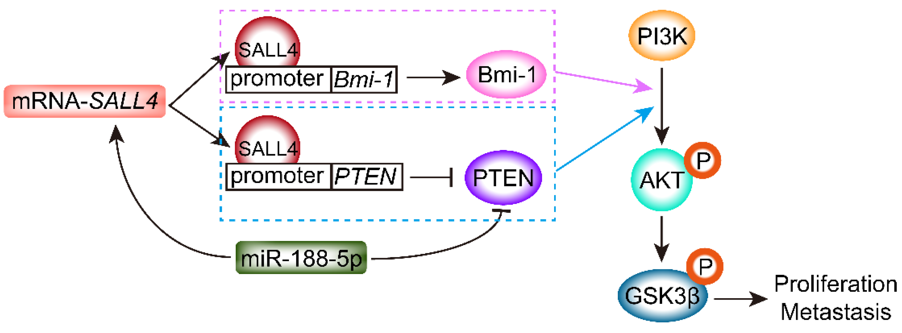
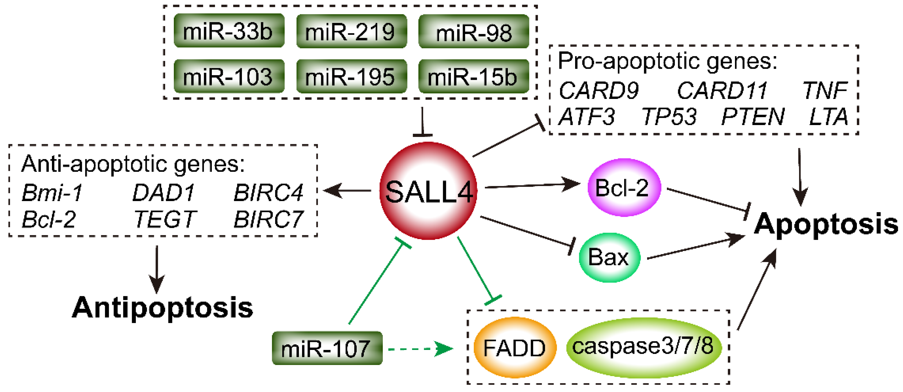
Table 1.
Function of different types of SALL family members in tumors.
| SALL4 Family Member | Function in Tumors | Mechanism | References |
|---|---|---|---|
| SALL1 | Anti-proliferative effects in glioma | Inhibit Wnt/β-catenin signaling | [11] |
| Induce angiogenesis | Active the VEGF-A gene | [12] | |
| SALL2 | Tumor suppressor | Inhibit c-Myc gene | [13] |
| SALL3 | Promote tumorigenesis | SALL3 promoter hypermethylation | [3] |
| SALL4 | Oncogene in glioma | Induce epithelial–mesenchymal transition | [14] |
| Promote progression in lung cancer | Activate EGFR by ERK1/2 signaling | [15] | |
| Promote proliferation, migration, and invasion in breast cancer | Activate Wnt/β-catenin signaling | [16] | |
| Promote growth and invasion in osteosarcoma | Sponge miR-107 | [17] | |
| Promote invasiveness in melanoma | Interact with the HDAC 2 and bind to invasiveness genes | [18] | |
| Promote development in hepatocellular carcinoma | Activate Wnt/β-catenin signaling | [19] | |
| Promote metastasis in gastric cancer | Activate the TGF-β/SMAD signaling | [20] |
Table 2.
Abbreviations.
| Abbreviation | Full Name |
|---|---|
| ALDH1 | aldehyde dehydrogenase 1 |
| ALL | acute lymphoblastic leukemia |
| AML | acute myeloid leukemia |
| APL | acute promyelocytic leukemia |
| AXIN2 | axis inhibition protein |
| B-ALL | B-cell lymphoblastic leukemia |
| Bax | Bcl-like-protein 4 |
| BC | breast cancer |
| Bcl-2 | B-cell lymphoma 2 |
| BM | bone marrow |
| Bmi-1 | B cell-specific Moloney murine leukemia virus integration site 1 |
| ccRCC | clear cell renal cell carcinoma |
| CEA | carcinoembryonic antigen |
| CLL | chronic lymphoblastic leukemia |
| CML | chronic myeloid leukemia |
| CRC | colorectal cancer |
| CTLA-4 | cytotoxic-T-lymphocyte-antigen-4 |
| DFS | disease-free survival |
| DLL1 | delta-like 1 |
| DNMTs | DNA methyltransferases |
| DNMT3A | DNA methyltransferase 3 alpha |
| DOT1 L | disruptor of telomeric silencing 1-like |
| DSS | disease-specific survival |
| EMT | epithelial–mesenchymal transition |
| EOC | epithelial ovarian carcinoma |
| ERK1/2 | extracellular signal-regulated kinase 1/2 |
| ESCC | esophageal squamous cell carcinoma |
| ESCs | embryonic stem cells |
| GBM | glioblastoma multiforme |
| GC | gastric cancer |
| GHCs | gastric hepatoid carcinomas |
| HB | hepatoblastoma |
| HCC | hepatocellular carcinoma |
| HDAC | high histone deacetylase |
| HE4 | human epididymis protein 4 |
| HEY2 | hairy/enhancer of split related to YRPW motif family member 2 |
| HK-2 | exokinase II |
| HNSCC | neck squamous cell carcinoma |
| HOXA9 | homeobox A9 |
| HOXA11-AS | homeobox A11 antisense |
| HPC | hematopoietic progenitor cell |
| HSC | hematopoietic stem cell |
| ICC | intrahepatic cholangiocarcinoma |
| IFN-γ | TNF-α and interferon-γ |
| IL-2 | interleukin-2 |
| KDMs | histone 3 lysine 9-specific demethylases |
| LSD1 | lysine-specific demethylase 1 |
| MAML1 | mastermind-like transcriptional coactivator 1 |
| MBD2 | methyl-CpG-binding domain 2 protein |
| MDS | myelodysplastic syndrome |
| MEIS1 | myeloid ecotropic viral insertion site 1 |
| miRNA/miR | microRNA |
| MLL | mixed lineage leukemia |
| NF-κB | transcription factor nuclear factor κB |
| NPC | nasopharyngeal carcinoma |
| NSCLC | non-small cell lung cancer |
| NuRD | nucleosome remodeling deacetylase |
| OC | ovarian cancer |
| OCT4 | octamer-binding transcription factor 4 |
| OS | osteosarcoma |
| OXPHOS | oxidative phosphorylation |
| PD-1 | programmed death ligand 1 |
| PFS | progression-free survival |
| POI | premature ovarian insufficiency |
| POLII | RNA polymerase II |
| PRC | polycomb repressive complex |
| PTEN | phosphatase and tension homolog |
| ROMA | risk of ovarian malignancy algorithm |
| SALLs | spalt-like transcription factors |
| SALL4TSS | SALL4 transcriptional start site TSS |
| SCLC | small cell lung cancer |
| SOC | serous ovarian carcinoma |
| SOX2 | sex-determining region Y (SRY)-Box 2 |
| STAT3 | activator of transcription 3 |
| TAMR | tamoxifen-resistant |
| TNF-α | tumor necrosis factor alpha |
| TNM | tumor node metastasis |
| TRIB3 | tribbles pseudokinase 3 |
| TRIM21 | tripartite motif-containing 21 |
| UTR | untranslated region |
| WHSC1 | Wolf–Hirschhorn syndrome candidate gene-1 |
Publisher’s Note: MDPI stays neutral with regard to jurisdictional claims in published maps and institutional affiliations. |
© 2022 by the authors. Licensee MDPI, Basel, Switzerland. This article is an open access article distributed under the terms and conditions of the Creative Commons Attribution (CC BY) license (https://creativecommons.org/licenses/by/4.0/).
Share and Cite
MDPI and ACS Style
Sun, B.; Xu, L.; Bi, W.; Ou, W.-B. SALL4 Oncogenic Function in Cancers: Mechanisms and Therapeutic Relevance. Int. J. Mol. Sci. 2022, 23, 2053. https://0-doi-org.brum.beds.ac.uk/10.3390/ijms23042053
AMA Style
Sun B, Xu L, Bi W, Ou W-B. SALL4 Oncogenic Function in Cancers: Mechanisms and Therapeutic Relevance. International Journal of Molecular Sciences. 2022; 23(4):2053. https://0-doi-org.brum.beds.ac.uk/10.3390/ijms23042053
Chicago/Turabian StyleSun, Boshu, Liangliang Xu, Wenhui Bi, and Wen-Bin Ou. 2022. "SALL4 Oncogenic Function in Cancers: Mechanisms and Therapeutic Relevance" International Journal of Molecular Sciences 23, no. 4: 2053. https://0-doi-org.brum.beds.ac.uk/10.3390/ijms23042053
Note that from the first issue of 2016, this journal uses article numbers instead of page numbers. See further details here.