Article

Overexpression of PpSnRK1α in Tomato Increased Autophagy Activity under Low Nutrient Stress

State Key Laboratory of Crop Biology, College of Horticulture Science and Engineering, Shandong Agricultural University, Tai’an 271018, China
*
Authors to whom correspondence should be addressed.
Int. J. Mol. Sci. 2022, 23(10), 5464; https://0-doi-org.brum.beds.ac.uk/10.3390/ijms23105464
Submission received: 22 April 2022 / Revised: 11 May 2022 / Accepted: 11 May 2022 / Published: 13 May 2022
(This article belongs to the Special Issue Response to Environmental Stress in Plants)

Abstract

:
Plants suffer from a variety of environmental stresses during their growth and development. The evolutionarily conserved sucrose nonfermenting kinase 1-related protein kinase 1 (SnRK1) plays a central role in the regulation of energy homeostasis in response to stresses. In plant cells, autophagy is a degradation process occurring during development or under stress, such as nutrient starvation. In recent years, SnRK1 signaling has been reported to be an upstream activator of autophagy. However, these studies all focused on the regulatory effect of SnRK1 on TOR signaling and the autophagy-related gene 1 (ATG1) complex. In this study, overexpression of the gene encoding the Prunus persica SnRK1 α subunit (PpSnRK1α) in tomato improved the photosynthetic rates and enhanced the resistance to low nutrient stress (LNS). Overexpression of PpSnRK1α increased autophagy activity and upregulated the expression of seven autophagy-related genes (ATGs). The transcriptional levels of SlSnRK2 family genes were altered significantly by PpSnRK1α, signifying that PpSnRK1α may be involved in the ABA signaling pathway. Further analysis showed that PpSnRK1α not only activated autophagy by inhibiting target of rapamycin (TOR) signaling but also enhanced ABA-induced autophagy. This indicates that PpSnRK1α regulates the photosynthetic rate and induces autophagy, and then responds to low nutrient stress.

1. Introduction

Different environmental challenges cause plants to suffer from various stresses and nutrient deprivation that disrupt metabolic and energy homeostasis, which are critical for plant development and growth. Plant sucrose nonfermenting kinase 1-related protein kinase 1 (SnRK1) is known as an energy and metabolism sensor that regulates the energy balance in response to nutritional and environmental stresses [1,2,3,4]. SnRK1 is a heterotrimeric complex composed of an α-catalytic subunit and β-and γ-regulatory subunits within plants [5,6]. Furthermore, evidence is accumulating that the α-subunit plays pivotal roles in regulating biotic and abiotic stress responses as well as plant growth and development [1,3,7,8,9].
In response to energy deficits, SnRK1 restores energy homeostasis by switching on ATP-producing catabolic pathways while also switching off ATP-consuming metabolic processes. KIN10 (the gene encoding an α-catalytic subunit in Arabidopsis thaliana) overexpression in seedlings was shown to promote plant survival in glucose (Glc)-free liquid medium [7]. KIN10 promotes stomatal development, thereby influencing many plant responses to changing environmental conditions [10]. PpSnRK1α has been shown to be involved in the abscisic acid (ABA) signaling pathway to improve tomato salt tolerance [11].
In plant cells, various complex catabolic pathways are upregulated to balance the nutrient cycle and restore energy homeostasis under nutrient starvation stress, and autophagy is an important part of this process. Autophagy is an evolutionarily conserved self-degradation process that recycles cellular nutrients or breaks down damaged components for the optimization of plant growth, development and stress responses [12,13,14]. Nutrient deficiency induces the upregulation of the transcription levels of a large number of autophagy-related genes (ATGs) in plants, accompanied by the formation of autophagosomes. In Arabidopsis, ATG3, ATG4a, ATG4b, ATG7 and ATG8a-ATG8i were shown to be induced by sugar starvation [15,16,17]. The ATG1 kinase complex, the main switch for autophagy initiation, is regulated by the upstream kinases target of rapamycin (TOR) and SnRK1 [18]. TOR is a phosphatidylinositol 3-kinase-related kinase that acts as a negative regulator of autophagy [19,20]. In general, TOR is activated and inhibits autophagy in nutrient-rich conditions, stimulating growth. Upon nutrient deprivation, TOR is inactivated, allowing for the activation of autophagy and reducing plant growth. Overexpression of KIN10 in Arabidopsis leaf cells induces the expression of ATG8s [7], which play a key role in autophagosome formation and are very sensitive markers of autophagy [21]. KIN10 activates autophagy by inhibiting the TOR signaling pathway [22] or by directly phosphorylating ATG1 [23].
Plant hormones are important endogenous signaling compounds that regulate plant growth, development and response to stress. In Arabidopsis, autophagy is induced by the salicylic acid (SA) functional analog benzothiadiazole (BTH) [24,25]. Zeatin (ZT) can reduce the formation of autophagy in root epidermal cells [16], suggesting that phytohormones can directly regulate autophagy. Ethylene response factor 5 (ERF5) can directly bind to the ATG8d and ATG18h promoters to induce autophagy formation under drought stress [26]. Brassinosteroid (BR) signaling transcription factor BZR1 (Brassinazole resistance 1) can upregulate the expression of ATGs to induce autophagy in response to low nitrogen stress [27]. Thus, plant hormone signal transduction pathways are also involved in regulating autophagy. In Arabidopsis, ABA has been shown to activate the expression of ATGs and the formation of autophagosomes [28]. ABA-activated SnRK2 can phosphorylate RAPTOR under osmotic stress, thereby inhibiting TOR signaling to induce autophagy [29]. ABA promoted autophagy by enhancing autophagosome formation in glioblastoma cells [30].
Some reports indicate that SnRK1 links stress with ABA signaling [5,8]. SnRK1.1 is involved in ABA signaling pathways, resulting in modifications of essential enzyme activities and gene expression in response to sugar starvation and darkness [7,31]. In Arabidopsis, SnRK1.1-overexpressing lines exhibit an ABA-sensitive phenotype [31]. ABA-insensitive 1 (ABI1) and clade A type 2C protein phosphatases (PP2CAs), negative regulators of the ABA pathway, interact with the SnRK1 α subunit to repress SnRK1 signaling [4]. Apple MdSnRK1.1 (α-catalytic subunit) interacts with MdCAIP1 to regulate ABA sensitivity [32]. However, the role of SnRK1 in ABA-induced autophagy is largely unknown.
Our previous study showed that overexpression of PpSnRK1α in tomato could promote fruit ripening and enhance salt tolerance. In this study, it was found that overexpressing PpSnRK1α in tomato increased the photosynthetic rate and autophagy activity under low nutrient tolerance. Further research found that PpSnRK1α not only enhanced autophagy through TOR signaling but also enhanced ABA-induced autophagy. Our research places PpSnRK1α as an important regulator controlling the balance between growth and stress responses via the direct regulation of autophagy.

2. Results

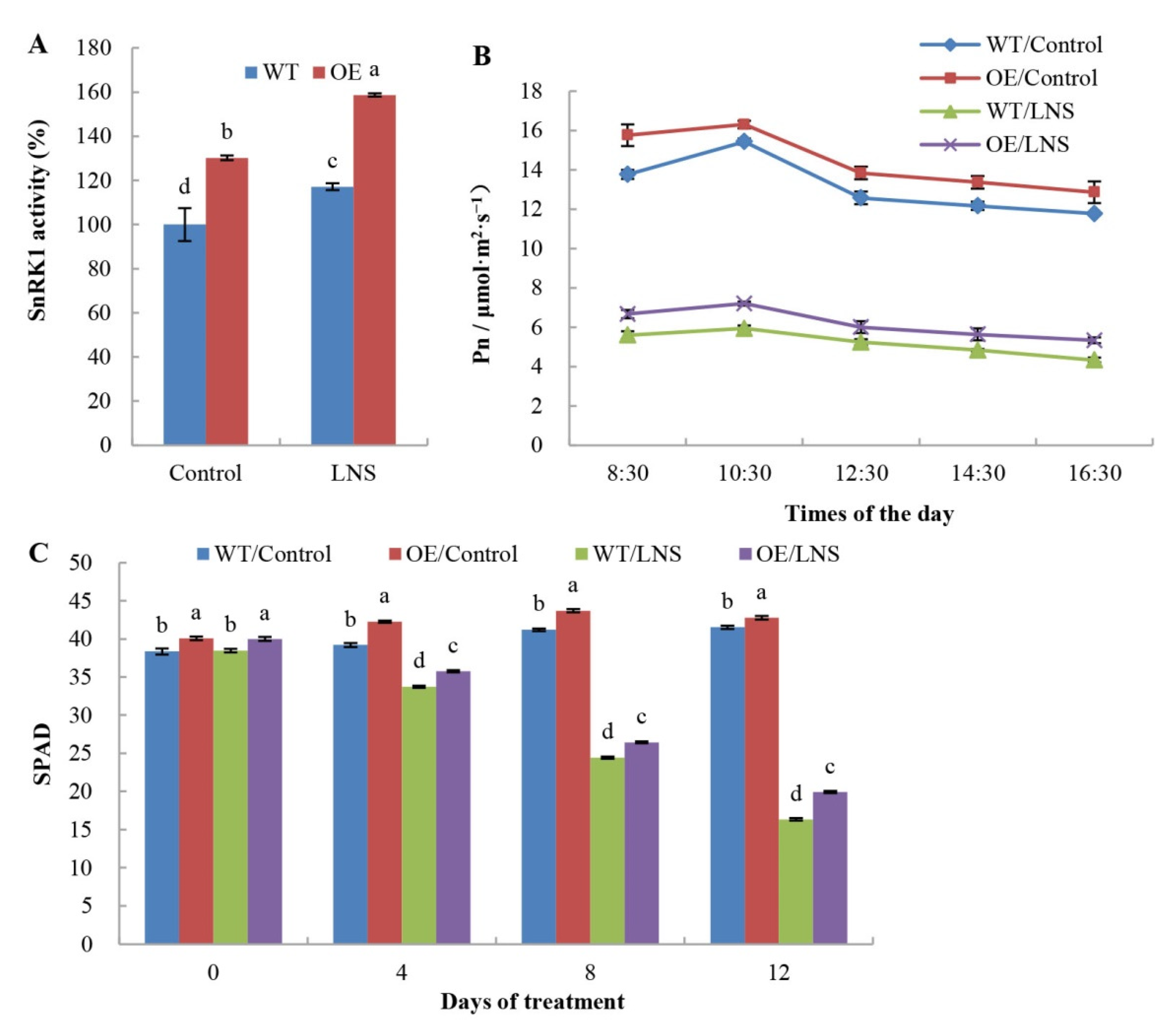
2.1. Effects of PpSnRK1α Overexpression on Tomato Photosynthetic Rate and Plant Growth under LNS

Plant SnRK1 protein kinase is a central energy and metabolism sensor that regulates the energy balance in response to biotic and abiotic stress. PpSnRK1α-overexpressing tomato (OE) and WT tomato were used to study the effects of SnRK1 on the growth of tomato plants under nutrient stress. In the control group, the SnRK1 activity in the leaves of OE plants was 30.23% higher than that of WT plants. Compared with tomato under normal nutrient levels (control), OE tomato under LNS had higher SnRK1 activity and was 39.46% higher than that of the WT (Figure 1A). Further, we analyzed the effects of PpSnRK1α on leaf photosynthesis. Compared with that of WT plants, the net photosynthetic rate of functional leaves of OE tomato increased by 9.79% on average under normal nutrient conditions (control) and 18.98% under LNS conditions (Figure 1B). As shown in Figure 1C, the relative chlorophyll content in leaves of OE plants was significantly higher than that of WT plants, which was 22.04% higher than that of WT plants after 12 days under LNS conditions. Compared with WT tomato plants, OE tomato plants had higher plant height and greater root and leaf weights (Table 1). These results indicated that PpSnRK1α could increase leaf photosynthesis and promote plant growth.

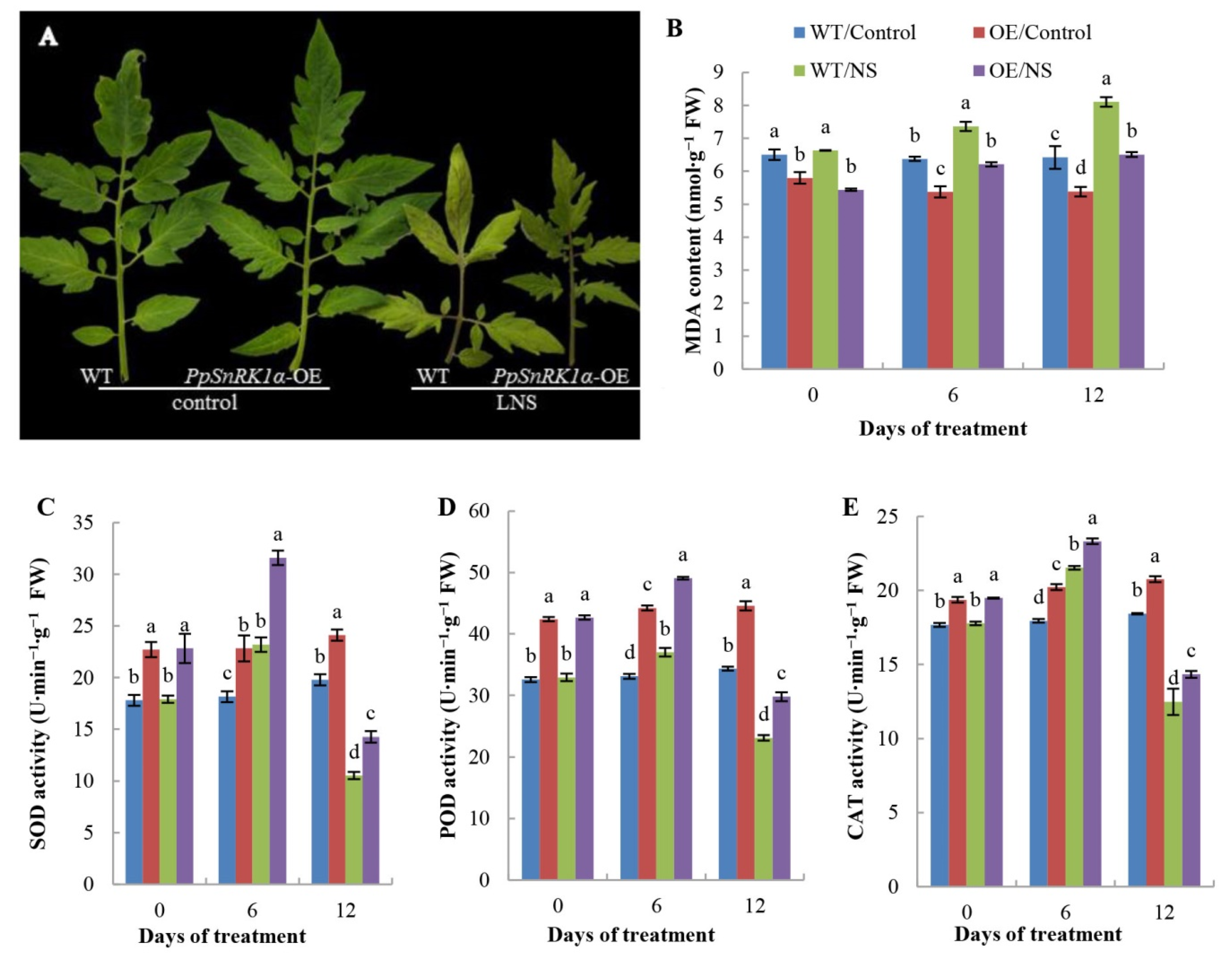
2.2. Effects of PpSnRK1α Overexpression on the Leaf Antioxidant Enzyme Activities

In the LNS group, the leaves of tomato plants were yellow and obviously smaller than those of plants in the control group, but the leaves of OE plants were obviously greener than those of WT plants, suggesting that PpSnRK1α may affect the process of leaf senescence under LNS (Figure 2A). Furthermore, we monitored changes in antioxidant enzyme activities and MDA content in both WT and OE plants before and after the LNS treatment. Compared with that in control leaves, the MDA contents in OE and WT leaves increased at 6 and 12 days after LNS treatment, but the MDA content in OE tomato leaves was lower than that in WT tomato leaves, indicating that PpSnRK1α overexpression leads to a decrease in the degree of cell membrane peroxidation in response to LNS (Figure 2B). The activities of antioxidant enzymes increased in response to stress at the initial stage of LNS and decreased as the stress time increased. Under both the control and LNS conditions, the activities of SOD, POD and CAT in the leaves of OE plants were significantly higher than those in the leaves of WT plants (Figure 2C–E). These results indicated that PpSnRK1α may play a role in antioxidant systems to protect plants under LNS.

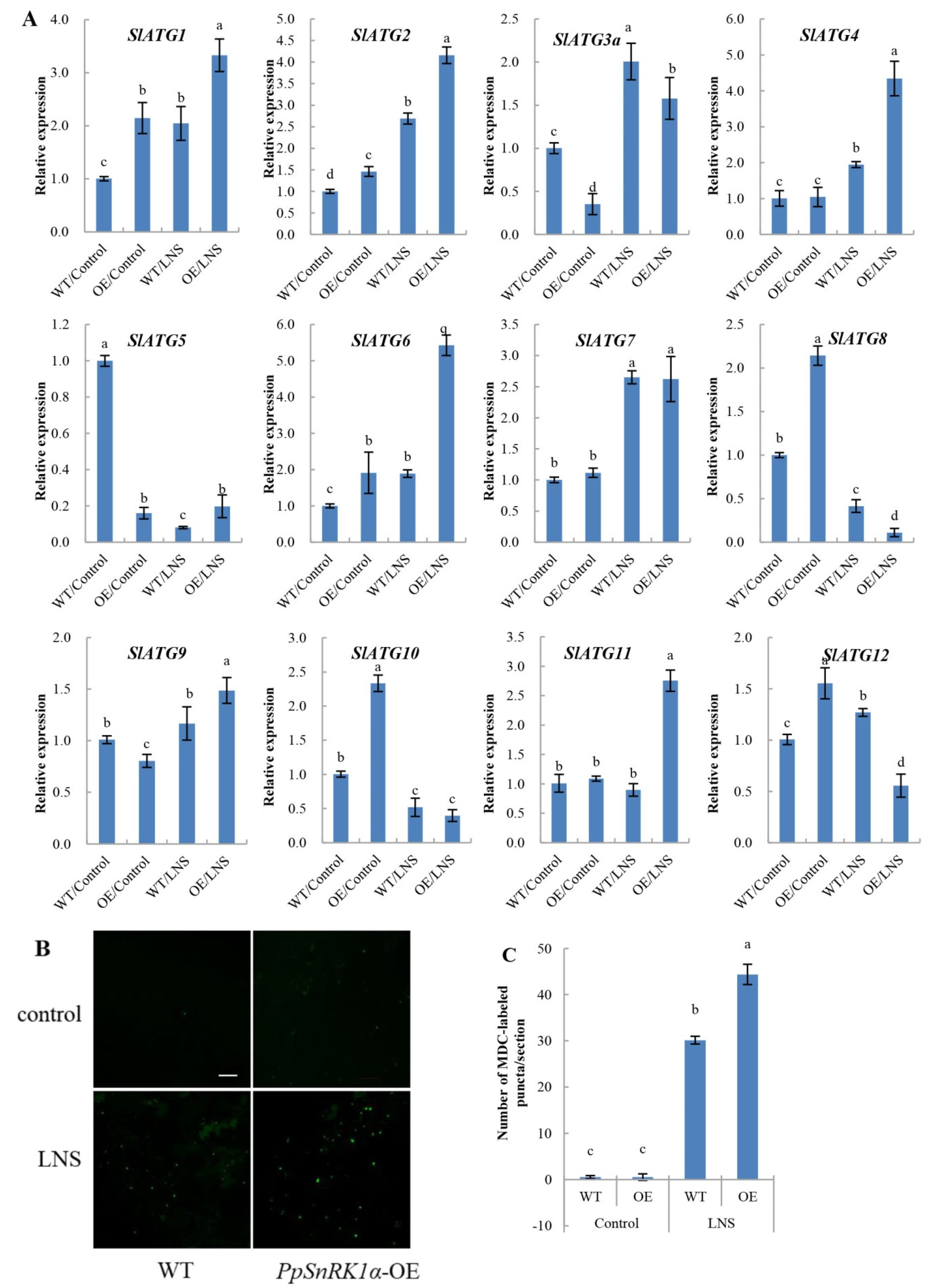
2.3. Overexpression of PpSnRK1α Enhances Autophagy Activity under LNS

Autophagy is a highly conserved self-degradation mechanism in eukaryotes. Under nutritional stress conditions, autophagy is largely induced, and plants can maintain cell homeostasis through autophagy. To investigate the effect of PpSnRK1α on autophagy under LNS, we measured the expression of ATGs and autophagy activity in OE and WT tomatoes. We found that 7 SlATGs were induced, and 3 SlATGs were inhibited by LNS relative to normal conditions (LNS vs. control). Under LNS conditions, the expression levels of SlATG1, SlATG2, SlATG4, SlATG5, SlATG6, SlATG9 and SlATG11 in OE plants were higher than those in WT plants, while the expression levels of SlATG3a, SlATG8 and SlATG12 were lower than those in WT plants (Figure 3A). These results suggest that autophagy in tomato leaves could be induced by LNS and that PpSnRK1α may mediate the formation of autophagy under LNS.
Furthermore, we used the fluorescent dye MDC to detect autophagic activity in WT and OE plants under control and LNS conditions (Figure 3B). Under the control condition, only a few autophagosomes were detected in WT and OE tomato leaves. After 7 days of LNS treatment, the number of autophagosomes in WT and OE leaves was significantly increased compared with that in the control group, and the number of autophagosomes in OE tomato leaves was 1.47 times that in WT leaves (Figure 3C), confirming that PpSnRK1α plays a positive role in low nutrient-induced autophagy.

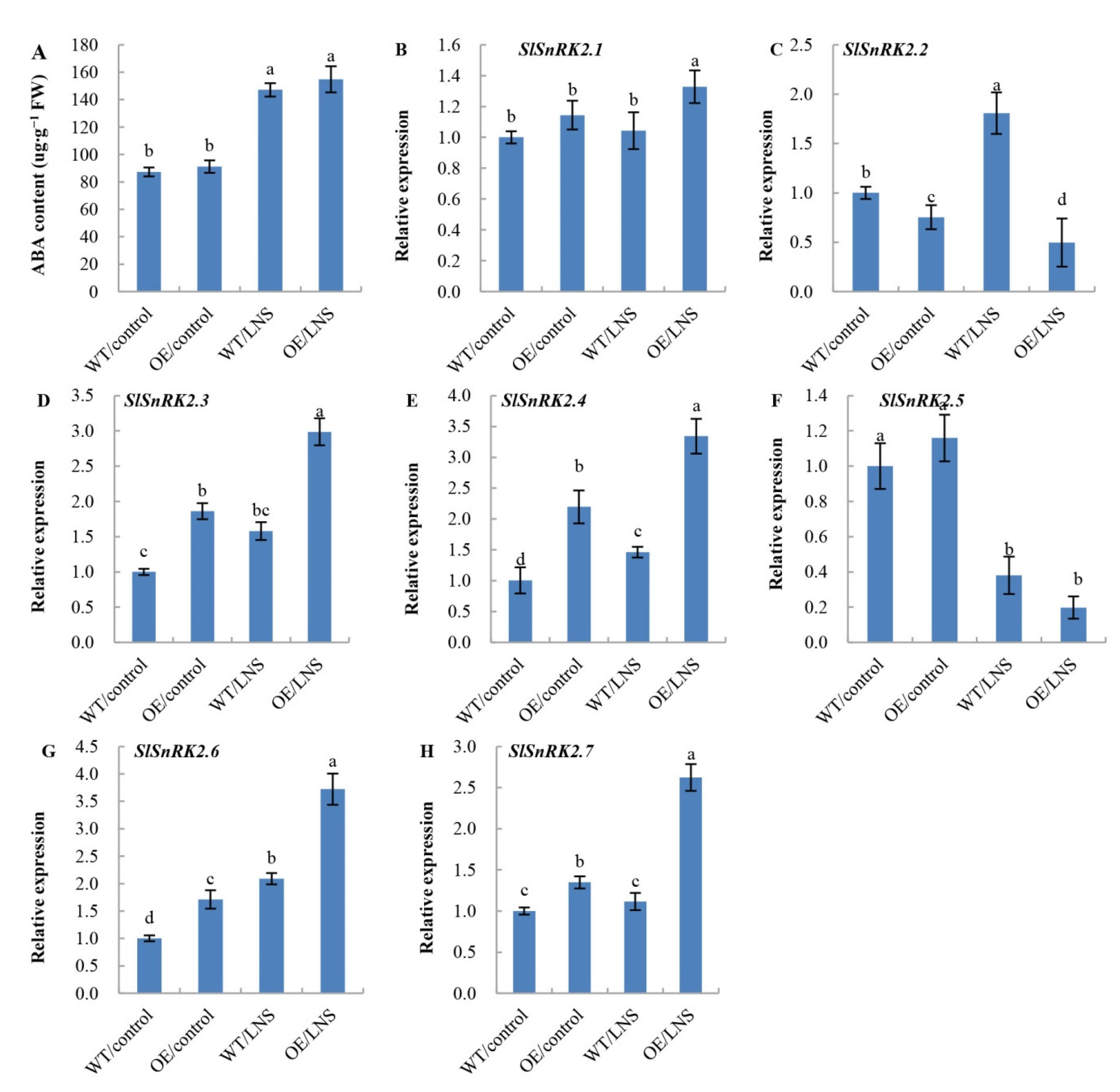
2.4. Overexpression of PpSnRK1α Affects the ABA Content and Transcription Level of SlSnRK2s under LNS

The ATG1 kinase complex, as the main switch of autophagy initiation, is regulated by the upstream kinases TOR and SnRK1 [18]. It was found that the overexpression of PpSnRK1α could improve the resistance of tomato to LNS, accompanied by an increase in autophagy activity. In Arabidopsis thaliana, downregulation of TOR activates autophagy under nutritional stress. Thus, we further explored the role of TOR in PpSnRK1α-induced autophagy. The TOR activator Glc was sprayed on the tomato leaves, and the autophagy activities in WT, WT + Glc, OE and OE + Glc leaves were measured by MDC staining. As shown in Figure 4A,B, the autophagy activity in WT and OE leaves significantly decreased after treatment with the TOR activator Glc. Under normal nutrient conditions, no MDC-labeled autophagosomes were detected in WT + Glc and OE + Glc leaves. Under LNS conditions, the number of MDC-stained autophagosomes in WT + Glc leaves was 36.4% lower than that in WT leaves, while the number of autophagosomes in OE + Glc leaves was 45.8% lower than that in OE leaves (Figure 4). Overall, these data suggested that the activation of PpSnRK1α on autophagy activity depended on TOR signaling and that PpSnRK1α was involved in the regulation of other signal-induced autophagy.
ABA, an essential plant hormone, plays a crucial role in response to abiotic stress, and the negative regulators of ABA signaling, ABI1 and PP2CA, directly inhibit the activity of SnRK1. Thus, we speculated that ABA signaling may induce autophagy and that SnRK1 may be involved in the ABA-induced autophagy process. SnRK2, a subfamily of kinases related to the SnRK1 subfamily, is an important component of the ABA signaling pathway. Therefore, the ABA content and the expression levels of seven SlSnRK2s were detected in both OE and WT plants (Figure 5). There was no significant difference in ABA content between WT and OE tomato leaves, but the ABA content was significantly increased under LNS conditions, which was 3.46 times that under control conditions (Figure 5A). This suggested that PpSnRK1α was downstream of ABA signaling. In OE plants, the transcript levels of SlSnRK2.3, SlSnRK2.4, SlSnRK2.6 and SlSnRK2.7 were increased under both control and LNS conditions, while the transcript level of SlSnRK2.2 was inhibited. Compared with the control, the expression level of SlSnRK2.1 was not significantly changed in the WT under LNS conditions but was increased significantly in the OE lines. Under LNS conditions, the expression levels of SlSnRK2.3, SlSnRK2.4, SlSnRK2.6 and SlSnRK2.7 were upregulated, while those of SlSnRK2.2 and SlSnRK2.5 were downregulated (Figure 5B–H). These results suggest that the regulation of SlSnRK2 transcription by PpSnRK1α is complex.

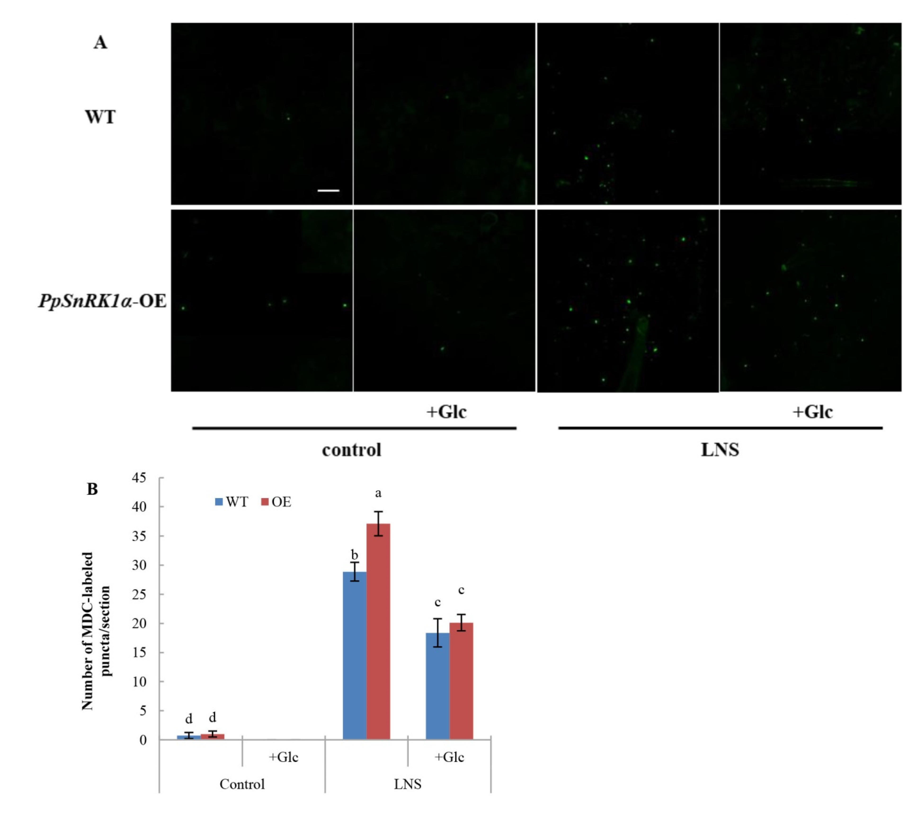
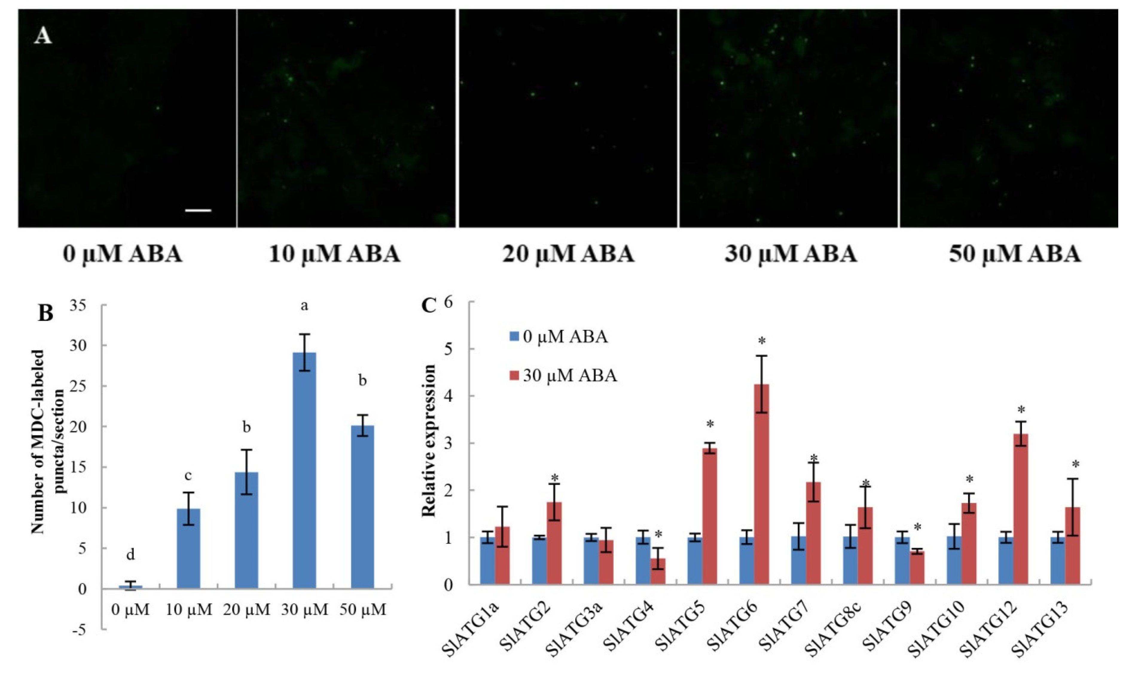
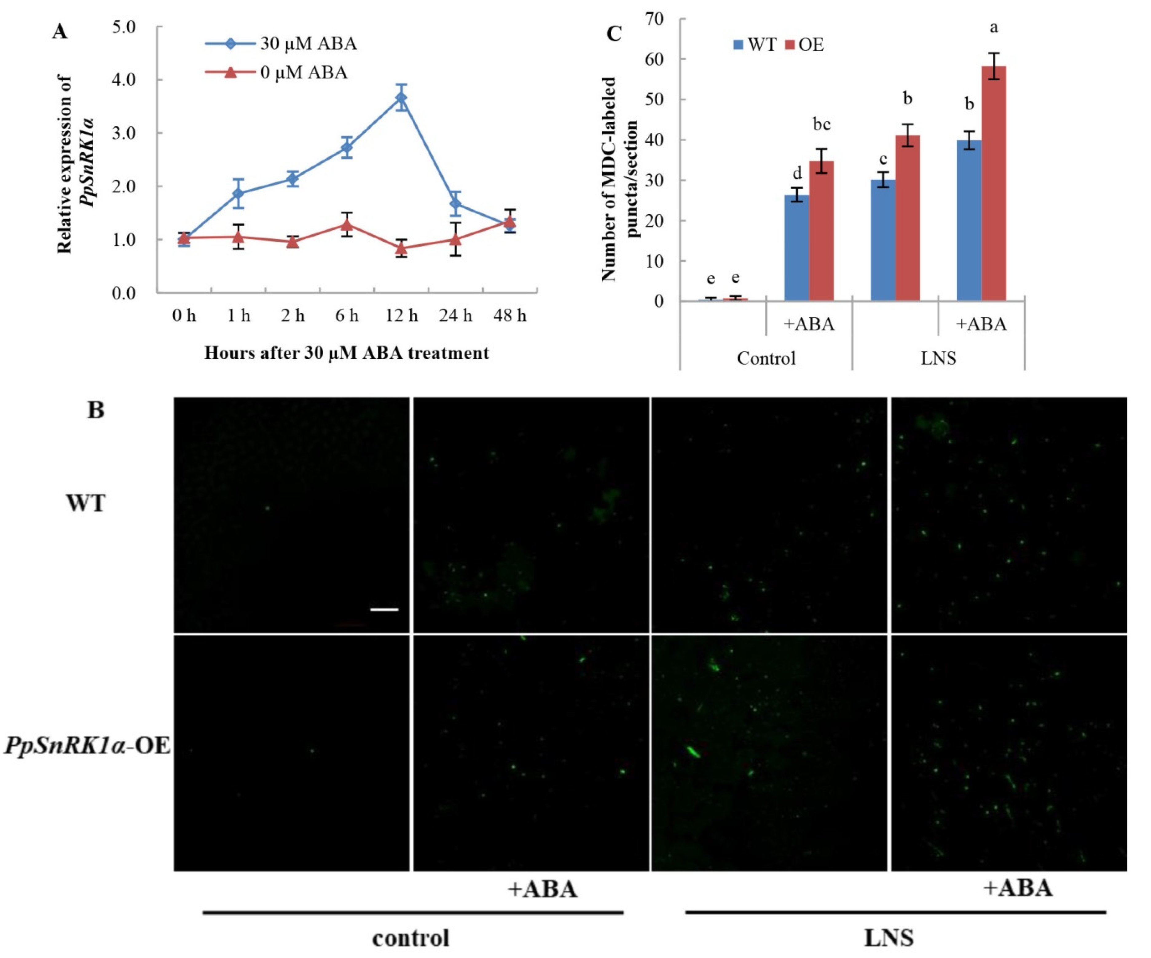
2.5. Overexpression of PpSnRK1α Enhances ABA-Induced Autophagy

To investigate whether ABA can induce autophagy in tomato leaves, we used MDC to detect autophagy activity after treatment with exogenous ABA. As shown in Figure 6, autophagy activity was significantly increased after ABA treatment, especially treatment with 30 µM ABA (Figure 6A,B). To gain further insight into the role of ABA in activating autophagy, we analyzed the transcript levels of 12 tomato ATGs after treatment with ABA (Figure 6C). The transcript levels of SlATG2, SlATG5, SlATG6, SlATG7, SlATG8c, SlATG10, SlATG12 and SlATG13 were significantly upregulated, and only two ATGs, SlATG4 and SlATG9, were downregulated. These results, taken together, confirm that exogenous ABA can induce the formation of autophagosomes in tomato leaves.
ABA can control SnRK1 activity via the interaction of SnRK1 with PP2C to regulate its phosphorylation [4]. In this study, the expression of PpSnRK1α in OE leaves was also detected at different time points after 0 µM and 30 µM ABA treatments. The expression of PpSnRK1α increased after ABA treatment and reached the highest level at 12 h, which was 3.7 times that before treatment (Figure 7A), indicating that ABA could also regulate PpSnRK1α transcription. Therefore, we speculated that PpSnRK1α might be involved in ABA-induced autophagy. To determine whether PpSnRK1α is involved in the regulation of ABA-induced autophagy, WT and OE tomato leaves were treated with 30 μM ABA, and autophagy activity was detected by MDC staining. As shown in Figure 7B,C, the number of autophagosomes in WT and OE leaves increased significantly after ABA treatment. Under LNS conditions, the number of autophagosomes in WT and OE leaves increased by 32.4% and 41.6%, respectively, suggesting that the overexpression of PpSnRK1α enhanced the ABA-dependent activation of autophagy under LNS.

3. Discussion

The role of SnRK1 has been researched mostly in A. thaliana and crop species, while the role of SnRK1 in horticultural crops has rarely been reported. In this study, we overexpressed peach PpSnRK1α in tomato to study the mechanism of PpSnRK1α in response to LNS. In Arabidopsis, SnRK1 can be activated by sugar starvation, energy deficiency, dark culture and oxygen deficiency. KIN10/11 can initiate multiple transcription cascades in response to sugar or energy depletion in darkness and under stress conditions. The role of KIN10 in the starvation response was most evident in Glc-free liquid medium [7]. SnRK1 activity was shown to be higher in seedlings grown under low nutrient conditions than in seedlings grown under optimal nutrient conditions [33]. Consistent with these findings, SnRK1 activity was higher in OE tomatoes grown under LNS conditions than in OE tomatoes grown under control conditions, and SnRK1 activity in OE lines increased more evidently.
Mounting evidence has shown that SnRK1 plays a key role in carbohydrate metabolism and maintains energy homeostasis [34,35]. SnRK1 is also involved in starch biosynthesis [36,37]. In our previous study, transgenic tomato plants overexpressing heterologous MhSnRK1 had greater soluble sugar and starch contents and greater photosynthesis rates than WT plants [37]. In the photosynthesis pathway, seven genes were upregulated in the MhSnRK1-overexpressing lines [38]. SnRK1 also plays a role in nitrogen metabolism [39]. In this study, the photosynthetic rate and the activity of antioxidant enzymes in mature leaves of OE were increased significantly compared with WT. After 12 days under LNS, the transgenic lines showed higher root biomass and better root activity. These results suggest that PpSnRK1α overexpression leads to enhanced resistance to low nutrient stress in tomatoes.
Autophagy is a highly conserved self-degradation mechanism in eukaryotes, and the cascade regulation of phosphorylation is the main switch to activate autophagy [40]. Autophagy usually begins with the perception of developmental and nutritional signals by the ATG1 complex, and ATG1 is regulated by the upstream kinases TOR and SnRK1 [18]. In Arabidopsis, autophagy is induced during senescence and nutrient deficiency [41,42] and stresses [43,44] by inhibiting TOR pathways. Overexpression of TOR inhibited autophagy activation by nutrient starvation and salt and osmotic stress [45]. SnRK1 can induce autophagy both via TOR-dependent and TOR-independent pathways in Arabidopsis [22]. In Arabidopsis, SnRK1 phosphorylation of RAPTOR represses TOR complex activity to activate autophagy in plants [46,47]. Overexpression of KIN10 activates the formation of autophagosomes by increasing the level of ATG1 phosphorylation, and KIN10 can interact with ATG1a and ATG13a in vitro, suggesting that SnRK1 can also regulate autophagy through the ATG1 complex. Transgenic Arabidopsis lines overexpressing KIN10 increased tolerance to nutrient starvation [23]. Tomato SnRK1.1 mediates the formation of ATG6-dependent autophagy and alleviates tomato LN stress [48]. In this study, PpSnRK1α-overexpressing lines exhibited higher autophagy activity than WT plants under LNS and showed stronger resistance to LNS. Moreover, overexpression of PpSnRK1α upregulated the expression levels of seven ATGs. Given that the TOR complex is also a major regulatory factor in nutrient sensing, we added the TOR activator Glc to verify whether SnRK1-mediated autophagy under LNS is related to the TOR pathway. After TOR was activated by Glc, autophagy activity was decreased in OE leaves; however, some autophagosomes were still observed, indicating that PpSnRK1α can activate autophagy activity through TOR-dependent and TOR-independent pathways. Although PpSnRK1α plays a positive role in autophagy regulation, whether autophagy induction is the cause of enhanced low nutrient tolerance requires further investigation, such as overexpressing PpSnRK1α in an autophagy-deficient context (mutant plants, treatment with inhibitors) to assess the role of autophagy in this phenotype.
Plants suffer from a variety of environmental stresses, during which ABA signaling and autophagy processes are activated to enhance stress tolerance. Therefore, we hypothesized that the ABA signaling pathway is involved in the regulation of autophagy under abiotic stress. In Arabidopsis, ABA activated the formation of autophagosomes and the expression of ATGs [28]. A recent study showed ABA-induced autophagy in glioblastoma cells [30]. Similarly, in this study, autophagosomes stained with MDC were observed in WT tomato leaves after treatment with different concentrations of ABA. Moreover, the expression levels of seven ATGs were significantly increased, and those of two ATGs were significantly decreased after 30 μM ABA treatment. These results confirmed that ABA activates autophagy.
ABA can respond to biotic and abiotic stresses [49], and ABI1 and PP2CA directly regulate SnRK1.1 activity [4]. Therefore, we hypothesized that SnRK1 may be involved in ABA-induced autophagy. The expression of PpSnRK1α was significantly increased after 30 μM ABA treatment, and peach seedlings overexpressing PpSnRK1α were sensitive to ABA [50]. Similar results were also found in apples, in which MdSnRK1.1 inhibited C2-domainABA insensitive protein1 (MdCAIP1)-mediated ABA sensitivity [32]. In this study, we sprayed 30 μM ABA on the leaves of WT and OE tomatoes growing under normal and LNS conditions and found that the autophagy activity in WT and OE leaves increased significantly after ABA treatment. More importantly, the autophagy activity increased more in OE leaves than in WT leaves after ABA treatment. These results suggested that SnRK1 is a positive regulator of ABA-activated autophagy.
SnRK2s play an essential role in response to biotic and abiotic stress [51]. In Arabidopsis, SnRK2.2, 2.3 and 2.6 play critical roles in ABA signaling [52,53]. Similar to ATGs, we found that the expression levels of SlSnRK2s were also regulated by PpSnRK1α. Under LNS conditions, the ABA content in tomato leaves increased significantly, and overexpression of PpSnRK1α significantly upregulated the expression levels of SlSnRK2.1, SlSnRK2.3, SlSnRK2.4, SlSnRK2.6 and SlSnRK2.7. In the absence of ABA, ABA-responsive SnRK2s are usually inhibited by PP2Cs. In the presence of ABA, SnRK2s are released and activated to regulate the expression of ABA-responsive genes [54,55,56]. Within the SlSnRK2 gene family, SlSnRK2.3 and SlSnRK2.4 belong to SnRK2 subclass III and are essential for transducing ABA signals [57,58]. In conclusion, PpSnRK1α and SlSnRK2s crosstalk can regulate the response of tomato to LNS, and the regulation of SlSnRK2s by PpSnRK1α may be related to the ABA signaling pathway. However, whether there is a direct or indirect interaction between PpSnRK1α and SnRK2 to regulate the plant response to LNS needs further study.

4. Summary

Overexpressing Prunus persica PpSnRK1α in tomato increased photosynthetic rate and induced autophagy under low nutrient tolerance. Moreover, PpSnRK1α not only enhanced autophagy through TOR signaling but also played a positive role in ABA-induced autophagy. However, the mechanism by which SnRK1 responds to LNS is complex, and the mechanism by which SnRK1 is involved in ABA-induced autophagy and whether SnRK1 directly regulates SlSnRK2 expression need further investigation.

5. Materials and Methods

5.1. Plant Materials and Treatments

In this study, wild-type (WT) tomato (Solanum lycopersicum ‘Sy12f’) and homozygous T2 lines overexpressing PpSnRK1α (ppa004347 m) (OE) [59] were germinated and grown in a plant growth chamber filled with a mixture of peat and vermiculite (1:1, v/v) at 23 °C for 3 weeks.
For the low nutrient stress (LNS) study, 1/2 Hoagland nutrient solution was used as the normal nutrient level (control), and 1/20 Hoagland nutrient solution was used as the LNS level. The 3-week-old plants were moved to a 10 L hydroponic system containing 8 L of 1/2 Hoagland nutrient solution, and the treatments were performed after 1 week of growth. The hydroponic system was ventilated 24 h a day, and the nutrient solution was replaced every 3 days.
For treatment with Glc, during the LNS treatment, the tomato plants were sprayed with 15 mM Glc solution or ddH2O every 2 days. Autophagy activity was detected on the 7th day of treatment.
For treatment with ABA, 6-week-old WT leaves were sprayed with 0, 10, 20, 30 or 50 µM ABA. Twelve hours after ABA treatment, the upper first fully expanded leaves were excised to detect autophagy activity, or they were sampled and frozen quickly in liquid N and stored at −80 °C before being used for gene expression.
For treatment with ABA under control and LNS conditions, the tomato plants were sprayed with 0 µM and 30 µM ABA solutions every 2 days. Samples were collected at different time points after ABA treatment to detect the expression level of PpSnRK1α. Autophagy activity was detected on the 7th day of treatment.

5.2. Determination of SnRK1 Activity

Approximately 1.0 g of frozen, fine tissue powder was homogenized in 1 mL of cold buffer solution that consisted of 100 mM HEPES (pH 8), 25 mM NaF, 2 mM sodium pyrophosphate, 0.5 mM EDTA, 0.5 mM EGTA, 5 mM DTT, 1 mM anisole, 1 mM phenylmethylsulfonyl fluoride, 1 mM protease inhibitor cocktail (Sigma P9599), phosphatase inhibitors (PhosStop; Roche) and insoluble polyvinylpyrrolidone (PVP). The tissue homogenate was transferred to a cold microfuge tube and clarified by centrifugation at 12,000× g for 5 min at 4 °C. The supernatant (750 µL) was desalted on a 2.5 mL pre-equilibrated centrifuge column (Sephadex G-25 medium columns; GE Healthcare) and maintained at 4 °C for the SnRK1 activity assay. SnRK1 activity was determined with a Universal Kinase Activity Kit (R&D Systems, Minneapolis, MN, USA, EA004) by using AMARA polypeptide as the substrate [59]. The procedure was carried out according to the manufacturer’s instructions. The optical density at 620 nm (OD620) was measured after the enzymatic reaction was over, and the relative SnRK1 activity was calculated based on OD620.5.3. Determination of antioxidant enzyme activities and the MDA content.
Proteins were extracted from approximately 0.5 g of sample with 4 mL of cold 50 mM phosphate buffer solution (pH = 7.8) [60]. Superoxide dismutase (SOD) activity was determined by the nitrogen blue tetrazole (NBT) method and calculated according to the method of Wang et al. [61]; catalase (CAT) activity was determined using the ultraviolet absorption method as previously described [62], and peroxidase (POD) activity was determined using the guaiacol method [63]. SOD, POD and CAT activities are expressed as U·min−1 g−1 (fresh weight, FW). The malondialdehyde (MDA) content was determined by the thiobarbituric acid (TBA) method [64].

5.3. Measurement of Photosynthetic Rate and Chlorophyll Content

The daily variation in the photosynthetic rate in tomato leaves was measured on clear days with a CIRAS-3 portable photosynthetic measurement system (Hansha Scientific Instruments Limited, Tai’an, China). The chlorophyll content was measured using a SPAD-502 chlorophyll meter (Tuopu Yunnong Technology). Five mature leaves were measured following the instrument instructions, and the mean of these measurements was used to represent the photosynthetic rate and chlorophyll content of that individual plant.

5.4. RT-qPCR

Total RNA was extracted from evenly mixed samples (1 g, the sample is a mixture of each separately treated sample) using a Fast Pure Plant Total RNA Isolation Kit (RC401, RC401, Vazyme, Nanjing, China) and reverse transcribed to generate the first-strand cDNA using HiScript III RT SuperMix for qPCR (+gDNA wiper) (R323-01, Vazyme, Nanjing, China) according to the manufacturer’s instructions. Quantitative real-time PCR (RT-qPCR) consisted of three biological and three technical replicates and was carried out using the Bole CFX96 system (Bio-Rad, Hercules, CA, USA) and Ultra SYBR mixture (CW2601M, CWBIO, Beijing, China,). The calculation method for RT–qPCR was 2−ΔΔCt with FaACTIN as the internal control. The specific primers used are listed in Table S1.

5.5. MDC Staining

Tomato leaves were stained with monodansylcadaverine (MDC) (Sigma–Aldrich, 30432) to determine autophagy activity, as described by Wang et al. [65]. Tomato leaves approximately 2 mm × 4 mm in size were excised, immediately vacuum infiltrated with 100 μM MDC (Sigma–Aldrich 30432) for 30 min and then washed twice with phosphate-buffered saline. MDC-incorporated structures were observed with a confocal laser scanning microscope.

5.6. ABA Extraction and Determination

Collected samples were homogenized using liquid nitrogen, and 0.3 g of plant material was extracted in a 10 mL centrifuge tube for 24 h using 80% methyl alcohol. The homogenate was centrifuged at 8000 rpm at 4 °C for 10 min, and the supernatant was transferred to a distillation bottle and concentrated to dryness under vacuum. The residue was dissolved in phosphoric acid buffer (pH = 8) and chloroform and transferred to a 10 mL centrifuge tube. The extract was shaken for 30 min and then incubated for at least 30 min. After stratification, the lower layer of trichloromethane was pipetted to remove the pigment. The upper phase was collected, and 150 mg of PVP was added and centrifuged at 8000 rpm at 4 °C for 10 min. The supernatant was transferred to a centrifuge tube and extracted with 5 mL of ethyl acetate. After stratification, the upper phase was collected and concentrated to dryness under vacuum. The residue was dissolved in 1 mL of 0.04% (v/v) acetic acid/methanol (55:45, v/v) and filtered through a 0.45 µm filter. ABA quantification was performed on three independent samples by high-performance liquid chromatography (HPLC) (10 μL of sample was injected). ABA quantification was performed on three independent samples by high-performance liquid chromatography (Agilent 1100). The extracts were filtered through 0.22-µm syringe filters and directly injected through a 10 µL fixed loop into a C18 guard column (4.6 mm × 250 mm, 5 µm, Kromasil, Sweden). The mobile phase was the mixture of 0.1% acetic acid: methanol (v:v = 1:1). The ABA contents were quantified from the areas of their peaks at 254 nm using external standard calibration curves.

Supplementary Materials

The following are available online at https://0-www-mdpi-com.brum.beds.ac.uk/article/10.3390/ijms23105464/s1, Table S1: Primers used in this study.

Author Contributions

F.P., Y.Z. and J.L. conceived and designed the research. J.L. and W.Y. performed and analyzed the experiments. J.L. and F.P. wrote the manuscript. Y.X. supervised the study and revised the manuscript. All authors read and approved the final version of the manuscript. All authors have read and agreed to the published version of the manuscript.

Funding

This work was supported by National Key R&D Program of China (Nos. 2019YFD1000103 and 2020YFD1000203) and the National Modern Agroindustry Technology Research System Fund (No. CARS-30-2-02).

Institutional Review Board Statement

Not applicable.

Informed Consent Statement

Not applicable.

Data Availability Statement

The data that support the findings of this study are available from the corresponding author upon reasonable request. All relevant data can be found within the manuscript and its supporting materials.

Conflicts of Interest

The work described here has not been published previously and is not under consideration for publication elsewhere before a decision is made by this journal. The authors declare that they have no competing financial interests.

References

  1. Baena-Gonza´lez, E.; Sheen, J. Convergent energy and stress signaling. Trends Plant Sci. 2008, 13, 474–482. [Google Scholar] [CrossRef] [PubMed] [Green Version]
  2. Hedbacker, K.; Carlson, M. SNF1/AMPK pathways in yeast. Front. Biosci. 2008, 13, 2408. [Google Scholar] [CrossRef] [PubMed] [Green Version]
  3. Polge, C.; Thomas, M. Snf1/ampk/snrk1 kinases, global regulators at the heart of energy control? Trends in Plant Sci. 2007, 12, 20–28. [Google Scholar] [CrossRef] [PubMed]
  4. Rodrigues, A.; Adamo, M.; Crozet, P.; Margalha, L.; Confraria, A.; Martinho, C.; Elias, A.; Rabissi, A.; Lumbreras, V.; González-Guzmán, M.; et al. ABI1 and PP2CA Phosphatases Are Negative Regulators of Snf1-Related Protein Kinase1 Signaling in Arabidopsis. Plant Cell 2013, 25, 3871–3884. [Google Scholar] [CrossRef] [Green Version]
  5. Emanuelle, S.; Doblin, M.S.; Stapleton, D.I.; Bacic, A.; Gooley, P. Molecular Insights into the Enigmatic Metabolic Regulator, SnRK1. Trends Plant Sci. 2016, 21, 341–353. [Google Scholar] [CrossRef]
  6. Crepin, N.; Rolland, F. SnRK1 activation, signaling, and networking for energy homeostasis. Curr. Opin. Plant Biol. 2019, 51, 29–36. [Google Scholar] [CrossRef]
  7. Baena-González, E.; Rolland, F.; Thevelein, J.; Sheen, J. A central integrator of transcription networks in plant stress and energy signalling. Nature 2007, 448, 938–942. [Google Scholar] [CrossRef]
  8. Hulsmans, S.; Rodriguez, M.S.; De Coninck, B.; Rolland, F. The SnRK1 Energy Sensor in Plant Biotic Interactions. Trends Plant Sci. 2016, 21, 648–661. [Google Scholar] [CrossRef]
  9. Wurzinger, B.; Nukarinen, E.; Nägele, T.; Weckwerth, W.; Teige, M. The SnRK1 Kinase as Central Mediator of Energy Signaling between Different Organelles. Plant Physiol. 2018, 176, 1085–1094. [Google Scholar] [CrossRef] [Green Version]
  10. Han, C.; Liu, Y.; Shi, W.; Qiao, Y.; Wang, L.; Tian, Y.; Fan, M.; Deng, Z.; Lau, O.S.; De Jaeger, G.; et al. KIN10 promotes stomatal development through stabilization of the SPEECHLESS transcription factor. Nat. Commun. 2020, 11, 4214. [Google Scholar] [CrossRef]
  11. Wang, W.-R.; Liang, J.-H.; Wang, G.-F.; Sun, M.-X.; Peng, F.-T.; Xiao, Y.-S. Overexpression of PpSnRK1α in tomato enhanced salt tolerance by regulating ABA signaling pathway and reactive oxygen metabolism. BMC Plant Biol. 2020, 20, 128. [Google Scholar] [CrossRef] [PubMed]
  12. Qin, G.; Ma, Z.; Zhang, L.; Xing, S.; Hou, X.; Deng, J.; Liu, J.; Chen, Z.; Qu, L.J.; Gu, H. Arabidopsis AtBECLIN 1/AtAtg6/AtVps30 is essential for pollen germination and plant development. Cell Res. 2007, 17, 249–263. [Google Scholar] [CrossRef] [PubMed] [Green Version]
  13. Liu, Y.; Bassham, D.C. Autophagy: Pathways for self-eating in plant cells. Annu. Rev. Plant Biol. 2012, 63, 215–237. [Google Scholar] [CrossRef] [PubMed] [Green Version]
  14. Zhou, J.; Wang, J.; Cheng, Y.; Chi, Y.-J.; Fan, B.; Yu, J.-Q.; Chen, Z. NBR1-Mediated Selective Autophagy Targets Insoluble Ubiquitinated Protein Aggregates in Plant Stress Responses. PLoS Genet. 2013, 9, e1003196. [Google Scholar] [CrossRef] [Green Version]
  15. Contento, A.L.; Xiong, Y.; Bassham, D.C. Visualization of autophagy in Arabidopsis using the fluorescent dye monodansylcadaverine and a GFP-AtATG8e fusion protein. Plant J. 2005, 42, 598–608. [Google Scholar] [CrossRef]
  16. Slavikova, S.; Ufaz, S.; Avin-Wittenberg, T.; Levanony, H.; Galili, G. An autophagy-associated Atg8 protein is involved in the responses of Arabidopsis seedlings to hormonal controls and abiotic stresses. J. Exp. Bot. 2008, 59, 4029–4043. [Google Scholar] [CrossRef] [Green Version]
  17. Rose, T.L.; Bonneau, L.; Der, C.; Marty, F.; Marty-Mazars, D. Starvation-induced expression of autophagy-related genes in Arabidopsis. Biol. Cell 2006, 98, 53–67. [Google Scholar] [CrossRef] [Green Version]
  18. Hurley, J.H.; Young, L.N. Mechanisms of Autophagy Initiation. Annu. Rev. Biochem. 2017, 86, 225–244. [Google Scholar] [CrossRef]
  19. Noda, T.; Ohsumi, Y. Tor, a phosphatidylinositol kinase homologue, controls autophagy in yeast. J. Biol. Chem. 1998, 273, 3963–3966. [Google Scholar] [CrossRef] [Green Version]
  20. Pattingre, S.; Espert, L.; Biard-Piechaczyk, M.; Codogno, P. Regulation of macroautophagy by mTOR and Beclin 1 complexes. Biochimie 2008, 90, 313–323. [Google Scholar] [CrossRef]
  21. Osuna, D.; Usadel, B.; Morcuende, R.; Gibon, Y.; Bläsing, O.E.; Höhne, M.; Günter, M.; Kamlage, B.; Trethewey, R.; Scheible, W.R.; et al. Temporal responses of transcripts, enzyme activities and metabolites after adding sucrose to car-bon-deprived Arabidopsis seedlings. Plant J. 2007, 49, 463–491. [Google Scholar] [CrossRef] [PubMed]
  22. Soto-Burgos, J.; Bassham, D.C. SnRK1 activates autophagy via the TOR signaling pathway in Arabidopsis thaliana. PLoS ONE 2017, 12, e0182591. [Google Scholar] [CrossRef] [PubMed] [Green Version]
  23. Chen, L.; Su, Z.-Z.; Huang, L.; Xia, F.-N.; Qi, H.; Xie, L.-J.; Xiao, S.; Chen, Q.-F. The AMP-Activated Protein Kinase KIN10 Is Involved in the Regulation of Autophagy in Arabidopsis. Front. Plant Sci. 2017, 8, 1201. [Google Scholar] [CrossRef] [PubMed] [Green Version]
  24. Yoshimoto, K. Plant autophagy puts the brakes on cell death by controlling salicylic acid signaling. Autophagy 2010, 6, 192–193. [Google Scholar] [CrossRef] [Green Version]
  25. Munch, D.; Rodriguez, E.; Bressendorff, S.; Park, O.K.; Hofius, D.; Petersen, M. Autophagy deficiency leads to accumulation of ubiquitinated proteins, ER stress, and cell death in Arabidopsis. Autophagy 2014, 10, 1579–1587. [Google Scholar] [CrossRef] [Green Version]
  26. Zhu, T.; Zou, L.; Li, Y.; Yao, X.; Xu, F.; Deng, X.; Zhang, D.; Lin, H. Mitochondrial alternative oxidase-dependent autophagy involved in ethylene-mediated drought tolerance in Solanum lycopersicum. Plant Biotechnol. J. 2018, 16, 2063–2076. [Google Scholar] [CrossRef] [Green Version]
  27. Wang, Y.; Cao, J.-J.; Wang, K.-X.; Xia, X.-J.; Shi, K.; Zhou, Y.-H.; Yu, J.-Q.; Zhou, J. BZR1 Mediates Brassinosteroid-Induced Autophagy and Nitrogen Starvation in Tomato. Plant Physiol. 2019, 179, 671–685. [Google Scholar] [CrossRef] [Green Version]
  28. Wang, K. ABA Regulation of Autophagy in Arabidopsis; Chongqing University: Chongqing, China, 2017; pp. 35–38. [Google Scholar]
  29. Belda-Palazón, B.; Adamo, M.; Valerio, C.; Ferreira, L.J.; Confraria, A.; Reis-Barata, D.; Rodrigues, A.; Meyer, C.; Rodriguez, P.L.; Baena-González, E. A dual function of SnRK2 kinases in the regulation of SnRK1 and plant growth. Nat. Plants 2020, 6, 1345–1353. [Google Scholar] [CrossRef]
  30. Zhou, N.; Wei, Z.; Qi, Z.; Chen, L. Abscisic Acid-Induced Autophagy Selectively via MAPK/JNK Signalling Pathway in Glioblastoma. Cell. Mol. Neurobiol. 2021, 41, 813–826. [Google Scholar] [CrossRef]
  31. Jossier, M.; Bouly, J.-P.; Meimoun, P.; Arjmand, A.; Lessard, P.; Hawley, S.; Hardie, D.G.; Thomas, M. SnRK1 (SNF1-related kinase 1) has a central role in sugar and ABA signalling in Arabidopsis thaliana. Plant J. 2009, 59, 316–328. [Google Scholar] [CrossRef]
  32. Liu, X.-J.; Liu, X.; An, X.-H.; Han, P.-L.; You, C.-X.; Hao, Y.-J. An Apple Protein Kinase MdSnRK1.1 Interacts with MdCAIP1 to Regulate ABA Sensitivity. Plant Cell Physiol. 2017, 58, 1631–1641. [Google Scholar] [CrossRef] [PubMed]
  33. Ananieva, E.A.; Gillaspy, G.E.; Ely, A.; Burnette, R.N.; Erickson, F.L. Interaction of the WD40 Domain of a Myoinositol Polyphosphate 5-Phosphatase with SnRK1 Links Inositol, Sugar, and Stress Signaling. Plant Physiol. 2008, 148, 1868–1882. [Google Scholar] [CrossRef] [PubMed] [Green Version]
  34. Harthill, J.E.; Meek, S.E.M.; Morrice, N.; Peggie, M.W.; Borch, J.; Wong, B.H.C.; Mackintosh, C. Phosphorylation and 14-3-3 binding of Arabidopsis trehalose-phosphate synthase 5 in response to 2-deoxyglucose. Plant J. 2006, 47, 211–223. [Google Scholar] [CrossRef] [PubMed]
  35. Sugden, C.; Donaghy, P.G.; Halford, N.G.; Hardie, D.G. Two SNF1-Related Protein Kinases from Spinach Leaf Phosphorylate and Inactivate 3-Hydroxy-3-Methylglutaryl-Coenzyme A Reductase, Nitrate Reductase, and Sucrose Phosphate Synthase in Vitro. Plant Physiol. 1999, 120, 257–274. [Google Scholar] [CrossRef] [PubMed] [Green Version]
  36. Tiessen, A.; Prescha, K.; Branscheid, A.; Palacios, N.; McKibbin, R.; Halford, N.G.; Geigenberger, P. Evidence that SNF1-related kinase and hexokinase are involved inseparate sugar-signalling pathways modulating post-translational redox acti-vation of ADP-glucose pyrophosphorylase in potato tubers. Plant J. 2003, 35, 490–500. [Google Scholar] [CrossRef]
  37. Wang, X.; Peng, F.; Li, M.; Yang, L.; Li, G. Expression of a heterologous SnRK1 in tomato increases carbon assimilation, nitrogen uptake and modifies fruit development. J. Plant Physiol. 2012, 169, 1173–1182. [Google Scholar] [CrossRef]
  38. Wang, G.; Peng, F.; Zhang, Y.; Dang, Z.; Wang, N. Effects of overexpressing Pingyi Tiancha MhSnRK1 on carbohydrate metabolism in tomato. Acta Hortriculturae Sin. 2014, 41, 2188–2195. [Google Scholar]
  39. Coello, P.; Hey, S.J.; Halford, N.G. The sucrose non-fermenting-1-related (SnRK) family of protein kinases: Potential for manipulation to improve stress tolerance and increase yield. J. Exp. Bot. 2011, 62, 883–893. [Google Scholar] [CrossRef] [Green Version]
  40. Antonioli, M.; Di Rienzo, M.; Piacentini, M.; Fimia, G.M. Emerging Mechanisms in Initiating and Terminating Autophagy. Trends Biochem. Sci. 2017, 42, 28–41. [Google Scholar] [CrossRef]
  41. Doelling, J.H.; Walker, J.M.; Friedman, E.M.; Thompson, A.R.; Vierstra, R.D. The APG8/12-activating enzyme APG7 is required for proper nutrient recycling and senescence in Arabidopsis thaliana. J. Biol. Chem. 2002, 277, 33105–33114. [Google Scholar] [CrossRef] [Green Version]
  42. Hanaoka, H.; Noda, T.; Shirano, Y.; Kato, T.; Hayashi, H.; Shibata, D.; Tabata, S.; Ohsumi, Y. Leaf Senescence and Starvation-Induced Chlorosis Are Accelerated by the Disruption of an Arabidopsis Autophagy Gene. Plant Physiol. 2002, 129, 1181–1193. [Google Scholar] [CrossRef] [PubMed] [Green Version]
  43. Liu, Y.; Xiong, Y.; Bassham, D.C. Autophagy is required for tolerance of drought and salt stress in plants. Autophagy 2009, 5, 954–963. [Google Scholar] [CrossRef] [PubMed] [Green Version]
  44. Xiong, Y.; Contento, A.L.; Nguyen, P.Q.; Bassham, D.C. Degradation of Oxidized Proteins by Autophagy during Oxidative Stress in Arabidopsis. Plant Physiol. 2007, 143, 291–299. [Google Scholar] [CrossRef] [PubMed] [Green Version]
  45. Pu, Y.; Luo, X.; Bassham, D.C. TOR-Dependent and -Independent Pathways Regulate Autophagy in Arabidopsis thaliana. Front. Plant Sci. 2017, 8, 1204. [Google Scholar] [CrossRef] [PubMed] [Green Version]
  46. Suttangkakul, A.; Li, F.; Chung, T.; Vierstra, R.D. The ATG1/ATG13 protein kinase complex is both a regulator and a target of autophagic recycling in Arabidopsis. Plant Cell 2011, 23, 3761–3779. [Google Scholar] [CrossRef] [Green Version]
  47. Nukarinen, E.; Nägele, T.; Pedrotti, L.; Wurzinger, B.; Mair, A.; Landgraf, R.; Börnke, F.; Hanson, J.; Teige, M.; Baena-González, E.; et al. Quantitative phosphoproteomics reveals the role of the AMPK plant ortholog SnRK1 as a metabolic master regulator under energy deprivation. Sci. Rep. 2016, 6, 31697. [Google Scholar] [CrossRef] [Green Version]
  48. Cao, J. Mechanisms and Functions of ATG6—Dependent Autophagy in Nitrogen Stress of Tomato; Zhejiang University: Hangzhou, China, 2021; pp. 91–105. [Google Scholar]
  49. Boneh, U.; Biton, I.; Schwartz, A.; Ben-Ari, G. Characterization of the ABA signal transduction pathway in Vitis vinifera. Plant Sci. 2012, 187, 89–96. [Google Scholar] [CrossRef]
  50. Wang, W.R. Functional Analysis of Peach PpSnRK1 Kinase α Subunit under Salt Stress; Shandong Agricultural University: Tai’an, China, 2021; pp. 38–40. [Google Scholar]
  51. Kulik, A.; Wawer, I.; Krzywinska, E.; Bucholc, M.; Dobrowolska, G. SnRK2 Protein Kinases—Key Regulators of Plant Response to Abiotic Stresses. OMICS A J. Integr. Biol. 2011, 15, 859–872. [Google Scholar] [CrossRef]
  52. Umezawa, T.; Nakashima, K.; Miyakawa, T.; Kuromori, T.; Tanokura, M.; Shinozaki, K.; Yamaguchi-Shinozaki, K. Molecular Basis of the Core Regulatory Network in ABA Responses: Sensing, Signaling and Transport. Plant Cell Physiol. 2010, 51, 1821–1839. [Google Scholar] [CrossRef]
  53. Waadt, R.; Manalansan, B.; Rauniyar, N.; Munemasa, S.; Booker, M.A.; Brandt, B.; Waadt, C.; Nusinow, D.A.; Kay, S.A.; Kunz, H.-H.; et al. Identification of Open Stomata1-Interacting Proteins Reveals Interactions with Sucrose Non-fermenting1-Related Protein Kinases2 and with Type 2A Protein Phosphatases That Function in Abscisic Acid Responses. Plant Physiol. 2015, 169, 760–779. [Google Scholar] [CrossRef]
  54. Fujii, H.; Chinnusamy, V.; Rodrigues, A.; Rubio, S.; Antoni, R.; Park, S.-Y.; Cutler, S.R.; Sheen, J.; Rodriguez, P.L.; Zhu, J.-K. In vitro reconstitution of an abscisic acid signalling pathway. Nature 2009, 462, 660–664. [Google Scholar] [CrossRef] [PubMed] [Green Version]
  55. Ma, Y.; Szostkiewicz, I.; Korte, A.; Moes, D.; Yang, Y.; Christmann, A.; Grill, E. Regulators of PP2C Phosphatase Activity Function as Abscisic Acid Sensors. Science 2009, 324, 1064–1068. [Google Scholar] [CrossRef] [PubMed]
  56. Park, S.-Y.; Fung, P.; Nishimura, N.; Jensen, D.R.; Fujii, H.; Zhao, Y.; Lumba, S.; Santiago, J.; Rodrigues, A.; Chow, T.-F.F.; et al. Abscisic Acid Inhibits Type 2C Protein Phosphatases via the PYR/PYL Family of START Proteins. Science 2009, 324, 1068–1071. [Google Scholar] [CrossRef] [Green Version]
  57. Yoshida, R.; Umezawa, T.; Mizoguchi, T.; Takahashi, S.; Takahashi, F.; Shinozaki, K. The Regulatory Domain of SRK2E/OST1/SnRK2.6 Interacts with ABI1 and Integrates Abscisic Acid (ABA) and Osmotic Stress Signals Controlling Stomatal Closure in Arabidopsis. J. Biol. Chem. 2006, 281, 5310–5318. [Google Scholar] [CrossRef] [PubMed] [Green Version]
  58. Sun, L.; Wang, Y.-P.; Chen, P.; Ren, J.; Ji, K.; Li, Q.; Li, P.; Dai, S.-J.; Leng, P. Transcriptional regulation of SlPYL, SlPP2C, and SlSnRK2 gene families encoding ABA signal core components during tomato fruit development and drought stress. J. Exp. Bot. 2011, 62, 5659–5669. [Google Scholar] [CrossRef] [PubMed] [Green Version]
  59. Yu, W.; Peng, F.; Xiao, Y.; Wang, G.; Luo, J. Overexpression of PpSnRK1α in Tomato Promotes Fruit Ripening by Enhancing RIPENING INHIBITOR Regulation Pathway. Front. Plant Sci. 2018, 9, 1856. [Google Scholar] [CrossRef] [Green Version]
  60. Luo, J.; Peng, F.; Zhang, S.; Xiao, Y.; Zhang, Y. The protein kinase FaSnRK1α regulates sucrose accumulation in strawberry fruits. Plant Physiol. Biochem. 2020, 151, 369–377. [Google Scholar] [CrossRef]
  61. Wang, Y.C.; Qu, G.Z.; Li, H.Y.; Wu, Y.J.; Wang, C.; Liu, G.F.; Yang, C.P. Enhanced salt tolerance of transgenic poplar plants expressing a manganese superoxide dismutase from Tamarix and rossowii. Mol. Biol. Rep. 2009, 37, 1119. [Google Scholar] [CrossRef]
  62. Aebi, H. Catalase. In Methods of Enzymatic Analysis, 2nd ed.; Bergmeyer, H.U., Ed.; Academic Press: New York, NY, USA, 1974; pp. 673–684. [Google Scholar]
  63. Pütter, J. Peroxidases. In Methods of Enzymatic Analysis, 2nd ed.; Bergmeyer, H.U., Ed.; Academic Press: New York, NY, USA, 1974; pp. 685–690. [Google Scholar]
  64. Zhao, S.J.; Shi, G.A.; Dong, X.C. Techniques of Plant Physiological Experiment, 1st ed.; China Agricultural Science and Technology Press: Beijing, China, 2002; pp. 47–75. [Google Scholar]
  65. Wang, Y.; Cai, S.; Yin, L.; Shi, K.; Xia, X.; Zhou, Y.; Yu, J.; Zhou, J. Tomato HsfA1a plays a critical role in plant drought tolerance by activating ATG genes and inducing autophagy. Autophagy 2015, 11, 2033–2047. [Google Scholar] [CrossRef] [Green Version]
Figure 1. Effects of PpSnRK1α overexpression on the photosynthetic capacity of tomatoes under LNS. (A) SnRK1 activity in the leaves of PpSnRK1α-OE and WT tomatoes 7 days after LNS treatment. (B) Photosynthetic rates in mature leaves of the WT and OE line tomatoes 7 days after LNS treatment. (C) The dynamics of leaf SPAD readings of the WT and OE tomato lines. The data are shown as the means ± SDs of three independent biological replicates. Different lowercase letters in the same days of treatment indicate significant differences according to Duncan’s multiple range test (p < 0.05). Control: 1/2 Hoagland nutrient solution (normal nutrient level); LNS: 1/20 Hoagland nutrient solution (low nutrient level).
Ijms 23 05464 g001
Figure 2. Response of wild-type (WT) and PpSnRK1α-overexpressing (OE) plants to low nutrient stress (LNS). (A) Phenotypes of the third functional leaf from the top of tomato plants after 12 days of treatment. (BE) The MDA content (B), SOD (C), POD (D) and CAT (E) activities in the leaves of WT and OE lines. Values are the mean ± SD of triplicate samples. Different lowercase letters on the same days of treatment indicate significant differences according to Duncan’s multiple range test (p < 0.05).
Ijms 23 05464 g002
Figure 3. Effects of PpSnRK1α overexpression on autophagy activity in WT and OE leaves under 7 days of LNS. (A) Changes in the transcription levels of tomato ATGs in the WT and OE lines. (B) Autophagosomes by MDC staining in leaves of WT and OE plants. Autophagosomes by MDC staining are shown as green signals. Bars: 50 μm. (C) The number of autophagosomes by MDC staining per image in (B) was quantified to calculate the autophagic activity. Different lowercase letters indicate significant differences according to Duncan’s multiple range test (p < 0.05).
Ijms 23 05464 g003
Figure 4. Activation of TOR by Glc inhibits autophagy. (A) Autophagosomes by MDC staining in leaves of WT and OE plants. Autophagosomes by MDC staining are shown as green signals. Bars: 50 μm. (B) The number of autophagosomes by MDC staining per image in (A) was quantified to calculate the autophagic activity. Different lowercase letters indicate significant differences according to Duncan’s multiple range test (p < 0.05).
Ijms 23 05464 g004
Figure 5. Effects of PpSnRK1α overexpression on ABA content (A) and the transcription level of SlSnRK2s (BH). Data are expressed as the mean ± SD of biological triplicates. Different lowercase letters indicate significant differences according to Duncan’s multiple range test (p < 0.05).
Ijms 23 05464 g005
Figure 6. Effects of ABA on the induction of autophagy in tomato leaves. (A) MDC-stained autophagosomes in the leaves of WT tomatoes. Six-week-old plants were treated with 0, 10, 20, 30 or 50 μM ABA, and the leaves were stained with MDC and visualized at 12 h by confocal microscopy. Autophagosomes by MDC staining are shown as green signals. Bars: 50 μm. (B) The number of autophagosomes by MDC staining per image in (A) was quantified to calculate the autophagy activity. Data are expressed as the mean ± SD of seven biological replicates. Different lowercase letters indicate significant differences according to Duncan’s multiple range test (p < 0.05). (C) Relative expression of SlATGs in WT tomato leaves after 0 μM and 30 μM ABA treatment. Data are expressed as the mean ± SD of biological triplicates. An asterisk (*) on top of the error bar indicates a significant difference between different treatments at p < 0.05.
Ijms 23 05464 g006
Figure 7. Effects of ABA on autophagy induction in tomato leaves. (A) Relative expression of PpSnRK1α in OE leaves after 0 μM and 30 μM ABA treatment. (B) MDC-stained autophagosomes in the WT and OE leaves under LNS conditions. Autophagosomes by MDC staining are shown as green signals. Bars: 50 μm. (C) The number of autophagosomes by MDC staining per image in (B) was quantified to calculate the autophagy activity. Data are expressed as the mean ± SD of seven biological replicates. Different lowercase letters indicate significant differences according to Duncan’s multiple range test (p < 0.05).
Ijms 23 05464 g007
Table 1. Growth indexes of tomato seedlings 12 days after low nutrient stress treatment.
TreatmentPlant Height (cm)Leaf Fresh Weight (g)Root Fresh Weight (g)Leaf Dry Weight (g)Root Dry Weight (g)
WT/control42.13 ± 0.31 b48.75 ± 0.86 b6.27 ± 0.43 b3.78 ± 0.03 b0.53 ± 0.02 b
OE/control45.80 ± 0.46 a54.34 ± 0.57 a8.77 ± 0.43 a4.57 ± 0.07 a0.71 ± 0.03 a
WT/LNS20.47 ± 0.25 d9.15 ± 0.12 d4.01 ± 0.21 c1.58 ± 0.07 d0.21 ± 0.03 d
OE/LNS23.00 ± 0.26 c11.18 ± 0.22 c4.93 ± 0.35 bc1.91 ± 0.03 c0.39 ± 0.02 c
The data are shown as the means ± SDs of three independent biological replicates. Different lowercase letters in the same column indicate significant differences according to Duncan’s multiple range test (p < 0.05).
Publisher’s Note: MDPI stays neutral with regard to jurisdictional claims in published maps and institutional affiliations.

