Article

Consequences of Acute or Chronic Methylphenidate Exposure Using Ex Vivo Neurochemistry and In Vivo Electrophysiology in the Prefrontal Cortex and Striatum of Rats †

1
Pharmacology and Neuroscience Research Group, Leicester School of Pharmacy, De Montfort University, The Gateway, Leicester LE1 9BH, UK
2
Worcester Biomedical Research Group, School of Science and the Environment, University of Worcester, Worcester WR2 6AJ, UK
*
Author to whom correspondence should be addressed.
This research is part of the doctoral thesis carried out by Mathieu Di Miceli.
Int. J. Mol. Sci. 2022, 23(15), 8588; https://0-doi-org.brum.beds.ac.uk/10.3390/ijms23158588
Submission received: 11 July 2022 / Revised: 29 July 2022 / Accepted: 1 August 2022 / Published: 2 August 2022
(This article belongs to the Collection Feature Papers in Molecular Neurobiology)

Abstract

:
Methylphenidate (MPH) is among the main drugs prescribed to treat patients with attention-deficit and hyperactivity disease (ADHD). MPH blocks both the norepinephrine and dopamine reuptake transporters (NET and DAT, respectively). Our study was aimed at further understanding the mechanisms by which MPH could modulate neurotransmitter efflux, using ex vivo radiolabelled neurotransmitter assays isolated from rats. Here, we observed significant dopamine and norepinephrine efflux from the prefrontal cortex (PFC) after MPH (100 µM) exposure. Efflux was mediated by both dopamine and norepinephrine terminals. In the striatum, MPH (100 µM) triggered dopamine efflux through both sodium- and vesicular-dependent mechanisms. Chronic MPH exposure (4 mg/kg/day/animal, voluntary oral intake) for 15 days, followed by a 28-day washout period, increased the firing rate of PFC pyramidal neurons, assessed by in vivo extracellular single-cell electrophysiological recordings, without altering the responses to locally applied NMDA, via micro-iontophoresis. Furthermore, chronic MPH treatment resulted in decreased efficiency of extracellular dopamine to modulate NMDA-induced firing activities of medium spiny neurons in the striatum, together with lower MPH-induced (100 µM) dopamine outflow, suggesting desensitization to both dopamine and MPH in striatal regions. These results indicate that MPH can modulate neurotransmitter efflux in brain regions enriched with dopamine and/or norepinephrine terminals. Further, long-lasting alterations of striatal and prefrontal neurotransmission were observed, even after extensive washout periods. Further studies will be needed to understand the clinical implications of these findings.

1. Introduction

The mechanism by which attention-deficit and hyperactivity disease (ADHD) drugs exert their therapeutic effects, particularly on attention and cognition processes, remains to be fully elucidated. The efficacy of ADHD drugs resides in the ability to dampen hyperactivity while also improving cognition [1,2]. Although apparently safe to use, ADHD drugs require adequate dosing to avoid side effects [3,4,5]. Many drugs are available to treat ADHD, such as methylphenidate (MPH), D-amphetamine (D-amph), atomoxetine (ATX), bupropion, clonidine and reboxetine, although not all have received approval from the Food and Drug Administration [2]. Both MPH and D-amph have immediate effects on ADHD symptoms, whereas ATX has a longer onset of action, usually between 4 and 8 weeks [6,7].
D-amphetamine and MPH are strong inhibitors of the synaptic reuptake of both dopamine and norepinephrine. MPH potently inhibits the dopamine reuptake transporter (Ki = 34 nM) as well as the norepinephrine reuptake transporter (Ki = 339 nM) [8], although these values are likely underestimated and/or biased [9]. Other effects of D-amph include the inhibition of monoamine oxidase and blockade of vesicular transport of catecholamines [4,10]. On the other hand, ATX interacts very selectively with the norepinephrine transporter [8]. It is believed that the therapeutic effects of these drugs are associated with their abilities to stimulate dopamine release in the prefrontal cortex (PFC) [11,12], which is considered as among the main brain regions involved in the behavioral-calming and cognition-enhancing effects of ADHD drugs [13]. It also plays a critical role in the control of higher cognitive function such as vigilance, attention, impulsivity and behavioral inhibition [14]. According to microdialysis studies, MPH, at therapeutic doses (1–3 mg/kg), increase dopamine release preferentially in prefrontal areas, with little or no effect in basal ganglia [15]. Prefrontal dopamine, at the adequate concentration range, is thought to play a major positive role in cognition, attention and working memory, mainly through stimulation of dopamine D1 receptors [16]. Nevertheless, dopamine innervations are relatively sparse in the PFC [17]. Because dopamine has an affinity for the norepinephrine transporter (NET) [18], it is believed that a significant part of the dopamine released in prefrontal areas is cleared by (or even originates from) norepinephrine terminals. Moreover, dopamine and norepinephrine can be simultaneously co-released in specific noradrenergic terminals [19], while dopamine can even be re-uptaken by the NET [18]. A recent study has elegantly demonstrated that both DAT and NET can be targeted by chronic MPH administration in rats, as well as vesicular monoamine transporter 2 (VMAT2) and dopamine D1 receptors [20], showing that long-term exposure to MPH can alter catecholamine functions, especially in developing brains [21].
In contrast, in the striatum, where dopamine innervations are dense [22], increased striatal dopamine transporters and low striatal activity have both been observed in adult patients with ADHD [23,24], two effects alleviated by MPH [25], although this is not observed in all patients [26]. A recent review has summarized the effect of MPH on cognitive functions in patients with ADHD [27]. Patients with ADHD present altered cortico-striatal functional connectivity [28], a characteristic that was also observed in rodents following early postnatal dopamine lesions [29]. Altered cortico-striatal connectivity has also been observed in a mouse model of Parkinson’s disease, when dopamine innervation in the dorsal striatum is low [30].
In the present study, we aim to characterize how acute MPH could alter neurotransmitter release in dopaminergic or adrenergic terminals using ex vivo neurotransmitter release experiments performed on both prefrontal cortex and striatum samples. Furthermore, we also investigated the impact of chronic MPH treatment on neurotransmitter release in the striatum as well as the electrical activities of both prefrontal pyramidal neurons and striatal GABAergic medium spiny neurons.

2. Results

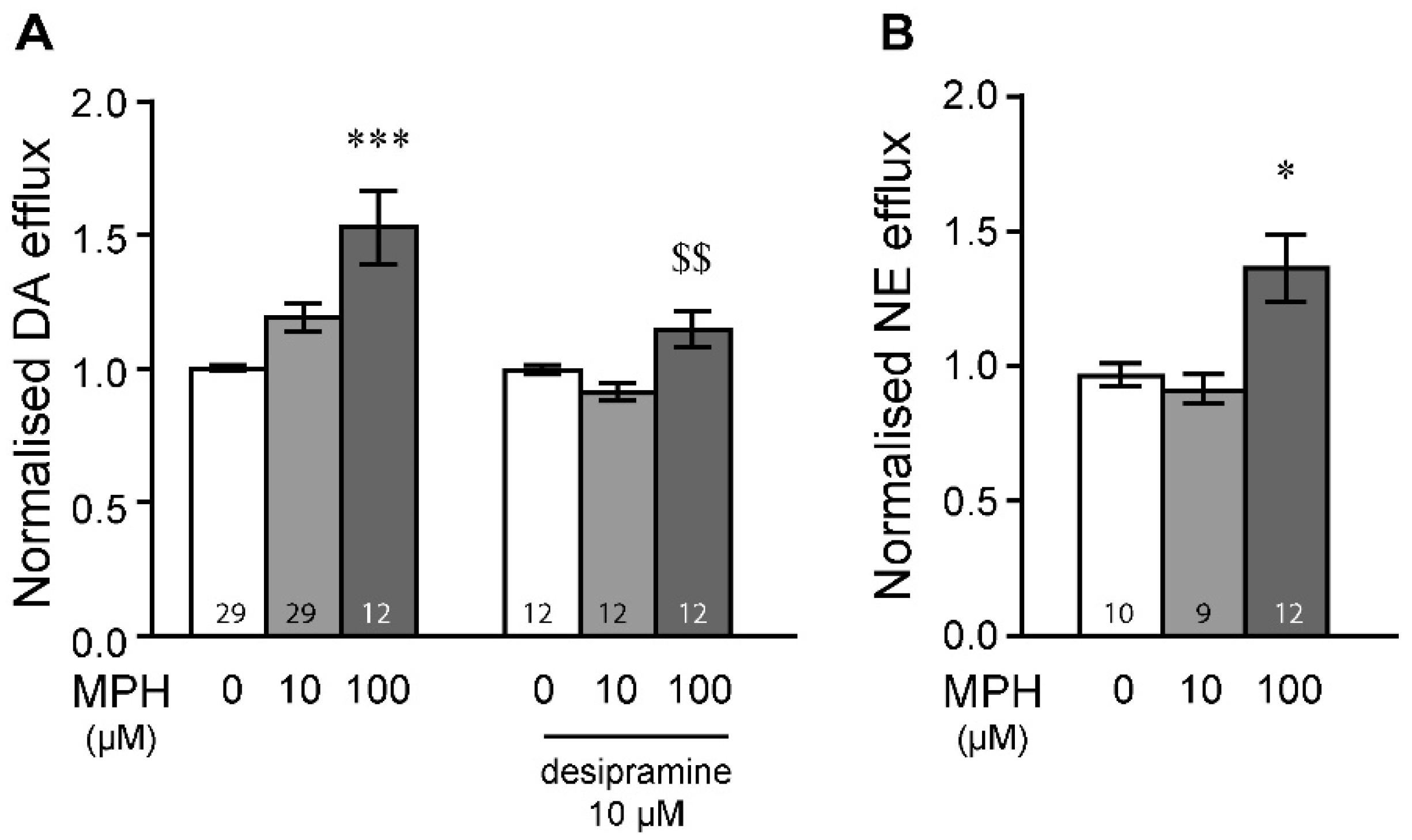
2.1. MPH Induces Dopamine and Norepinephrine Efflux in the Prefrontal Cortex

In the prefrontal cortex (PFC), application of methylphenidate (MPH) at 100 µM (Figure 1A) triggered significant ex vivo dopamine release (Bonferroni post hoc test after significant two-way ANOVA, Supplementary Table S1). This effect was dependent on norepinephrine terminals, since incubation of radiolabelled dopamine (35 nM) in the presence of desipramine significantly dampened dopamine efflux induced by 100 µM of MPH (Figure 1A, Bonferroni post hoc test after significant two-way ANOVA). This was furthermore confirmed by assessing radiolabelled norepinephrine (67–83 nM) efflux following MPH exposure in PFC samples. Indeed, MPH, at 100 µM, induced significant norepinephrine efflux in the PFC (Figure 1B, Tukey’s post hoc test after significant one-way ANOVA). A lower dose of 10 µM of MPH did not induce dopamine efflux under any conditions. These results indicate that MPH can induce both dopamine and norepinephrine efflux in the PFC, an effect that is arising from both dopamine and norepinephrine terminals when applied at 100 µM.

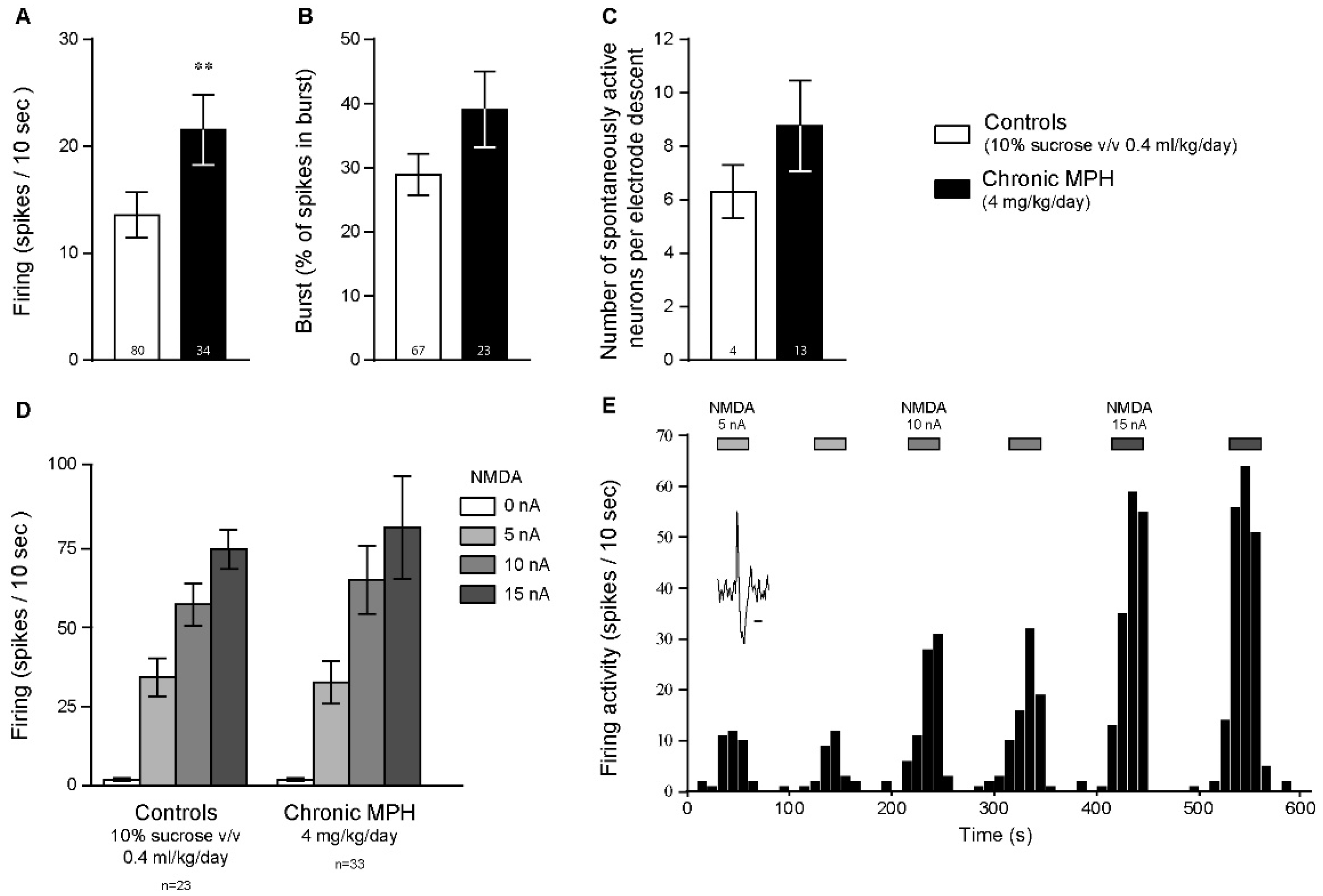
2.2. Chronic MPH Increases the Firing Activity of PFC Pyramidal Neurons without Altering Glutamatergic Neurotransmission

We have demonstrated in a previous study that acute exposure to MPH increases the firing activity of PFC pyramidal neurons and potentiates NMDA-induced neurotransmission [31]. Since acute exposure to MPH can induce dopamine or norepinephrine efflux within the PFC (Figure 1), we examined if chronic MPH exposure could modulate PFC neurotransmission. In animals chronically treated with MPH (voluntary oral intake of 4 mg/kg/day, in two separate doses, dissolved in 10% v/v sucrose solution, followed by a washout period of 28 days, [32]), a significant long-term increase in firing activity of PFC pyramidal neurons was observed (Figure 2A), compared to control animals (10% v/v sucrose vehicle, 4 mL/kg/day in two separate doses; Mann–Whitney test). However, compared to control animals, no difference was found concerning the bursting activities of these neurons (Figure 2B, Mann–Whitney test) nor the total number of spontaneously discharging neurons (Figure 2C, unpaired t-test). Moreover, chronic MPH exposure did not alter glutamatergic neurotransmission in these neurons, since iontophoretic NMDA induced similar firing activity enhancements in control and chronically treated animals (Figure 2D, Supplementary Table S1). A recording example time-course of such an experiment is illustrated in Figure 2E, where local iontophoretic NMDA induce reversible dose-dependent potentiation of firing activities. Thus, these results suggest that chronic MPH can modulate the excitability of cortical pyramidal neurons, without altering the responses of such neurons to locally applied NMDA.

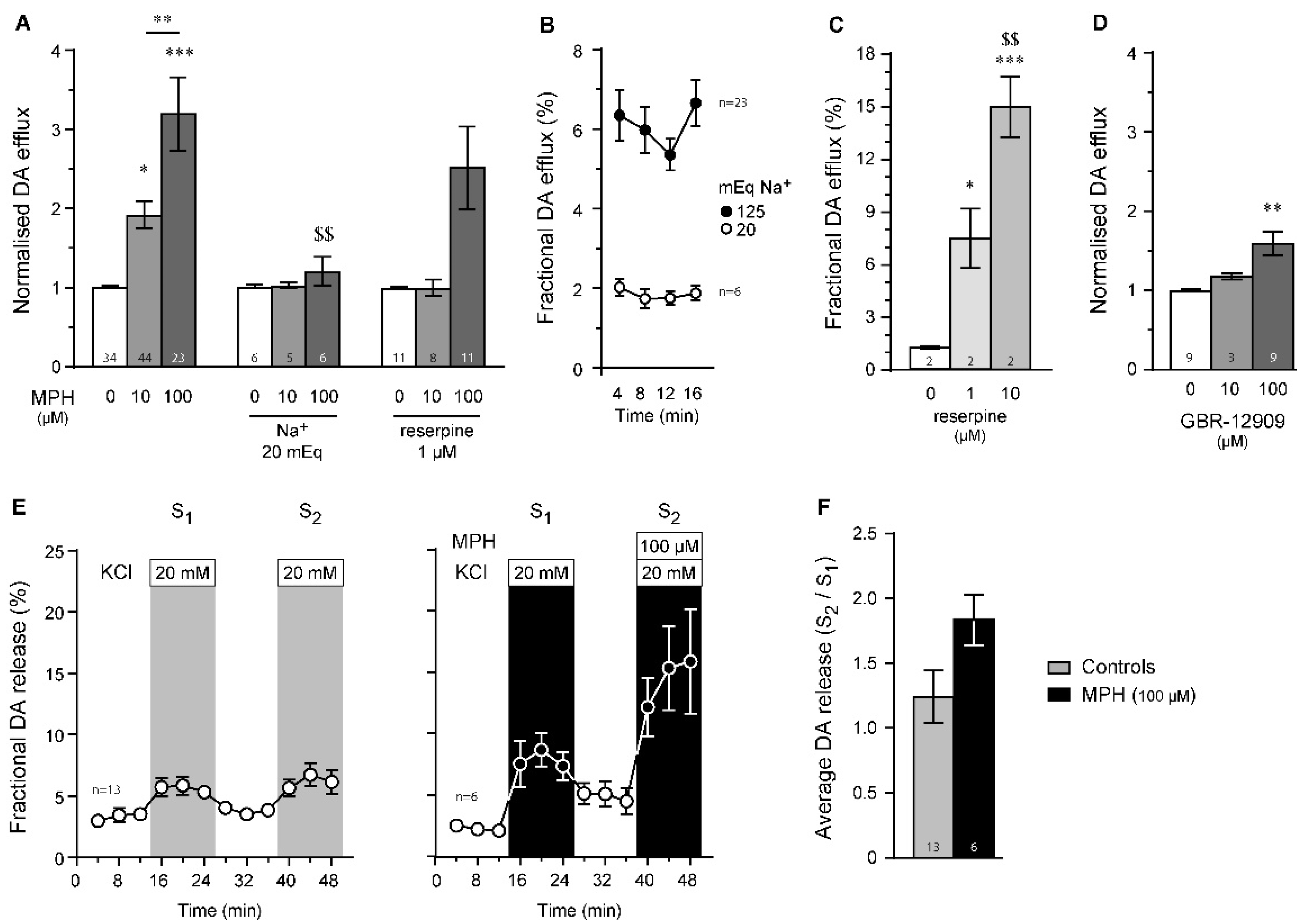
2.3. Dopamine Efflux, but Not Dopamine Release, Is Modulated by MPH in the Striatum

Here, we used different ex vivo experimental conditions to determine the different mechanisms involved (Figure 3A). First, decreasing sodium concentration in the superfusate from 125 mEq to 20 mEq (by isotonic substitution with choline chloride, Figure 3B) significantly reduced MPH-induced dopamine efflux (Figure 3A, Supplementary Table S1), suggesting that electrochemical gradients are necessary for MPH-induced dopamine efflux. When vesicular content was depleted using a 20-min period pre-incubation in the presence of 1 µM of reserpine (Figure 3B, Supplementary Table S1), dopamine efflux induced by 100 µM of MPH was significantly reduced (Figure 3A, Supplementary Table S1), suggesting that MPH requires catecholamine vesicular integrity. The sodium substitution used in this study is known to induce rapid membrane hyperpolarization [33], which should decrease dopamine efflux. Our results are compatible with the sodium dependency of dopamine reuptake. Indeed, under extracellular sodium depletion conditions, dopamine cannot be re-uptaken by the DAT, as sodium gradients are known to be the driving forces of dopamine transport [34]. In fact, a reduction in the sodium driving force induced, in itself, significantly dampened dopamine efflux (Figure 3B). Further, to confirm that 1 µM of reserpine is sufficient to induce vesicular content depletion (Figure 3A), we confirmed on a few samples that application of reserpine at 1 µM can induce successful dopamine depletion, assessed by significant striatal dopamine efflux (Figure 3C), reflecting vesicular emptying. Increasing reserpine concentration to 10 µM dose dependently increased this effect (Figure 3C). In comparison to the above results, the selective dopamine reuptake transporter inhibitor GBR-12909 also induced dopamine efflux from striatal samples (Figure 3D, Supplementary Table S1), but to lower levels than previously observed (Figure 3A).
To further confirm such results, we measured dopamine efflux from striatal samples while superfusing a buffer rich in KCl (20 mM), which is known to induce neuronal membrane depolarization [35], thus producing dopamine release (arising from vesicular fusion). Raising KCl concentrations from 2.5 to 20 mM produced temporary dopamine releases (Figure 3E). When MPH (100 µM) was applied concomitantly to KCl, only non-significant dopamine release was observed (Figure 3E,F, p = 0.07, Mann–Whitney test). Similar results were found following data resampling with 1000 replicates (not shown), validating our previous results. Altogether, these results suggest that MPH, in the striatum, induces dopamine efflux by reversing the dopamine reuptake transporter.

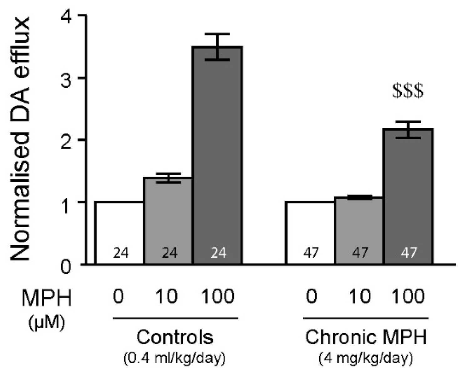
2.4. Tolerance to MPH and Dopamine in Chronically Treated Animals

When MPH was superfused ex vivo in striatal tissue from animals chronically treated with MPH (4 mg/kg/day, please see [36,37,38] for related pharmacokinetics), it induced significantly lower dopamine efflux than in control animals (Figure 4), suggesting tolerance to MPH by the DAT (Bonferroni post hoc test after significant two-way ANOVA, Supplementary Table S1).
To confirm these data, we recorded a small sample of GABAergic medium spiny neurons (MSN) in control and MPH-treated animals. In the striatum, MSN are mostly silent during in vivo extracellular recordings [39], due to their low resting membrane potentials [40]. We observed that iontophoretically applied dopamine could dampen or potentiate NMDA-induced firing activities of MSN, likely reflecting direct and indirect striatal pathways [41,42,43]. Following chronic MPH treatment, the efficacy of dopamine in modulating in vivo NMDA-induced firing activities was significantly altered (not shown). These preliminary data suggest that plastic mechanisms can occur in MSN following chronic MPH treatment.
Altogether, these results suggest that acute MPH strongly modulates neurotransmission and that chronic exposure to MPH could exert long-term effects, as observed before in another region of the basal ganglia [37].
Finally, to compare the results acquired with MPH, we also investigated the potential of ATX, another drug approved for ADHD treatment, to modulate ex vivo dopamine and norepinephrine efflux. In the PFC, 100 µM of ATX produced significant dopamine efflux (Figure 5A). This was significantly reduced when slices were pretreated with 10 µM of desipramine, suggesting that this effect could originate in adrenergic terminals. To confirm this, we measured norepinephrine efflux in the PFC and observed a significant norepinephrine efflux from PFC slices when 100 µM of ATX is applied (Figure 5B), confirming our previous results. In the striatum, 100 µM of ATX induced large dopamine efflux, an effect that was strongly prevented by superfusing a low-Na+ buffer or by reserpine (1 µM) pre-treatment (Figure 5C), suggesting that ATX might modulate dopamine vesicular release in the striatum.

3. Discussion

We have demonstrated that MPH, at 100 µM, could induce 3H-dopamine efflux in the PFC (Figure 1). This effect was partly due to dopamine efflux arising from norepinephrine terminals, as desipramine dampened MPH-induced dopamine release. This was confirmed by observing norepinephrine efflux after superfusion with MPH (Figure 1). These results suggest an involvement of the NET in MPH-dependent dopamine efflux, at least in the PFC. Our data support previous assumptions that in the PFC, MPH elicits dopamine efflux mainly via an inhibition of the NET, suggesting that extracellular dopamine in the PFC originates not only from dopaminergic terminals but also from noradrenergic ones, where dopamine can act both as a precursor for norepinephrine and as a co-transmitter [19,44]. Some studies have suggested that both dopamine and norepinephrine are located within the same dense core vesicles in noradrenergic terminals [45]. Ex vivo, the IC50 for epinephrine uptake inhibition by MPH was 0.85 µM in the PFC and 0.12 µM in the striatum [46]. In vitro, MPH has a similar Ki for the human NET (0.1 µM) and DAT (0.06 µM) [47]. This was also observed for the mouse NET (0.17 µM) and DAT (0.26 µM) [47]. Furthermore, when DAT levels are low, dopamine can be uptaken by the NET, at least ex vivo [18]. Previous microdialysis and neurochemical studies on DAT knockout mice, as well as on naive rats, have shown that selective NET inhibitors increase prefrontal dopamine efflux [8,18]. We remain unable to evaluate the exact contribution of the DAT in the effects of MPH to induce dopamine efflux in the PFC in our experimental conditions. The DAT probably contributes to increasing dopamine efflux as MPH can still exert its effects when the slices were previously loaded in the presence of desipramine. However, following the loading of the slices, redistribution of tritiated dopamine within both dopamine and norepinephrine terminals could have occurred.
The efficacy of MPH in inducing dopamine efflux in the PFC under our conditions is lower than under in vivo conditions using microdialysis techniques. Systemic administration of low doses of MPH (1–2 mg/kg), probably reaching a concentration in the low micromolar range near the catecholamine synapse [48,49], could stimulate dopamine efflux by more than 300% [8], although this could be explained by a detection bias, as reuptake blockade will artificially drive high neurotransmitter release detection. Thus, comparing ex vivo and in vivo results could be challenging. When administered on the intact brain, these drugs are likely to activate other neuronal circuitries, which will further potentiate the release of dopamine in the PFC. Such activation may occur at local levels, as applications of MPH by reverse microdialysis in the PFC or in the nucleus accumbens can still produce consistent large increases in dopamine efflux in vivo [50,51].
In the striatum, however, noradrenergic innervations as well as norepinephrine reuptake transporter levels are low [18,52,53], which is in line with previous published observation, showing that, in the striatum, the NET is responsible for dopamine uptake only when DAT levels reach critically low levels, as observed in Parkinson’s disease [54,55]. In the present study, we have shown that MPH triggers dopamine efflux only through sodium-dependent mechanisms (Figure 3). The fact that pre-incubation with reserpine partially affected (p = 0.08) the ability of MPH to induce striatal dopamine efflux (Figure 3) suggests that inhibition of the DAT by MPH might involve vesicular integrity, at least to some extent. In our experimental design of sodium depletion, no osmolarity shock could have occurred due to the isotonic addition of choline chloride, thus preventing astrocyte swelling [56]. This sodium substitution is known to induce rapid membrane hyperpolarization [33], which should decrease dopamine efflux. Our results are compatible with the sodium dependency of dopamine reuptake [34]. Indeed, under extracellular sodium depletion conditions, dopamine cannot be re-uptaken by the DAT [57], as the sodium gradient is known to be the driving force of dopamine transport.
Our previous study has also demonstrated that MPH increases the firing discharges of PFC pyramidal neurons and can modulate NMDA neurotransmission in the PFC [31]. In the present study, chronic MPH induced long-lasting increases in the firing rates of PFC pyramidal neurons (Figure 2), an effect that was not linked to altered NMDA neurotransmission (Figure 2). In the striatum, chronic MPH exposure induced long-lasting desensitization to an acute 100 µM dose of MPH (Figure 4) or extracellularly applied dopamine (preliminary data not shown). These results are in line with one study witnessing either behavioral sensitization or tolerance (in a 1:1 ratio) to chronic MPH exposure (0.6–10 mg/kg for 5 days), correlated with electrophysiological sensitization or tolerance in MSN of the nucleus accumbens [58] and in PFC pyramidal neurons [59]. Interestingly, a recent study has pointed out that re-exposure to MPH can trigger behavioral sensitization or tolerance in rats, an effect not linked to the electrophysiological properties of dopaminergic neurons in the ventral tegmental area [60]. Further, adolescent or adult animals presented different responses to MPH [60], highlighting the importance of the exposure window. Chronic methylphenidate exposure may reduce striatal plasticity and might not be without long-term consequences, even after washout periods. In fact, chronic MPH exposure significantly decreased dopamine D2 receptor availability in the striatum [61,62], in line with our previous study showing dopamine D2 receptor desensitization in the midbrain following chronic MPH exposure during adolescence [32]. Altogether, these result may suggest long-term consequences of MPH on dopamine neurotransmission. Additional studies have also witnessed long-term consequence of MPH on brain metabolism [63], inflammation [64] and peripheral hemostasis [65]. Our results need to be studied further in order to determine the role of D1- or D2-like receptors after chronic exposure to MPH.
Previous studies have demonstrated that chronic MPH disrupts DA function in the striatum and synaptic plasticity, an effect that was dependent on dopamine D2 receptors [66]. Our previous study also observed similar results [32]. Chronic MPH is known to induce long-term consequences in the prefrontal cortex and striatum, such as sensitization/tolerance [59,67,68,69,70,71,72]. In the prefrontal cortex, these effects appear to be age-dependent [73] and can last for more than 10 weeks [74], which may warrant long-term monitoring in patients withdrawing from MPH. Furthermore, MPH is known to disrupt the expression of DAT and NET following chronic (but not acute) MPH exposure. Indeed, chronic MPH exposure increases DAT and NET levels in both striatal and prefrontal regions [20], as recently reviewed [75]. These results can explain, at least in part, the results observed in the present study. Furthermore, imaging studies in humans [76,77,78,79,80,81] have also observed significant effects of MPH on DAT and NET availability. In patients with ADHD, MPH treatment decreases DAT availability [82,83,84,85], likely indicating altered baseline DAT levels in these patients [24,83], although this is not always observed [81]. Finally, long-term MPH was shown to alter dopamine levels in striatal regions of patients with ADHD [86], highlighting altered dopamine neurotransmission following chronic MPH exposure.
In the current study, serotonergic terminals have not been examined. Since striatal dopamine terminals are known to co-release dopamine and serotonin [87] and that dopamine release in the striatum is dependent on serotonergic terminals [88], assessing striatal serotonergic-mediated release of dopamine following MPH exposure would be of interest. Due to the low concentrations of tritiated dopamine (35 nM) and norepinephrine (67–83 nM) used, recruitment of serotonergic terminals could not be achieved in the present study [89]. Thus, we were unable to determine if serotonergic terminals could be involved in dopamine and/or norepinephrine efflux following MPH exposure.
The present study also observed DA efflux following ATX exposure, an effect that was stronger than previously observed with MPH. Since ATX has a non-negligible affinity for the dopamine vesicle transporter (Ki ~ 3.5 µM) compared to MPH (Ki ~ 39.3 µM) [90], ATX could be uptaken inside the dopaminergic terminals through a relatively low affinity transporter, likely the DAT, where it will then interact with vesicular transporters (VMAT2, vesicular monoamine transporter 2) to promote intracellular dopamine efflux. Such dopamine efflux, observed at high ATX concentrations, may result from reverse dopamine transporter by the DAT as well as passive diffusion directly across phospholipid bilayers, which has been previously observed following amphetamine exposure [91].
To conclude, we have shown that acute exposure to the two ADHD drugs MPH or ATX can modulate dopamine and norepinephrine efflux in cortical and striatal structures. While norepinephrine terminals are likely the preferential targets within the PFC, dopamine terminals within the striatum can trigger dopamine outflows through sodium- and vesicular-dependent mechanisms. Chronic exposure to MPH induced desensitization of medium spiny neurons to locally applied NMDA, dampened striatal dopamine outflows during MPH exposure, increased firing rates of PFC pyramidal neurons in a long-lasting manner and desensitization to both MPH and dopamine in striatal regions. Further studies need to be conducted to evaluate the clinical implications of the current findings.

4. Materials and Methods

4.1. Animals and Drug Treatments

All animal experiments were conducted in strict accordance with the UK Home Office guidelines and the Animal Scientific Procedures Act (1986). A total of 27 male Sprague Dawley rats were housed in groups of 2–4 per cage, maintained at 20–22 °C with humidity rates above 40% under a 12:12 light/dark cycle with lights on at 07:00. Food and water were both provided ad libitum. Animals were allowed a three-day acclimatization period after delivery. All experiments were performed during the light phase and with permission from the UK Home Office (60/4333) and De Montfort University Ethics Committee. No adverse effects were reported.
In the present study, we used either naïve animals or late adolescent animals (150–180 g, PND 42) which were orally (per os) administered twice a day a sucrose solution (10% v/v, 2 mL/kg) with or without MPH (2 mg/kg), for 15 consecutive days, followed by a 28-day washout period, as detailed previously [32]. Daily MPH dose was 4 mg/kg/day/animal; given into two separate per os doses, each of 2 mg/kg. This protocol was chosen to best mimic peak plasma levels observed in patients treated with therapeutic MPH [36,37,49,92,93]. Furthermore, this regimen has also been used in our previous study [32]. Drug administration was therefore voluntary and stress free, which was adapted from a previously published protocol [94]. A washout period of 28 days was allowed before any experiments (neurochemistry or electrophysiology). For details about the pharmacokinetics of MPH, please see [38]. All experiments were performed on adult animals.

4.2. Ex Vivo Radiolabelled Neurotransmitter Efflux

Animals were sacrificed by cervical dislocation. The brain was quickly dissected out and immersed into ice-cold oxygenated Krebs buffer (NaCl 125 mM, MgSO4 1.2 mM, KCl 2.5 mM, CaCl2 2.5 mM, KH2PO4 1.2 mM, NaHCO3 25 mM, glucose 10 mM and pargyline 10 μM, to inhibit monoamine catabolism, pH 7.4). The brain was then placed on an ice-cold platform for further dissection of either the prefrontal cortex or the striatum. The tissue was then sliced into 350 × 350 µm prisms using a McIlwain tissue chopper (Campden Instruments LTD, Loughborough, UK). Constant oxygenation was maintained after this step (95% O2, 5% CO2). Prisms were then left for 20 min to rest at room temperature. Tissue prisms were then loaded for 40 min at 37 °C with either radiolabelled 3H-dopamine (1.0 µCi/mL, specific activity 28.7 Ci/mmol, 35 nM) or 3H-norepinephrine (1.0 µCi/mL, specific activity 12–15 Ci/mmol, 67–83 nM; Perkin-Elmer, Waltham, MA, USA) in the presence or absence of 10 µM of desipramine (to prevent norepinephrine uptake if necessary). Once the loading completed, the prisms were then washed 3 times with fresh Krebs buffer before being divided into perfusion chambers. Throughout the experiment, all samples and superfusion buffers were maintained at 37 °C. An equilibrating period of 40 min was initiated by superfusion of the chambers with Krebs buffer at 0.6 mL/min.
In order to determine baseline outflow of dopamine, 3–4 samples were collected per chamber at 4 min intervals. Sample were collected into vials and each sample would hold 2.4 mL of perfusion liquid, to which scintillation liquid was added up to a total volume of 7 mL per vial. At the end of the experiment, all tissues were collected and dissolved with 1 mL of tissue solubilizer. Total release quantities of tritium (3H) were measured in a liquid scintillation counter (Hidex 300 SL), from which disintegrations per minute were extracted. A minimum of 3 animals were used per experimental condition, except in Figure 3B,C (1 animal in each; internal controls).
If necessary, the composition of the superfused Krebs buffer was altered. Low-Na+ Krebs buffer consisted of 20 mEq of NaCl (instead of 125 mEq, substituted by isotonic concentration of choline chloride). A depolarizing buffer was also tested by increasing KCl concentration from 2.5 to 20 mM. Superfusion of such a K+-rich buffer (substituted by decreasing NaCl from 125 to 107 mM) is known to induce sudden membrane depolarization and neurotransmitter release [35].

4.3. In Vivo Extracellular Single-Unit Electrophysiology

Animals were initially deeply anaesthetized with urethane (1.2–1.7 g/kg, intraperitoneal, with additional doses administered if necessary), secured to a stereotaxic frame and maintained at 36–37 °C with a heating pad. A catheter was inserted into the lateral tail vein to perform systemic drug administration. An incision was made across the top of the head and the edges of the skin drawn back to reveal the cranium. Bregma was identified and a hole was drilled through the bone at the coordinates of the prefrontal cortex (PFC) or the striatum, according to the atlas of Paxinos and Watson [95]. Electrodes were manufactured in house from borosilicate capillaries (1.5 mm, Harvard Apparatus Ltd., Waterbeach, UK), pulled on a PP-830 vertical electrode puller (Narishige, Tokyo, Japan) and filled by hand with an electrolyte solution of NaCl 147 mM. The tip of the electrode was broken down under a microscope to an external diameter of 1–1.5 μm. Typical electrode resistance was in the range of 4–8 MΩ. Single-unit recordings with iontophoresis drug application were made using five-barrel glass micropipettes (World Precision Instruments, Hitchin, UK). The central recording barrel was filled with NaCl 147 mM. The side barrels were filled with: N-methyl-D-aspartate (NMDA) 30 mM and NaCl 2 M for current balancing. Outputs from the electrode were sent to a Neurolog AC pre-amplifier and amplifier (Digitimer Ltd., Welwyn Garden City, UK). If necessary, signal amplification was manually adjusted to record whole neuronal action potential amplitudes. Signals were filtered and sent to an audio amplifier, a Tektronix 2201 digital storage oscilloscope and a 1401 interface connected to a computer running Spike 2 v5.21 (Cambridge Electronic Design Ltd., Cambridge, UK) for data capture and analysis. Descent of the electrode was carried out using a hydraulic micromanipulator (MO-103, Narishige, Tokyo, Japan).
Putative glutamatergic pyramidal neurons were identified according to previous electrophysiological criteria: a broad action potential (1 ms), with a biphasic or triphasic, large waveform, starting with a positive inflection, a relatively slow firing rate, typically between 1 and 50 spikes/10 s and an irregular firing pattern [96,97].
Putative GABAergic medium spiny neurons were identified according to previous electrophysiological criteria such as a very low level or absence of spontaneous activity [39], in combination with a long-lasting action potential waveform, usually above 1 ms [98]. These neurons were detected by microiontophoretic applications of NMDA.
A minimum of 3 animals were used per experimental condition.

4.4. Drugs and Reagents

All drugs were purchased from Sigma (Sigma-Aldrich, Gillingham, UK). For neurotransmitter release assays, drugs were dissolved into normal or modified Krebs buffers, as appropriate. For intravenous administration during in vivo extracellular electrophysiology, all drugs were dissolved into saline (NaCl 0.9% w/v). For micro-iontophoresis experiments, all drugs (except NaCl 2 M) were dissolved into NaCl 147 mM.

4.5. Resampling

Resampling of means was performed with the Visual Inference Tool (VIT), a component of iNZight [99,100], which is encoded in R [101].

4.6. Data Analysis

All data are expressed as the mean ± standard error of the mean (S.E.M.). Statistical analyses were performed using paired/unpaired Student’s t-tests, Mann–Whitney tests (non-normal distribution) or one/two-way analysis of variance (ANOVA), followed by appropriate post hoc Tukey’s (one-way ANOVA) or Bonferroni tests (two-way ANOVA). Normal distributions were assessed with Shapiro–Wilk tests. The significance threshold was set at p < 0.05 and n values refer to the number of samples used. All statistical results are presented in Supplementary Table S1 and were performed with R [101].
Fractional efflux for each superfusate sample was calculated by dividing the amount of tritium in each sample by the total tritium left thereafter. The effect of a tested condition was assessed on at least 3 subsequent sample collections and averaged. Normalized efflux values are calculated for each chamber as the ratio between the mean tested values (generally from at least 3 collections) and average baseline values (usually 3–4 collections). In the present study, we make a distinction between dopamine release and dopamine efflux/outflow. Indeed, dopamine release arises from vesicular exocytosis under in vivo or artificially stimulated conditions (e.g., perfusions of KCl), while dopamine efflux/outflow occurs when samples are not under stimulated conditions.
For resampling, 1000 replicates (r = 1000) of the initial data were generated. Output means (n = 1000 means) were analyzed using the 95% confidence interval (CI) spread.

Supplementary Materials

The following supporting information can be downloaded at: https://0-www-mdpi-com.brum.beds.ac.uk/article/10.3390/ijms23158588/s1.

Author Contributions

Conceptualization, M.D.M. and B.G.; methodology, M.D.M. and B.G.; formal analysis, M.D.M.; investigation, A.D. and M.D.M.; data curation, M.D.M.; writing—original draft preparation, M.D.M.; writing—review and editing, M.D.M. and B.G.; visualization, M.D.M.; supervision, B.G.; funding acquisition, B.G. All authors have read and agreed to the published version of the manuscript.

Funding

M.D.M. was supported by an internal grant from De Montfort University.

Institutional Review Board Statement

This study was approved by the UK Home Office (60/4333) and De Montfort University Ethics Committee on 5 February 2012.

Informed Consent Statement

Not applicable.

Data Availability Statement

The data presented in this study are available on request from the corresponding author.

Acknowledgments

The authors are grateful for generous financial support from De Montfort University (stipend to M.D.M.). The authors thank Anita O’Donoghue and Stephen Bowen for excellent technical support in taking care of the animals, together with David Reeder, Amrat Khorana, Claire West, Jo Tonkin, Leonie Hough and Mike Storer for their assistance with consumables and laboratory equipment. The authors also thank Elisabeth (Liz) O’Brien, Nazmin Juma and Ketan Ruparelia for support in the handling and disposal of radioactive compounds. The authors also thank Adèle Bousquet for acquiring preliminary results.

Conflicts of Interest

The authors declare no conflict of interest.

References

  1. Swanson, J.; Baler, R.D.; Volkow, N.D. Understanding the Effects of Stimulant Medications on Cognition in Individuals with Attention-Deficit Hyperactivity Disorder: A Decade of Progress. Neuropsychopharmacology 2011, 36, 207–226. [Google Scholar] [CrossRef] [PubMed]
  2. De Sousa, A.; Kalra, G. Drug Therapy of Attention Deficit Hyperactivity Disorder: Current Trends. Mens Sana Monogr. 2012, 10, 45–69. [Google Scholar] [CrossRef] [PubMed] [Green Version]
  3. Vitiello, B. Understanding the Risk of Using Medications for Attention Deficit Hyperactivity Disorder with Respect to Physical Growth and Cardiovascular Function. Child Adolesc. Psychiatr. Clin. N. Am. 2008, 17, 459–474. [Google Scholar] [CrossRef] [PubMed] [Green Version]
  4. Heal, D.J.; Cheetham, S.C.; Smith, S.L. The Neuropharmacology of ADHD Drugs in Vivo: Insights on Efficacy and Safety. Neuropharmacology 2009, 57, 608–618. [Google Scholar] [CrossRef]
  5. Spiller, H.A.; Hays, H.L.; Aleguas, A. Overdose of Drugs for Attention-Deficit Hyperactivity Disorder: Clinical Presentation, Mechanisms of Toxicity, and Management. CNS Drugs 2013, 27, 531–543. [Google Scholar] [CrossRef] [PubMed]
  6. Kolar, D.; Keller, A.; Golfinopoulos, M.; Cumyn, L.; Syer, C.; Hechtman, L. Treatment of Adults with Attention-Deficit/Hyperactivity Disorder. Neuropsychiatr. Dis. Treat. 2008, 4, 389–403. [Google Scholar] [CrossRef] [PubMed] [Green Version]
  7. Bushe, C.J.; Savill, N.C. Systematic Review of Atomoxetine Data in Childhood and Adolescent Attention-Deficit Hyperactivity Disorder 2009-2011: Focus on Clinical Efficacy and Safety. J. Psychopharmacol. 2014, 28, 204–211. [Google Scholar] [CrossRef] [PubMed]
  8. Bymaster, F.P.; Katner, J.S.; Nelson, D.L.; Hemrick-Luecke, S.K.; Threlkeld, P.G.; Heiligenstein, J.H.; Morin, S.M.; Gehlert, D.R.; Perry, K.W. Atomoxetine Increases Extracellular Levels of Norepinephrine and Dopamine in Prefrontal Cortex of Rat: A Potential Mechanism for Efficacy in Attention Deficit/Hyperactivity Disorder. Neuropsychopharmacology 2002, 27, 699–711. [Google Scholar] [CrossRef]
  9. Reith, M.E.A.; Wang, L.C.; Dutta, A.K. Pharmacological Profile of Radioligand Binding to the Norepinephrine Transporter: Instances of Poor Indication of Functional Activity. J. Neurosci. Methods 2005, 143, 87–94. [Google Scholar] [CrossRef] [PubMed]
  10. Erickson, J.D.; Schafer, M.K.; Bonner, T.I.; Eiden, L.E.; Weihe, E. Distinct Pharmacological Properties and Distribution in Neurons and Endocrine Cells of Two Isoforms of the Human Vesicular Monoamine Transporter. Proc. Natl. Acad. Sci. USA 1996, 93, 5166–5171. [Google Scholar] [CrossRef] [Green Version]
  11. Kalivas, P.W. Cocaine and Amphetamine-like Psychostimulants: Neurocircuitry and Glutamate Neuroplasticity. Dialogues Clin. Neurosci. 2007, 9, 389–397. [Google Scholar] [CrossRef] [PubMed]
  12. Dela Peña, I.; Gevorkiana, R.; Shi, W.-X. Psychostimulants Affect Dopamine Transmission through Both Dopamine Transporter-Dependent and Independent Mechanisms. Eur. J. Pharmacol. 2015, 764, 562–570. [Google Scholar] [CrossRef] [Green Version]
  13. Gamo, N.J.; Wang, M.; Arnsten, A.F.T. Methylphenidate and Atomoxetine Enhance Prefrontal Function through A2-Adrenergic and Dopamine D1 Receptors. J. Am. Acad. Child Adolesc. Psychiatry 2010, 49, 1011–1023. [Google Scholar] [CrossRef] [PubMed] [Green Version]
  14. Kieling, C.; Goncalves, R.R.F.; Tannock, R.; Castellanos, F.X. Neurobiology of Attention Deficit Hyperactivity Disorder. Child Adolesc. Psychiatr. Clin. N. Am. 2008, 17, 285–307. [Google Scholar] [CrossRef] [PubMed]
  15. Koda, K.; Ago, Y.; Cong, Y.; Kita, Y.; Takuma, K.; Matsuda, T. Effects of Acute and Chronic Administration of Atomoxetine and Methylphenidate on Extracellular Levels of Noradrenaline, Dopamine and Serotonin in the Prefrontal Cortex and Striatum of Mice. J. Neurochem. 2010, 114, 259–270. [Google Scholar] [CrossRef] [PubMed]
  16. Floresco, S.B. Prefrontal Dopamine and Behavioral Flexibility: Shifting from an “Inverted-U” toward a Family of Functions. Front. Neurosci. 2013, 7, 62. [Google Scholar] [CrossRef] [PubMed] [Green Version]
  17. Devoto, P.; Flore, G. On the Origin of Cortical Dopamine: Is It a Co-Transmitter in Noradrenergic Neurons? Curr. Neuropharmacol. 2006, 4, 115–125. [Google Scholar] [CrossRef] [Green Version]
  18. Morón, J.A.; Brockington, A.; Wise, R.A.; Rocha, B.A.; Hope, B.T. Dopamine Uptake through the Norepinephrine Transporter in Brain Regions with Low Levels of the Dopamine Transporter: Evidence from Knock-out Mouse Lines. J. Neurosci. 2002, 22, 389–395. [Google Scholar] [CrossRef] [Green Version]
  19. Devoto, P.; Flore, G.; Pani, L.; Gessa, G.L. Evidence for Co-Release of Noradrenaline and Dopamine from Noradrenergic Neurons in the Cerebral Cortex. Mol. Psychiatry 2001, 6, 657–664. [Google Scholar] [CrossRef] [Green Version]
  20. Quansah, E.; Zetterström, T.S.C. Chronic Methylphenidate Preferentially Alters Catecholamine Protein Targets in the Parietal Cortex and Ventral Striatum. Neurochem. Int. 2019, 124, 193–199. [Google Scholar] [CrossRef] [PubMed]
  21. Quansah, E.; Sgamma, T.; Jaddoa, E.; Zetterström, T.S.C. Chronic Methylphenidate Regulates Genes and Proteins Mediating Neuroplasticity in the Juvenile Rat Brain. Neurosci. Lett. 2017, 654, 93–98. [Google Scholar] [CrossRef]
  22. Matsuda, W.; Furuta, T.; Nakamura, K.C.; Hioki, H.; Fujiyama, F.; Arai, R.; Kaneko, T. Single Nigrostriatal Dopaminergic Neurons Form Widely Spread and Highly Dense Axonal Arborizations in the Neostriatum. J. Neurosci. 2009, 29, 444–453. [Google Scholar] [CrossRef] [PubMed] [Green Version]
  23. Lou, H.C.; Henriksen, L.; Bruhn, P.; Børner, H.; Nielsen, J.B. Striatal Dysfunction in Attention Deficit and Hyperkinetic Disorder. Arch. Neurol. 1989, 46, 48–52. [Google Scholar] [CrossRef] [PubMed]
  24. Fusar-Poli, P.; Rubia, K.; Rossi, G.; Sartori, G.; Balottin, U. Striatal Dopamine Transporter Alterations in ADHD: Pathophysiology or Adaptation to Psychostimulants? A Meta-Analysis. Am. J. Psychiatry 2012, 169, 264–272. [Google Scholar] [CrossRef]
  25. Krause, K.H.; Dresel, S.H.; Krause, J.; Kung, H.F.; Tatsch, K. Increased Striatal Dopamine Transporter in Adult Patients with Attention Deficit Hyperactivity Disorder: Effects of Methylphenidate as Measured by Single Photon Emission Computed Tomography. Neurosci. Lett. 2000, 285, 107–110. [Google Scholar] [CrossRef]
  26. Krause, J.; la Fougere, C.; Krause, K.-H.; Ackenheil, M.; Dresel, S.H. Influence of Striatal Dopamine Transporter Availability on the Response to Methylphenidate in Adult Patients with ADHD. Eur. Arch. Psychiatry Clin. Neurosci. 2005, 255, 428–431. [Google Scholar] [CrossRef]
  27. Mckenzie, A.; Meshkat, S.; Lui, L.M.W.; Ho, R.; Di Vincenzo, J.D.; Ceban, F.; Cao, B.; McIntyre, R.S. The Effects of Psychostimulants on Cognitive Functions in Individuals with Attention-Deficit Hyperactivity Disorder: A Systematic Review. J. Psychiatr. Res. 2022, 149, 252–259. [Google Scholar] [CrossRef]
  28. Hong, S.-B.; Harrison, B.J.; Fornito, A.; Sohn, C.-H.; Song, I.-C.; Kim, J.-W. Functional Dysconnectivity of Corticostriatal Circuitry and Differential Response to Methylphenidate in Youth with Attention-Deficit/Hyperactivity Disorder. J. Psychiatry Neurosci. JPN 2015, 40, 46–57. [Google Scholar] [CrossRef] [PubMed] [Green Version]
  29. Braz, B.Y.; Galiñanes, G.L.; Taravini, I.R.E.; Belforte, J.E.; Murer, M.G. Altered Corticostriatal Connectivity and Exploration/Exploitation Imbalance Emerge as Intermediate Phenotypes for a Neonatal Dopamine Dysfunction. Neuropsychopharmacology 2015, 40, 2576–2587. [Google Scholar] [CrossRef] [PubMed] [Green Version]
  30. Alberquilla, S.; Gonzalez-Granillo, A.; Martín, E.D.; Moratalla, R. Dopamine Regulates Spine Density in Striatal Projection Neurons in a Concentration-Dependent Manner. Neurobiol. Dis. 2020, 134, 104666. [Google Scholar] [CrossRef] [PubMed]
  31. Di Miceli, M.; Gronier, B. Psychostimulants and Atomoxetine Alter the Electrophysiological Activity of Prefrontal Cortex Neurons, Interaction with Catecholamine and Glutamate NMDA Receptors. Psychopharmacology 2015, 232, 2191–2205. [Google Scholar] [CrossRef] [PubMed]
  32. Di Miceli, M.; Omoloye, A.; Gronier, B. Chronic Methylphenidate Treatment during Adolescence Has Long-Term Effects on Monoaminergic Function. J. Psychopharmacol. 2019, 33, 109–121. [Google Scholar] [CrossRef] [PubMed] [Green Version]
  33. Cvetkovic-Lopes, V.; Eggermann, E.; Uschakov, A.; Grivel, J.; Bayer, L.; Jones, B.E.; Serafin, M.; Mühlethaler, M. Rat Hypocretin/Orexin Neurons Are Maintained in a Depolarized State by TRPC Channels. PLoS ONE 2010, 5, e15673. [Google Scholar] [CrossRef]
  34. Wheeler, D.D.; Edwards, A.M.; Chapman, B.M.; Ondo, J.G. A Model of the Sodium Dependence of Dopamine Uptake in Rat Striatal Synaptosomes. Neurochem. Res. 1993, 18, 927–936. [Google Scholar] [CrossRef] [PubMed]
  35. Khvotchev, M.; Lonart, G.; Südhof, T.C. Role of Calcium in Neurotransmitter Release Evoked by Alpha-Latrotoxin or Hypertonic Sucrose. Neuroscience 2000, 101, 793–802. [Google Scholar] [CrossRef]
  36. Thanos, P.K.; Robison, L.S.; Steier, J.; Hwang, Y.F.; Cooper, T.; Swanson, J.M.; Komatsu, D.E.; Hadjiargyrou, M.; Volkow, N.D. A Pharmacokinetic Model of Oral Methylphenidate in the Rat and Effects on Behavior. Pharmacol. Biochem. Behav. 2015, 131, 143–153. [Google Scholar] [CrossRef] [Green Version]
  37. Berridge, C.W.; Devilbiss, D.M.; Andrzejewski, M.E.; Arnsten, A.F.T.; Kelley, A.E.; Schmeichel, B.; Hamilton, C.; Spencer, R.C. Methylphenidate Preferentially Increases Catecholamine Neurotransmission within the Prefrontal Cortex at Low Doses That Enhance Cognitive Function. Biol. Psychiatry 2006, 60, 1111–1120. [Google Scholar] [CrossRef]
  38. Wargin, W.; Patrick, K.; Kilts, C.; Gualtieri, C.T.; Ellington, K.; Mueller, R.A.; Kraemer, G.; Breese, G.R. Pharmacokinetics of Methylphenidate in Man, Rat and Monkey. J. Pharmacol. Exp. Ther. 1983, 226, 382–386. [Google Scholar]
  39. el Mansari, M.; Blier, P. In Vivo Electrophysiological Characterization of 5-HT Receptors in the Guinea Pig Head of Caudate Nucleus and Orbitofrontal Cortex. Neuropharmacology 1997, 36, 577–588. [Google Scholar] [CrossRef]
  40. Chuhma, N.; Tanaka, K.F.; Hen, R.; Rayport, S. Functional Connectome of the Striatal Medium Spiny Neuron. J. Neurosci. 2011, 31, 1183–1192. [Google Scholar] [CrossRef] [Green Version]
  41. Gertler, T.S.; Chan, C.S.; Surmeier, D.J. Dichotomous Anatomical Properties of Adult Striatal Medium Spiny Neurons. J. Neurosci. 2008, 28, 10814–10824. [Google Scholar] [CrossRef]
  42. Frederick, A.L.; Yano, H.; Trifilieff, P.; Vishwasrao, H.D.; Biezonski, D.; Mészáros, J.; Urizar, E.; Sibley, D.R.; Kellendonk, C.; Sonntag, K.C.; et al. Evidence against Dopamine D1/D2 Receptor Heteromers. Mol. Psychiatry 2015, 20, 1373–1385. [Google Scholar] [CrossRef] [Green Version]
  43. Soares-Cunha, C.; Coimbra, B.; Sousa, N.; Rodrigues, A.J. Reappraising Striatal D1- and D2-Neurons in Reward and Aversion. Neurosci. Biobehav. Rev. 2016, 68, 370–386. [Google Scholar] [CrossRef] [PubMed]
  44. Devoto, P.; Flore, G.; Pira, L.; Longu, G.; Gessa, G.L. Alpha2-Adrenoceptor Mediated Co-Release of Dopamine and Noradrenaline from Noradrenergic Neurons in the Cerebral Cortex. J. Neurochem. 2004, 88, 1003–1009. [Google Scholar] [CrossRef] [PubMed] [Green Version]
  45. De Potter, W.P.; Partoens, P.; Schoups, A.; Llona, I.; Coen, E.P. Noradrenergic Neurons Release Both Noradrenaline and Neuropeptide Y from a Single Pool: The Large Dense Cored Vesicles. Synapse 1997, 25, 44–55. [Google Scholar] [CrossRef]
  46. Hendley, E.D.; Snyder, S.H.; Fauley, J.J.; LaPidus, J.B. Stereoselectivity of Catecholamine Uptake by Brain Synaptosomes: Studies with Ephedrine, Methylphenidate and Phenyl-2-Piperidyl Carbinol. J. Pharmacol. Exp. Ther. 1972, 183, 103–116. [Google Scholar] [PubMed]
  47. Han, D.D.; Gu, H.H. Comparison of the Monoamine Transporters from Human and Mouse in Their Sensitivities to Psychostimulant Drugs. BMC Pharmacol. 2006, 6, 6. [Google Scholar] [CrossRef] [PubMed] [Green Version]
  48. Gerasimov, M.R.; Franceschi, M.; Volkow, N.D.; Rice, O.; Schiffer, W.K.; Dewey, S.L. Synergistic Interactions between Nicotine and Cocaine or Methylphenidate Depend on the Dose of Dopamine Transporter Inhibitor. Synapse 2000, 38, 432–437. [Google Scholar] [CrossRef]
  49. Balcioglu, A.; Ren, J.-Q.; McCarthy, D.; Spencer, T.J.; Biederman, J.; Bhide, P.G. Plasma and Brain Concentrations of Oral Therapeutic Doses of Methylphenidate and Their Impact on Brain Monoamine Content in Mice. Neuropharmacology 2009, 57, 687–693. [Google Scholar] [CrossRef] [Green Version]
  50. Nomikos, G.G.; Damsma, G.; Wenkstern, D.; Fibiger, H.C. In Vivo Characterization of Locally Applied Dopamine Uptake Inhibitors by Striatal Microdialysis. Synapse 1990, 6, 106–112. [Google Scholar] [CrossRef]
  51. Schmeichel, B.E.; Berridge, C.W. Neurocircuitry Underlying the Preferential Sensitivity of Prefrontal Catecholamines to Low-Dose Psychostimulants. Neuropsychopharmacology 2013, 38, 1078–1084. [Google Scholar] [CrossRef] [PubMed] [Green Version]
  52. Swanson, L.W.; Hartman, B.K. The Central Adrenergic System. An Immunofluorescence Study of the Location of Cell Bodies and Their Efferent Connections in the Rat Utilizing Dopamine-Beta-Hydroxylase as a Marker. J. Comp. Neurol. 1975, 163, 467–505. [Google Scholar] [CrossRef] [PubMed]
  53. Berridge, C.W.; Stratford, T.L.; Foote, S.L.; Kelley, A.E. Distribution of Dopamine Beta-Hydroxylase-like Immunoreactive Fibers within the Shell Subregion of the Nucleus Accumbens. Synapse 1997, 27, 230–241. [Google Scholar] [CrossRef]
  54. Arai, A.; Tomiyama, M.; Kannari, K.; Kimura, T.; Suzuki, C.; Watanabe, M.; Kawarabayashi, T.; Shen, H.; Shoji, M. Reuptake of L-DOPA-Derived Extracellular DA in the Striatum of a Rodent Model of Parkinson’s Disease via Norepinephrine Transporter. Synapse 2008, 62, 632–635. [Google Scholar] [CrossRef]
  55. Chotibut, T.; Apple, D.M.; Jefferis, R.; Salvatore, M.F. Dopamine Transporter Loss in 6-OHDA Parkinson’s Model Is Unmet by Parallel Reduction in Dopamine Uptake. PLoS ONE 2012, 7, e52322. [Google Scholar] [CrossRef] [Green Version]
  56. Lauderdale, K.; Murphy, T.; Tung, T.; Davila, D.; Binder, D.K.; Fiacco, T.A. Osmotic Edema Rapidly Increases Neuronal Excitability through Activation of NMDA Receptor-Dependent Slow Inward Currents in Juvenile and Adult Hippocampus. ASN Neuro 2015, 7, 1759091415605115. [Google Scholar] [CrossRef]
  57. Roitman, M.F.; Patterson, T.A.; Sakai, R.R.; Bernstein, I.L.; Figlewicz, D.P. Sodium Depletion and Aldosterone Decrease Dopamine Transporter Activity in Nucleus Accumbens but Not Striatum. Am. J. Physiol. 1999, 276, R1339–R1345. [Google Scholar] [CrossRef]
  58. Claussen, C.M.; Chong, S.L.; Dafny, N. Nucleus Accumbens Neuronal Activity Correlates to the Animal’s Behavioral Response to Acute and Chronic Methylphenidate. Physiol. Behav. 2014, 129, 85–94. [Google Scholar] [CrossRef] [Green Version]
  59. Salek, R.L.; Claussen, C.M.; Pérez, A.; Dafny, N. Acute and Chronic Methylphenidate Alters Prefrontal Cortex Neuronal Activity Recorded from Freely Behaving Rats. Eur. J. Pharmacol. 2012, 679, 60–67. [Google Scholar] [CrossRef] [Green Version]
  60. Medina, A.C.; Reyes-Vasquez, C.; Kharas, N.; Dafny, N. Adolescent Rats Respond Differently to Methylphenidate as Compared to Adult Rats-Concomitant VTA Neuronal and Behavioral Recordings. Brain Res. Bull. 2022, 183, 1–12. [Google Scholar] [CrossRef] [PubMed]
  61. Thanos, P.K.; Michaelides, M.; Benveniste, H.; Wang, G.J.; Volkow, N.D. Effects of Chronic Oral Methylphenidate on Cocaine Self-Administration and Striatal Dopamine D2 Receptors in Rodents. Pharmacol. Biochem. Behav. 2007, 87, 426–433. [Google Scholar] [CrossRef] [PubMed]
  62. Caprioli, D.; Jupp, B.; Hong, Y.T.; Sawiak, S.J.; Ferrari, V.; Wharton, L.; Williamson, D.J.; McNabb, C.; Berry, D.; Aigbirhio, F.I.; et al. Dissociable Rate-Dependent Effects of Oral Methylphenidate on Impulsivity and D2/3 Receptor Availability in the Striatum. J. Neurosci. 2015, 35, 3747–3755. [Google Scholar] [CrossRef] [PubMed] [Green Version]
  63. Richer, K.; Hamilton, J.; Delis, F.; Martin, C.; Fricke, D.; Yao, R.; Sajjad, M.; Blum, K.; Hadjiargyrou, M.; Komatsu, D.; et al. Chronic Treatment and Abstinence from Methylphenidate Exposure Dose-Dependently Changes Glucose Metabolism in the Rat Brain. Brain Res. 2022, 1780, 147799. [Google Scholar] [CrossRef] [PubMed]
  64. Foschiera, L.N.; Schmitz, F.; Wyse, A.T.S. Evidence of Methylphenidate Effect on Mitochondria, Redox Homeostasis, and Inflammatory Aspects: Insights from Animal Studies. Prog. Neuropsychopharmacol. Biol. Psychiatry 2022, 116, 110518. [Google Scholar] [CrossRef] [PubMed]
  65. Alam, N.; Ikram, R.; Naeem, S.; Khan, S.S.; Siddiqui, T.; Khatoon, H.; Kashif, S.S. Effect of Methylphenidate and Buspirone-Methylphenidate Co-Administration on Biochemical and Hematological Parameters in Rats: Implications for Safe and Confrontational Use. Pak. J. Pharm. Sci. 2021, 34, 2131–2139. [Google Scholar]
  66. Crowley, N.A.; Cody, P.A.; Davis, M.I.; Lovinger, D.M.; Mateo, Y. Chronic Methylphenidate Exposure during Adolescence Reduces Striatal Synaptic Responses to Ethanol. Eur. J. Neurosci. 2014, 39, 548–556. [Google Scholar] [CrossRef] [Green Version]
  67. Yang, P.B.; Swann, A.C.; Dafny, N. Chronic Administration of Methylphenidate Produces Neurophysiological and Behavioral Sensitization. Brain Res. 2007, 1145, 66–80. [Google Scholar] [CrossRef] [Green Version]
  68. Jones, Z.; Dafny, N. Acute and Chronic Dose Response Effect of Methylphenidate on Ventral Tegmental Area Neurons Correlated with Animal Behavior. J. Neural Transm. 2014, 121, 327–345. [Google Scholar] [CrossRef] [Green Version]
  69. Venkataraman, S.S.; Joseph, M.; Dafny, N. Concomitant Behavioral and Prefrontal Cortex Neuronal Responses Following Acute and Chronic Methylphenidate Exposure in Adolescent and Adult Rats. Brain Res. Bull. 2019, 144, 200–212. [Google Scholar] [CrossRef]
  70. King, N.; Floren, S.; Kharas, N.; Thomas, M.; Dafny, N. Glutaminergic Signaling in the Caudate Nucleus Is Required for Behavioral Sensitization to Methylphenidate. Pharmacol. Biochem. Behav. 2019, 184, 172737. [Google Scholar] [CrossRef]
  71. Venkataraman, S.S.; Claussen, C.M.; Kharas, N.; Dafny, N. The Prefrontal Cortex and the Caudate Nucleus Respond Conjointly to Methylphenidate (Ritalin). Concomitant Behavioral and Neuronal Recording Study. Brain Res. Bull. 2020, 157, 77–89. [Google Scholar] [CrossRef] [PubMed]
  72. Broussard, E.; Reyes-Vazquez, C.; Dafny, N. Methylphenidate Dose-Response Behavioral and Neurophysiological Study of the Ventral Tegmental Area and Nucleus Accumbens in Adolescent Rats. Eur. J. Neurosci. 2019, 50, 2635–2652. [Google Scholar] [CrossRef]
  73. Dos Santos Pereira, M.; Sathler, M.F.; Valli, T.; da, R.; Marques, R.S.; Ventura, A.L.M.; Peccinalli, N.R.; Fraga, M.C.; Manhães, A.C.; Kubrusly, R. Long Withdrawal of Methylphenidate Induces a Differential Response of the Dopaminergic System and Increases Sensitivity to Cocaine in the Prefrontal Cortex of Spontaneously Hypertensive Rats. PLoS ONE 2015, 10, e0141249. [Google Scholar] [CrossRef] [PubMed] [Green Version]
  74. Urban, K.R.; Waterhouse, B.D.; Gao, W.-J. Distinct Age-Dependent Effects of Methylphenidate on Developing and Adult Prefrontal Neurons. Biol. Psychiatry 2012, 72, 880–888. [Google Scholar] [CrossRef] [PubMed] [Green Version]
  75. Zetterström, T.S.C.; Quansah, E.; Grootveld, M. Effects of Methylphenidate on the Dopamine Transporter and Beyond. In Current Topics in Behavioral Neurosciences; Springer: Berlin/Heidelberg, Germany, 2022. [Google Scholar] [CrossRef]
  76. Dipasquale, O.; Martins, D.; Sethi, A.; Veronese, M.; Hesse, S.; Rullmann, M.; Sabri, O.; Turkheimer, F.; Harrison, N.A.; Mehta, M.A.; et al. Unravelling the Effects of Methylphenidate on the Dopaminergic and Noradrenergic Functional Circuits. Neuropsychopharmacology 2020, 45, 1482–1489. [Google Scholar] [CrossRef]
  77. Krause, K.H.; Dresel, S.H.; Krause, J.; la Fougere, C.; Ackenheil, M. The Dopamine Transporter and Neuroimaging in Attention Deficit Hyperactivity Disorder. Neurosci. Biobehav. Rev. 2003, 27, 605–613. [Google Scholar] [CrossRef]
  78. Spencer, T.J.; Biederman, J.; Madras, B.K.; Faraone, S.V.; Dougherty, D.D.; Bonab, A.A.; Fischman, A.J. In Vivo Neuroreceptor Imaging in Attention-Deficit/Hyperactivity Disorder: A Focus on the Dopamine Transporter. Biol. Psychiatry 2005, 57, 1293–1300. [Google Scholar] [CrossRef]
  79. Volkow, N.D.; Fowler, J.S.; Wang, G.; Ding, Y.; Gatley, S.J. Mechanism of Action of Methylphenidate: Insights from PET Imaging Studies. J. Atten. Disord. 2002, 6 (Suppl. S1), S31–S43. [Google Scholar] [CrossRef]
  80. Hannestad, J.; Gallezot, J.-D.; Planeta-Wilson, B.; Lin, S.-F.; Williams, W.A.; van Dyck, C.H.; Malison, R.T.; Carson, R.E.; Ding, Y.-S. Clinically Relevant Doses of Methylphenidate Significantly Occupy Norepinephrine Transporters in Humans In Vivo. Biol. Psychiatry 2010, 68, 854–860. [Google Scholar] [CrossRef] [Green Version]
  81. Wang, G.-J.; Volkow, N.D.; Wigal, T.; Kollins, S.H.; Newcorn, J.H.; Telang, F.; Logan, J.; Jayne, M.; Wong, C.T.; Han, H.; et al. Long-Term Stimulant Treatment Affects Brain Dopamine Transporter Level in Patients with Attention Deficit Hyperactive Disorder. PLoS ONE 2013, 8, e63023. [Google Scholar] [CrossRef] [Green Version]
  82. Aster, H.-C.; Romanos, M.; Walitza, S.; Gerlach, M.; Mühlberger, A.; Rizzo, A.; Andreatta, M.; Hasenauer, N.; Hartrampf, P.E.; Nerlich, K.; et al. Responsivity of the Striatal Dopamine System to Methylphenidate—A within-Subject I-123-β-CIT-SPECT Study in Male Children and Adolescents with Attention-Deficit/Hyperactivity Disorder. Front. Psychiatry 2022, 13, 804730. [Google Scholar] [CrossRef] [PubMed]
  83. Dresel, S.; Krause, J.; Krause, K.H.; LaFougere, C.; Brinkbäumer, K.; Kung, H.F.; Hahn, K.; Tatsch, K. Attention Deficit Hyperactivity Disorder: Binding of [99mTc]TRODAT-1 to the Dopamine Transporter before and after Methylphenidate Treatment. Eur. J. Nucl. Med. 2000, 27, 1518–1524. [Google Scholar] [CrossRef] [PubMed]
  84. Szobot, C.M.; Shih, M.C.; Schaefer, T.; Júnior, N.; Hoexter, M.Q.; Fu, Y.K.; Pechansky, F.; Bressan, R.A.; Rohde, L.A.P. Methylphenidate DAT Binding in Adolescents with Attention-Deficit/ Hyperactivity Disorder Comorbid with Substance Use Disorder—A Single Photon Emission Computed Tomography with [Tc(99m)]TRODAT-1 Study. NeuroImage 2008, 40, 1195–1201. [Google Scholar] [CrossRef] [PubMed]
  85. Akay, A.P.; Kaya, G.Ç.; Kose, S.; Yazıcıoğlu, Ç.E.; Erkuran, H.Ö.; Güney, S.A.; Oğuz, K.; Keskin, D.; Baykara, B.; Emiroğlu, N.İ.; et al. Genetic Imaging Study with [Tc-99m] TRODAT-1 SPECT in Adolescents with ADHD Using OROS-Methylphenidate. Prog. Neuropsychopharmacol. Biol. Psychiatry 2018, 86, 294–300. [Google Scholar] [CrossRef]
  86. Volkow, N.D.; Wang, G.-J.; Tomasi, D.; Kollins, S.H.; Wigal, T.L.; Newcorn, J.H.; Telang, F.W.; Fowler, J.S.; Logan, J.; Wong, C.T.; et al. Methylphenidate-Elicited Dopamine Increases in Ventral Striatum Are Associated with Long-Term Symptom Improvement in Adults with Attention Deficit Hyperactivity Disorder. J. Neurosci. 2012, 32, 841–849. [Google Scholar] [CrossRef] [PubMed] [Green Version]
  87. Zhou, F.-M.; Liang, Y.; Salas, R.; Zhang, L.; De Biasi, M.; Dani, J.A. Corelease of Dopamine and Serotonin from Striatal Dopamine Terminals. Neuron 2005, 46, 65–74. [Google Scholar] [CrossRef] [Green Version]
  88. Navailles, S.; Bioulac, B.; Gross, C.; De Deurwaerdère, P. Serotonergic Neurons Mediate Ectopic Release of Dopamine Induced by L-DOPA in a Rat Model of Parkinson’s Disease. Neurobiol. Dis. 2010, 38, 136–143. [Google Scholar] [CrossRef]
  89. Larsen, M.B.; Sonders, M.S.; Mortensen, O.V.; Larson, G.A.; Zahniser, N.R.; Amara, S.G. Dopamine Transport by the Serotonin Transporter: A Mechanistically Distinct Mode of Substrate Translocation. J. Neurosci. 2011, 31, 6605–6615. [Google Scholar] [CrossRef] [Green Version]
  90. Easton, N.; Steward, C.; Marshall, F.; Fone, K.; Marsden, C. Effects of Amphetamine Isomers, Methylphenidate and Atomoxetine on Synaptosomal and Synaptic Vesicle Accumulation and Release of Dopamine and Noradrenaline in Vitro in the Rat Brain. Neuropharmacology 2007, 52, 405–414. [Google Scholar] [CrossRef]
  91. Wallace, L.J. Effects of Amphetamine on Subcellular Distribution of Dopamine and DOPAC. Synapse 2012, 66, 592–607. [Google Scholar] [CrossRef]
  92. Kuczenski, R.; Segal, D.S. Stimulant Actions in Rodents: Implications for Attention-Deficit/Hyperactivity Disorder Treatment and Potential Substance Abuse. Biol. Psychiatry 2005, 57, 1391–1396. [Google Scholar] [CrossRef]
  93. Schiffer, W.K.; Volkow, N.D.; Fowler, J.S.; Alexoff, D.L.; Logan, J.; Dewey, S.L. Therapeutic Doses of Amphetamine or Methylphenidate Differentially Increase Synaptic and Extracellular Dopamine. Synapse 2006, 59, 243–251. [Google Scholar] [CrossRef]
  94. Atcha, Z.; Rourke, C.; Neo, A.H.; Goh, C.W.; Lim, J.S.; Aw, C.-C.; Browne, E.R.; Pemberton, D.J. Alternative Method of Oral Dosing for Rats. J. Am. Assoc. Lab. Anim. Sci. 2010, 49, 335–343. [Google Scholar] [PubMed]
  95. Paxinos, G.; Watson, C. The Rat Brain in Stereotaxic Coordinates, 6th ed.; Academic Press: San Diego, CA, USA, 2007. [Google Scholar]
  96. Gronier, B. In Vivo Electrophysiological Effects of Methylphenidate in the Prefrontal Cortex: Involvement of Dopamine D1 and Alpha 2 Adrenergic Receptors. Eur. Neuropsychopharmacol. 2011, 21, 192–204. [Google Scholar] [CrossRef] [PubMed]
  97. Wang, Y.; Liu, J.; Gui, Z.H.; Ali, U.; Fan, L.L.; Hou, C.; Wang, T.; Chen, L.; Li, Q. A2-Adrenoceptor Regulates the Spontaneous and the GABA/Glutamate Modulated Firing Activity of the Rat Medial Prefrontal Cortex Pyramidal Neurons. Neuroscience 2011, 182, 193–202. [Google Scholar] [CrossRef] [PubMed]
  98. Mallet, N.; Ballion, B.; Moine, C.L.; Gonon, F. Cortical Inputs and GABA Interneurons Imbalance Projection Neurons in the Striatum of Parkinsonian Rats. J. Neurosci. 2006, 26, 3875–3884. [Google Scholar] [CrossRef] [Green Version]
  99. Wild, C.J.; Elliott, T.; Sporle, A. On Democratizing Data Science: Some INZights Into Empowering the Many. Harv. Data Sci. Rev. 2021, 3. [Google Scholar] [CrossRef]
  100. Elliott, T.; Soh, Y.H.; Barnett, D.; Anastasiadis, S. INZightPlots: Graphical Tools for Exploring Data with “INZight” 2022. Available online: https://inzight.nz (accessed on 1 April 2022).
  101. R Core Team. R: A Language and Environment for Statistical Computing; R Foundation for Statistical Computing: Vienna, Austria, 2013. [Google Scholar]
Figure 1. Methylphenidate induce dopamine and norepinephrine efflux in the prefrontal cortex. (A) Application of 100 µM of MPH, but not 10 µM, significantly induced dopamine efflux in the PFC, which was efficiently reduced when the norepinephrine transporter inhibitor desipramine (10 µM) was applied as a pre-treatment. (B) MPH at 100 µM induced significant norepinephrine efflux in the PFC. * p < 0.05 and *** p < 0.001 vs. respective 0 µM conditions; $$ p < 0.01 vs. 100 µM without desipramine. Bonferroni post hoc tests after significant two-way ANOVAs (A) or Tukey’s post hoc tests after significant one-way ANOVAs (B). n values are given for each condition and represent number of tissue samples used. DA: dopamine, NE: norepinephrine.
Ijms 23 08588 g001
Figure 2. Chronic MPH treatment increases the firing activities of PFC pyramidal neurons, without altering NMDA neurotransmission. Chronic MPH (4 mg/kg/day/animal for 15 consecutive days followed by a 28-day washout period) significantly increased the firing discharges of PFC pyramidal neurons (A) but had no influence on the bursting discharges of these neurons (B) or the total number of spontaneously active neurons encountered during electrode descent (C). ** p < 0.01 vs. controls, unpaired t-tests. (D) Chronic MPH treatment did not influence the responses of PFC pyramidal neurons to locally applied NMDA (5–15 nA, 30 mM NMDA solution), via micro-iontophoresis (F(3,162) = 0.26, p > 0.8). (E) Single-unit extracellular electrophysiological recording time course example, where NMDA increased the firing activities of a PFC pyramidal neuron in a current-dependent manner. A typical action potential waveform in such neurons is also illustrated. The horizontal bar represents 1 ms. n values are given for each group and represent number of neurons recorded, except in (C) where n values represent number of electrode descents.
Ijms 23 08588 g002
Figure 3. Effects of MPH on striatal dopamine efflux or dopamine release. (A) MPH-induced (10 and 100 µM) striatal dopamine efflux depended on sodium gradients and vesicular content. * p < 0.05, ** p < 0.01 and *** p < 0.001 vs. respective 0 µM conditions; $$ p < 0.01 vs. respective 100 µM conditions. Bonferroni post hoc tests after significant two-way ANOVAs. (B) Sodium depletion decreases dopamine efflux per se (Bonferroni post hoc test after significant two-way ANOVA (F(1,27) = 17.67, p < 0.001). (C) The vesicle depleting agent reserpine successfully induces dopamine efflux in a dose-dependent manner. * p < 0.05 and *** p < 0.001 vs. 0 µM; $$ p < 0.01 vs. 1 µM. Tukey’s post hoc tests after significant one-way ANOVA. (D) In contrast, the selective DAT inhibitor GBR-12909 induced small but significant dopamine efflux when applied at 100 µM (** p < 0.01 vs. respective 0 µM, Tukey’s post hoc test after significant one-way ANOVA). (E) Dopamine release can be triggered by exposure to a KCl-rich Krebs buffer (20 mM instead of 2.5 mM). (F) However, MPH (100 µM) failed to significantly increase such dopamine releases (Mann–Whitney test, p = 0.07). n values are given for each condition and represent number of tissue samples used. DA: dopamine.
Ijms 23 08588 g003
Figure 4. Chronic MPH treatment induces ex vivo long-term desensitization to subsequent MPH exposure in the striatum. Chronic treatment with MPH (4 mg/kg/day/animal for 15 days, followed by 28 days of washout) induced tolerance to subsequently applied MPH. Bonferroni post hoc tests after significant two-way ANOVAs, $$$ p < 0.001 vs. 100 µM in controls (two-way ANOVA results: [doses of MPH]: F(2,138) = 276.4, p < 0.001; [chronic treatment]: F(1,169) = 34.04, p < 0.001, [doses of MPH x chronic treatment]: F(2,138) = 33.55, p < 0.001). n values are given for each condition and represent number of tissue samples used. DA: dopamine.
Ijms 23 08588 g004
Figure 5. ATX modulates neurotransmitter efflux in the PFC and striatum. (A) Application of 100 µM of ATX significantly induced dopamine efflux in the PFC, which was significantly reduced when the norepinephrine transporter inhibitor desipramine (10 µM) was applied as a pre-treatment. Bonferroni post hoc tests after significant two-way ANOVAs. (B) ATX at 100 µM also induced significant norepinephrine efflux in the PFC. (C) Striatal dopamine efflux induced by ATX under baseline, low-Na+ buffer and pre-treatment with reserpine. n values are given for each condition and represent number of tissue samples used. Please refer to Supplementary Table S1 for statistical results. *** p < 0.001, ** p < 0.01 vs. 0 µM. $$$ p < 0.001, $$ p < 0.01 vs. 100 µM in control.
Ijms 23 08588 g005
Publisher’s Note: MDPI stays neutral with regard to jurisdictional claims in published maps and institutional affiliations.

