Review

Bacterial Infections and Cancer: Exploring This Association And Its Implications for Cancer Patients

1
Department of Surgery, University of Kansas Medical Center, Kansas City, KS 66160, USA
2
Department of Cancer Biology, University of Kansas Medical Center, Kansas City, KS 66160, USA
3
Department of Pediatrics and Gastroenterology, Children’s Mercy Hospital, Kansas City, KS 66160, USA
*
Author to whom correspondence should be addressed.
Int. J. Mol. Sci. 2023, 24(4), 3110; https://0-doi-org.brum.beds.ac.uk/10.3390/ijms24043110
Submission received: 5 January 2023 / Revised: 31 January 2023 / Accepted: 1 February 2023 / Published: 4 February 2023

Abstract

:
Bacterial infections are common in the etiology of human diseases owing to the ubiquity of bacteria. Such infections promote the development of periodontal disease, bacterial pneumonia, typhoid, acute gastroenteritis, and diarrhea in susceptible hosts. These diseases may be resolved using antibiotics/antimicrobial therapy in some hosts. However, other hosts may be unable to eliminate the bacteria, allowing them to persist for long durations and significantly increasing the carrier's risk of developing cancer over time. Indeed, infectious pathogens are modifiable cancer risk factors, and through this comprehensive review, we highlight the complex relationship between bacterial infections and the development of several cancer types. For this review, searches were performed on the PubMed, Embase, and Web of Science databases encompassing the entirety of 2022. Based on our investigation, we found several critical associations, of which some are causative: Porphyromonas gingivalis and Fusobacterium nucleatum are associated with periodontal disease, Salmonella spp., Clostridium perfringens, Escherichia coli, Campylobacter spp., and Shigella are associated with gastroenteritis. Helicobacter pylori infection is implicated in the etiology of gastric cancer, and persistent Chlamydia infections present a risk factor for the development of cervical carcinoma, especially in patients with the human papillomavirus (HPV) coinfection. Salmonella typhi infections are linked with gallbladder cancer, and Chlamydia pneumoniae infection is implicated in lung cancer, etc. This knowledge helps identify the adaptation strategies used by bacteria to evade antibiotic/antimicrobial therapy. The article also sheds light on the role of antibiotics in cancer treatment, the consequences of their use, and strategies for limiting antibiotic resistance. Finally, the dual role of bacteria in cancer development as well as in cancer therapy is briefly discussed, as this is an area that may help to facilitate the development of novel microbe-based therapeutics as a means of securing improved outcomes.

1. Introduction

Cancer is a disabling, challenging, and frightening disease that can affect any body part [1]. The global cancer epidemic remains a public health concern, as cancer is established as the second foremost cause of death in the United States, with close to 2 million new cases and a little over 600,000 deaths expected to occur in 2022 [2]. Mortality from lung cancer remains the most common type of cancer-related death, accounting for approximately 350 deaths daily [2]. The public health burden associated with these high numbers has fueled massive research efforts to uncover the preventable causes of cancer [3]. In a series of excellent reviews published recently, expert analysts reflect on the fact that the global burden of cancer is growing, especially in the most vulnerable sociodemographic populations, and that significant global effort is required not only to reduce the incidence of cancer but also to provide more equally distributed cancer control [4,5,6].
Cancer results from a series of genetic and epigenetic changes that disrupt regular cell growth, control, and survival. A wide range of intrinsic and extrinsic factors influence these changes. Intrinsic factors may include genetic mutations, random errors in DNA replication, immune and inflammatory responses along with other modifiable factors, including aging; external factors may include diet type, smoking/tobacco use, radiation, and infectious organisms [7].
Infectious pathogens such as bacteria and viruses are modifiable causes of cancer, accounting for 20% of all human tumors [8,9]. Pathogens associated with cancer can exhibit mechanisms that include persistent infection, evasion of the immune response, chronic inflammation leading to continued cell proliferation, and an increased risk of oncogenic transformation, even in immune-competent individuals [10].
The human body is home to many microbes that form complex ecological habitats and influence the physiology of human health and disease, the totality of which may be summarized as the human microbiome [11,12]. The most effective way to describe the human microbiome is as a complex collection of microorganisms found in different body parts, including the skin, the oral cavity and saliva, the respiratory system, the reproductive tract, and the gastrointestinal system. These microorganisms include bacteria, eukaryotes, archaea, fungi, and viruses [12,13]. Since the population of bacteria in the microbiome vastly outnumbers that of other microorganisms, researchers sometimes simply refer to the microbiome as bacteria [13]. According to Curtis and Sperandio, there are 100 trillion bacteria in the human body, with 500–1000 different bacterial species living mostly (but not exclusively) in the gut [14]. The resulting interactions are beneficial to our homeostasis and well-being [15].
Commensal bacteria colonize the host shortly after birth, forming at first a small community that progressively transforms into a diversified ecosystem. Over time, the host-bacterial associations develop into a mutually beneficial relationship [14,16]. The gut, for instance, provides nutrients to resident bacteria, which in turn assists food digestion, the absorption of nutrients, and the metabolism of indigestible substrates. This coexistence can also help to modulate immune system activity, maintain the intestinal architecture, and prevent the colonization of pathogenic microorganisms [14,16]. Despite this, an imbalance in host-bacterial interactions (dysbiosis) can change the physiological equilibrium in the host cells. This discrepancy can facilitate the progression of many conditions including inflammatory bowel disease, malnutrition, obesity, diabetes, and cancer [15,17].
Given the intrinsic relationship between humans and bacteria, specific bacterial pathogens responsible for cancer morbidity, mortality, and treatment resistance must be highlighted to help identify new therapeutic approaches.

2. Methods

In preparation for this review, articles describing bacterial infections and cancer were retrieved using these search terms: Microbes (MeSH Terms), Bacteria (MeSH Terms, AND Cancer (MeSH Terms)). Articles related to the use of antibiotics, antibiotic resistance, or adaptation, etc., were retrieved using these search terms: Antibiotics (MeSH Terms) OR Resistance (MeSH Terms) OR Adaptation (MeSH Terms). All these searches were performed on the PubMed, Embase, and Web of Science databases and care was taken to include all of the recent discoveries relating to this topic. The results were further screened by title and abstract. Articles not directly relevant were excluded. Overall, the intent was to provide readers with a review of cutting-edge science on the role of bacterial infections in cancer in general and the ways in which some of these infections might stimulate inflammatory signals to promote neoplastic transformation.

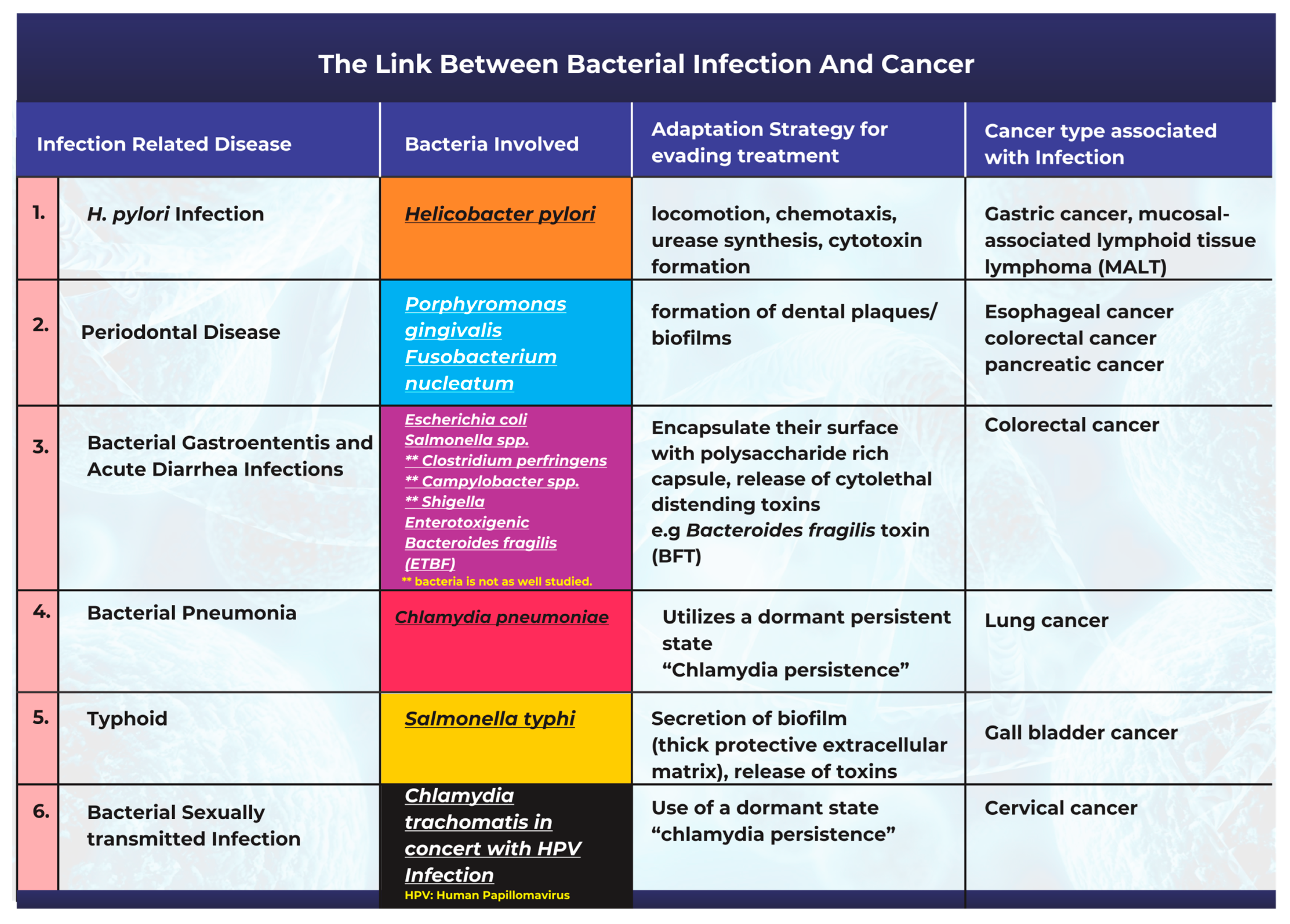
3. The Link between Bacterial Infection and the Onset of Cancer

Bacteria and bacterial infections can act as potential carcinogens and tumor promoters [18]. The production of bacterial toxins, enzymes, and oncogenic peptides can all significantly contribute to tumor development by promoting inflammation, interfering with cell cycle control, and disrupting cell signaling pathways [18,19]. Other studies have also corroborated the fact that microbiota-mediated infection stimulates cancer cell proliferation by targeting host cell DNA, altering the immune system, and promoting epithelial-to-mesenchymal transition [17]. A description of the most frequently studied cases highlighting the link between bacterial infections and cancer is summarized in Figure 1.

3.1. Helicobacter pylori Infection and Gastric Cancer

One of the most well-known and researched connections associating cancer with a bacterial origin is the link between Helicobacter pylori infection and gastric cancer [8,20,21]. H. pylori has been extensively studied in this context with comprehensive epidemiological data supporting the existence of its causal relationship with carcinogenesis [20,22]. H. pylori is a gram-negative spiral-shaped bacterium that lives in a neutral pH niche between the stomach mucus layer and the gastric epithelium [23,24,25]. Studies have opined that there are two potential explanations for the association between H. pylori and gastric cancer. The first of these is that H. pylori infection causes persistent gastric mucosal inflammation, resulting in atrophy and eventual intestinal metaplasia; the second is that H. pylori can create, alter, or release bacterial virulence factors that play a significant role in cancer progression [20,23,25]. Studies have also shown that H. pylori infection contributes to an increase in the bacteria populations from the Proteobacteria, Spirochaetes, and Acidobacteria phyla, alongside a decline in the abundance of Bacteroidetes, Actinobacteria, and Firmicutes [26]. H. pylori infection promotes persistent inflammation of the gastric mucosa and facilitates the progression of both mucosal-associated lymphoid tissue lymphoma and gastric cancer [27,28].
Causes and incidence: Helicobacter pylori is a ubiquitous bacterium with a long history. Its colonization rates vary widely, with the infection rates in developing countries being higher than those in developed regions [29]. Most infections are reported as being contracted in childhood through the feces-to-mouth or the mouth-to-mouth routes [30]. Different outcomes of H. pylori infection may also depend on the specific strain of the infection and its bacterial properties, the host’s response to the infection, and environmental factors; all of these influence pathogen–host interactions [31].
Adaptation strategies for survival and the evasion of treatment: H. pylori has proven its ability to infiltrate the stomach and survive there, even though the stomach is an acidic environment to many species. It is almost impossible for the host to completely eliminate the infection, as H. pylori uses locomotion, chemotaxis, urease synthesis (Figure 1), and other strategies to adapt to the harsh stomach environment and to persist for decades [32]. Furthermore, H. pylori secrete VacA, a vacuolating pore-forming cytotoxin [33]. VacA pores can disrupt mitochondria and endosomes, modify plasma membrane permeability, and cause leakage in epithelial monolayers [33]. VacA also promotes apoptosis in many cells by interfering with mitochondrial activity. In addition to being cytotoxic, VacA can suppress the immune system and prevent the proliferation of T-cells [32,33]. Although all H. pylori strains possess the VacA gene [34], certain species of the bacteria have mutations in their VacA sequences, and patients infected with strains harboring the s1, i1, or m1 variants of VacA have an increased risk of developing gastric cancer [34].

3.2. Periodontal Disease Caused by Bacterial Infections Can Promote Tumorigenesis

Periodontal disease is a common oral bacterial infection that causes inflammation of the gums (gingivitis) and the tooth-supporting structures (periodontitis) [35]. Periodontal disease can promote long-term and persistent inflammation that can eventually lead to systemic diseases such as cancer of the esophagus [36]. Several types of bacteria have been identified as periodontal pathogens; the most extensively studied are Porphyromonas gingivalis and Fusobacterium nucleatum, which have been shown to stimulate oral tumor proliferation [37]. These extensively studied organisms in the gums have become the focus of the emerging link between oral bacteria and cancer [38].
Fusobacterium nucleatum is an anaerobic, gram-negative, disease-causing bacterium in the oral cavity [39]. Studies have suggested that dysbiosis of the oral microbiome allows F. nucleatum to become an opportunistic bacterium that can cause gum disease and human cancers [38,39]. A research study revealed that the abundance of F. nucleatum in the tissues of patients with esophageal squamous carcinoma was linked to shorter patient survival times [39].
Porphyromonas gingivalis is a gram-negative bacterium that is an apparent pathogen in periodontal diseases and other systemic conditions [40]. In 2011, a detailed study found that the concentration of P. gingivalis was higher in cancer cells than in normal mouth tissues. The authors suggested that P. gingivalis promoted oral carcinogenesis by transforming epithelial cells [41]. Correspondingly, a meta-analysis discovered that P. gingivalis significantly increased the risk of periodontal disease and cancer [42].
According to recent research, pathogenic bacteria that cause periodontitis may actively enter the bloodstream from periodontal tissues, become dislodged and carried to numerous organs, and increase the risk of inflammatory processes in those organs [43]. In addition to this, some researchers have found promising evidence linking periodontitis-related bacteria to other cancers [38,44]. According to the reports, F. nucleatum is prevalent in colorectal adenomas and advanced-stage colorectal cancer (CRC) [45]. It was discovered that F. nucleatum causes an inflammatory response that enables the persistent proliferation of colorectal cancer cells [45]. In a similar study, in comparison with healthy volunteers, patients with pancreatic cancer produced more elevated levels of antibodies against P. gingivalis. The study revealed that patients with more elevated levels of P. gingivalis antibodies had a twofold higher risk of developing pancreatic cancer [36]. Investigators from the Harvard School of Public Health conducted a more recent study analyzing the link between periodontal disease, tooth loss, and the risk of gastric and esophageal cancer in approximately 98,000 women (1992 to 2014) and approximately 49,000 men from the Health Professionals Follow-Up Study (1988 to 2016). According to the findings, the risk of gastric cancer was elevated by 52% and the risk of esophageal cancer by 43% in people with periodontal disease [46].
Causes and incidence: Periodontal disease is a well-established and common oral condition in the human population [47]. Periodontal diseases are associated with several risk factors: smoking habits, poor oral hygiene, medication regimens, age, heredity, and stress [47]. These factors can hasten the dysbiosis of the oral microbiome and the multiplication of pathogenic bacteria, which drive disease progression.
Adaptation strategies for survival and the evasion of treatment: One major mechanism related to the persistence and adaptability observed in oral pathogenic bacteria is their ability to form dental plaques or biofilms (Figure 1). These structures make them resistant to mechanical stress or antibiotic treatment [47,48]. The plaque biofilm acts as a protective barrier by isolating them from harmful agents and maintaining distinct phenotypic characteristics that promote their survival. Once the niche is established, the bacteria can release toxins such as lipopolysaccharide (LPS) and produce proinflammatory cytokines that promote chronic inflammation in periodontal tissues [49].

3.3. Bacterial Pathogenesis in the Onset of Gastroenteritis, Acute Diarrhea, and Colon Cancer

Bacterial gastroenteritis is a kind of inflammation in the stomach and small intestine caused by a bacterial infection. In affected patients, the disease is often accompanied by severe diarrhea [50]. Although gastroenteritis can be caused by viruses, fungi, or parasites, most cases are caused by bacterial pathogens [50]. Bacteria associated with gastroenteritis include Escherichia coli, Salmonella spp., Clostridium perfringens, Campylobacter spp., and Shigella [50]. Salmonella and E. coli are the most frequently studied regarding their potential role in carcinogenesis.
Salmonella is an intracellular infection that affects a variety of animals and humans. The results of a Salmonella infection can range significantly from a moderate, self-limiting gastroenteritis to a severe and potentially fatal systemic infection [51]. Salmonella enterica serovar Enteritidis has been implicated in several gastroenteritis outbreaks linked to contaminated food [52,53]. An exhaustive study found that episodes of bacterial gastroenteritis caused by Salmonella or Campylobacter infection resulted in an increased risk of inflammatory bowel disease only one year after acute bacterial gastroenteritis [54].
A more recent epidemiological investigation supported these findings when it discovered a link between severe Salmonella infection and an elevated risk of colon cancer [55]. According to a further study, Salmonella encourages the development of colonic tumors; its AvrA protein can stimulate the Wnt and STAT3 signaling pathways in colonic tumor cells [56].
E. coli is a gram-negative bacterium, ubiquitous in nature, that is found in the human gut microbiome [57]. Several studies have found that patients with colorectal cancer have higher levels of colon mucosa-associated E. coli colonization than those found in healthy individuals [57,58]. Pathogenic E. coli strains have been shown to produce toxins (Figure 1) such as cyclomodulin, cytotoxic necrosis factor (CNF), circulation inhibitory factor (Cif), colibactin, and cytolethal distending toxins (CDT). By disrupting the cell cycle and/or promoting DNA damage, these toxins can affect cell differentiation, apoptosis, and cell proliferation [57,59,60].
Enterotoxigenic Bacteroides fragilis (ETBF) is another common bacterium associated with acute diarrhea infections [61]. Bacteroides fragilis (B. fragilis) is an obligate anaerobic gram-negative bacillus bacterium [62] that makes up 0.1% of the normal flora of the colon [25]. However, ETBF levels are elevated in CRC patients’ feces and colonic mucosal tissues [25]. According to the “alpha-bug” theory, a critical pathogenic species, such as (ETBF), remodels the microbiota to promote CRC, most probably through an IL-17 and Th17-mediated inflammatory response [63]. Similarly, the bacterial driver–passenger view proposes that “driver” bacteria, such as ETBF, impel or exacerbate inflammation while producing toxins that cause cell proliferation and mutations. Consequently, an adenoma develops and is colonized by “passenger” bacteria, such as Fusobacterium spp., thus promoting tumor growth [64].
Causes and incidence: Acute gastroenteritis affects millions of people worldwide and is characterized by symptoms such as vomiting, diarrhea, and abdominal pain [65]. Acute gastroenteritis caused by bacterial pathogens is acquired through contaminated foods. Researchers also believe that the widespread use of antibiotics [66] in recent years has aided pathogenic bacterial colonization [51].
Adaptation strategies for survival and the evasion of treatment: Pathogenic bacteria have evolved adaptations to their outer surfaces that permit immunological escape and opportunities to survive and withstand the host’s defense mechanisms against infection [8]. Gram-negative bacteria such as E. coli encapsulate their intricate outer-surface macromolecules in a polysaccharide-rich capsule to prevent immune-system detection and clearance. These capsules are protective and help minimize the host’s complement activation [8,67]. The release of protein toxins with cytolytic characteristics is a further adaptive techniques used by pathogenic bacteria [8]. Shigella dysenteriae, Campylobacter jejuni, Salmonella typhi, and E. coli all secrete cytolethal distending toxins (CDTs); these toxins promote DNA strand breaks that eventually promote cancer progression [8,68]. In addition, B. fragilis releases a toxin (BFT, Figure 1) that attaches to gut epithelial cell receptors and promotes cell proliferation by cleavage of E-cadherin [62,69].

3.4. Chlamydia pneumoniae Infection and Lung Cancer

Chlamydia pneumoniae (Cpn) is an obligate intracellular bacterium that causes various respiratory diseases in humans, including pneumonia [70]. Chronic obstructive pulmonary disease, asthma, and lung cancer may result from repeated or prolonged exposure to Chlamydia antigens [71].
Previous research has found serological evidence of a link between Chlamydia pneumoniae infection and lung cancer [72]. A report also reviewed previous epidemiological studies on the link between C. pneumoniae and lung cancer. It concluded that earlier investigations supported a causative relationship between C. pneumoniae infection and lung cancer [73]. Correspondingly, a group of researchers conducted a meta-analysis on 12 published articles that studied the link between C. pneumoniae infection and lung cancer. The study concluded that C. pneumoniae infection is correlated with an elevated risk of lung cancer, implying that a higher serological titer may be an efficient predictor of lung cancer risk [74]. A systematic review of published articles was recently conducted on C. pneumoniae infection and the development of lung cancer. The authors of this study discovered that twenty-four articles describing one animal model positively supported the hypothesis, while three of the articles did not [70].
More exhaustive studies on different animal models and follow-up studies in humans with Cpn are urgently needed to assist in validating these findings.
Causes and incidence: Cpn, a gram-negative bacillus, is an obligate intracellular bacterium responsible for respiratory infections [75,76]. Cpn is aerosolized by individuals coughing or sneezing, which produces tiny respiratory droplets that contain bacteria. Others then inhale these droplets and the bacteria that cause the infection [75].
Adaptation strategies for survival and the evasion of treatment: Chlamydia’s most common adaptative strategy is to enter a persistent dormant state (Figure 1). If Chlamydia is exposed to immune system stress or antibiotics, the pathogen can suspend replication by entering a dormant but viable state called “Chlamydia persistence”. During this time, Chlamydia prioritizes the cellular activities required for extended survival; this is often associated with the presence of enlarged abnormal reticulate bodies. When conditions improve and normal replication resumes, the durable state is reversed [77].

3.5. Salmonella typhi Infections and Gallbladder Cancer

Salmonella typhi is a gram-negative, rod-shaped, flagellated bacterium that is well documented as being responsible for typhoid infections [78,79,80]. After infection, S. typhi invades the gallbladder, causing persistent infection in susceptible carriers that serve as a reservoir for the spread of the disease; this persistent chronic infection has also been linked to cancer [79,81]. The ability of the pathogen to survive and persist for an extended time in the gallbladders of patients after infection creates a suitable environment for its tumor-promoting effect [81]. It has also been reported that S. typhi produces typhoid toxins and carcinogenic toxins such as nitroso-chemical compounds. These compounds play a crucial role in the progression of cancer [80]. A long-term follow-up study of infected mice discovered the presence of precancerous lesions and dysplasia of the gallbladder, reflecting an association between Salmonella infection and gallbladder cancer [82]. Other large-scale studies have also found a link between S. typhi infection and gallbladder cancer [80,81,83].
Causes and incidence: Infection by S. typhi is usually acquired through the ingestion of food or water contaminated with the feces of a person carrying the organism [78]. Salmonella typhi is suspected to be responsible for approximately 22 million cases of typhoid fever, slightly more than 5 million cases of paratyphoid fever, and approximately 200,000 deaths worldwide each year [79]. According to research, some 90% of chronic infection carriers suffer from gallstone infections over time. This association has been described as a significant risk factor for developing gallbladder cancer [82,84,85].
Adaptation strategies for survival and the evasion of treatment: Through the secretion of biofilm, S. typhi appears to survive and become well adapted to thrive in the gallbladder. Biofilm formation (Figure 1) is a pathological adaptive response that allows the bacteria to aggregate, adhere to surfaces, and develop antimicrobial resistance through the utilization of a thick protective extracellular matrix [79,85].

3.6. Bacterial Infection and Cervical Cancer

Chlamydia trachomatis is a gram-negative bacterium and a prevalent cause of curable sexually-transmitted bacterial infections (STIs) worldwide [86]. The infection presents as urethritis in men and endocervicitis in women [87]. Persistent Chlamydia infections have also been identified as a risk factor for developing cervical carcinoma [87], especially in patients with the human papillomavirus (HPV) coinfection [86,88]. Recent elaborate meta-analysis studies have also corroborated the link between C. trachomatis and cervical cancer [89]. The mechanism of the C. trachomatis action is proposed as resulting from the dysregulation of DNA damage responses, and the disruption of cell-cycle control, which promotes malignant transformation in infected patients [88].
The association between C. trachomatis and cervical cancer is usually seen in the presence of HPV infections [86]. Since HPV infections have long been implicated in the etiology of cervical cancer [90], it is difficult to directly classify the bacterial infection as solely causative, even though it contributes significantly to an increased incidence.

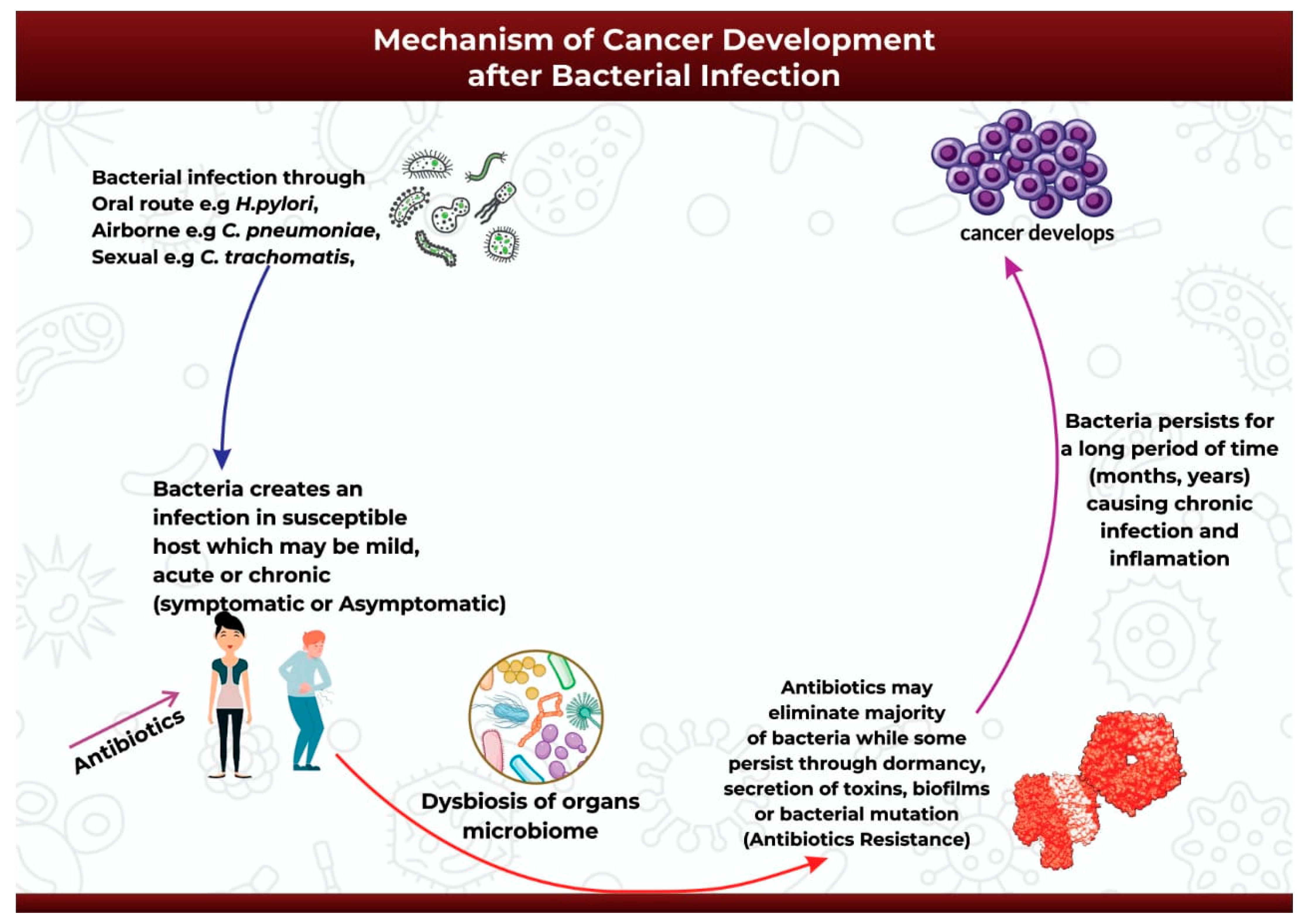
4. Mechanism of Cancer Development after Bacterial Infection

The onset of carcinogenesis after exposure to bacterial infection occurs over an extended period and depends on a wide range of factors such as host susceptibility, genetic background, and suitable environmental conditions [91].
After the successful infection of the host either through oral, airborne, or sexually-transmitted routes (Figure 2), the infection may start as mild, acute, or chronic and may be symptomatic or asymptomatic [92]. Antibiotics might be introduced at this point to help ease the infection [92]. The initial infection and subsequent antibiotic use may then initiate bacterial dysbiosis in the microbiome of the infected organ, in this case promoting the bloom of opportunistic and pathogenic bacteria species [93,94,95]. Antibiotic use may completely eradicate the infection in some cases, or it may be ineffective against the application of alternative survival strategies by the bacteria to facilitate its continued existence over extended periods of time (Figure 2). Pathogenic bacteria can survive and persist in the host environment through dormancy, secretion of cyotethal toxins, the formation of protective biofilms, or mutations to form antibiotic resistant strains [17,18]. This adaptive technique enables bacteria to promote persistent chronic inflammation or chronic infection, and ultimately carcinogenesis [17] (Figure 2).

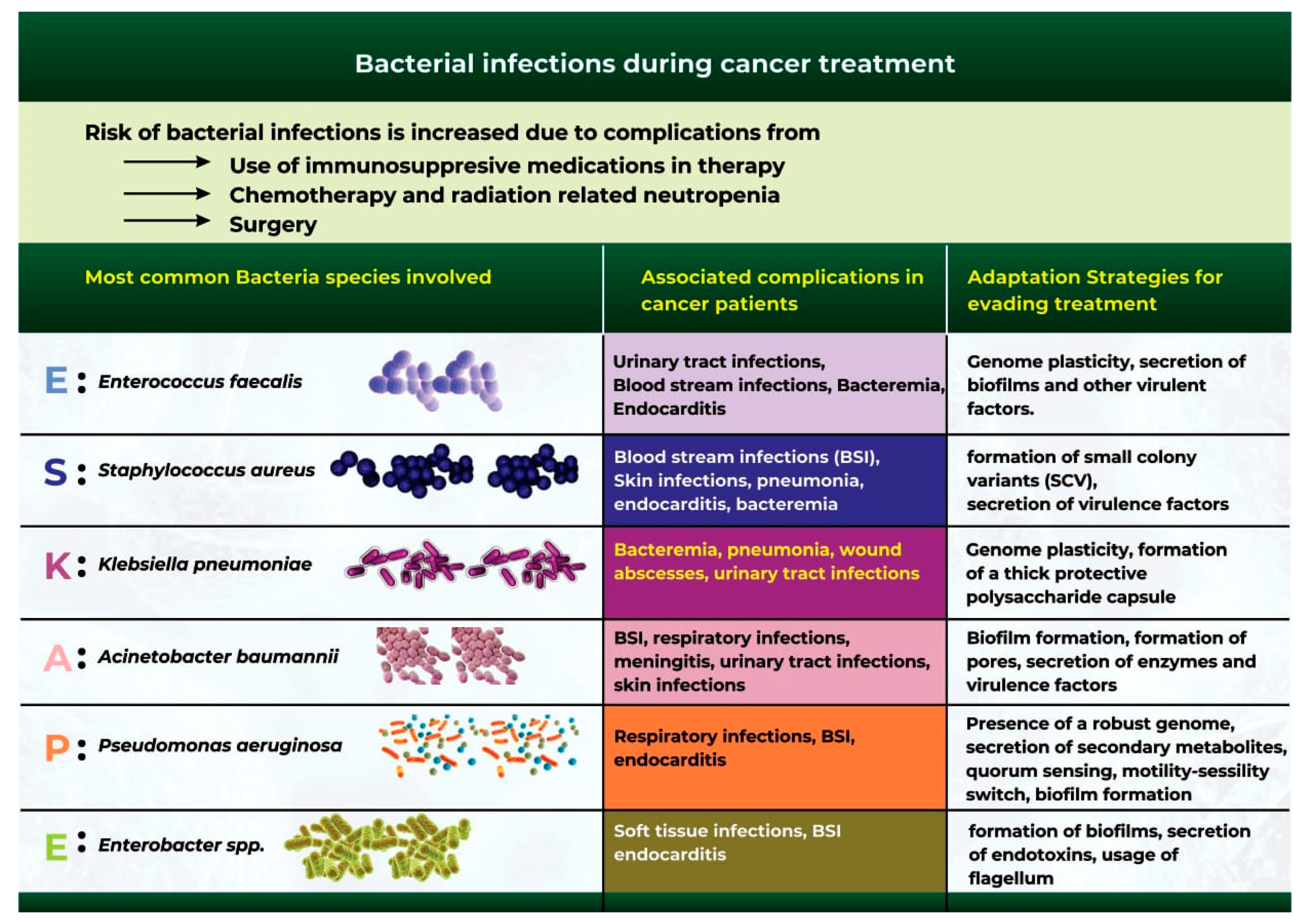
5. Bacterial Infections during Cancer Treatment

Bacterial infection is one of the most common complications during cancer treatment [96]. Although cancer mortality rates have continued to decline in recent years, bacterial infections remain a significant cause of infection-related mortality in patients [97]. Cancer patients are at a high risk of bacterial infection due to surgical complications, chemotherapy and radiotherapy-related neutropenia, and the use of immunosuppressive drugs during cancer treatment [97,98].
Several reports highlight that bacterial infections are prevalent in patients with blood cancers [99,100,101], owing mainly to prolonged neutropenia during treatment [101]. Although patients with solid tumors are also susceptible to bacterial infections, an extensive epidemiological study reported an eightfold higher incidence in patients with hematological cancers than in patients with solid tumors [102]. A more recent study also revealed that the levels of Pseudomonas aeruginosa bloodstream infections were higher in hematological malignancies than in solid tumors [103].
Extensive studies have identified a group of six bacterial microbes otherwise classified as the “ESKAPE” pathogens, which are at the forefront of bacterial infection and antibiotic resistance during cancer treatment (Figure 3) [104,105]. These organisms include Acinetobacter baumannii, Staphylococcus aureus, Enterococcus faecalis, Klebsiella pneumoniae, Pseudomonas aeruginosa, and Enterobacter spp. [104,105].
An extensive study conducted by a group of researchers at Cairo’s National Cancer Institute aimed to investigate the effect of ESKAPE pathogens on the course of infectious incidents in pediatric cancer patients. According to the study, the ESKAPE pathogens were significantly associated with more prolonged infections, longer duration of infection, and higher mortality. The study also found that Pseudomonas aeruginosa and Klebsiella pneumoniae infections caused the highest mortality rates, at 43% and 30%, respectively [106].
A similar rigorous study was carried out over a five-year period in hospitalized adults with cancer from 2006 to 2011. All bacteremia episodes in hospitalized cancer patients were included in the investigation. The study concluded that the ESKAPE pathogens were responsible for 34% of bacteremia episodes in cancer patients, with Pseudomonas aeruginosa being the most frequently isolated organism [107].
Most enterococcal infections in cancer patients are bacteremia caused by Enterococcus faecalis [108]. Prior exposure to antibiotics and long-term neutropenia are factors that predispose cancer patients to enterococcal infections [108]. Urinary tract infections (UTIs), bloodstream infections (BSIs), and endocarditis are common complications for cancer patients exposed to enterococcal infections during therapy [109,110]. Reports have also highlighted that Enterococcus faecalis often evades treatment owing to its ability to secrete virulent factors and biofilms, and the plasticity of its genome [111].
Staphylococcus aureus has a significant clinical impact on mortality rate in patients with malignancy [107]. A systematic analysis of bacteremia infections in cancer patients highlights that Staphylococcus aureus infections accounts for 1.3% to 12% of all bacteremia cases [112]. Infections caused by Staphylococcus aureus often reportedly led to BSIs, skin infections, pneumonia, and endocarditis [113]. Staphylococcus aureus is of clinical interest because the bacterium can form small colony variants (SCV) in addition to releasing virulence factors (e.g., enterotoxins and proteases) that interfere with the host’s innate immune response [114,115].
Klebsiella pneumoniae is a leading cause of sepsis and the most common cause of bacteremia, pneumonia, wound infections, abscesses, and urinary tract infections in cancer patients [116]. K pneumoniae causes a significant threat to health because the bacteria rely on the presence of an accessory genome that allows for genetic plasticity, the release of virulence factors, and the formation of a thick protective polysaccharide capsule that enables them to overcome the host immune system and clearance [117,118].
A. baumannii infections are nosocomial infections that can be fatal in patients with compromised immune systems, especially cancer patients. Patients infected with Acinetobacter baumannii have a significantly increased mortality rate of up to 80% [119]. Patients also develop complications from BSI, respiratory tract infections, meningitis, UTIs, and skin infections [113,120]. The pathogenesis of A. baumannii infection is severe because the bacterium can adapt to the host environment in several ways, which include the secretion of biofilms (thick polysaccharide capsules) and porins, alongside the release of enzymes and virulence factors. The bacterium also utilizes micronutrient acquisition and protein secretion systems that facilitate its survival even under harsh environmental conditions [120].
Pseudomonas aeruginosa is an increasingly prevalent opportunistic pathogen that can cause serious and life-threatening nosocomial infections [116]. P. aeruginosa has been implicated in causing BSI, respiratory infections, and endocarditis [113,121]. The bacterium is highly adaptive to the host environment owing to its robust genome and its ability to form biofilms, activate a motile-sessility switch, and secrete secondary metabolites [122].
Enterobacter spp. can also infect the respiratory tract, surgical wounds, the urinary tract, and the bloodstream of cancer patients [123]. These bacteria facilitate soft-tissue infections, BSIs, and endocarditis [113]. Often, the bacteria population thrives in its host environment through the use of flagellum, the formation of biofilms, and the secretion of endotoxins [124].

6. Antibiotic Use and Antimicrobial Resistance in Cancer Patients

Antibiotics are secondary metabolites produced by microorganisms, higher animals, and plants, and they have antipathogenic effects that can interfere with the growth of other living cells [125]. Antibiotics are increasingly being used to treat cancers, often because of their pro-apoptotic, antiproliferative, and antimetastatic potentials [125]. Since infections are also common in cancer patients, it is essential to use antibiotics to prevent and treat bacterial infections [98].
A major limitation of antibiotic use is the causation of dysbiosis due to the elimination of beneficial bacterial groups, such as Lactobacillus and Bifidobacterium, in addition to the pathogenic bacteria [125]. A further limitation is the ability of pathogenic bacteria to evade killing and induce antibiotic resistance [104].
It is also worth noting that the microbiome plays a crucial role in the development of antibiotic resistance [126,127]. Microbiota dysbiosis induced by antibiotic treatment contributes to the development of resistance that is often due to increased numbers of opportunistic bacteria secreting high levels of antimicrobial resistance genes [128,129].
Antibiotic resistance in cancer patients often correlates with increased susceptibility to infections that eventually reduce the patient’s survival time; it also poses a significant threat to the accomplishments achieved in cancer treatment, and it highlights the importance of monitoring cancer patients and protecting them against antibiotic resistance [104].

Potential Approaches for Reducing Antibiotic Resistance in Cancer Patients

  • Avoiding bacterial infections (prevention is always better than cure): The first stage of bacterial-driven carcinogenesis is usually an initial infection that allows the pathogen to infect the host. Hosts can prevent such infections by being vaccinated and by adopting proper hygiene measures [130]. Antibiotic prophylaxis is another common practice used for cancer patients in neutropenic settings to prevent infection and infection-related complications. According to reports, the prophylactic use of quinolones reduced the incidence of fever, infection, hospitalization, and overall mortality [104].
  • The discovery and design of novel antibiotics that are pathogen-specific: Most antibiotics are designed for broad-spectrum use and can severely impact the human microbiome, leading to complications such as antibiotic-associated colitis and the rapid spread of antibiotic resistance. Utilizing more targeted approaches or using narrow-spectrum antibiotics may help address this limitation and provide additional resources for expanding the antibiotic pool, which is at risk owing to the inevitable rise in resistance [131].
  • Optimizing Antibiotic Use (more does not guarantee better): Excessive antibiotic use is a significant driver of resistance [132]. To promote the effective use of antibiotics, the concept of “antimicrobial stewardship” was developed. Antimicrobial stewardship refers to the practice of establishing the antibiotic treatment that is most appropriate and then administering it for a short time and at the lowest effective dose to achieve the best possible clinical outcome in combating infection and preventing its spread [133]. In clinical settings, antimicrobial stewardship teams comprise an infectious disease doctor, an infectious disease pharmacist, and clinical microbiologists who monitor the patient’s dosage and response [104]. A research study evaluated the association between patient mortality and antibiotic stewardship outcomes in patients who developed febrile neutropenia during their cancer treatment. The study showed that adherence to antibiotic stewardship was associated with reduced mortality [134].

7. Conclusions

The link between infectious agents and the risk of cancer has been studied for decades. Nevertheless, there are limited reports on the causal relationship between bacterial infection and tumorigenesis. The link between bacterial infections acquired during cancer treatment and mortality rates is yet to be explored. This comprehensive review highlights the association between bacterial infection and cancer growth, and it also demonstrates the impact of nosocomial bacterial infection on the survival of cancer patients. We also provide crucial information on how bacterial infections can persist for long durations, contribute to tumorigenesis, and affect cancer patients during their treatment. The article correspondingly provides recommendations for minimizing antibiotic resistance in cancer patients. To better understand the direct link between bacterial infections and cancer development, it will be essential to conduct further long-term follow-up studies on patients with bacterial infections and on the ways in which these infections impact on their risks. Finally, while we have focused in this review on the role of bacteria in cancer, recent studies have also shed light on bacteriotherapy as a potential anticancer therapeutic strategy. Live attenuated strains, toxins, peptides or bacteriocins could be used; alternatively, bacteria could be developed as a drug-delivery system for cancer therapy with the assistance of microbe engineering for tumor targeting. The therapeutic effects of engineered microorganisms in cancer treatment hold immense promise for biomedical applications in targeted cancer therapy. These areas, while cutting edge, are beyond the scope of this review and will be deferred for future discussion.

Author Contributions

Conceptualization, K.Y. and S.U.; Methodology, K.Y.; Writing—Original Draft Preparation, K.Y.; Writing—Review and Editing, K.Y., V.S. and S.U.; Supervision, V.S. and S.U.; Funding Acquisition, S.U. All authors have read and agreed to the published version of the manuscript.

Funding

This study was funded by an R01 (R01DK117296) from the National Institute of Diabetes and Digestive and Kidney Diseases (NIDDK).

Conflicts of Interest

The authors declare no conflict of interest.

References

  1. Hausman, D.M. What Is Cancer? Perspect. Biol. Med. 2019, 62, 778–784. [Google Scholar] [CrossRef] [PubMed]
  2. Siegel, R.L.; Miller, K.D.; Fuchs, H.E.; Jemal, A. Cancer statistics, 2022. CA Cancer J. Clin. 2022, 72, 7–33. [Google Scholar] [CrossRef] [PubMed]
  3. Golemis, E.A.; Scheet, P.; Beck, T.N.; Scolnick, E.M.; Hunter, D.J.; Hawk, E.; Hopkins, N. Molecular mechanisms of the preventable causes of cancer in the United States. Genes Dev. 2018, 32, 868–902. [Google Scholar] [CrossRef]
  4. GBD 2019 Cancer Risk Factors Collaborators. The global burden of cancer attributable to risk factors, 2010–2019: A systematic analysis for the Global Burden of Disease Study 2019. Lancet 2022, 400, 563–591. [Google Scholar] [CrossRef]
  5. GBD 2019 Adolescent Young Adult Cancer Collaborators. The global burden of adolescent and young adult cancer in 2019: A systematic analysis for the Global Burden of Disease Study 2019. Lancet Oncol. 2022, 23, 27–52. [Google Scholar] [CrossRef]
  6. Kocarnik, J.M.; Compton, K.; Dean, F.E.; Fu, W.; Gaw, B.L.; Harvey, J.D.; Henrikson, H.J.; Lu, D.; Pennini, A.; Xu, R.; et al. Cancer Incidence, Mortality, Years of Life Lost, Years Lived With Disability, and Disability-Adjusted Life Years for 29 Cancer Groups From 2010 to 2019: A Systematic Analysis for the Global Burden of Disease Study 2019. JAMA Oncol. 2022, 8, 420–444. [Google Scholar] [CrossRef]
  7. Anand, P.; Kunnumakkara, A.B.; Sundaram, C.; Harikumar, K.B.; Tharakan, S.T.; Lai, O.S.; Sung, B.; Aggarwal, B.B. Cancer is a preventable disease that requires major lifestyle changes. Pharm. Res. 2008, 25, 2097–2116. [Google Scholar] [CrossRef] [PubMed]
  8. van Elsland, D.; Neefjes, J. Bacterial infections and cancer. EMBO Rep. 2018, 19, e46632. [Google Scholar] [CrossRef]
  9. de Martel, C.; Georges, D.; Bray, F.; Ferlay, J.; Clifford, G.M. Global burden of cancer attributable to infections in 2018: A worldwide incidence analysis. Lancet Glob. Health 2020, 8, e180–e190. [Google Scholar] [CrossRef] [PubMed]
  10. Dalton-Griffin, L.; Kellam, P. Infectious causes of cancer and their detection. J. Biol. 2009, 8, 67. [Google Scholar] [CrossRef]
  11. Dekaboruah, E.; Suryavanshi, M.V.; Chettri, D.; Verma, A.K. Human microbiome: An academic update on human body site specific surveillance and its possible role. Arch. Microbiol. 2020, 202, 2147–2167. [Google Scholar] [CrossRef] [PubMed]
  12. Shreiner, A.B.; Kao, J.Y.; Young, V.B. The gut microbiome in health and in disease. Curr. Opin. Gastroenterol. 2015, 31, 69–75. [Google Scholar] [CrossRef] [PubMed]
  13. Sender, R.; Fuchs, S.; Milo, R. Revised Estimates for the Number of Human and Bacteria Cells in the Body. PLoS Biol. 2016, 14, e1002533. [Google Scholar] [CrossRef] [PubMed]
  14. Curtis, M.M.; Sperandio, V. A complex relationship: The interaction among symbiotic microbes, invading pathogens, and their mammalian host. Mucosal Immunol. 2011, 4, 133–138. [Google Scholar] [CrossRef]
  15. Conrad, R.; Vlassov, A.S. The Human Microbiota: Composition, Functions, and Therapeutic Potential. Med. Sci. Rev. 2015, 2, 92–103. [Google Scholar] [CrossRef]
  16. Wang, B.; Yao, M.; Lv, L.; Ling, Z.; Li, L. The human microbiota in health and disease. Engineering 2017, 3, 71–82. [Google Scholar] [CrossRef]
  17. Khatun, S.; Appidi, T.; Rengan, A.K. The role played by bacterial infections in the onset and metastasis of cancer. Curr. Res. Microb. Sci. 2021, 2, 100078. [Google Scholar] [CrossRef]
  18. Laliani, G.; Sorboni, S.G.; Lari, R.; Yaghoubi, A.; Soleimanpour, S.; Khazaei, M.; Hasanian, S.M.; Avan, A. Bacteria and cancer: Different sides of the same coin. Life Sci. 2020, 246, 117398. [Google Scholar] [CrossRef]
  19. Lax, A.J. Opinion: Bacterial toxins and cancer—A case to answer? Nat. Rev. Microbiol. 2005, 3, 343–349. [Google Scholar] [CrossRef]
  20. Parsonnet, J. Bacterial infection as a cause of cancer. Environ. Health Perspect. 1995, 103 (Suppl. 8), 263–268. [Google Scholar] [CrossRef] [Green Version]
  21. Song, S.; Vuai, M.S.; Zhong, M. The role of bacteria in cancer therapy—Enemies in the past, but allies at present. Infect. Agent Cancer 2018, 13, 9. [Google Scholar] [CrossRef]
  22. Zella, D.; Gallo, R.C. Viruses and Bacteria Associated with Cancer: An Overview. Viruses 2021, 13, 1039. [Google Scholar] [CrossRef] [PubMed]
  23. Asaka, M.; Sepulveda, A.R.; Sugiyama, T.; Graham, D.Y. Chapter 40: Gastric Cancer. In Helicobacter pylori: Physiology and Genetics; Mobley, H.L.T., Mendz, G.L., Hazell, S.L., Eds.; ASM Press: Washington, DC, USA, 2001. [Google Scholar]
  24. Khosravi, Y.; Dieye, Y.; Poh, B.H.; Ng, C.G.; Loke, M.F.; Goh, K.L.; Vadivelu, J. Culturable bacterial microbiota of the stomach of Helicobacter pylori positive and negative gastric disease patients. Sci. World J. 2014, 2014, 610421. [Google Scholar] [CrossRef] [PubMed]
  25. Elsalem, L.; Jum’ah, A.A.; Alfaqih, M.A.; Aloudat, O. The Bacterial Microbiota of Gastrointestinal Cancers: Role in Cancer Pathogenesis and Therapeutic Perspectives. Clin. Exp. Gastroenterol. 2020, 13, 151–185. [Google Scholar] [CrossRef] [PubMed]
  26. Maldonado-Contreras, A.; Goldfarb, K.C.; Godoy-Vitorino, F.; Karaoz, U.; Contreras, M.; Blaser, M.J.; Brodie, E.L.; Dominguez-Bello, M.G. Structure of the human gastric bacterial community in relation to Helicobacter pylori status. ISME J. 2011, 5, 574–579. [Google Scholar] [CrossRef] [PubMed]
  27. Elagan, S.K.; Almalki, S.J.; Alharthi, M.R.; Mohamed, M.S.; El-Badawy, M.F. Role of Bacteria in the Incidence of Common GIT Cancers: The Dialectical Role of Integrated Bacterial DNA in Human Carcinogenesis. Infect. Drug Resist. 2021, 14, 2003–2014. [Google Scholar] [CrossRef]
  28. Vogiatzi, P.; Cassone, M.; Luzzi, I.; Lucchetti, C.; Otvos, L.; Giordano, A. Helicobacter pylori as a class I carcinogen: Physiopathology and management strategies. J. Cell. Biochem. 2007, 102, 264–273. [Google Scholar] [CrossRef]
  29. Blaser, M.J.; Berg, D.E. Helicobacter pylori genetic diversity and risk of human disease. J. Clin. Investig. 2001, 107, 767–773. [Google Scholar] [CrossRef]
  30. Wroblewski, L.E.; Peek, R.M., Jr.; Wilson, K.T. Helicobacter pylori and Gastric Cancer: Factors That Modulate Disease Risk. Clin. Microbiol. Rev. 2010, 23, 713–739. [Google Scholar] [CrossRef]
  31. Cover, T.L. Helicobacter pylori Diversity and Gastric Cancer Risk. mBio 2016, 7, e01869-15. [Google Scholar] [CrossRef] [Green Version]
  32. Amieva, M.; Peek, R.M. Pathobiology of Helicobacter pylori—Induced Gastric Cancer. Gastroenterology 2016, 150, 64–78. [Google Scholar] [CrossRef] [PubMed]
  33. Kim, I.J.; Blanke, S.R. Remodeling the host environment: Modulation of the gastric epithelium by the Helicobacter pylori vacuolating toxin (VacA). Front. Cell. Infect. Microbiol. 2012, 2, 37. [Google Scholar] [CrossRef] [PubMed]
  34. Foegeding, N.J.; Caston, R.R.; McClain, M.S.; Ohi, M.D.; Cover, T.L. An Overview of Helicobacter pylori VacA Toxin Biology. Toxins 2016, 8, 173. [Google Scholar] [CrossRef] [PubMed]
  35. Kawasaki, M.; Ikeda, Y.; Ikeda, E.; Takahashi, M.; Tanaka, D.; Nakajima, Y.; Arakawa, S.; Izumi, Y.; Miyake, S. Oral infectious bacteria in dental plaque and saliva as risk factors in patients with esophageal cancer. Cancer 2021, 127, 512–519. [Google Scholar] [CrossRef]
  36. Michaud, D.S.; Lu, J.; Peacock-Villada, A.Y.; Barber, J.R.; Joshu, C.E.; Prizment, A.E.; Beck, J.D.; Offenbacher, S.; Platz, E.A. Periodontal Disease Assessed Using Clinical Dental Measurements and Cancer Risk in the ARIC Study. J. Natl. Cancer Inst. 2018, 110, 843–854. [Google Scholar] [CrossRef]
  37. Öğrendik, M. Periodontal Pathogens in the Etiology of Pancreatic Cancer. Gastrointest. Tumors 2017, 3, 125–127. [Google Scholar] [CrossRef]
  38. Whitmore, S.E.; Lamont, R.J. Oral bacteria and cancer. PLoS Pathog. 2014, 10, e1003933. [Google Scholar] [CrossRef]
  39. Yamamura, K.; Baba, Y.; Nakagawa, S.; Mima, K.; Miyake, K.; Nakamura, K.; Sawayama, H.; Kinoshita, K.; Ishimoto, T.; Iwatsuki, M.; et al. Human Microbiome Fusobacterium Nucleatum in Esophageal Cancer Tissue Is Associated with Prognosis. Clin. Cancer Res. 2016, 22, 5574–5581. [Google Scholar] [CrossRef]
  40. Gao, S.; Liu, Y.; Duan, X.; Liu, K.; Mohammed, M.; Gu, Z.; Ren, J.; Yakoumatos, L.; Yuan, X.; Lu, L.; et al. Porphyromonas gingivalis infection exacerbates oesophageal cancer and promotes resistance to neoadjuvant chemotherapy. Br. J. Cancer 2021, 125, 433–444. [Google Scholar] [CrossRef]
  41. Katz, J.; Onate, M.D.; Pauley, K.M.; Bhattacharyya, I.; Cha, S. Presence of Porphyromonas gingivalis in gingival squamous cell carcinoma. Int. J. Oral Sci. 2011, 3, 209–215. [Google Scholar] [CrossRef] [Green Version]
  42. Sayehmiri, F.; Sayehmiri, K.; Asadollahi, K.; Soroush, S.; Bogdanovic, L.; Jalilian, F.A.; Emaneini, M.; Taherikalani, M. The prevalence rate of Porphyromonas gingivalis and its association with cancer: A systematic review and meta-analysis. Int. J. Immunopathol. Pharmacol. 2015, 28, 160–167. [Google Scholar] [CrossRef]
  43. Nocini, R.; Lippi, G.; Mattiuzzi, C. Periodontal disease: The portrait of an epidemic. J. Public Health Emerg. 2020, 4–10. [Google Scholar] [CrossRef]
  44. Gallimidi, A.B.; Fischman, S.; Revach, B.; Bulvik, R.; Maliutina, A.; Rubinstein, A.M.; Nussbaum, G.; Elkin, M. Periodontal pathogens Porphyromonas gingivalis and Fusobacterium nucleatum promote tumor progression in an oral-specific chemical carcinogenesis model. Oncotarget 2015, 6, 22613–22623. [Google Scholar] [CrossRef]
  45. Rubinstein, M.R.; Wang, X.; Liu, W.; Hao, Y.; Cai, G.; Han, Y.W. Fusobacterium nucleatum promotes colorectal carcinogenesis by modulating E-cadherin/β-catenin signaling via its FadA adhesin. Cell Host Microbe 2013, 14, 195–206. [Google Scholar] [CrossRef] [PubMed]
  46. Printz, C. Study adds evidence to link between gum disease and cancer risk: Researchers observe connection with gastric, esophageal cancer. Cancer 2021, 127, 495–496. [Google Scholar] [CrossRef]
  47. Nazir, M.A. Prevalence of periodontal disease, its association with systemic diseases and prevention. Int. J. Health Sci. 2017, 11, 72–80. [Google Scholar]
  48. Lovegrove, J.M. Dental plaque revisited: Bacteria associated with periodontal disease. J. N. Z. Soc. Periodontol. 2004, 87, 7–21. [Google Scholar]
  49. Ishida, N.; Ishihara, Y.; Ishida, K.; Tada, H.; Funaki-Kato, Y.; Hagiwara, M.; Ferdous, T.; Abdullah, M.; Mitani, A.; Michikawa, M.; et al. Periodontitis induced by bacterial infection exacerbates features of Alzheimer’s disease in transgenic mice. Npj Aging Mech. Dis. 2017, 3, 15. [Google Scholar] [CrossRef]
  50. Sattar, S.B.A.; Singh, S. Bacterial Gastroenteritis; StatPearls Publishing: Treasure Island, FL, USA, 2022.
  51. Sun, J. Impact of bacterial infection and intestinal microbiome on colorectal cancer development. Chin. Med. J. 2022, 135, 400–408. [Google Scholar] [CrossRef]
  52. Jiang, M.; Zhu, F.; Yang, C.; Deng, Y.; Kwan, P.S.L.; Li, Y.; Lin, Y.; Qiu, Y.; Shi, X.; Chen, H.; et al. Whole-Genome Analysis of Salmonella enterica Serovar Enteritidis Isolates in Outbreak Linked to Online Food Delivery, Shenzhen, China, 2018. Emerg. Infect. Dis. 2020, 26, 789–792. [Google Scholar] [CrossRef]
  53. Zhang, Y.; Liu, K.; Zhang, Z.; Tian, S.; Liu, M.; Li, X.; Han, Y.; Zhu, K.; Liu, H.; Yang, C.; et al. A Severe Gastroenteritis Outbreak of Salmonella enterica Serovar Enteritidis Linked to Contaminated Egg Fried Rice, China, 2021. Front. Microbiol. 2021, 12, 779749. [Google Scholar] [CrossRef] [PubMed]
  54. Ternhag, A.; Törner, A.; Svensson, A.; Ekdahl, K.; Giesecke, J. Short- and long-term effects of bacterial gastrointestinal infections. Emerg. Infect. Dis. 2008, 14, 143–148. [Google Scholar] [CrossRef] [PubMed]
  55. Mughini-Gras, L.; Schaapveld, M.; Kramers, J.; Mooij, S.; Neefjes-Borst, E.A.; Pelt, W.V.; Neefjes, J. Increased colon cancer risk after severe Salmonella infection. PLoS ONE 2018, 13, e0189721. [Google Scholar] [CrossRef] [PubMed]
  56. Lu, R.; Wu, S.; Zhang, Y.-G.; Xia, Y.; Zhou, Z.; Kato, I.; Dong, H.; Bissonnette, M.; Sun, J. Salmonella Protein AvrA Activates the STAT3 Signaling Pathway in Colon Cancer. Neoplasia 2016, 18, 307–316. [Google Scholar] [CrossRef]
  57. Hernández-Luna, M.A.; López-Briones, S.; Luria-Pérez, R. The Four Horsemen in Colon Cancer. J. Oncol. 2019, 2019, 5636272. [Google Scholar] [CrossRef]
  58. Pleguezuelos-Manzano, C.; Puschhof, J.; Huber, A.R.; van Hoeck, A.; Wood, H.M.; Nomburg, J.; Gurjao, C.; Manders, F.; Dalmasso, G.; Stege, P.B.; et al. Mutational signature in colorectal cancer caused by genotoxic pks. Nature 2020, 580, 269–273. [Google Scholar] [CrossRef]
  59. Wassenaar, T.M. E. coli and colorectal cancer: A complex relationship that deserves a critical mindset. Crit. Rev. Microbiol. 2018, 44, 619–632. [Google Scholar] [CrossRef]
  60. Veziant, J.; Gagnière, J.; Jouberton, E.; Bonnin, V.; Sauvanet, P.; Pezet, D.; Barnich, N.; Miot-Noirault, E.; Bonnet, M. Association of colorectal cancer with pathogenic Escherichia coli: Focus on mechanisms using optical imaging. World J. Clin. Oncol. 2016, 7, 293–301. [Google Scholar] [CrossRef]
  61. Sears, C.L.; Islam, S.; Saha, A.; Arjumand, M.; Alam, N.H.; Faruque, A.S.; Salam, M.A.; Shin, J.; Hecht, D.; Weintraub, A.; et al. Association of enterotoxigenic Bacteroides fragilis infection with inflammatory diarrhea. Clin. Infect. Dis. 2008, 47, 797–803. [Google Scholar] [CrossRef]
  62. Elsaghir, H.; Reddivari, A.K.R. Bacteroides Fragilis; StatPearls Publishing: Treasure Island, FL, USA, 2022.
  63. Hajishengallis, G.; Darveau, R.P.; Curtis, M.A. The keystone-pathogen hypothesis. Nat. Rev. Microbiol. 2012, 10, 717–725. [Google Scholar] [CrossRef]
  64. Tjalsma, H.; Boleij, A.; Marchesi, J.R.; Dutilh, B.E. A bacterial driver-passenger model for colorectal cancer: Beyond the usual suspects. Nat. Rev. Microbiol. 2012, 10, 575–582. [Google Scholar] [CrossRef] [PubMed]
  65. Cardemil, C.V.; Balachandran, N.; Kambhampati, A.; Grytdal, S.; Dahl, R.M.; Rodriguez-Barradas, M.C.; Vargas, B.; Beenhouwer, D.O.; Evangelista, K.V.; Marconi, V.C.; et al. Incidence, Etiology, and Severity of Acute Gastroenteritis Among Prospectively Enrolled Patients in 4 Veterans Affairs Hospitals and Outpatient Centers, 2016–2018. Clin. Infect. Dis. 2021, 73, e2729–e2738. [Google Scholar] [CrossRef]
  66. Fleming-Dutra, K.E.; Hersh, A.L.; Shapiro, D.J.; Bartoces, M.; Enns, E.A.; File, T.M.; Finkelstein, J.A.; Gerber, J.S.; Hyun, D.Y.; Linder, J.A.; et al. Prevalence of Inappropriate Antibiotic Prescriptions Among US Ambulatory Care Visits, 2010–2011. JAMA 2016, 315, 1864–1873. [Google Scholar] [CrossRef] [PubMed]
  67. Costa, T.R.; Felisberto-Rodrigues, C.; Meir, A.; Prevost, M.S.; Redzej, A.; Trokter, M.; Waksman, G. Secretion systems in Gram-negative bacteria: Structural and mechanistic insights. Nat. Rev. Microbiol. 2015, 13, 343–359. [Google Scholar] [CrossRef] [PubMed]
  68. Lai, Y.R.; Chang, Y.F.; Ma, J.; Chiu, C.H.; Kuo, M.L.; Lai, C.H. From DNA Damage to Cancer Progression: Potential Effects of Cytolethal Distending Toxin. Front. Immunol. 2021, 12, 760451. [Google Scholar] [CrossRef]
  69. Wu, S.; Rhee, K.J.; Zhang, M.; Franco, A.; Sears, C.L. Bacteroides fragilis toxin stimulates intestinal epithelial cell shedding and gamma-secretase-dependent E-cadherin cleavage. J. Cell Sci. 2007, 120, 1944–1952. [Google Scholar] [CrossRef]
  70. Premachandra, N.M.; Jayaweera, J.A.A.S. Chlamydia pneumoniae infections and development of lung cancer: Systematic review. Infect. Agents Cancer 2022, 17, 11. [Google Scholar] [CrossRef]
  71. Xu, X.; Liu, Z.; Xiong, W.; Qiu, M.; Kang, S.; Xu, Q.; Cai, L.; He, F. Combined and interaction effect of Chlamydia pneumoniae infection and smoking on lung cancer: A case-control study in Southeast China. BMC Cancer 2020, 20, 903. [Google Scholar] [CrossRef]
  72. Laurila, A.L.; Anttila, T.; Läärä, E.; Bloigu, A.; Virtamo, J.; Albanes, D.; Leinonen, M.; Saikku, P. Serological evidence of an association between Chlamydia pneumoniae infection and lung cancer. Int. J. Cancer 1997, 74, 31–34. [Google Scholar] [CrossRef]
  73. Littman, A.J.; Jackson, L.A.; Vaughan, T.L. Chlamydia pneumoniae and lung cancer: Epidemiologic evidence. Cancer Epidemiol. Biomark. Prev. 2005, 14, 773–778. [Google Scholar] [CrossRef]
  74. Zhan, P.; Suo, L.J.; Qian, Q.; Shen, X.K.; Qiu, L.X.; Yu, L.K.; Song, Y. Chlamydia pneumoniae infection and lung cancer risk: A meta-analysis. Eur. J. Cancer 2011, 47, 742–747. [Google Scholar] [CrossRef]
  75. Gautam, J.; Krawiec, C. Chlamydia Pneumonia; StatPearls Publishing: Treasure Island, FL, USA, 2022.
  76. Cummins, J.; Tangney, M. Bacteria and tumours: Causative agents or opportunistic inhabitants? Infect. Agents Cancer 2013, 8, 11. [Google Scholar] [CrossRef] [PubMed] [Green Version]
  77. Panzetta, M.E.; Valdivia, R.H.; Saka, H.A. Persistence: A Survival Strategy to Evade Antimicrobial Effects. Front. Microbiol. 2018, 9, 3101. [Google Scholar] [CrossRef] [PubMed]
  78. Ashurst, J.V.; Truong, J.; Woodbury, B. Salmonella Typhi; StatPearls Publishing: Treasure Island, FL, USA, 2022.
  79. Di Domenico, E.G.; Cavallo, I.; Pontone, M.; Toma, L.; Ensoli, F. Biofilm Producing Salmonella typhi: Chronic Colonization and Development of Gallbladder Cancer. Int. J. Mol. Sci. 2017, 18, 1887. [Google Scholar] [CrossRef] [PubMed]
  80. Upadhayay, A.; Pal, D.; Kumar, A. Salmonella typhi induced oncogenesis in gallbladder cancer: Co-relation and progression. Adv. Cancer Biol. Metastasis 2022, 4, 100032. [Google Scholar] [CrossRef]
  81. Scanu, T.; Spaapen, R.M.; Bakker, J.M.; Pratap, C.B.; Wu, L.E.; Hofland, I.; Broeks, A.; Shukla, V.K.; Kumar, M.; Janssen, H.; et al. Salmonella Manipulation of Host Signaling Pathways Provokes Cellular Transformation Associated with Gallbladder Carcinoma. Cell Host Microbe 2015, 17, 763–774. [Google Scholar] [CrossRef]
  82. Gonzalez-Escobedo, G.; La Perle, K.M.; Gunn, J.S. Histopathological analysis of Salmonella chronic carriage in the mouse hepatopancreatobiliary system. PLoS ONE 2013, 8, e84058. [Google Scholar] [CrossRef]
  83. Shukla, R.; Shukla, P.; Behari, A.; Khetan, D.; Chaudhary, R.K.; Tsuchiya, Y.; Ikoma, T.; Asai, T.; Nakamura, K.; Kapoor, V.K. Roles of Salmonella typhi and Salmonella paratyphi in Gallbladder Cancer Development. Asian Pac. J. Cancer Prev. 2021, 22, 509–516. [Google Scholar] [CrossRef]
  84. Nath, G.; Singh, Y.K.; Kumar, K.; Gulati, A.K.; Shukla, V.K.; Khanna, A.K.; Tripathi, S.K.; Jain, A.K.; Kumar, M.; Singh, T.B. Association of carcinoma of the gallbladder with typhoid carriage in a typhoid endemic area using nested PCR. J. Infect. Dev. Ctries 2008, 2, 302–307. [Google Scholar] [CrossRef]
  85. Gunn, J.S.; Marshall, J.M.; Baker, S.; Dongol, S.; Charles, R.C.; Ryan, E.T. Salmonella chronic carriage: Epidemiology, diagnosis, and gallbladder persistence. Trends Microbiol. 2014, 22, 648–655. [Google Scholar] [CrossRef]
  86. Franchini, A.P.A.; Iskander, B.; Anwer, F.; Oliveri, F.; Fotios, K.; Panday, P.; Hamid, P. The Role of Chlamydia trachomatis in the Pathogenesis of Cervical Cancer. Cureus 2022, 14, e21331. [Google Scholar] [CrossRef]
  87. Malhotra, M.; Sood, S.; Mukherjee, A.; Muralidhar, S.; Bala, M. Genital Chlamydia trachomatis: An update. Indian J. Med. Res. 2013, 138, 303–316. [Google Scholar] [PubMed]
  88. Yang, X.; Siddique, A.; Khan, A.A.; Wang, Q.; Malik, A.; Jan, A.T.; Rudayni, H.A.; Chaudhary, A.A.; Khan, S. Infection: Their potential implication in the Etiology of Cervical Cancer. J. Cancer 2021, 12, 4891–4900. [Google Scholar] [CrossRef]
  89. Zhu, H.; Shen, Z.; Luo, H.; Zhang, W.; Zhu, X. Chlamydia trachomatis Infection-Associated Risk of Cervical Cancer: A Meta-Analysis. Medicine 2016, 95, e3077. [Google Scholar] [CrossRef] [PubMed]
  90. Malik, S.; Sah, R.; Muhammad, K.; Waheed, Y. Tracking HPV Infection, Associated Cancer Development, and Recent Treatment Efforts—A Comprehensive Review. Vaccines 2023, 11, 102. [Google Scholar] [CrossRef] [PubMed]
  91. Bhatt, A.P.; Redinbo, M.R.; Bultman, S.J. The role of the microbiome in cancer development and therapy. CA Cancer J. Clin. 2017, 67, 326–344. [Google Scholar] [CrossRef]
  92. Grant, S.S.; Hung, D.T. Persistent bacterial infections, antibiotic tolerance, and the oxidative stress response. Virulence 2013, 4, 273–283. [Google Scholar] [CrossRef]
  93. Pham, T.A.; Lawley, T.D. Emerging insights on intestinal dysbiosis during bacterial infections. Curr. Opin. Microbiol. 2014, 17, 67–74. [Google Scholar] [CrossRef] [PubMed]
  94. Carding, S.; Verbeke, K.; Vipond, D.T.; Corfe, B.M.; Owen, L.J. Dysbiosis of the gut microbiota in disease. Microb. Ecol. Health Dis. 2015, 26, 26191. [Google Scholar] [CrossRef]
  95. Langdon, A.; Crook, N.; Dantas, G. The effects of antibiotics on the microbiome throughout development and alternative approaches for therapeutic modulation. Genome Med. 2016, 8, 39. [Google Scholar] [CrossRef]
  96. Rolston, K.V. Infections in Cancer Patients with Solid Tumors: A Review. Infect. Dis. Ther. 2017, 6, 69–83. [Google Scholar] [CrossRef] [PubMed]
  97. Bhat, S.; Muthunatarajan, S.; Mulki, S.S.; Bhat, K.A.; Kotian, K.H. Bacterial Infection among Cancer Patients: Analysis of Isolates and Antibiotic Sensitivity Pattern. Int. J. Microbiol. 2021, 2021, 8883700. [Google Scholar] [CrossRef] [PubMed]
  98. Perez, K.K.; Olsen, R.J.; Musick, W.L.; Cernoch, P.L.; Davis, J.R.; Peterson, L.E.; Musser, J.M. Integrating rapid diagnostics and antimicrobial stewardship improves outcomes in patients with antibiotic-resistant Gram-negative bacteremia. J. Infect. 2014, 69, 216–225. [Google Scholar] [CrossRef] [PubMed]
  99. Gedik, H.; Simşek, F.; Kantürk, A.; Yildirmak, T.; Arica, D.; Aydin, D.; Demirel, N.; Yokuş, O. Bloodstream infections in patients with hematological malignancies: Which is more fatal—Cancer or resistant pathogens? Ther. Clin. Risk Manag. 2014, 10, 743–752. [Google Scholar] [CrossRef]
  100. Moreno-Sanchez, F.; Gomez-Gomez, B. Antibiotic Management of Patients with Hematologic Malignancies: From Prophylaxis to Unusual Infections. Curr. Oncol. Rep. 2022, 24, 835–842. [Google Scholar] [CrossRef]
  101. Choi, H.; Ahn, H.; Lee, R.; Cho, S.Y.; Lee, D.G. Bloodstream Infections in Patients with Hematologic Diseases: Causative Organisms and Factors Associated with Resistance. Infect. Chemother. 2022, 54, 340–352. [Google Scholar] [CrossRef]
  102. Marin, M.; Gudiol, C.; Ardanuy, C.; Garcia-Vidal, C.; Calvo, M.; Arnan, M.; Carratalà, J. Bloodstream infections in neutropenic patients with cancer: Differences between patients with haematological malignancies and solid tumours. J. Infect. 2014, 69, 417–423. [Google Scholar] [CrossRef]
  103. Royo-Cebrecos, C.; Laporte-Amargós, J.; Peña, M.; Ruiz-Camps, I.; Puerta-Alcalde, P.; Abdala, E.; Oltolini, C.; Akova, M.; Montejo, M.; Mikulska, M.; et al. Bloodstream Infections in Patients with Cancer: Differences between Patients with Hematological Malignancies and Solid Tumors. Pathogens 2022, 11, 1132. [Google Scholar] [CrossRef]
  104. Nanayakkara, A.K.; Boucher, H.W.; Fowler, V.G.; Jezek, A.; Outterson, K.; Greenberg, D.E. Antibiotic resistance in the patient with cancer: Escalating challenges and paths forward. CA Cancer J. Clin. 2021, 71, 488–504. [Google Scholar] [CrossRef]
  105. Rice, L.B. Federal funding for the study of antimicrobial resistance in nosocomial pathogens: No ESKAPE. J. Infect. Dis. 2008, 197, 1079–1081. [Google Scholar] [CrossRef]
  106. El-Mahallawy, H.A.; Hassan, S.S.; El-Wakil, M.; Moneer, M.M. Bacteremia due to ESKAPE pathogens: An emerging problem in cancer patients. J. Egypt. Natl. Cancer Inst. 2016, 28, 157–162. [Google Scholar] [CrossRef] [PubMed]
  107. Bodro, M.; Gudiol, C.; Garcia-Vidal, C.; Tubau, F.; Contra, A.; Boix, L.; Domingo-Domenech, E.; Calvo, M.; Carratalà, J. Epidemiology, antibiotic therapy and outcomes of bacteremia caused by drug-resistant ESKAPE pathogens in cancer patients. Support. Care Cancer 2014, 22, 603–610. [Google Scholar] [CrossRef] [PubMed]
  108. Holland, T.; Fowler, V.G.; Shelburne, S.A. Invasive gram-positive bacterial infection in cancer patients. Clin. Infect. Dis. 2014, 59 (Suppl. 5), S331–S334. [Google Scholar] [CrossRef] [PubMed]
  109. Said, M.S.; Tirthani, E.; Lesho, E. Enterococcus Infections; StatPearls Publishing: Treasure Island, FL, USA, 2022.
  110. Awadh, H.; Chaftari, A.M.; Khalil, M.; Fares, J.; Jiang, Y.; Deeba, R.; Ali, S.; Hachem, R.; Raad, I.I. Management of enterococcal central line-associated bloodstream infections in patients with cancer. BMC Infect. Dis. 2021, 21, 643. [Google Scholar] [CrossRef] [PubMed]
  111. Bhardwaj, S.B. Enterococci: An Important Nosocomial Pathogen. In Pathogenic Bacteria; IntechOpen: London, UK, 2019. [Google Scholar]
  112. Montassier, E.; Batard, E.; Gastinne, T.; Potel, G.; de La Cochetière, M.F. Recent changes in bacteremia in patients with cancer: A systematic review of epidemiology and antibiotic resistance. Eur. J. Clin. Microbiol. Infect. Dis. 2013, 32, 841–850. [Google Scholar] [CrossRef] [PubMed]
  113. Oliveira, C.S.; Torres, M.T.; Pedron, C.N.; Andrade, V.B.; Silva, P.I.; Silva, F.D.; de la Fuente-Nunez, C.; Oliveira, V.X. Synthetic Peptide Derived from Scorpion Venom Displays Minimal Toxicity and Anti-infective Activity in an Animal Model. ACS Infect. Dis. 2021, 7, 2736–2745. [Google Scholar] [CrossRef]
  114. Pietrocola, G.; Nobile, G.; Rindi, S.; Speziale, P. Manipulates Innate Immunity through Own and Host-Expressed Proteases. Front. Cell. Infect. Microbiol. 2017, 7, 166. [Google Scholar] [CrossRef]
  115. Goldmann, O.; Medina, E. Staphylococcus aureus strategies to evade the host acquired immune response. Int. J. Med. Microbiol. 2018, 308, 625–630. [Google Scholar] [CrossRef]
  116. Worku, M.; Belay, G.; Tigabu, A. Bacterial profile and antimicrobial susceptibility patterns in cancer patients. PLoS ONE 2022, 17, e0266919. [Google Scholar] [CrossRef]
  117. Bengoechea, J.A.; Pessoa, J.S. Klebsiella pneumoniae infection biology: Living to counteract host defences. FEMS Microbiol. Rev. 2019, 43, 123–144. [Google Scholar] [CrossRef]
  118. Gomez-Simmonds, A.; Uhlemann, A.C. Clinical Implications of Genomic Adaptation and Evolution of Carbapenem-Resistant Klebsiella pneumoniae. J. Infect. Dis. 2017, 215, S18–S27. [Google Scholar] [CrossRef]
  119. Katip, W.; Uitrakul, S.; Oberdorfer, P. Clinical outcomes and nephrotoxicity of colistin loading dose for treatment of extensively drug-resistant. Infect. Drug Resist. 2017, 10, 293–298. [Google Scholar] [CrossRef] [Green Version]
  120. Moubareck, C.A.; Halat, D.H. Insights into Acinetobacter baumannii: A Review of Microbiological, Virulence, and Resistance Traits in a Threatening Nosocomial Pathogen. Antibiotics 2020, 9, 119. [Google Scholar] [CrossRef] [PubMed]
  121. Yu, S.; Wei, Q.; Zhao, T.; Guo, Y.; Ma, L.Z. A Survival Strategy for Pseudomonas aeruginosa that Uses Exopolysaccharides to Sequester and Store Iron To Stimulate Psl-Dependent Biofilm Formation. Appl. Environ. Microbiol. 2016, 82, 6403–6413. [Google Scholar] [CrossRef]
  122. Moradali, M.F.; Ghods, S.; Rehm, B.H. Pseudomonas aeruginosa Lifestyle: A Paradigm for Adaptation, Survival, and Persistence. Front. Cell. Infect. Microbiol. 2017, 7, 39. [Google Scholar] [CrossRef] [PubMed]
  123. Ramirez, D.; Giron, M. Enterobacter Infections; StatPearls Publishing: Treasure Island, FL, USA, 2022.
  124. Davin-Regli, A.; Lavigne, J.P.; Pagès, J.M. Enterobacter spp.: Update on Taxonomy, Clinical Aspects, and Emerging Antimicrobial Resistance. Clin. Microbiol. Rev. 2019, 32, e00002-19. [Google Scholar] [CrossRef] [PubMed]
  125. Gao, Y.; Shang, Q.; Li, W.; Guo, W.; Stojadinovic, A.; Mannion, C.; Man, Y.G.; Chen, T. Antibiotics for cancer treatment: A double-edged sword. J. Cancer 2020, 11, 5135–5149. [Google Scholar] [CrossRef]
  126. Pandey, K.; Umar, S. Microbiome in drug resistance to colon cancer. Curr. Opin. Physiol. 2021, 23, 100472. [Google Scholar] [CrossRef]
  127. Casals-Pascual, C.; Vergara, A.; Vila, J. Intestinal microbiota and antibiotic resistance: Perspectives and solutions. Hum. Microbiome J. 2018, 9, 11–15. [Google Scholar] [CrossRef]
  128. Baron, S.A.; Diene, S.M.; Rolain, J.-M. Human microbiomes and antibiotic resistance. Hum. Microbiome J. 2018, 10, 43–52. [Google Scholar] [CrossRef]
  129. Gargiullo, L.; Del Chierico, F.; D’Argenio, P.; Putignani, L. Gut Microbiota Modulation for Multidrug-Resistant Organism Decolonization: Present and Future Perspectives. Front. Microbiol. 2019, 10, 1704. [Google Scholar] [CrossRef]
  130. Doron, S.; Gorbach, S.L. Bacterial Infections: Overview. In International Encyclopedia of Public Health; Elsevier Inc.: Amsterdam, The Netherlands, 2008; pp. 273–282. [Google Scholar]
  131. Maxson, T.; Mitchell, D.A. Targeted Treatment for Bacterial Infections: Prospects for Pathogen-Specific Antibiotics Coupled with Rapid Diagnostics. Tetrahedron 2016, 72, 3609–3624. [Google Scholar] [CrossRef] [PubMed] [Green Version]
  132. Diaz, A.; Antonara, S.; Barton, T. Prevention Strategies to Combat Antimicrobial Resistance in Children in Resource-Limited Settings. Curr. Trop. Med. Rep. 2018, 5, 5–15. [Google Scholar] [CrossRef]
  133. Doron, S.; Davidson, L.E. Antimicrobial stewardship. Mayo Clin. Proc. 2011, 86, 1113–1123. [Google Scholar] [CrossRef]
  134. Rosa, R.G.; Goldani, L.Z.; dos Santos, R.P. Association between adherence to an antimicrobial stewardship program and mortality among hospitalised cancer patients with febrile neutropaenia: A prospective cohort study. BMC Infect. Dis. 2014, 14, 286. [Google Scholar] [CrossRef] [PubMed] [Green Version]
Figure 1. An illustration of the links between common bacterial infections and cancer.
Ijms 24 03110 g001
Figure 2. An illustration of the mechanism of cancer development after bacterial infection.
Ijms 24 03110 g002
Figure 3. The most common bacterial species that cause bacterial infections during cancer therapy.
Ijms 24 03110 g003
Disclaimer/Publisher’s Note: The statements, opinions and data contained in all publications are solely those of the individual author(s) and contributor(s) and not of MDPI and/or the editor(s). MDPI and/or the editor(s) disclaim responsibility for any injury to people or property resulting from any ideas, methods, instructions or products referred to in the content.

