Review

Can Similarities between the Pathogenesis of Preeclampsia and COVID-19 Increase the Understanding of COVID-19?

by
Valentina Giardini
1,*,
Carlo Gambacorti-Passerini
2,
Marco Casati
3,
Andrea Carrer
2 and
Patrizia Vergani
1
1
Department of Obstetrics and Gynecology, MBBM Foundation, San Gerardo Hospital, University of Milano-Bicocca, 20900 Monza, Italy
2
Hematology Division, ASST-Monza, San Gerardo Hospital, University of Milano-Bicocca, 20900 Monza, Italy
3
Laboratory Medicine, ASST-Monza, San Gerardo Hospital, University of Milano-Bicocca, 20900 Monza, Italy
*
Author to whom correspondence should be addressed.
Int. J. Transl. Med. 2022, 2(2), 186-197; https://0-doi-org.brum.beds.ac.uk/10.3390/ijtm2020016
Submission received: 16 March 2022 / Revised: 1 May 2022 / Accepted: 3 May 2022 / Published: 6 May 2022
(This article belongs to the Special Issue Translational Medicine Approach against the COVID-19 Pandemic)

Abstract

:
COVID-19 has been primarily identified as a respiratory infection characterized by signs and symptoms associated with the dysfunction of the renin-angiotensin system (RAS). This is attributed to the SARS-CoV-2 virus invading the respiratory mucosa via angiotensin-converting enzyme 2 (ACE2), which is an important element of the RAS. Meanwhile, preeclampsia is an obstetric pathology that, surprisingly, resembles the pathology of COVID-19. It is a systemic syndrome that occurs during the second half of pregnancy and is determined to be a major cause of maternal and perinatal morbidity and mortality. This disease typically presents with new-onset hypertension and proteinuria or other specific end-organ dysfunctions. RAS-mediated mechanisms may explain its primary clinical-pathological features, which are suggestive of an underlying microvascular dysfunction in both diseases, with induction of vasculopathy, coagulopathy, and inflammation. In this report, we review the medical literature on this subject. Further, the underlying similarities between the two conditions are discussed to assess preeclampsia as a model for COVID-19. These considerations are valid in the case of original SARS-CoV-2 primary infection. Emerging SARS-CoV-2 variants as well as the vaccination could alter various aspects of the virus biology, including human ACE-2 receptor binding affinity and therefore the RAS mediated consequences.

1. Introduction

COVID-19 is a pandemic infection caused by the severe acute respiratory syndrome coronavirus 2 (SARS-CoV-2) [1]. The pathogenesis of COVID-19 is not yet fully understood. COVID-19 is primarily a respiratory infection with signs and symptoms associated with the dysfunction of the renin-angiotensin system (RAS). In fact, the virus invades the respiratory mucosa via the angiotensin-converting enzyme 2 (ACE2), which is an important element of the RAS. The resulting loss of function of the RAS explains the vasospasm, microvascular thrombosis, platelet activation, and reduced tissue perfusion [2]. Meanwhile, preeclampsia (PE) is an obstetric pathology that, surprisingly, resembles COVID-19. PE is a pregnancy-specific hypertensive disorder with multisystem involvement which occurs during the second half of pregnancy in approximately 2–8% of pregnant women. It has been determined as a major cause of maternal and perinatal morbidity and mortality. In recognition of the syndromic nature of PE, in 2013, the American College of Obstetricians and Gynecologists’ Task Force on Hypertension in Pregnancy eliminated the dependence of the diagnosis on proteinuria when a new-onset hypertension is associated with any of the following signs of organ failure: thrombocytopenia, hypertransaminasemia, elevated serum creatinine in the absence of other kidney disease, pulmonary edema, or new-onset cerebral or visual disturbances [3]. Therefore, PE is a complex disease that involves different biochemical and pathophysiological pathways, which include endothelial dysfunction (ED), inflammation, oxidative stress and activation of coagulation. The RAS plays a crucial role in the activation of these pathways [4].
A study showed that a PE-like syndrome can be induced with severe COVID-19 during pregnancy [5]. Moreover, a recent sub-analysis from the INTERCOVID study population has shown that COVID-19 during pregnancy is independently associated with PE. Interestingly, this association is not modified by COVID-19 severity [6]. Evidence has suggested that PE is caused by a disproportion of anti-angiogenic and pro-angiogenic soluble plasmatic factors, which are vital in the preservation of the vascular endothelium. PE-affected women have lower placental growth factor (PlGF), a potent angiogenic factor, and a higher level of soluble FMS-like tyrosine kinase 1 (sFlt-1), which is the major anti-angiogenic factor, even before clinical presentation [7]. An angiogenic imbalance is also noted in COVID-19, as shown by our group, initially in non-pregnant patients with COVID-19 pneumonia [8], and recently also in pregnancies complicated by SARS-CoV-2 infection [9].
RAS components, such as angiotensin II (Ang II) and angiotensin 1–7 (Ang 1–7), have been shown to regulate angiogenesis [10]. In this paper, similarities between PE and SARS-CoV-2 infection are described to evaluate PE as an impactful study model, which may increase our understanding of COVID-19.

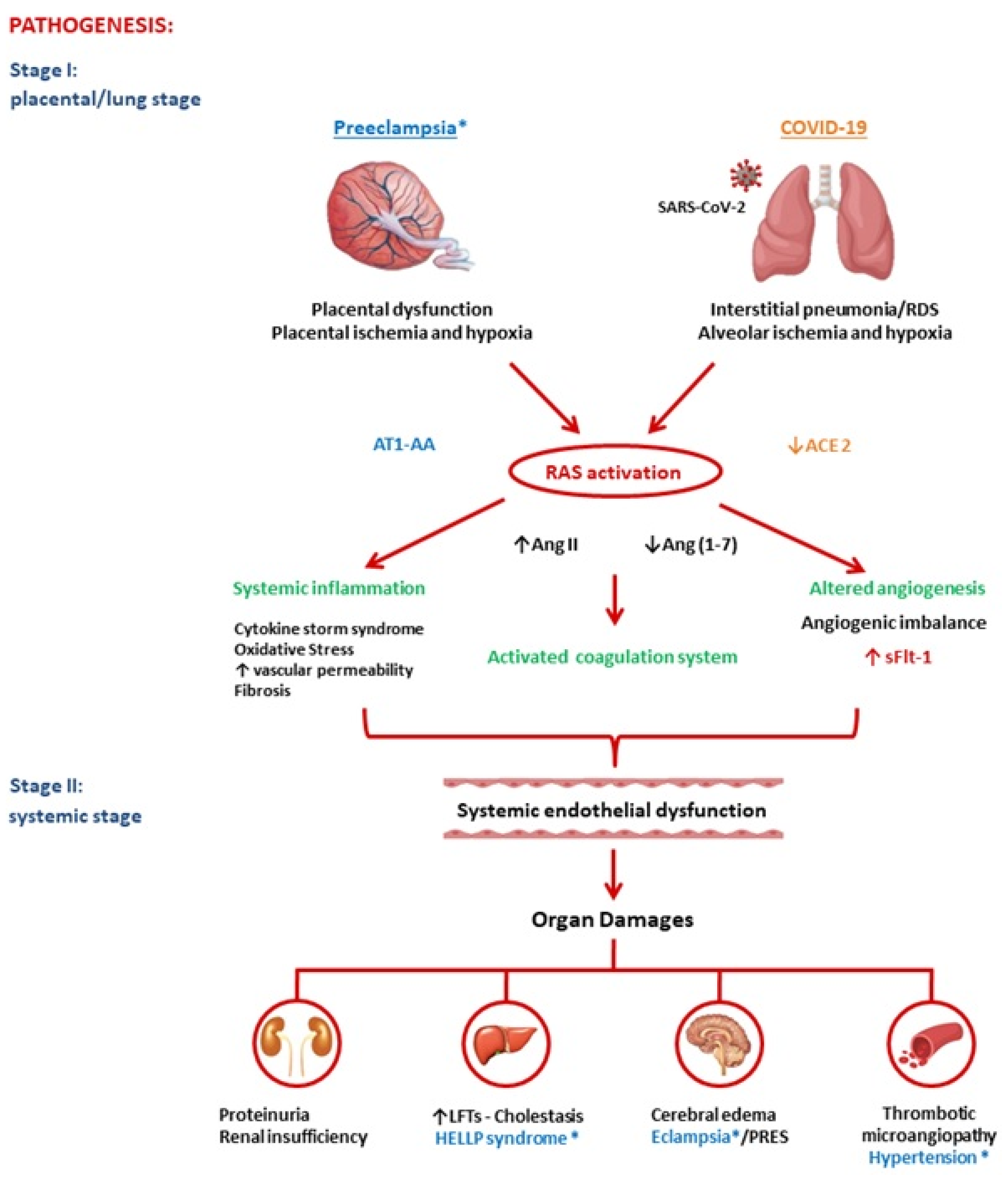
2. Pathogenesis of Preeclampsia and COVID-19

In both pathologies, we were able to identify two main phases. In PE, a placental dysfunction is followed by maternal syndrome (systemic vascular inflammation). First, placental ischemia occurs most commonly because of vascular damage due to anomalous placentation or intraplacental malperfusion. In the next step, the hypoxic placenta then liberates bioactive mediators including sFlt-1, reactive oxygen species, and inflammatory cytokines (e.g., TNF-α, IL-1, -6, and -8) into the maternal circulation, which has been determined to be responsible for the consequent endothelial damage and related clinical manifestations [11].
In COVID-19 infection, the virus triggers an interstitial pneumonia. This causes alveolar hypoxia, which can quickly evolve into a severe respiratory distress syndrome. Moreover, inflammatory cells and cytokines can enter the bloodstream and play a relevant role in ED and in a multiple organ dysfunction syndrome (“cytokine storm syndrome”) [12]. Endotheliitis, in turn, is an important precursor of a generalized hypercoagulable state that results in macro- and microvascular thrombosis in the pulmonary vascular system and beyond [13].
In conclusion, placental/alveolar hypoxia could lead to PE and severe COVID-19, respectively, through an angiogenic imbalance and subsequent exacerbated systemic inflammatory reaction due to RAS activation, as explained below in detail and in Figure 1.

2.1. Renin-Angiotensin System

The RAS has been identified as playing a key role in the pathogenesis of both diseases. The classical RAS as it appeared in the mid-1970s is a hormone system that regulates blood pressure and fluid-electrolyte balance. When renal blood flow is reduced, an enzymatic cascade is activated: the juxtaglomerular cells in the kidneys transform the precursor prorenin into renin that converts angiotensinogen, released by the liver, into angiotensin I (Ang I). Ang I is subsequently turned into Ang II by the angiotensin-converting enzyme (ACE) located on the surface of, mostly, lung vascular endothelial cells. Ang II leads to an increase in blood pressure through the vasoconstriction and the secretion of the hormone aldosterone from the adrenal cortex. This increases the volume of extracellular fluid in the body. In the renal tubules, aldosterone provokes the reabsorption of sodium, and therefore of water into the blood, simultaneously causing the excretion of potassium, to maintain electrolyte balance. Since then, a broader view on RAS has gradually emerged [14,15]. Local tissue RAS systems have been recognized in most organs. Recently, evidence for an intracellular RAS has been reported. The new expanded view of RAS therefore covers endocrine, paracrine, and intracrine functions. Other RAS peptides have been shown to have biological actions, for example, the Ang (1–7) generated from angiotensin I (Ang I) or Ang II by ACE2 and other peptidases. This seems to play an important role in offsetting many of Ang II’s actions (Figure 2). Lung tissue has high RAS activity and is the main site of Ang II synthesis. The local RAS is also present in the placenta and is one of the major extrarenal RAS sites during pregnancy [16]. Various organ systems have a predilection for the involvement in COVID-19 and PE, and each of these organ systems can be a site of a tissue-based RAS, such as the brain, heart, and kidneys [15]. The RAS undergoes important changes in response to pregnancy and plays a crucial role in placentation. It is upregulated in normal pregnant women and downregulated in women with PE. Although Ang II levels are enhanced during pregnancy, normotensive pregnant women are refractory to its vasopressor effects. Trophoblasts are rich in angiotensin type 1 receptors (AT1Rs) and thus respond to changes in Ang II concentrations that occur during pregnancy. While plasma Ang I, Ang II, Ang (1–7), and plasma renin activity are all noted to decrease in the circulation of preeclamptic women and in the chorionic villi of preeclamptic placentas, Ang II peptide, angiotensinogen, and AT1R mRNA levels are observed to increase, showing an excessive pressor response to Ang II, which is also due to the presence of agonistic autoantibodies to the angiotensin type 1 receptor (AT1-AA). These antibodies have been reported to facilitate the interaction of Ang II with its receptor and may play a role in increasing vascular sensitivity to Ang II in preeclamptic women [17]. In COVID-19, ACE2 acts as a functional SARS-CoV-2 receptor, which leads to the downregulation of ACE2, which catalyzes and inactivates Ang II and produces Ang (1–7), a potent vasodilator. This serves as a negative regulator of the RAS. Recently, Liu et al. have also revealed that serum Ang II levels were directly proportional to the viral load and extent of lung damage in COVID-19 [18].

2.2. Angiotensin II

Ang II is a key vasoactive hormone. Recently, a large number of experimental studies have shown that it mediates several events of the inflammatory processes via binding to AT1Rs located in the heart, lungs, blood vessels, kidneys, adrenal glands, and placenta. It also plays an essential role in myocardial hypertrophy, fibrosis, inflammation, vascular remodeling, angiogenesis, atherosclerosis, and microvascular thrombosis and therefore ED [19].
Ang II-mediated mechanisms may explain the primary clinical-pathological features of COVID-19 and PE [20]. Indeed, both PE and COVID-19 could be due to an excess of Ang II or AT1 receptor activation.
ACE2 knockout mice have showed more severe lung damage caused by increased hydrostatic pressure, reduced perfusion, and severe pulmonary edema [21]. Activation of coagulation and prothrombotic events is a well-known phenomenon in PE and COVID-19 [22,23]. Fibrosis is one of the most salient pathologic features of preeclamptic placentas [24]. Autopsy of the lungs after severe COVID-19 showed fibrin deposition [25].
The novel observation that Ang II modulates T-cell responses suggests a possible role for the peptide in autoimmune diseases. In COVID-19, as in PE, it has been shown that CD4 T lymphocytes are quickly activated to become pathogenic T helper-1 cells. This subsequently triggers a “cytokine storm” through increased expression of interleukin-6 (IL-6) and many other cytokines [26]. In the late 1990s, Wallukat et al. proposed that PE is a pregnancy-induced autoimmune disease in which abnormalities in placentation result from circulating autoantibodies, which, in turn, react to and activate the AT1R (AT1-AA). However, it has been hypothesized that AT1-AA may appear in a variety of pathological circumstances related to vascular damage; for example, parvovirus B19 also causes the generation of AT1-AA during pregnancy [17].

2.3. Link between RAS and sFlt-1

The RAS is an important mediator of angiogenesis. Recent studies have demonstrated sFlt-1 is regulated by AT1 receptor signaling, along with multiple genes [27]. Ang II has also been shown to regulate angiogenesis [28].

2.4. sFlt1 and Endothelial Dysfunction

sFlt-1 (sVEGFR-1) has been determined to be an anti-angiogenic factor expressed as an alternate junction variant of VEGFR-1 that lacks both the transmembrane and cytoplasmic domains. sFlt-1 antagonizes VEGF and PlGF (pro-angiogenic factors) in the circulation by binding and preventing interaction with their endothelial receptors, creating an anti-angiogenic state and ED [29], Figure 3. Clinical tests and experimental research have suggested that endothelial cell damage reduces the synthesis of vasorelaxant agents, increases the production of vasoconstrictors, impairs the synthesis of endogenous anticoagulants, and increases procoagulant production [30]. VEGF and PlGF are important in both angiogenesis and the maintenance of endothelial cell health at baseline [31].
Preeclamptic women show an imbalance between sFlt-1, VEGF, and PlGF in serum. sFlt1 levels are increased, and placental sFlt1 mRNA is upregulated, whereas free (unbound) PlGF and free VEGF levels are suppressed [32]. Changes in these markers precede the onset of clinical disease. It has been suggested that sFlt-1 could provoke generalized endotheliosis in blood vessels, leading to hypertension and proteinuria [29]. A causal relationship between sFlt1 and the clinical manifestations of PE was suggested in a study by Levine et al., which showed that an increase in circulating levels of sFlt1 was correlated with greater severity of PE [33]. Currently, the sFlt1/PlGF ratio is used as a clinical biomarker for the early detection and prognosis of PE [34]. An angiogenic imbalance also seems to be present in COVID-19, as shown by our group [8]. Levels of sFlt-1 were also noted to be significantly higher in patients with pneumonia due to COVID-19, compared to those with pneumonia due to other causes and to healthy controls. PlGF values were not significantly affected by COVID-19, but the sFlt1/PlGF ratio was higher in COVID-19-positive pneumonia compared with COVID-19-negative pneumonia (14.1 vs. 5.0). Subsequently, other authors confirmed increased sFlt-1 values in severe COVID-19 and identified sFlt-1 as a biomarker to predict survival and thrombotic accidents in COVID-19 patients [35,36]. ED also contributes to the pathogenesis of a variety of serious diseases, including sepsis and acute pancreatitis, which are conditions with elevated sFlt-1 levels and poor outcomes [37,38]. Ang II and excess sFlt-1-mediated vascular ED may explain the consistent involvement of several organs with local RAS in PE as well as in COVID-19, e.g., kidney, liver, and brain. In the kidney, endothelial damage causes proteinuria and produces characteristic pathological lesions and glomerular endotheliosis with fibrinogen and fibrin deposits within and under the endothelial cells. In the liver, vascular actions of Ang II and Ang II-mediated mitochondrial injury may contribute to the mild cholestasis and release of hepatic enzymes. In the brain, vascular dysfunction and ED in the central vasculature may result in impaired dynamic cerebral autoregulation, neuronal cell injury and cerebral edema and may explain the development of grand mal seizures, i.e., eclampsia, and, occasionally, even coma [27,39,40].

3. Risk and Protective Factors of Preeclampsia and COVID-19

PE is a clinical syndrome with several underlying risk factors; i.e., maternal constitutional factors (genetics, obesity, diet, and comorbid conditions) in combination with normal inflammatory changes in pregnancy can lead directly to ED [41,42]. Pro-inflammatory factors play a central role in COVID-19 severity, especially in patients with comorbidities. SARS-CoV-2 infection could further exacerbate sFlt-1 release in these patients in which sFlt-1 levels are already high, as in those with cardiovascular disease [43]. We therefore hypothesize that every person has a threshold for angiogenic imbalance, which, when crossed, may lead to PE in pregnancy or severe COVID-19 in SARS-CoV-2 infection. It is possible that angiogenic factors interact with susceptibility elements in complex ways to produce a disease that varies greatly in timing and severity. Current research has shown that the poor prognosis and mortality in patients with COVID-19 are related to factors such as gender (male), age (>60 years), ethnicity, low vitamin D levels, blood type, hyperinflammation, hyperandrogenism, and underlying diseases (hypertension, diabetes, cardiovascular and cerebrovascular diseases, respiratory system disease, renal disease, and obesity) Table 1 [44,45,46,47]. These risk factors support our hypothesis regarding the presence of a preexisting ED at baseline and indicate that suboptimal cardiovascular health and ED may predispose patients to PE/COVID-19 severe disease.

3.1. Blood Type

A significant association has been found between the AB0 blood group and COVID-19 severity as well as hypertensive disorders of pregnancy. It is currently known that the AB0 blood group may affect hemostatic balance. Type blood 0 is protective against the development of PE and severe COVID-19 as it is associated with lower ACE levels and higher ACE2 activity. Blood A type confers greater risk for developing hypertensive disorders of pregnancy and severe COVID-19 due to its positive association with ACE activity [48,49,50,51,52,53].

3.2. Obesity

Overweight adults are most at risk during the COVID-19 pandemic, as most women who are obese have a higher risk of PE. Obese patients show marked adipose tissue dysfunction and dysregulated adipokine/cytokine secretion, resulting in a chronic pro-inflammatory state [54,55].

3.3. Diabetes

Diabetes has been identified as a risk factor for PE and COVID-19. It has been suggested that angiogenic and insulin-dependent pathways may affect each other. Furthermore, the pro-angiogenic effect of PlGF is strongly influenced and modified by insulin resistance, leading to widespread endothelial activation and injury culminating in vascular dysfunction [56,57,58].

3.4. Vascular Diseases

Hypertension, cardiovascular diseases (CVDs), and cerebrovascular diseases are the most common underlying diseases in COVID-19 patients. PE, COVID-19, and CVD patients exhibit inflammation and endothelial stress that often results in increased production of pro-inflammatory factors [41,59,60,61].

3.5. Vitamin D

Recent studies support a link between vitamin D deficiency and worse COVID-19 outcomes. Vitamin D deficiency has also been shown to be related to a higher risk of maternal complications including PE. Vitamin D is a powerful immunomodulator. It can regulate IL-6 activity and suppress the pro-inflammatory cytokine response of macrophages and respiratory epithelial cells to various viruses, promoting angiogenesis [62,63,64,65,66].

3.6. Smoking

A potential protective effect of smoking and nicotine on SARS-CoV-2 infection has also been noted. Smoking is the only environmental exposure known to consistently reduce the risk of PE and gestational hypertension. It has been associated with lower circulating concentrations of anti-angiogenic proteins, such as sFlt1, and higher levels of pro-angiogenic proteins, such as PlGF [67]. An unexpectedly low prevalence of current smoking was also observed among COVID-19 patients. The suggested hypothesis is that nicotine is a relevant inhibitor of pro-inflammatory cytokines [68].

4. Clinical Features of Preeclampsia and COVID-19

PE and COVID-19 are multisystem disorders with heterogeneous presentations and their manifestations reflect widespread ED, often resulting in vasoconstriction and end-organ ischemia. Various organ systems that are characteristically involved in PE and COVID-19 can be possible sites of a tissue-based RAS [27]. PE is defined as new-onset hypertension with proteinuria or other specific end-organ dysfunction after 20 weeks of pregnancy [3]. Extreme variability is reported in the gestational age at onset, the rate of progression, and the implication of several compartments leading to eclampsia, hemorrhagic stroke, hemolysis, elevated liver function, low platelet counts, thrombotic microangiopathy, renal failure, pulmonary edema, placental infarction, abruptio placentae, fetal growth restriction, and preterm birth [69].
SARS-CoV-2 can cause various symptoms ranging from mild flu-like manifestations, such as dry cough, phlegm, ageusia, anosmia, myalgia, or diarrhea, to severe pneumonia or even acute respiratory distress syndrome, and it may display and cause lasting harm to other organs [70]. The transition from mild to severe disease in COVID-19 patients can occur rapidly without the presentation of signals [71]. Although initially considered a respiratory disease, rapidly accumulating data suggests that COVID-19 occurs in a unique, deeply prothrombotic environment leading to both arterial and venous thrombosis [72,73].
COVID-19-associated acute kidney injury has a prevalence rate reported as high as 46% in large cohorts of hospitalized patients. Signs of renal involvement in COVID-19 patients include proteinuria [74,75]. To date, there is a scarcity of published data regarding new-onset hypertension during COVID-19. In a single-centered, retrospective, observational study, the prevalence of new-onset hypertension was significantly higher in severe COVID-19 patients compared to non-serious ones during hospitalization [76]. Finally, ED in the context of COVID-19 could contribute to posterior reversible encephalopathy syndrome, as described in some case reports, in a similar way to eclampsia [77].

5. Laboratory Abnormalities in PE and COVID-19

It has been determined that COVID-19 and PE are both associated with hypocalcemia [78,79], high levels of IL-6 [80,81], and hypoalbuminemia [82,83,84,85]. Several laboratory test results that are indicative of endotheliosis in the systemic, renal, and hepatic circulation are present in PE and COVID-19 infections. They include thrombocytopenia, high levels of D-dimer, proteinuria, hypertransaminasemia, and an elevation in lactate dehydrogenase and sFlt1—Table 2, [18,86].

6. Long-Term Effects of PE and COVID-19

Evidence is emerging of an increased long-term risk of cardiovascular, cerebrovascular, and renal disease in women who have had PE, and in their babies [87]. Long-term outcomes in these women suggest that the endothelial changes are not limited to pregnancy. Serum sFlt-1 is higher in women with previous PE compared to women with a previous normal pregnancy, and increases can be detected up to 6 months or more after childbirth [88]. Meanwhile, long-term consequences following a SARS-CoV-2 infection are yet to be determined; however, preliminary data suggest that these patients could have persistent microvascular dysfunction post-infection, mediated in part by increased sensitivity to Ang II, in a similar way to PE. The most common problem leading to COVID-19-induced mortality is respiratory failure due to extensive, accelerating lung fibrogenesis. Thus, the acute acceleration of lung fibrosis in COVID-19 can be explained by ACE-AngII-AT1 overactivation caused by the SARS-CoV-2 virus [89,90].

7. Conclusions

PE and COVID-19 have common pathogenic pathways. Both diseases are characterized by significant alterations in the RAS with an imbalanced proportion of anti-angiogenic and pro-angiogenic soluble plasmatic factors. In summary, we believe that both PE and COVID-19 are due to a state of ED secondary to increased Ang II and ensuing excessive levels of circulating anti-angiogenic factors, such as sFlt1. COVID-19 and PE are defined to be diseases that begin, respectively, in the lungs and in the placenta, and both end in the endothelium. In conclusion, SARS-CoV-2 infection could be defined an angiogenic-pneumo-syndrome.
A better understanding of the biological and molecular mechanisms of COVID-19 and PE, as presented here, may offer a window for future research into exploiting these pathways to improve our medical treatment. We hope that this comparison with PE may shed light and increase knowledge for the management of both conditions.
These considerations are valid in the case of original SARS-CoV-2 primary infection. Emerging SARS-CoV-2 variants as well as the vaccination could alter various aspects of the virus biology, including human ACE-2 receptor binding affinity and therefore the RAS mediated consequences [91,92].

Author Contributions

Conceptualization, V.G.; writing—original draft preparation, V.G.; writing—review and editing, V.G., P.V., C.G.-P., M.C. and A.C. All authors have read and agreed to the published version of the manuscript.

Funding

This research received no external funding.

Conflicts of Interest

The authors declare no conflict of interest.

References

  1. Guo, Y.R.; Cao, Q.D.; Hong, Z.S.; Tan, Y.Y.; Chen, S.D.; Jin, H.J.; Tan, K.S.; Wang, D.Y.; Yan, Y. The origin, transmission and clinical therapies on coronavirus disease 2019 (COVID-19) outbreak—An update on the status. Mil. Med. Res. 2020, 7, 11. [Google Scholar] [CrossRef] [Green Version]
  2. Cheng, H.; Wang, Y.; Wang, G.Q. Organ-protective effect of angiotensin-converting enzyme 2 and its effect on the prognosis of COVID-19. J. Med. Virol. 2020, 92, 726–730. [Google Scholar] [CrossRef] [PubMed]
  3. Roberts, J.M.; August, P.A.; Bakris, G.; Barton, J.R.; Bernstein, I.M.; Druzin, M.; Gaiser, R.R.; Granger, J.P.; Jeyabalan, A.; Johnson, D.D.; et al. Hypertension in pregnancy. Report of the American College of Obstetricians and Gynecologists’ Task Force on Hypertension in Pregnancy. Obstet. Gynecol. 2013, 122, 1122–1131. [Google Scholar]
  4. Mayrink, J.; Costa, M.L.; Cecatti, J.G. Preeclampsia in 2018: Revisiting Concepts, Physiopathology, and Prediction. Sci. World J. 2018, 2018, 6268276. [Google Scholar] [CrossRef] [Green Version]
  5. Mendoza, M.; Garcia-Ruiz, I.; Maiz, N.; Rodo, C.; Garcia-Manau, P.; Serrano, B.; Lopez-Martinez, R.M.; Balcells, J.; Fernandez-Hidalgo, N.; Carreras, E.; et al. Pre-eclampsia-like syndrome induced by severe COVID-19: A prospective observational study. BJOG 2020, 127, 1374–1380. [Google Scholar] [CrossRef] [PubMed]
  6. Papageorghiou, A.T.; Deruelle, P.; Gunier, R.B.; Rauch, S.; García-May, P.K.; Mhatre, M.; Usman, M.A.; Abd-Elsalam, S.; Etuk, S.; Simmons, L.E.; et al. Preeclampsia and COVID-19: Results from the INTERCOVID prospective longitudinal study. Am. J. Obstet. Gynecol. 2021, 225, 289.e1–289.e17. [Google Scholar] [CrossRef] [PubMed]
  7. Stepan, H.; Hund, M.; Andraczek, T. Combining Biomarkers to Predict Pregnancy Complications and Redefine Preeclampsia: The Angiogenic-Placental Syndrome. Hypertension 2020, 75, 918–926. [Google Scholar] [CrossRef] [PubMed]
  8. Giardini, V.; Carrer, A.; Casati, M.; Contro, E.; Vergani, P.; Gambacorti-Passerini, C. Increased sFLT-1/PlGF ratio in COVID-19: A novel link to angiotensin II-mediated endothelial dysfunction. Am. J. Hematol. 2020, 95, E188–E191. [Google Scholar] [CrossRef]
  9. Giardini, V.; Ornaghi SAcampora, E.; Vasarri, M.V.; Arienti, F.; Gambacorti-Passerini, C.; Casati, M.; Carrer, A.; Vergani, P. Letter to the Editor: SFlt-1 and PlGF Levels in Pregnancies Complicated by SARS-CoV-2 Infection. Viruses 2021, 13, 2377. [Google Scholar] [CrossRef]
  10. Murphy, S.R.; Cockrell, K. Regulation of soluble fms-like tyrosine kinase-1 production in response to placental ischemia/hypoxia: Role of angiotensin II. Physiol. Rep. 2015, 3, e12310. [Google Scholar] [CrossRef]
  11. Staff, A.C. The two-stage placental model of preeclampsia: An update. J. Reprod. Immunol. 2019, 134–135, 1–10. [Google Scholar] [CrossRef] [PubMed]
  12. Lippi, G.; Sanchis-Gomar, F.; Henry, B.M. COVID-19: Unravelling the clinical progression of nature’s virtually perfect biological weapon. Ann. Transl. Med. 2020, 8, 693. [Google Scholar] [CrossRef] [PubMed]
  13. Varga, Z.; Flammer, A.J.; Steiger, P.; Haberecker, M.; Andermatt, R.; Zinkernagel, A.S.; Mehra, M.R.; Schuepbach, R.A.; Ruschitzka, F.; Moch, H. Endothelial cell infection and endotheliitis in COVID-19. Lancet 2020, 395, 1417–1418. [Google Scholar] [CrossRef]
  14. Patel, S.; Rauf, A.; Khan, H.; Abu-Izneid, T. Renin-angiotensin-aldosterone (RAAS): The ubiquitous system for homeostasis and pathologies. Biomed. Pharmacother. 2017, 94, 317–325. [Google Scholar] [CrossRef]
  15. Santos, R.A.S.; Oudit, G.Y.; Verano-Braga, T.; Canta, G.; Steckelings, U.M.; Bader, M. The renin-angiotensin system: Going beyond the classical paradigms. Am. J. Physiol. Heart Circ. Physiol. 2019, 316, H958–H970. [Google Scholar] [CrossRef]
  16. Verdonk, K.; Visser, W.; Van Den Meiracker, A.H.; Danser, A.H. The renin-angiotensin-aldosterone system in pre-eclampsia: The delicate balance between good and bad. Clin. Sci. 2014, 126, 537–544. [Google Scholar] [CrossRef]
  17. Walther, T.; Stepan, H. Agonist autoantibodies against the angiotensin AT1 receptor in renal and hypertensive disorders. Curr. Hypertens. Rep. 2007, 9, 128–132. [Google Scholar] [CrossRef]
  18. Liu, Y.; Yang, Y.; Zhang, C.; Huang, F.; Wang, F.; Yuan, J.; Wang, Z.; Li, J.; Li, J.; Feng, C.; et al. Clinical and biochemical indexes from 2019-nCoV infected patients linked to viral loads and lung injury. Sci. China Life Sci. 2020, 63, 364–374. [Google Scholar] [CrossRef] [Green Version]
  19. Benigni, A.; Cassis, P.; Remuzzi, G. Angiotensin II revisited: New roles in inflammation, immunology and aging. EMBO Mol. Med. 2010, 2, 247–257. [Google Scholar] [CrossRef]
  20. Miesbach, W. Pathological Role of Angiotensin II in Severe COVID-19. TH Open 2020, 4, e138–e144. [Google Scholar] [CrossRef]
  21. Wang, L.; Li, Y.; Qin, H.; Xing, D.; Su, J.; Hu, Z. Crosstalk between ACE2 and PLGF regulates vascular permeability during acute lung injury. Am. J. Transl. Res. 2016, 8, 1246–1252. [Google Scholar]
  22. Egan, K.; Kevane, B.; Ní Áinle, F. Elevated venous thromboembolism risk in preeclampsia: Molecular mechanisms and clinical impact. Biochem. Soc. Trans. 2015, 43, 696–701. [Google Scholar] [CrossRef]
  23. Bikdeli, B.; Madhavan, M.V.; Jimenez, D.; Chuich, T.; Dreyfus, I.; Driggin, E.; Nigoghossian, C.; Ageno, W.; Madjid, M.; Guo, Y.; et al. Global COVID-19 Thrombosis Collaborative Group, Endorsed by the ISTH, NATF, ESVM, and the IUA, Supported by the ESC Working Group on Pulmonary Circulation and Right Ventricular Function. COVID-19 and Thrombotic or Thromboembolic Disease: Implications for Prevention, Antithrombotic Therapy, and Follow-Up: JACC State-of-the-Art Review. J. Am. Coll. Cardiol. 2020, 75, 2950–2973. [Google Scholar] [CrossRef]
  24. Ohmaru-Nakanishi, T.; Asanoma, K.; Fujikawa, M.; Fujita, Y.; Yagi, H.; Onoyama, I.; Hidaka, N.; Sonoda, K.; Kato, K. Fibrosis in Preeclamptic Placentas Is Associated with Stromal Fibroblasts Activated by the Transforming Growth Factor-β1 Signaling Pathway. Am. J. Pathol. 2018, 188, 683–695. [Google Scholar] [CrossRef] [Green Version]
  25. Carsana, L.; Sonzogni, A.; Nasr, A.; Rossi, R.S.; Pellegrinelli, A.; Zerbi, P.; Rech, R.; Colombo, R.; Antinori, S.; Corbellino, M.; et al. Pulmonary post-mortem findings in a series of COVID-19 cases from northern Italy: A two-centre descriptive study. Lancet Infect. Dis. 2020, 20, 1135–1140. [Google Scholar] [CrossRef]
  26. Mehta, P.; McAuley, D.F.; Brown, M.; Sanchez, E.; Tattersall, R.S.; Manson, J.J.; HLH Across Speciality Collaboration, UK. COVID-19: Consider cytokine storm syndromes and immunosuppression. Lancet 2020, 395, 1033–1034. [Google Scholar] [CrossRef]
  27. Irani, R.A.; Xia, Y. The functional role of the renin-angiotensin system in pregnancy and preeclampsia. Placenta 2008, 29, 763–771. [Google Scholar] [CrossRef] [Green Version]
  28. Anton, L.; Merrill, D.C.; Neves, L.A.; Gruver, C.; Moorefield, C.; Brosnihan, K.B. Angiotensin II and angiotensin-(1-7) decrease sFlt1 release in normal but not preeclamptic chorionic villi: An in vitro study. Reprod. Biol. Endocrinol. 2010, 8, 135. [Google Scholar] [CrossRef] [PubMed] [Green Version]
  29. Maynard, S.E.; Min, J.Y.; Merchan, J.; Lim, K.H.; Li, J.; Mondal, S.; Libermann, T.A.; Morgan, J.P.; Sellke, F.W.; Stillman, I.E.; et al. Excess placental soluble fms-like tyrosine kinase 1 (sFlt1) may contribute to endothelial dysfunction, hypertension, and proteinuria in preeclampsia. J. Clin. Investig. 2003, 111, 649–658. [Google Scholar] [CrossRef] [Green Version]
  30. Roberts, J.M. Objective evidence of endothelial dysfunction in preeclampsia. Am. J. Kidney Dis. 1999, 33, 992–997. [Google Scholar] [CrossRef]
  31. He, Y.; Smith, S.K.; Day, K.A.; Clark, D.E.; Licence, D.R.; Charnock-Jones, D.S. Alternative splicing of vascular endothelial growth factor (VEGF)-R1 (FLT-1) pre-mRNA is important for the regulation of VEGF activity. Mol. Endocrinol. 1999, 13, 537–545. [Google Scholar] [CrossRef] [PubMed]
  32. Shanes, E.D.; Mithal, L.B.; Otero, S.; Azad, H.A.; Miller, E.S.; Goldstein, J.A. Placental Pathology in COVID-19. Am. J. Clin. Pathol. 2020, 154, 23–32. [Google Scholar] [CrossRef] [PubMed]
  33. Levine, R.J.; Maynard, S.E.; Qian, C.; Lim, K.H.; England, L.J.; Yu, K.F.; Schisterman, E.F.; Thadhani, R.; Sachs, B.P.; Epstein, F.H.; et al. Circulating angiogenic factors and the risk of preeclampsia. N. Engl. J. Med. 2004, 350, 672–683. [Google Scholar] [CrossRef] [PubMed] [Green Version]
  34. Zeisler, H.; Llurba, E.; Chantraine, F.; Vatish, M.; Staff, A.C.; Sennström, M.; Olovsson, M.; Brennecke, S.P.; Stepan, H.; Allegranza, D.; et al. Predictive Value of the sFlt-1:PlGF Ratio in Women with Suspected Preeclampsia. N. Engl. J. Med. 2016, 374, 13–22. [Google Scholar] [CrossRef]
  35. Negro, A.; Fama, A.; Penna, D.; Belloni, L.; Zerbini, A.; Giuri, P.G. SFLT-1 levels in COVID-19 patients: Association with outcome and thrombosis. Am. J. Hematol. 2021, 96, E41–E43. [Google Scholar] [CrossRef]
  36. Dupont, V.; Kanagaratnam, L.; Goury, A.; Poitevin, G.; Bard, M.; Julien, G.; Bonnivard, M.; Champenois, V.; Noel, V.; Mourvillier, B.; et al. Excess Soluble fms-like Tyrosine Kinase 1 Correlates with Endothelial Dysfunction and Organ Failure in Critically Ill Coronavirus Disease 2019 Patients. Clin. Infect. Dis. 2021, 72, 1834–1837. [Google Scholar] [CrossRef]
  37. Shapiro, N.I.; Schuetz, P.; Yano, K.; Sorasaki, M.; Parikh, S.M.; Jones, A.E.; Trzeciak, S.; Ngo, L.; Aird, W.C. The association of endothelial cell signaling, severity of illness, and organ dysfunction in sepsis. Crit. Care 2010, 14, R182. [Google Scholar] [CrossRef] [Green Version]
  38. Dumnicka, P.; Sporek, M.; Mazur-Laskowska, M.; Ceranowicz, P.; Kuźniewski, M.; Drożdż, R.; Ambroży, T.; Olszanecki, R.; Kuśnierz-Cabala, B. Serum Soluble Fms-Like Tyrosine Kinase 1 (sFlt-1) Predicts the Severity of Acute Pancreatitis. Int. J. Mol. Sci. 2016, 17, 2038. [Google Scholar] [CrossRef] [Green Version]
  39. Maynard, S.E.; Karumanchi, S.A. Angiogenic factors and preeclampsia. Semin. Nephrol. 2011, 31, 33–46. [Google Scholar] [CrossRef] [Green Version]
  40. van Veen, T.R.; Panerai, R.B.; Haeri, S.; Griffioen, A.C.; Zeeman, G.G.; Belfort, M.A. Cerebral autoregulation in normal pregnancy and preeclampsia. Obstet. Gynecol. 2013, 122, 1064–1069. [Google Scholar] [CrossRef] [Green Version]
  41. Fox, R.; Kitt, J.; Leeson, P.; Aye, C.Y.L.; Lewandowski, A.J. Preeclampsia: Risk Factors, Diagnosis, Management, and the Cardiovascular Impact on the Offspring. J. Clin. Med. 2019, 8, 1625. [Google Scholar] [CrossRef] [Green Version]
  42. Raymond, D.; Peterson, E. A critical review of early-onset and late-onset preeclampsia. Obstet. Gynecol. Surv. 2011, 66, 497–506. [Google Scholar] [CrossRef]
  43. Ky, B.; French, B.; Ruparel, K.; Sweitzer, N.K.; Fang, J.C.; Levy, W.C.; Sawyer, D.B.; Cappola, T.P. The vascular marker soluble fms-like tyrosine kinase 1 is associated with disease severity and adverse outcomes in chronic heart failure. J. Am. Coll. Cardiol. 2011, 58, 386–394. [Google Scholar] [CrossRef] [Green Version]
  44. Albitar, O.; Ballouze, R.; Ooi, J.P.; Sheikh Ghadzi, S.M. Risk factors for mortality among COVID-19 patients. Diabetes Res. Clin. Pract. 2020, 166, 108293. [Google Scholar] [CrossRef]
  45. Chaudhary, M. COVID-19 susceptibility: Potential of ACE2 polymorphisms. Egypt J. Med. Hum. Genet. 2020, 21, 54. [Google Scholar] [CrossRef]
  46. Swärd, P.; Edsfeldt, A.; Reepalu, A.; Jehpsson, L.; Rosengren, B.E.; Karlsson, M.K. Age and sex differences in soluble ACE2 may give insights for COVID-19. Crit. Care 2020, 24, 221. [Google Scholar] [CrossRef]
  47. Gargaglioni, L.H.; Marques, D.A. Let’s talk about sex in the context of COVID-19. J. Appl. Physiol. 2020, 128, 1533–1538. [Google Scholar] [CrossRef]
  48. Lee, B.; Zhang, Z.; Wikman, A.; Lindqvist, P.; Reilly, M. ABO and RhD blood groups and gestational hypertensive disorders: A population-based cohort study. BJOG 2012, 119, 1232–1237. [Google Scholar] [CrossRef]
  49. Franchini, M.; Mengoli, C.; Lippi, G. Relationship between ABO blood group and pregnancy complications: A systematic literature analysis. Blood Transfus. 2016, 14, 441–448. [Google Scholar] [CrossRef]
  50. Li, H.; Liu, Z.; Ge, J. Scientific research progress of COVID-19/SARS-CoV-2 in the first five months. J. Cell. Mol. Med. 2020, 24, 6558–6570. [Google Scholar] [CrossRef]
  51. Dai, X. ABO blood group predisposes to COVID-19 severity and cardiovascular diseases. Eur. J. Prev. Cardiol. 2020, 27, 1436–1437. [Google Scholar] [CrossRef] [PubMed]
  52. Zaidi, F.Z.; Zaidi, A.R.Z.; Abdullah, S.M.; Zaidi, S.Z.A. COVID-19 and the ABO blood group connection. Transfus. Apher. Sci. 2020, 59, 102838. [Google Scholar] [CrossRef] [PubMed]
  53. Gérard, C.; Maggipinto, G.; Minon, J.M. COVID-19 and ABO blood group: Another viewpoint. Br. J. Haematol. 2020, 190, e93–e94. [Google Scholar] [CrossRef] [PubMed]
  54. Stefan, N.; Birkenfeld, A.L.; Schulze, M.B.; Ludwig, D.S. Obesity and impaired metabolic health in patients with COVID-19. Nat. Rev. Endocrinol. 2020, 16, 341–342. [Google Scholar] [CrossRef] [Green Version]
  55. Spradley, F.T. Metabolic abnormalities and obesity’s impact on the risk for developing preeclampsia. Am. J. Physiol. Regul. Integr. Comp. Physiol. 2017, 312, R5–R12. [Google Scholar] [CrossRef] [Green Version]
  56. Weissgerber, T.L.; Mudd, L.M. Preeclampsia and diabetes. Curr. Diab. Rep. 2015, 15, 9. [Google Scholar] [CrossRef] [Green Version]
  57. Ghosh, A.; Freestone, N.S.; Anim-Nyame, N.; Arrigoni, F.I.F. Microvascular function in pre-eclampsia is influenced by insulin resistance and an imbalance of angiogenic mediators. Physiol. Rep. 2017, 5, e13185. [Google Scholar] [CrossRef]
  58. Bloomgarden, Z.T. Diabetes and COVID-19. J. Diabetes 2020, 12, 347–348. [Google Scholar] [CrossRef] [Green Version]
  59. Nishiga, M.; Wang, D.W.; Han, Y.; Lewis, D.B.; Wu, J.C. COVID-19 and cardiovascular disease: From basic mechanisms to clinical perspectives. Nat. Rev. Cardiol. 2020, 17, 543–558. [Google Scholar] [CrossRef]
  60. Fan, H.; Tang, X.; Song, Y.; Liu, P.; Chen, Y. Influence of COVID-19 on Cerebrovascular Disease and its Possible Mechanism. Neuropsychiatr. Dis. Treat. 2020, 16, 1359–1367. [Google Scholar] [CrossRef]
  61. Leon, L.J.; McCarthy, F.P.; Direk, K.; Gonzalez-Izquierdo, A.; Prieto-Merino, D.; Casas, J.P.; Chappell, L. Preeclampsia and Cardiovascular Disease in a Large UK Pregnancy Cohort of Linked Electronic Health Records: A CALIBER Study. Circulation 2019, 140, 1050–1060. [Google Scholar] [CrossRef] [Green Version]
  62. Ali, N. Role of vitamin D in preventing of COVID-19 infection, progression and severity. J. Infect. Public Health 2020, 13, 1373–1380. [Google Scholar] [CrossRef]
  63. Achkar, M.; Dodds, L.; Giguère, Y.; Forest, J.C.; Armson, B.A.; Woolcott, C.; Agellon, S.; Spencer, A.; Weiler, H.A. Vitamin D status in early pregnancy and risk of preeclampsia. Am. J. Obstet. Gynecol. 2015, 212, 511.e1-7. [Google Scholar] [CrossRef]
  64. Kim, H.; Baek, S.; Hong, S.M.; Lee, J.; Jung, S.M.; Lee, J.; Cho, M.; Kwok, S.K.; Park, S.H. 1,25-dihydroxy Vitamin D3 and Interleukin-6 Blockade Synergistically Regulate Rheumatoid Arthritis by Suppressing Interleukin-17 Production and Osteoclastogenesis. J. Korean Med. Sci. 2020, 35, e40. [Google Scholar] [CrossRef]
  65. Ferrari, D.; Locatelli, M.; Briguglio, M.; Lombardi, G. Is there a link between vitamin D status, SARS-CoV-2 infection risk and COVID-19 severity? Cell Biochem. Funct. 2020. Epub ahead of printing. [Google Scholar] [CrossRef]
  66. Brodowski, L.; Burlakov, J.; Myerski, A.C.; von Kaisenberg, C.S.; Grundmann, M.; Hubel, C.A.; von Versen-Höynck, F. Vitamin D prevents endothelial progenitor cell dysfunction induced by sera from women with preeclampsia or conditioned media from hypoxic placenta. PLoS ONE 2014, 9, e98527. [Google Scholar] [CrossRef] [Green Version]
  67. Karumanchi, S.A.; Levine, R.J. How does smoking reduce the risk of preeclampsia? Hypertension 2010, 55, 1100–1101. [Google Scholar] [CrossRef] [Green Version]
  68. Farsalinos, K.; Niaura, R.; Le Houezec, J.; Barbouni, A.; Tsatsakis, A.; Kouretas, D.; Vantarakis, A.; Poulas, K. Editorial: Nicotine and SARS-CoV-2: COVID-19 may be a disease of the nicotinic cholinergic system. Toxicol. Rep. 2020, 7, 658–663. [Google Scholar] [CrossRef]
  69. American College of Obstetricians and Gynecologists. ACOG Practice Bulletin No. 202: Gestational Hypertension and Preeclampsia. Obstet. Gynecol. 2019, 133, e1–e25. [Google Scholar] [CrossRef]
  70. Machhi, J.; Herskovitz, J.; Senan, A.M.; Dutta, D.; Nath, B.; Oleynikov, M.D.; Blomberg, W.R.; Meigs, D.D.; Hasan, M.; Patel, M.; et al. The Natural History, Pathobiology, and Clinical Manifestations of SARS-CoV-2 Infections. J. Neuroimmune Pharmacol. 2020, 15, 359–386. [Google Scholar] [CrossRef]
  71. Dhont, S.; Derom, E.; Van Braeckel, E.; Depuydt, P.; Lambrecht, B.N. The pathophysiology of ’happy’ hypoxemia in COVID-19. Respir. Res. 2020, 21, 198. [Google Scholar] [CrossRef]
  72. Connors, J.M.; Levy, J.H. COVID-19 and its implications for thrombosis and anticoagulation. Blood 2020, 135, 2033–2040. [Google Scholar] [CrossRef]
  73. Wichmann, D.; Sperhake, J.P.; Lütgehetmann, M.; Steurer, S.; Edler, C.; Heinemann, A.; Heinrich, F.; Mushumba, H.; Kniep, I.; Schröder, A.S.; et al. Autopsy Findings and Venous Thromboembolism in Patients With COVID-19: A Prospective Cohort Study. Ann. Intern. Med. 2020, 173, 268–277. [Google Scholar] [CrossRef]
  74. Hassanein, M.; Radhakrishnan, Y.; Sedor, J.; Vachharajani, T.; Vachharajani, V.T.; Augustine, J.; Demirjian, S.; Thomas, G. COVID-19 and the kidney. Clevel. Clin. J. Med. 2020, 87, 619–631. [Google Scholar] [CrossRef]
  75. Farouk, S.S.; Fiaccadori, E.; Cravedi, P.; Campbell, K.N. COVID-19 and the kidney: What we think we know so far and what we don’t. J. Nephrol. 2020, 33, 1213–1218. [Google Scholar] [CrossRef] [PubMed]
  76. Xiong, S.; Liu, L.; Lin, F.; Shi, J.; Han, L.; Liu, H.; He, L.; Jiang, Q.; Wang, Z.; Fu, W.; et al. Clinical characteristics of 116 hospitalized patients with COVID-19 in Wuhan, China: A single-centered, retrospective, observational study. BMC Infect. Dis. 2020, 20, 787. [Google Scholar] [CrossRef]
  77. Kishfy, L.; Casasola, M.; Banankhah, P.; Parvez, A.; Jan, Y.J.; Shenoy, A.M.; Thomson, C.; AbdelRazek, M.A. Posterior reversible encephalopathy syndrome (PRES) as a neurological association in severe Covid-19. J. Neurol. Sci. 2020, 414, 116943. [Google Scholar] [CrossRef]
  78. Cappellini, F.; Brivio, R.; Casati, M.; Cavallero, A.; Contro, E.; Brambilla, P. Low levels of total and ionized calcium in blood of COVID-19 patients. Clin. Chem. Lab. Med. 2020, 58, e171–e173. [Google Scholar] [CrossRef]
  79. Almaghamsi, A.; Almalki, M.H.; Buhary, B.M. Hypocalcemia in Pregnancy: A Clinical Review Update. Oman Med. J. 2018, 33, 453–462. [Google Scholar] [CrossRef]
  80. Paces, J.; Strizova, Z.; Smrz, D.; Cerny, J. COVID-19 and the immune system. Physiol. Res. 2020, 69, 379–388. [Google Scholar] [CrossRef]
  81. Aggarwal, R.; Jain, A.K.; Mittal, P.; Kohli, M.; Jawanjal, P.; Rath, G. Association of pro- and anti-inflammatory cytokines in preeclampsia. J. Clin. Lab. Anal. 2019, 33, e22834. [Google Scholar] [CrossRef] [PubMed] [Green Version]
  82. Violi, F.; Cangemi, R.; Romiti, G.F.; Ceccarelli, G.; Oliva, A.; Alessandri, F.; Pirro, M.; Pignatelli, P.; Lichtner, M.; Carraro, A.; et al. Is Albumin Predictor of Mortality in COVID-19? Antioxid. Redox Signal. 2021, 35, 139–142. [Google Scholar] [CrossRef]
  83. de la Rica, R.; Borges, M.; Aranda, M.; Del Castillo, A.; Socias, A.; Payeras, A.; Rialp, G.; Socias, L.; Masmiquel, L.; Gonzalez-Freire, M. Low Albumin Levels Are Associated with Poorer Outcomes in a Case Series of COVID-19 Patients in Spain: A Retrospective Cohort Study. Microorganisms 2020, 8, 1106. [Google Scholar] [CrossRef] [PubMed]
  84. Martell-Claros, N.; Abad-Cardiel, M.; García-Donaire, J.; De Los Santos, C.; Gonzalez, V.; Fuentes, M.; De La Fuente, J.A.; Perez-Perez, N.; Sosa, L. Hypoalbuminemia as a Risk Factor for Preeclampsia in the Pregnant Hypertensive Population. J. Hypertens. 2019, 37, e240. [Google Scholar] [CrossRef]
  85. Chen, H.; Tao, F.; Fang, X.; Wang, X. Association of hypoproteinemia in preeclampsia with maternal and perinatal outcomes: A retrospective analysis of high-risk women. J. Res. Med. Sci. 2016, 21, 98. [Google Scholar] [CrossRef]
  86. Duan, Z.; Li, C.; Leung, W.T.; Wu, J.; Wang, M.; Ying, C.; Wang, L. Alterations of Several Serum Parameters Are Associated with Preeclampsia and May Be Potential Markers for the Assessment of PE Severity. Dis. Markers 2020, 2020, 7815214. [Google Scholar] [CrossRef] [Green Version]
  87. Anderson, C.M. Preeclampsia: Exposing future cardiovascular risk in mothers and their children. J. Obstet. Gynecol. Neonatal. Nurs. 2007, 36, 3–8. [Google Scholar] [CrossRef]
  88. Powe, C.E.; Levine, R.J.; Karumanchi, S.A. Preeclampsia, a disease of the maternal endothelium: The role of antiangiogenic factors and implications for later cardiovascular disease. Circulation 2011, 123, 2856–2869. [Google Scholar] [CrossRef]
  89. Stanhewicz, A.E.; Jandu, S.; Santhanam, L.; Alexander, L.M. Increased Angiotensin II Sensitivity Contributes to Microvascular Dysfunction in Women Who Have Had Preeclampsia. Hypertension 2017, 70, 382–389. [Google Scholar] [CrossRef]
  90. Venkataraman, T.; Frieman, M.B. The role of epidermal growth factor receptor (EGFR) signaling in SARS coronavirus-induced pulmonary fibrosis. Antiviral. Res. 2017, 143, 142–150. [Google Scholar] [CrossRef]
  91. Mittal, A.; Khattri, A.; Verma, V. Structural and antigenic variations in the spike protein of emerging SARS-CoV-2 variants. PLoS Pathog. 2022, 18, e1010260. [Google Scholar] [CrossRef]
  92. Hirabara, S.M.; Serdan, T.D.A.; Gorjao, R.; Masi, L.N.; Pithon-Curi, T.C.; Covas, D.T.; Curi, R.; Durigon, E.L. SARS-COV-2 Variants: Differences and Potential of Immune Evasion. Front. Cell. Infect. Microbiol. 2022, 11, 781429. [Google Scholar] [CrossRef]
Figure 1. Schematic summary of the pathogenesis of preeclampsia and COVID-19. Legend: AT1-AA: Angiotensin II Type 1 Receptor Agonistic Autoantibody; ACE2: Ang-Converting Enzyme 2; RAS: Renin-Angiotensin System; Ang II: Angiotensin II; Ang 1–7: Angiotensin 1–7; sFlt-1: soluble FMS-like tyrosine kinase-1; LFTs: Liver Function Tests; HELLP Syndrome: Hemolysis, Elevated Liver enzymes, Low Platelets; PRES: Posterior Reversible Encephalopathy Syndrome.
Ijtm 02 00016 g001
Figure 2. The renin-angiotensin system (RAS) with the two different axes, the classical one with the effector peptide Angiotensin II and the new one with the effector peptide Angiotensin 1–7.
Ijtm 02 00016 g002
Figure 3. Angiogenic factor interactions.
Ijtm 02 00016 g003
Table 1. Risk and protective factors of preeclampsia and COVID-19.
Risk FactorsProtective Factors
Blood A typeBlood 0 type
ObesitySmoking
Diabetes
Vascular diseases
Vitamin D deficiency
Table 2. Laboratory tests of preeclampsia and COVID-19.
TransaminasesCalcemia
Lactate dehydrogenaseAlbuminemia
D-dimerPlatelets
Interleukin-6
sFlt-1
Proteinuria
Publisher’s Note: MDPI stays neutral with regard to jurisdictional claims in published maps and institutional affiliations.

